cell interactions in the differentiation of a melanotic
TRANSCRIPT

Differentiation 12, 167-178 (1979) Differentiation 0 Springer-Verlag 1979
Cell Interactions in the Differentiation of a Melanotic Tumor in Drosophila ROSE M. RIZKI and T. M. RIZKI Division of Biological Sciences, University of Michigan, Ann Arbor, Michigan 48109, USA
The cellular events in the formation of melanotic tumors in the tu- W mutant larva of Drosophila melanoguster are described. The fvst step is the differentiation of spherical hemocytes to flattened cells, the lamellocyte variants. Subsequently, the surface of the caudal fat body undergoes changes to which the hemocytes respond by forming cellular capsules. The hemocytes utilize two mecha- nisms in this process: (1) phagocytosis of small particulate materials escaping from the adipose cells, (2) adhesion to form a multilayered wall of lamellocytes.
Differentiating hemocytes in the vicinity of the tumor-forming site extrude membrane-bound vesicles that tend to adhere to the hemocyte surfaces. These vesicles are trapped between the lamel- locytes as they pile in layers to form the capsule wall. It is suggested that the vesicles play a role in lamellocyte-to-lamellocyte adhesion during the initial stages of hemocyte aggregation at the tumor- forming site.
Introduction
The first case of a hereditary melanotic tumor in Droso- phila melanogaster was discovered in 1915 [ll. Since then many mutant strains showing aberrant internal black masses have been isolated, and the term ‘melanot- ic tumor’ has been used in a generic sense to identify this phenotype associated with the expression of a mutant gene. Whether this phenotypic similarity among mela- notic tumor mutants is superficial or these non-allelic mutant genes block different steps in a sequential chain of molecular and cellular events has not been explored. Such an analysis requires information on the cell types involved in melanotic tumor formation, the changes oc- curring in these cell types prior to tumorigenesis, and the interactions between these cells as tumorigenesis prog- resses.
The cellular components of the melanotic tumors in the tumor-W (tu-tv) mutant have been examined in greater detail than melanotic tumors in any other mu-
tant. The phenogenetics of this mutant isolated by Gowen was described by Wilson et al. [21 who con- cluded that a region on the second chromosome is the cause of the abnormality. Sparrow [31 recently sug- gested that the tu- W phenotype is due to two different mutant genes, but our data indicate that tu- W is a single gene in the second linkage group (manuscript in prepa- ration). Therefore, the eventual goal of determining whether the initiation of the tu- W cellular syndrome of melanotic tumors results from a loss of gene regulation and/or lack of a single gene product is still reasonable. The large melanotic masses in tu- W larvae result from an interaction between hemocytes and the caudal adi- pose tissue [41. Cell proliferation is not involved in their formation. The present report examines the details of the cellular events in the differentiation of this mutant phe- notype.
In appreciation of his encouragement in the initiation of this research and three decades of friendship, we respectfully dedicate this article to our esteemed colleague, Professor Donald F. Poulson
Larvae from the tu-W (2-63.8 k) and the Ore-R wild-type (nontu- morous) strains were collected within 2 h after hatching and raised on cream of wheat-molasses medium seeded with Fleischmana’s
0301-468 1/79/00 12/0167/$ 02.40

168 R. M. Rizki and T. M. Rizki: Melanotic Tumors in Drosophila
dorsal vessel of tu- W larvae examined with the light mi- croscope show the same profile.
Hemocytes in various stages of transition to the la- mellocyte form have also been observed in the SEM (Figs. 5-7). These cells have globules of various sizes adhering to their surfaces by membranous attachments and filamentous borders. Globules as large as 2.5 p in diameter have been observed. The association between the globules and the differentiating cells suggests that the morphologic transition from a spherical to flattened shape is accompanied by extrusion of intracellular fluids. Small knobs on the transforming hemocytes mea- sure 0.1-0.2 p and are in the same size range as knobs on Ore-R plasmatocytes of the same age larvae.
Melanotic tumor formation in tu- W is limited to the caudal region of the larval fat body. The fat body is covered by a basement membrane and the surfaces of Ore-R and tu- W adipose tissue are alike when examined in the SEM (Fig. 11). The first sign of deviation from normality at the surface of the prospective tumor-form- ing site appears at approximately 60 h with a rounding up of the caudal fat body cells (Fig. 12). Globular and membranous materials appear at the intercellular boun- daries of the adipose cells. At this stage an occasional hemocyte can be seen resting on the surface of the adi- pose tissue near the accumulations. In the O r e 4 larval fat bodies no hemocytes were encountered adhering to the tissue surfaces. Either the adherence of hemocytes to normal fat body is rare or absent.
Thin sections through the tumor-forming site of fu- W larvae at 60 h reveal that the hemocytes are actively engulfing materials oozing from the surfaces of the adi- pose cells (Fig. 15). Basement membrane is broken or absent in some regions of the adipose cell surfaces. A section through undisturbed basement membrane of tu- W fat body is included to illustrate how this surface material between the tracheal cell and the adipose cell inhibits direct contact between the surfaces of the two cell types (Fig. 16). Ruthenium red treated material shows high electron density at the surface of the base- ment membrane and less dense material with disoriented fine fibrils underneath. When the basement membrane of the caudal fat body undergoes dissolution, the tra- cheal cells become decemented from the adipose tissue surface and some of the globular and membranous ma- terial seen by SEM at the intercellular boundaries must include this material (compare SEM Fig. 12 and TEM Fig. 15). At higher magnification the absence of base- ment membrane over the surface of adipose cells can be seen more clearly (Fig. 21). The phagocytic activity by the hemocytes resting on the adipose cell surfaces is also evident. The material being engulfed by the hemocyte in
yeast at 24.5"C. Under these conditions the second larval molt occurs at approximately 48 h and pupariation at about 96 h. Larvae of appropriate ages were fixed in buffered formaldehyde fixative fol- lowed by post-furation in osmium fixative for electron microscopy. Specimens for scanning electron microscopy (SEM) were dehydrated through a graded series of alcohols, amyl acetate, and processed through critical point drying. They were mounted on aluminum stubs, sputter-coated with gold, and examined in a JEOL JSM-U3 SEM. The caudal fat bodies for transmission electron microscopy (TEM) were dehydrated in alcohol, transferred to propylene oxide, and embedded in epon. Thin sections were stained with uranyl ace- tate and Reynold's lead citrate, and examined in a Philips 300 TEM.
Results
Classification of the larval hemocytes of D. melanogas- ter is based on studies of hemolymph samples by phase microscopy [51. There are two types of spherical cells: those with large crystal-like inclusions are referred to as crystal cells, and cells lacking these cytoplasmic gran- ules are the plasmatocytes. Two variants of the plasma- tocyte category occur: cells with filamentous surface projections and membranous folds are designated podo- cytes while the flattened, disk-like variant is called a la- mellocyte [61.
Both plasmatocytes and podocytes are found within the body cavities of Ore-R and tu- Wlarvae examined in the SEM (Figs. 1-4). Plasmatocytes in Ore-R show a relatively smooth surface dotted with small surface blebs, but the surfaces of tu- W hemocytes appear more active generally with a variety of membranous folds and filaments as well as large and small knobs. The morpho- logic characteristics of the lamellocyte variant are best demonstrated by examining two views of the same cell from an early third instar tu-W larva (Figs. 8 and 9). There remains no doubt as to the shape of these cells which have often been reported as spindle shaped in studies of Drosophila melanotic tumors employing sec- tioned material. The fully differentiated cells are extremely flattened. Although prominent filaments have not been seen on the upper and lower surfaces of these cells, the borders may retain short filaments (Fig. 10). Small surface knobs or blebs, however, are present on both sides of the flattened surface. The thickness of the lamella at the border of the lamellocyte in Figure 10 is about 0.1 p and agrees with measurements on sectioned lamellocytes under TEM. The boat-shaped lateral pro- file is characteristic of lamellocytes resting on various surfaces of the internal organs in the fxed preparations. That these contours are not artifacts of faation is sug- gested by the fact that circulating lamellocytes in the

R. M. Rizki and T. M. Rizki: Melanotic Tumors in Drosophila 169
Fig. 1. Two Ore-R plasmatocytes resting on the surface of segmental muscles show knobs which vary from 0.1-0.2 p in diameter. Larval age, 86 h. x 10,000 Fig. 2. A group of hemocytes in the body of a tu- W larva. The hemocytes are pleomorphic; their surfaces show numerous filaments, membra- nous folds, and bubbles. Bubbles are also free in the surrounding region. Larval age, 80 h. x 3,000 Fig. 3. An Ore-R podocyte resting on the ventral surface of the caudal hemocoel (anal organ region). There is a regional differentiation of the hemocyte surface with respect to the morphology of the fdamentous extensions; those near the substratum are membranous at their proximal ends and tend to spread over the surface. Those on other parts of the cell are blunt. There are a few clusters of filaments which appear as ‘clenched’ fmgers (arrows). Larval age, 96 h. x l0,OOO Fig. 4. A tu-W podocyte in the caudal hemocoel. Note the high density of filamentous projections. Larval age, 86 h. x 10,OOO

170
Figs. 5-10. (Legends see page 172)
R. M. R i k i and T. M. Rizki: Melanotic Tumors in Drosophila
R. M. Rizki and T. M. Rizki: Melanotic Tumors in Drosophila
Figs. 11-14. (Legends see page 172)
171

I72 R. M. R i k i and T. M. Rizki: Melanotic Tumors in Drosophila
Fig. 5. A hl-W podocyte differentiating into a lamellocyte. Note the membranous material with adhesive tendency for the substrate and globu- lar extensions which vary in size from 0.7-2.3 p. In this lateral profde of the cell the fdamentous extensions arising from the cell surface are free as opposed to those seen in the normal podocyte in Figure 3 where they made contact with the substrate. Larval age, 60 h. x 10,000
Fig. 6. Same cell as Figure 5 tilted to examine the ventral surface. Note the small globule between the substratum and the cell surface, and the absence of filaments from this surface. x l0,OOO
Fig. 7. A polar view of a nearly transformed tu-W blood cell showing fdaments and small knobs of material on the surface. Large globules with membranous extensions appear in the process of detachment from the cell body. As in the cell above, the fdaments are more prominent at the periphery of the cell. Larval age, 86 h. x 10,000
Fig. 8. A polar view of a fully transformed blood cell, the lamellocyte variant. Note the presence of small knobs and globules on the cell surface; fdaments are extremely reduced and can be seen only at the edges of the cell or at the borders of the pleated surface of the lamellocyte. Lar- val age, 12 h. x 4,500
Fig. 9. Same cell as Figure 8 viewed from the side. In the lateral profde the discoid cell appears boat-shaped
Fig. 10. Part of the frame in Figure 9 magnified to resolve surface details of the lamellocyte. Small protrusions can be seen on both surfaces; fine filaments extending from the edge of the cell body can also be seen. The large globule sitting on the upper surface measured 1.5 p in diame- ter; the thickness of the cell body is between 0.15-0.2 p. It should be noted that the substrate is the basement membrane of an internal organ and the two surfaces, the cell and substratum, are in repulsion. A similar behavior of the surface in profile was observed with lamellocytes resting on a glass surface [61. x 16,000
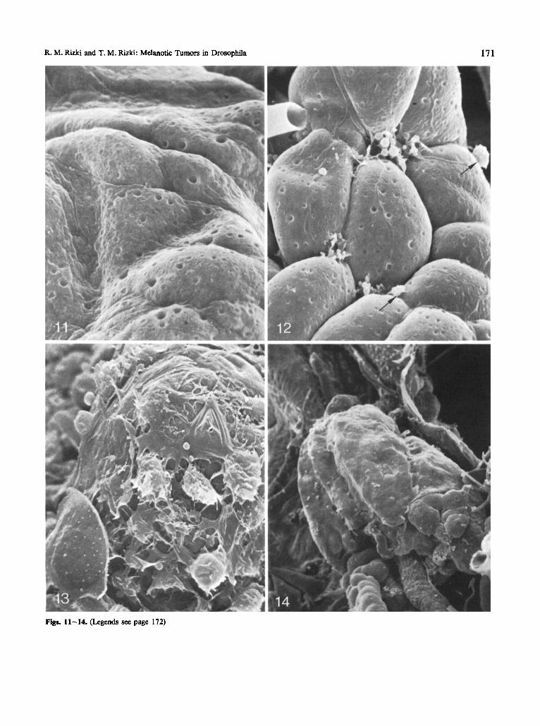
Fig. 11. The caudal fat body surface of an Ore-R larva. The adipose cells are covered by basement membrane and the major prominences of the surface contour correspond to the underlying adipose cells. The pits on the surfaces are also covered by membrane. Larval age, 64 h. x 1,500
Fig. 12. The caudal fat body of tu- W illustrating the early changes in the topography. Each adipose cell is rounded up; the contours of the tra- cheoles overlying the cells are clearly discernible. The basement membrane shows interruptions at the points where three or four cells are com- ing together, and membranous fragments and globular materials are seen at these intercellular spaces. Hemocytes (arrows). Larval age, 64 h. x 1,500
Fig. 13. A region of tu-Wcaudal fat body in the process of capsule formation by hemocytes. A lamellocyte can be seen in the lower left. Some of the blood cells are in transition form an ellipsoidal to flattened form; the thinness of these spreading cells is apparent. Note also the membranous extensions and the apparent stickiness of the hemocytes. Large and small globules are scattered over the surface. At the high- er magnification at least two or more layers of flattened cells can be resolved in this specimen. Larval age, 96 h. x 3,000
Flg. 14. A fully formed melanotic tumor. The adipose cells of the caudal fat body are completely covered by encapsulating hemocytes. Larval age, 96 h. x 200
Fig. 15. A phagocytic hemocyte on the surface of the fat body where three adipose cells are undergoing loss of basement membrane. The alveo- lar contours appear to arise from the lifting of the outer surface (0s) of the basement membrane (6). Projections @) arising from the hemo- cyte are extending between the intercellular space and also apposing the surface of the tracheoles (0. Larval age, 60 h. x 7,400
Fig. 16. Section showing the surface of an unaffected adipose cell in a tu- Wlarva. Note how the basement membrane material (b) separates the tracheal cell (r) from the surface of the adipose cell. In this ruthenium red preparation the surface of the basement membrane shows greater electron density than the inner matrix. Lipid globule (L) within the adipose cell. Larval age, 86 h. x 18,900
Fig. 17. Two hemocytes in the process of engulfing extruded ‘protein globules’ (& from tu-W adipose cells @. Note one globule between the two affected adipose cells, one within the cell body of a hemocyte, and one adhering to the hemocyte surface probably in the process of being engulfed. Ruthenium red stain. Larval age, 86 h. x 24,000
Fig. 18. Section through a group of transforming hemocytes in a tu- Wlarva showing the extrusion of surface vesicles. The one from the end of a surface filament is 0.25 p in diameter; the smallest vesicle profile measures 0.08 p in diameter. Larval age, 60 h. x 60,000
Fig. 19. Section through lamellocytes near the tumor-forming site. Note the free round vesicles and the collapsed vesicles adhering to the surfaces of the hemocytes. Larval age, 86 h. x 24,900
Fig. 20. The wall of the tumor formed by the layer of lamellocytes. The electron dense regions (arrows) are remnants of the flattened vesicles sandwiched between the surfaces of the lamellocytes. Larval age, 96 h. x 49,000

R. M. Rizki and T. M. Rizki: Melanotic Tumors in Drosophila
Figs. 15-20. (Legends see page 172)
173

174
Figure 21 is similar in electron density to basement membrane material. Material of a similar nature can also be seen within one of the adipose cells in this photo- graph, and such material has frequently been seen in the intercellular spaces between the dissociating adipose cells. In older larvae (80 h) large globules similar to the protein inclusion bodies normally found in the fat body of mid third instar larvae [7,81 are also lost from the tu- W adipose cells, and these are phagocytized by the he- mocytes as well (Fig. 17).
Mitochondria and endoplasmic reticulum (ER) in the affected fat body cells show no apparent structural deviations from normal adipose cells when the first signs of surface changes appear. Synthetic activity as viewed by intracisternal accumulations is also pronounced in some of the tumorous cells at this stage (study in prog- ress).
The encapsulation process follows the initial period of phagocytic activity by the hemocytes (Fig. 13). La- mellocytes and hemocytes in transition to the lamello- cytic form land on the adipose cell surfaces which ap- parently lack basement membrane. Layering of hemo- cyte upon hemocyte continues until a laminated capsu- lar wall surrounds the entire area of afllicted adipose cells. There does not appear to be a distinction between the type of cell adhering to the outermost surface of the capsule; some cells are fully differentiated lamellocytes and some cells are in the stage of flattening from the podocyte to lamellocyte variant. Heterogeneous sized globules are scattered over the surface of the developing capsule. A fully formed melanized capsule has a smooth surface where the outlines of the flattened cells can be discerned (Fig. 14).
Sections through the capsule show the laminated nature of the wall formed by the layering of the lamello- cytes (Fig. 22). Lamellocytes within this wall may be as thin as 0.1 p, but the regions containing the nuclei, which are somewhat thicker, are about 1.5 k, in thick-
R. M. Rizki and T. M. Rizki: Melanotic Tumors in Drosophila
ness. The plasma membranes of apposing lamellocytes are in close contact in some regions whereas other re- gions show a space between the membranes of the two cells. The latter may be long or short stretches and are traversed by electron dense fibrillar material which ex- tends into the cytoplasm of the cells on either side (Fig. 22B). These regions are about 200 A in width. The surface layer of lamellocytes facing the adipose cell sur- face does not show this feature (Fig. 22A). The cyto- plasm of the lamellocytes contains many microtubules which can be seen in cross and longitudinal section. Some of the lamellocytes within the capsule wall contain protein globules similar to those found in the adipose cells. Presumably these inclusions represent phagocytic materials within the hemocytes. If this is so, then a cell engaged in the initial phagocytic reaction may subse- quently participate in capsule formation.
In addition to the large globules in the hemocoel that are extruded from the disturbed surfaces of the adipose cells, there are also small vesicles that originate from the surfaces of the hemocytes. The vesicles seen in Figure 18 measure 0.08-0.25 p in diameter. These vesicles have a tendency to adhere to the surfaces of the plasma- tocytes at the time of active encapsulation, and can also be seen flattening and spreading over the hemocyte sur- faces (Fig. 19). This adhesiveness between the vesicles and the hemocyte surfaces suggests that the vesicular materials play a role in the initial lamellocyte-to-lamello- cyte binding during capsule formation. We therefore searched for comparable material in laminated capsules and found such entrapments between the cells in some instances (Fig. 20). It appears that the electron-dense flattened vesicles of various sizes may form regions of physical hindrance preventing direct contact of the plas- ma membranes of two apposing lamellocyte surfaces whereas regions lacking this material may form a close lamellocyte-lamellocyte contact. The lipid bilayers of the vesicles and the lamellocyte plasma membranes may
Fig. 21. A hemocyte actively engaged in phagocytosis at the junction of two tu- W adipose cells. This section of caudal fat body corresponds to the region illustrated in Figure 12 where membranous globules are accumulated. Note that the basement membrane of the adipose tissue is dis- rupted at the level of a projection arising from the hemocyte @) and the amorphous material engulfed by the hemocyte has an electron den- sity similar to basement membrane. This material (a) is seen within and between the adipose cells, in the hemocyte, and between the projec- tions extending from the hemocyte. L, lipid globules; b, basement membrane; t, tracheal cell. Larval age, 60 h. x 20,900
Fig. 22. The laminated structure of the tumor capsule resulting from close packing of the lamellocyte surfaces and the wedging developed be- tween the membranous folds of the lamellocyte surfaces. Note the close apposition of the cell membranes and the regions of electron dense gaps (gp) of varying lengths. In some regions of the lamellocytes there is a high density of microtubules in cross section whereas in other regions they are longitudinally disposed. Larval age, 96 h. x 20,900. Inset A: An enlarged view of the first layer of lamellocytes (I) against the adipose cell ().I surface. Note the absence of gaps between the two surfaces. x 51,200. Inset B: An enlargement of the area marked by asterisks showing the close apposition of lamellocyte surfaces and the gaps (gp). x 5 1,200

R. M. Rizki and T. M. Rizki: Melanotic Tumors in Drosophila 175
Fig. 21. (Legend see page 174)

176 R. M. Rizki and T. M. Rizki: Melanotic Tumors in Drosophila
Fig. 22. (Legend see page 174)

R. M. Rizki and T. M. Rizki: Melanotic Tumors in Drosophila
eventually amalgamate leaving a dilated region filled with electron dense material extending on either side into the cell cytoplasms as seen in Figure22B.
177
The fact that lamellocytes appear in circulation prior to visible changes in the adipose cells suggests that the transition from the spherical to flattened hemocyte form may be independent of tumor site specific contact. It is likely that biochemical factors released into the hemo- lymph provide a stimulus for hemocyte differentiation. Such biochemical stimuli may be emitted from the cau- dal fat body cells in tu- W larvae or may be due to fac- tors external to the tumor site, such as hormonal modifi- cations [13, 141. These considerations do not exclude hemocyte differentiation at the tumor-forming site, how- ever, since differentiating cells can be seen at the aggre- gation surface.
The role of the vesicles observed adhering to the sur- faces of the lamellocytes in the vicinity of the tumor- forming site is not certain. These vesicles may represent cytosol byproducts associated with the morphologic change of the spherical hemocyte to an extremely flat- tened shape. On the other hand, these vesicles may func- tion in the adhesion of the lamellocytes to other lamello- cytes in the initial stages of aggregation. In this regard, the difference between the adipose cell-to-lamellocyte and lamellocyte-to-lamellocyte interfaces may be signifi- cant since interference with the adhesiveness between lamellocytes is obtained by glucosamine treatment whereas the attachment of lamellocytes to adipose cells remains undisturbed [151.
Acknowledgement: This investigation was supported by Grant No. CA-16619 awarded by the National Cancer Institute, DHEW, and in part by NIH Biomedical Sciences Grant No. RR-07050.
Discussion
The first morphologic deviation from normality in tu- W larvae is the precocious appearance of many lamello- cytes in the early third instar [61. These cells circulate throughout the hemocoel and are not associated with the caudal fat body. Only after changes are apparent in the surface of the adipose tissue does hemocyte activity at this site begin. That surface modification is a prereq- uisite for the encapsulation reaction has recently been tested by implantation studies using host larvae in which lamellocytes were present 191. While the lamellocytes re- mained neutral to intact, unmodified fat body implants, fat bodies with mechanically injured surfaces or where the basement membrane was removed by collagenase treatment became encapsulated by lamellocytes.
Two mechanisms are utilized by the hemocytes to confiie the dissemination of products from the aberrant adipose cells. The hemocytes appearing fvst at the cau- dal fat body phagocytize surface materials and debris lost from the adipose cells. Phagocytosis is the typical defense response of insect hemocytes against small for- eign objects in the hemocoel [lo], and this behavior of the plasmatocytes in Drosophila can be readily demon- strated by injection of bacteria (Rizki and Rizki, in prep- aration). The phenomenon of melanotic tumor forma- tion in the tu-W mutant is hereditary, and neither ultra- structural studies nor parabiosis of normal and tumor- ous larvae have demonstrated the presence of an infec- tive agent [l 11. Phagocytosis in these larvae is directed against components that are normally within the adi- pose cells. These ‘in situ’ observations on phagocytosis are of particular interest since the hemocoel is undis- turbed by external stresses or experimental manipula- tions.
The second response of the hemocytes is encapsula- tion of the caudal adipose cells to produce compact cap- sules that later undergo melanization. What initiates the encapsulation reaction is not clear. The disturbances at the adipose cell surfaces may serve as the direct stimulus for the encapsulation response as well as the phagocytic reaction. On the other hand, it is possible that the stimu- lus for the encapsulation process by the lamellocytes is evoked by the activity of the phagocytes establishing first contact with the adipose cells since some of these hemocytes appear to exude material over the substrate [ la .
References
1. Bridges, C. B.: Non-disjunction as proof of the chromosome theory of heredity. Genetics 1, 53 (1916)
2. Wilson, L. P., King, R. C., Lowry, J. L.: Studies on the tu-W strain of Drosophila melanogaster I. Phenotypic and genotypic characterization. Growth 19, 215 (1955)
3. Sparrow, J. C.: The genetics of some second chromosome mela- notic tumour mutants of Drosophila melanogaster. Genet. Res. 23, 13 (1974)
4. Rizki, T. M.: Tumor formation in relation to metamorphosis in Drosophila melanogaster. J. Morphol. 100, 459 (1957)
5. Rizki, T. M.: Alterations in the haemocyte population of Droso- phila melanogaster. J. Morphol. 100, 437 (1957)
6. Rizki, T. M.: Experimental analysis of hemocyte morphology in insects. Am. Zool. 2, 247 (1962)
7. Butterworth, F. M., Bodenstein, D., King, R. C.: Adipose tissue of Drosophila melanogaster I. An experimental study of larval fat body. J. Exp. Zool. 158, 141 (1965)
8. Gaudecker, B., von: Uber den Formwechsel einiger Zellorganel- len bei der Bildung der Reservestoffe in Fettkorper von Droso- phila-Larven. Z. Zellforsch. 61, 56 (1963)

178
9. Rizki, R. M., Rizki, T. M.: Tissue encapsulation in melanotic tumor mutants of Drosophila. Proc. XI Int. Colloq. Invert. Pa- thol. (In press)
10. Salt, G.: The cellular defence reactions of insects. Cambridge: University Press 1970
11. Rizki, T. M.: Telobiosis of normal and tumorous larvae of Drosophila melanogaster. Drosophila Inform. Sew. 32, 153 (1958)
12. Rizki, R. M., Rizki, T. M.: Basement membrane abnormalities in melanotic tumor formation of Drosophila. Experientia 30, 543 (1974)
R. M. Rizki and T. M. Rizki: Melanotic Tumors in Drosophila
13. Rizki, T. M.: Melanotic tumor formation in Drosophfla. J. Mor- phol. 106, 147 (1960)
14. Madhavan, K.: Induction of melanotic pseudotumors in Droso- philu rnelanogaster by juvenile hormone. Wilhelm Row’ Arch. Entwick1.-Mech. Org. 169, 345 (1972)
15. Rizki, T. M.: The influence of glucosamine-hydrochloride on cellular adhesiveness in Drosophflu melunogaster. Exp. Cell Res. 24, 111 (1961)
Received April 1978/Accepted November 1978