cellulase make-up of certain facultative marine fungi …mbai.org.in/uploads/manuscripts/note_05...
TRANSCRIPT

I. mar. biol. Ass. India, 42 (1&2) 2000 : 153 - 156
Cellulase make-up of certain facultative marine fungi isolated from Bhavnagar coast
Anjna K. Vala, Shruti Y. Vaidya and H. C. Dube
Department of Life Sciences, Bhavnagar University, Bhavnagar - 364 002, Gujarat
Abstract
Four facultative marine fungi (Aspergillus nidulans, A. versicolor, Paecilomyces variotii and Pencillium citrinum), isolated from Bhavnagar coast were examined for cellulase production using four different media. All the test isolates produced exoglucanase, endoglucanase and b- glucosidase and showed complete cellulase activity in all the four media. Mandels & Sterberg medium supported highest enzyme production and the least preferred media for enzyme production were Park's and Zobell.
Cellulose is the most abundant organic matter on earth and also highly recalci- trant to enzymic degradation. Marine cellulolytic microorganisms are found in relatively high numbers in the near shore waters, where dead plant residues and live plant materials are accumulated due to action of the tide (D' Souza & Frietas, 1976). Zobell (1946) found thousand cel- lulose digesters per gram of marine mud samples and observed that "Marine cellu- lolytic microorganisms may be demon- strated in most 10-100 ml samples of botoom deposites". This early lead, how- ever, was not kept up and interest in cellulose degradation shifted to prepara- tion of glucose syrups from agricultural wastes and bagasse. Elucidation of enzy- mes of the cellulase complex is rarely done for marine organisms. In the present study, we have examined cellulase make-up (exoglucanase, endoglucanase, b-glucosi- dase and complete cellulase activity) of four fungi isolated from Bhavnagar coast.
We are grateful to Prof. BPR Vittal,
CAS in Botany, University of Madras for identifying the fungi and to Department of Ocean Development, Govt. of India, New Delhi, for sponsoring a research project under which this work was done.
Material and methods
The fungi used in this study were As- pergill us nid ulans (isolated from sediment), A. versicolor (from water), Paecilomyces variotii (from sediment) and Pencillium citrinum (from water). These were grown on four media viz. Kadota medium (Kadota, 1956), Mandels and Sternberg medium (Mandels and Sternberg, 1989), Park's medium (Vardavakis, 1976) and Zobell 2216E medium (Schlieper, 1972) contaning carboxyrnethyl cellulose (CMC) or cellulose powder as the carbon source. The organisms were grown at 28 + 2°C on a rotary shaker at 150 rpm, and cell- free culture filtrates at different intervals of growth (2, 4, 6, 8 and 10 days) were used as enzyme samples or assay of vari- ous cellulase enzymes viz. endoglucanase,

exoglucanase, and b - glucosidase and, complete cellulase activity.
Standard assay methods as described by Mandels (1982), were used in which reducing groups released from enzyme- specific substrates were measured spec- trophotometrically by Dinitro salicylic acid (DNS) method of Miller (1959). A glucose standard curve was used to quantify the reducing groups released. Assay for each enzyme was done as given below.
Endoglucanase (EC 3.2.1.4) : Enzyme sample (0.5 ml) was mixed with 0.5 ml of 1% CMC in 0.05 M Citrate buffer (pH 6.8) and incubated at 50°C for 30 min. The reducing groups released were measured by DNS method.
Exoglucanase (EC 3.2.1.91) : 50 mg ab- sorbent cotton, used as substrate, was mixed with 1 ml of 0.05 M Citrate buffer (pH 6.8) and 1 ml of enzyme sample. It was incubated at 50°C for 24 h and the reducing groups released were measured by DNS method.
B-glucosidase (EC 3.2.1.21) : B- glu- cosidase activity was assayed by using salicin as the substrate. The reaction mix- ture containing 0.5 ml of culture filtrates and 0.5 ml of 1% salicin in 0.05 M Citrate buffer (pH 6.8) was incubated for 30 min at 50°C. The reducing groups released were measured by DNS method.
Complete cellulase : 50 mg of Whatman No. 1 filter paper strip (rolled) was used as the substrate. 1 ml of enzyme and filter paper strip buffered at pH 6.8, were in- cubated for l h at 50°C. The reducing groups released were measured by DNS method.
All enzyme activities were expressed in International Units (IU) in which one enzyme unit equals one micromole of substrate hydrolyzed per minute (for cel- lulose, the micromole glucose released per minute).
Results and discussion
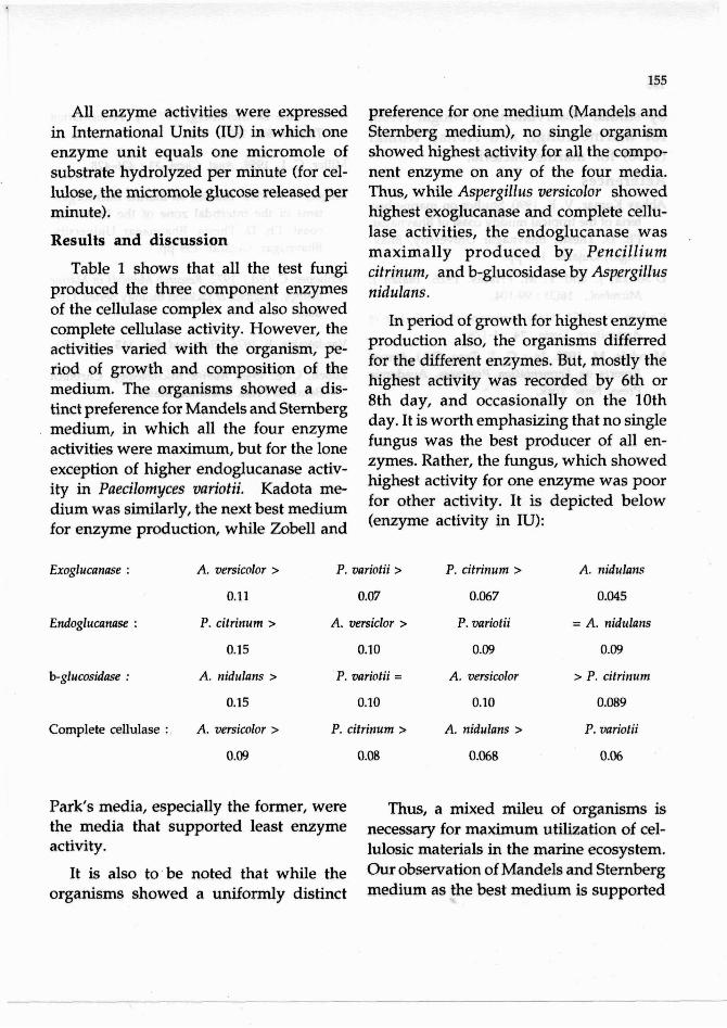
Table 1 shows that all the test fungi produced the three component enzymes of the cellulase complex and also showed complete cellulase activity. However, the activities varied with the organism, pe- riod of growth and composition of the medium. The organisms showed a dis- tinct preference for Mandels and Stemberg medium, in which all the four enzyme activities were maximum, but for the lone exception of higher endoglucanase activ- ity in Paecilomyces variotii. Kadota me- dium was similarly, the next best medium for enzyme production, while Zobell and
preference for one medium (Mandels and Sternberg medium), no single organism showed highest activity for all the compo- nent enzyme on any of the four media. Thus, while Aspergillus versicolor showed highest exoglucanase and complete cellu- lase activities, the endoglucanase was ,
maximally produced by Pencillium citrinum, and b-glucosidase by Aspergillus nidulans.
In period of growth for highest enzyme production also, the organisms differred for the different enzymes. But, mostly the highest activity was recorded by 6th or 8th day, and occasionally on the 10th day. It is worth emphasizing that no single fungus was the best producer of all en- zymes. Rather, the fungus, which showed highest activity for one enzyme was poor for other activity. It is depicted below (enzyme activity in IU):
Exoglucanase : A. versicolor >
0.11
Endoglucanase : P. citrinum >
0.15
b-glucosidase : A. nidulans >
0.15
Complete cellulase :. A. versicolor >
0.09
P. variotii >
0.07
A. m i c l o r >
0.10
P. variotii =
0.10
P. citrinum >
0.08
P. citrinum >
0.067
P. variotii
0.09
A. versicolor
0.10
A. nidulans >
0.068
A. nidulans
0.045
= A. nidulans
0.09
> P. citrinum
0.089
P. variotii
0.06
Park's media, especially the former, were Thus, a mixed mileu of organisms is the media that supported least enzyme necessary for maximum utilization of cel- activity. lulosic materials in the marine ecosystem.
~t is also to, be noted that while the o u r observation of Mandels and Sternberg organisms showed a distinct medium as the best medium is supported

by similar observations of Mogal (1993) for marine fungi, and Abhay Kumar (1990) for marine bacteria.
References Abhay Kumar, V. K. 1990. Studies on marine bac-
teria of the tropical muddy coast of Bhavnagar. Ph. D. Thesis, Bhavnagar University, Bhav- nagar, Gujarat. 143 pp.
D'Souza, J. and Y. M. Frietas. 1976. Indian J. Microbiol., 16(3) : 99-104.
Kadota, H. 1956. Univ Memories of the College of Agriculture, Kyoto., 74 : 1-178.
Mandels, M. 1982. In : G. T. Tsao (Ed.) Annual Reports on Fermentation Processes. Academic Press, New York.
and D. Sternberg, 1976. J. Fermentation Technol. 54 : 267.
Miller, G. L. 1959. Anal. Chem. 31 : 426-428.
Mogal, H. F. 1993. Studies on marine microorgan- isms of the intertidal zone of the Dandi sea coast. Ph. D. Thesis, Bhavnagar University, Bhavnagar, Gujarat. 159 pp.
Schlieper, C. (Ed.). 1972. Research Methods in Marine Biology. S idp ick & Jackson Biology Series, Lon- don.
Vardavakis, E. 1976. Plant and Soil. 115 : 145-150.
Zobell, C. E. 1946. Marine Microbiology. Chronica Botanica Press, Walthan, Maso.