cesarean section for placenta previa and placenta …...cesarean section for placenta previa and...
TRANSCRIPT

Cesarean Section for Placenta Previa andPlacenta Previa Accreta SpectrumSatoru Takeda, MD, PhD1,2 Jun Takeda, MD, PhD1 Shintaro Makino, MD, PhD1
1Department of Obstetrics and Gynecology, Faculty of Medicine,Juntendo University, Tokyo, Japan
2Aiiku Research Institute for Maternal, Child Health and Welfare,Imperial Gift Foundation Boshi-Aiiku-Kai, Tokyo, Japan
Surg J
Address for correspondence Satoru Takeda, MD, PhD, Department ofObstetrics and Gynecology, Faculty of Medicine, Juntendo University,2-1-1, Hongo, Bunkyo-ku, Tokyo 113-8421, Japan(e-mail: [email protected]).
In placenta previa cases, abundant blood flow enters theuterus not only from the internal iliac artery but also viaanastomosis of the external iliac artery, interior mesenter-ic artery, lumbar artery, median sacral artery, etc. There-fore, it is difficult to control bleeding; arterial ligationexerts a poor hemostatic effect. It is necessary to under-stand various hemostatic procedures, damage control sur-gery and resuscitation for massive hemorrhage, andsystemic management. Placenta increta and percreta arediagnosed by ultrasonography, Doppler ultrasonography,magnetic resonance imaging (MRI), and cystoscopy, etc.However, it is difficult to obtain a definitive diagnosis ofplacenta accreta.
In cases of placenta previawith previous cesarean section,the operation should be performed in a tertiary medicalfacility with sufficient staff and blood available for transfu-sion. Preoperative placement of intra-arterial balloon occlu-sion catheter in the common iliac artery or the aorta is usefulfor the prevention of massive hemorrhage. In cases withplacenta previa accrete spectrum, procedures following ce-sarean section include hysterectomy, preservation of theuterus with hemostasis of the placental separation surface,conservative treatment while leaving the placenta in situ,and delayed hysterectomy. Massive hemorrhage occurs atthe placental separation surface (spontaneous separation
and manual separation) and around the bladder (placentapercreta and placenta increta).
Cesarean Section for Placenta Previa
1. Laparotomy↓
2. Incision in the peritoneum of the vesicouterinepouch and separation of the bladder↓
3. Transverse incision on the uterine lower segment orthe lower uterine corpus↓
4. Delivery of the fetus↓
5. Separation of the placenta and uterotonicmedication↓
6. Hemostasis of the placental separation surface andmyometrial suture↓
7. Abdominal closure
Keywords
► balloon tamponade► cesarean section► compression sutures► hysterectomy► placenta previa► placenta accreta
spectrum
Abstract According to the increase in the rate of cesarean section and the increase of high-agedpregnancy, we seem to more often encounter cases with placenta previa and placentaprevia accrete spectrum. There are concerns about these cases, such as difficulty incontrolling bleeding from the separation surface of placenta previa, the need forhysterectomy as a life-saving procedure, systemic management and hemostasis duringmassive hemorrhage, and treatment of disseminated intravascular coagulation (DIC).These cases are most frequently associated with cesarean hysterectomy.
Surgical Steps
DOI https://doi.org/10.1055/s-0039-3402036.ISSN 2378-5128.
Copyright © by Thieme MedicalPublishers, Inc., 333 Seventh Avenue,New York, NY 10001, USA.Tel: +1(212) 760-0888.
THIEME
Precision Surgery in Obstetrics and Gynecology
Published online: 2020-03-09
Tips and Warnings
Risk Factors for Placenta Accrete SpectrumPlacentapreviawithapriorcesareansection isahighrisk
for placenta accreta spectrum (►Fig. 1; placenta accreta,increta, and percreta), that encompasses clinical adherenceandpathologicalfindings, andamajor factorcontributing tomaternal death.1,2 Along with the increase in the rate ofcesarean section, the incidence of placenta accreta spec-trum increased to 1 out of 2,500 deliveries in 1997 (10-foldcompared with that 50years ago).3 The frequency of pla-centa accreta spectrum in cases of placenta previa is gen-erally 1 out of 2,065 cases, but it increases to 25% in caseswith one prior cesarean section and 40% in cases with twoprior cesarean sections.4 According to the report by Sumi-gama et al5 in Japan, the frequency of placenta accretaspectrum in placenta previa is 1.1% in cases with a firstcesarean section, 37.8% in cases with one prior cesareansection, and38.5% incaseswith twopriorcesareansections.In our institution, the corresponding frequencywas 41% forcaseswithonepriorcesareansectionand54% for thosewithtwo prior cesarean sections.1
Preoperative EvaluationThe state of the cervical os, fetal presentation, position of thepresentation, and placental location should be confirmed bytransvaginal and transabdominal ultrasonography prior tocesarean section. Placenta previa cases, particularly thosewith previous cesarean section, should be examined for pla-centa increta and placenta percreta by ultrasonography, color-Doppler ultrasonography, MRI, cystoscopy, etc. (►Figs. 2–4,►Table 1).1,2,6–12 Diagnostic imaging of placenta accreta cancreate many false-positive results, yielding a low rate ofaccurate diagnosis. It is, furthermore, difficult to obtain adefinitive diagnosis of partial placenta accreta.1
Tips and Warnings
Management of Placenta Previa Accrete SpectrumPlacenta previa increta or percreta and suspected
placenta previa accreta (such as in cases of placentaprevia with prior cesarean section) should be managedand undergo cesarean section in a tertiary medical fa-cility.1–4,6 In cases of suspected placenta accreta spec-trum, the obstetrician should have a good knowledge ofpelvic anatomy, obstetric hemostasis and damage control
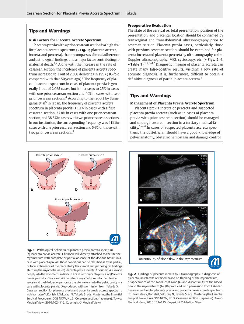
Fig. 1 Pathological definition of placenta previa accreta spectrum.(a) Placenta previa accrete. Chorionic villi directly attached to the uterinemyometrium with complete or partial absence of the decidua basalis in acasewith placenta previa. Those conditions can be classified as total, partial,or focal adherence of the placenta by the clinical and pathological findingsabutting themyometrium. (b) Placenta previa increta. Chorionic villi invadedeeply into themyometrium layer in a casewithplacentaprevia. (c) Placentaprevia percreta. Chorionic villi penetrate myometrium into the uterineserosaand thebladder, orperforate theuterinewall into thepelvic cavity in acase with placenta previa. (Reproduced with permission from Takeda S.Cesarean section for placenta previa and placenta previa accrete spectrum.In: Hiramatsu Y, Konishi I, Sakuragi N, Takeda S, eds.Mastering the EssentialSurgical Procedures OGS NOW, No.3. Cesarean section. (Japanese). Tokyo:Medical View; 2010:102–115. Copyright © Medical View).
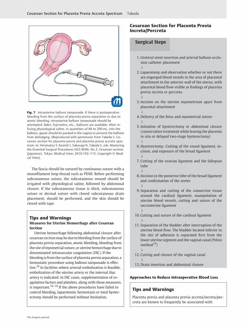
Fig. 2 Findings of placenta increta by ultrasonography. A diagnosis ofplacenta increta was obtained based on thinning of the myometrium,disappearance of the sonolucent zone (a) and discontinuity of the bloodflow in the myometrium (b). (Reproduced with permission from Takeda S.Cesarean section for placenta previa and placenta previa accrete spectrum.In: Hiramatsu Y, Konishi I, Sakuragi N, Takeda S, eds.Mastering the EssentialSurgical Procedures OGS NOW, No.3. Cesarean section. (Japanese). Tokyo:Medical View; 2010:102–115. Copyright © Medical View).
The Surgery Journal
Cesarean Section for Placenta Previa Accreta Spectrum Takeda

surgery for massive hemorrhage, and he should also beskilled in performing total hysterectomy. In each case ofplacenta previawith/without findings of placenta accretespectrum, the operators should discuss about approachesof cesarean delivery, management for placenta accretespectrum, selection of treatment and prevention ofmassive hemorrhage, and therefore, should make a finaldecision of surgical strategies for placenta previa accretespectrum (►Fig. 4).1 Preoperative preparation, includingsecuring of staff, an anesthesiologist, and sufficient bloodfor transfusion, is important in cases undergoing cesareansection for placenta previa.
Preoperative PreparationIt is necessary to make sure that the patient and family under-stand placenta previa and placenta previa accreta spectrum, toobtain their consent forpossiblebloodtransfusionand that theycomprehend the risks of massive hemorrhage and hysterecto-my. Autologous blood should be stored preoperatively to pre-vent allogeneic blood transfusion.13 Sufficient blood fortransfusion, autologous blood recovery system (Cell Saver),and staff should be secured, and discussions with the depart-ments of anesthesiology, blood transfusion, urology, and ob-stetrics, as well as with the operating room staff, should beconducted in advance.
The positions of the fetus and placenta should be con-firmed by ultrasonography, allowing the location of the
uterine incision to be simulated. If total hysterectomy isscheduled for placenta previa increta or percreta, preopera-tive ureteral stent or hemostatic intra-arterial balloon occlu-sion catheter placement should be considered.1
Tips and WarningsEnhancement of Cooperation with Health CareProfessionals
In-hospital cooperation and hospital-clinic coopera-tion are important for prompt and appropriate patienttransport and treatment. Discussions of the actions andmeasures against massive hemorrhage and obstetricemergencies should be made on a regular basis not onlywithin the department of obstetrics but also with thedepartments of anesthesiology, clerical work, nursing,blood transfusion, and critical care, and necessary actionsshould be simulated within the hospital. It is necessary tounderstand actions to be taken for critical hemorrhageand not crossmatched compatible red blood cell (RBC)such as typeO RBC and type AB fresh-frozen plasma (FFP),allowing consensus among the departments concerned tobe reached.14 Cooperation between primary medicalfacilities and secondary and tertiary facilities should bedeepened. Feedback regarding cases should be providedto facilities from which patients are transferred throughcase conferences or other suitable occasions.1,2
Explanation of Procedures
LaparotomyA transverse skin incision may be used. If the field of view isnarrow, the Maylard incision should be combined with thePfannenstiel incision. In cases of intrauterine infection orrupture of the membranes, the use of a wound protector(Alexis) after laparotomy reduces the occurrence of abdomi-nal wall wound disruption and wound infection. The perito-neum of the vesicouterine pouch is incised transversely andthen the bladder is gently dissected.
Whentherearemanyengorgedbloodvessels in thesurfaceofthe myometrium or in the pericystic area, incision in theperitoneum of the vesicouterine pouch and separation of thebladder should be avoided (►Figs. 5 and 6).Making a transverseincision, an oblique incision, or a J-shaped incision in the uterinecorpus is recommended, that is, preoccupationwitha transverseincision on the lower uterine segment is unwarranted.
Tips and WarningsContraindications of Placenta Manual Removal
The placenta should never be separated whenengorged blood vessels are found in the area of placental
Fig. 3 MRI findings of placenta previa increta. The placenta isobserved to invade the bladder from the anterior wall of the uterus.(Reproduced with permission from Takeda S. Cesarean section forplacenta previa and placenta previa accrete spectrum. In: HiramatsuY, Konishi I, Sakuragi N, Takeda S, eds. Mastering the Essential SurgicalProcedures OGS NOW, No.3. Cesarean section. (Japanese). Tokyo:Medical View; 2010:102–115. Copyright © Medical View).
The Surgery Journal
Cesarean Section for Placenta Previa Accreta Spectrum Takeda

attachment to the anterior wall of the uterus with pla-cental blood flow visible after laparotomy and when adiagnosis of placenta percreta or placenta increta hasbeenmade (►Fig. 6).1Not only primary hysterectomy butalso the conservative therapy or delayed hysterectomy(the two-stage hysterectomy) should be considered.
Incision of the Lower Uterine SegmentTo avoid excessive splitting, it is recommended that a knife beused for myometrial incision, followed by the use of Cooperscissors to add any necessary incisions after the maternalsurface of the placenta is observed. In reference to theplacental location determined by preoperative ultrasonog-raphy, the incision should be extended in the direction thatreaches the amniotic cavity by the shortest path, and thematernal surface of the placenta should be pressed
Table 1 Diagnostic methods and its findings of placenta accreta spectrum
• Ultrasonography
� The presence of many low-intensity areas (placental lacunae) in the placenta; a Swiss cheese-like appearance.
� Loss of the low-intensity area (sonolucent zone, retroplacental clear space) between the placenta and myometrium.
� Irregularity and interruption of the bladder border.
� Thinning (�1mm) of the myometrium, protrusion of the placenta toward the bladder.
• Color-Doppler and pulse-Doppler ultrasonography
� There are diffuse blood vessels (lacunar flow) in the placenta with pulsating rapid venous flow.
� There are myometrial or subplacental blood vessels with pulsating turbulent venous flow.
� There are dilated blood vessels in the bladder and myometrium with arterial flow showing a low resistance index.
� Markedly dilated subplacental blood vessels with pulsating venous flow.
• Biomarker testing
� Elevated α-fetoprotein levels.
� Cell-free fetal DNA, placental mRNA, DNA microarray assay.
Source: Reproduced with permission of Takeda S. Cesarean section for placenta previa and placenta previa accrete spectrum. In: Takeda S,Hiramatsu Y, Konishi I, Sakuragi N, eds. OGS NOW, No.3. Cesarean Section. Mastering the Essential and Practical Surgical Procedures. Tokyo:Medical View; 2010:102–115. Copyright © Medical View.
Fig. 4 Surgical strategies for placenta previa accrete spectrum and its management.
The Surgery Journal
Cesarean Section for Placenta Previa Accreta Spectrum Takeda

downward to expose the bulging fetal membranes. Afterrupture of the membranes, the fetus should be deliveredwhile pressing the placenta downward while avoiding pla-cental separation. It is not necessary to deliver the placentafirst or to deliver the fetus transplacentally. In placentaprevia cases in which the placenta is mainly attached tothe anterior wall, the myometrial incision should be cutupward toward the right or left amniotic cavity, or a longitu-dinal incision in the uterine corpus may be made in an areawhere there is no placenta.
Delivery of the FetusThe fetus should be delivered promptly after the rupture ofthe membranes, and the umbilical cord should be clampedto prevent anemia of the neonate. At the same time, the firstassistant should hold the bleeding site as well as both endsof the uterine incision on the right and left sides withserrated forceps (At least eight pairs of the forceps should beplaced on the operating table; each pair should be dis-infected and ready for additional use) to achieve hemostasisto the maximum extent possible. When there is massivehemorrhage, the uterine incisional wound located down-ward may be unobservable and not possible to hold. In thiscase, the lower myometrial cut end of one lateral end of theuterine wound should be held and elevated with serratedforceps while aspirating blood, and the site of clampingshould gradually be shifted to the center by alternating theforceps.
Whenhemorrhage is severe, pressurehemostasis shouldbeemployed with two or three rolled towels placed on theplacental separation surface. A uterotonic agent should beadministered locally and intravenously to achieve uterinecontraction. When bleeding from the placental separationsurface is severe, direct Z suturewith1–0 synthetic absorbablethread, or U-shaped suture or enclosing suture interruptedsuture piercing the myometrium, should be performed.1 Ifthese procedures fail to achieve hemostasis, compressionsutures such as a vertical suture15,16 or an arterial ligationshould be used. If hemorrhage is minor, gauze packing oruterine balloon tamponade is performed (►Fig. 7); the gauzeor balloon is removed transvaginally the followingday.16
Closure of the Uterine Wound and the AbdomenThe right and left ends of the myometrial incisional woundshould be sutured by interrupted or Z-suture, making surethat the thread is exposed to the uterine cavity. Caution isnecessary because the inner surface may be deeply cleaved.The incisional wound is then sutured by interrupted suturewith synthetic absorbable thread, while keeping the endo-metrial surface consistent. A thick myometrium may besutured in two layers. Even when the bladder is separated,it is better not to suture the vesicouterine pouch; the bladdermay otherwise be elevated. After intraperitoneal irrigationwith physiological saline, the hemostatic status is confirmed,and a closed low-pressure continuous drain is inserted intothe Douglas pouch and the site of myometrial suture.Absorbable adhesion barrier, such as Seprafilm is appliedto the wounds on the uterus.
Fig. 5 Findings of the anterior wall of the uterus in placenta previaincreta. The site of placental attachment to the anterior wall of theuterus is purpuric, and placental blood flow is seen through the wall. Itis apparent that there are many engorged blood vessels, and that theplacenta invades the area just beneath the serosa. No incision shouldbe made around this area at the time of delivery of the fetus. Atransverse incision in the uterine body, a J-shaped incision, or atransverse incision in the uterine fundus should be made at a sitesufficiently distant from the placenta. (Reproduced with permissionfrom Takeda S. Cesarean section for placenta previa and placentaprevia accrete spectrum. In: Hiramatsu Y, Konishi I, Sakuragi N, TakedaS, eds. Mastering the Essential Surgical Procedures OGS NOW, No.3.Cesarean section. (Japanese). Tokyo: Medical View; 2010:102–115.Copyright © Medical View).
Fig. 6 Placenta previa with previous cesarean section. Because of aprior cesarean section, the bladder is elevated and adherent. Manyblood vessels in the uterine surface around the incisional wound andin area around the bladder are engorged. !: There are engorgedblood vessels in the bladder surface. (Reproduced with permissionfrom Takeda S. Cesarean section for placenta previa and placentaprevia accrete spectrum. In: Hiramatsu Y, Konishi I, Sakuragi N, TakedaS, eds. Mastering the Essential Surgical Procedures OGS NOW, No.3.Cesarean section. (Japanese). Tokyo: Medical View; 2010:102–115.Copyright © Medical View).
The Surgery Journal
Cesarean Section for Placenta Previa Accreta Spectrum Takeda
The fascia should be sutured by continuous suture with amonofilament loop thread such as PDSII. Before performingsubcutaneous suture, the subcutaneous wound should beirrigated with physiological saline, followed by abdominalclosure. If the subcutaneous tissue is thick, subcutaneoussuture or dermal suture with closed subcutaneous drainplacement, should be performed, and the skin should beclosed with tape.
Tips and WarningsMeasures for Uterine Hemorrhage after CesareanSection
Uterine hemorrhage following abdominal closure aftercesarean sectionmaybedue tobleeding fromthe surface ofplacenta previa separation, atonic bleeding, bleeding fromthesite ofmyometrial suture, or uterinehemorrhagedue todisseminated intravascular coagulation (DIC). If thebleeding is from the surface of placenta previa separation, ahemostatic procedure using balloon tamponade is effec-tive.16 In facilities where arterial embolization is feasible,embolization of the uterine artery or the internal iliacartery is indicated. In DIC cases, supplementation of co-agulation factors and platelets, along with these measures,is important.16–18 If the above procedures have failed tocontrol bleeding, laparotomic hemostasis or total hyster-ectomy should be performed without hesitation.
Cesarean Section for Placenta PreviaIncreta/Percreta
1. Ureteral stent insertion and arterial balloon occlu-sion catheter placement↓
2. Laparotomy and observation whether or not thereare engorged blood vessels in the area of placentalattachment to the anterior wall of the uterus, withplacental blood flow visible as findings of placentaprevia increta or percreta↓
3. Incision on the uterine myometrium apart fromplacental attachment↓
4. Delivery of the fetus and myometrial suture↓
5. Initiation of hysterectomy or abdominal closure(conservative treatment while leaving the placentain situ or delayed two-stage hysterectomy)↓
6. Hysterectomy: Cutting of the round ligament, in-cision, and exposure of the broad ligament↓
7. Cutting of the ovarian ligament and the fallopiantube↓
8. Incision in the posterior lobe of the broad ligamentand confirmation of the ureter↓
9. Separation and cutting of the connective tissuearound the cardinal ligament, manipulation ofuterine blood vessels, cutting and suture of thesacrouterine ligament↓
10. Cutting and suture of the cardinal ligament↓
11. Separation of the bladder after interruption of theuterine blood flow. The bladder located inferior tothe site of adhesion is separated first from thelower uterine segment and the vaginal canal (Pelosimethod19)↓
12. Cutting and closure of the vaginal canal↓
13. Drain insertion and abdominal closure
Approaches to Reduce Intraoperative Blood Loss
Tips and Warnings
Placenta previa and placenta previa accreta/increta/per-creta are known to frequently be associated with
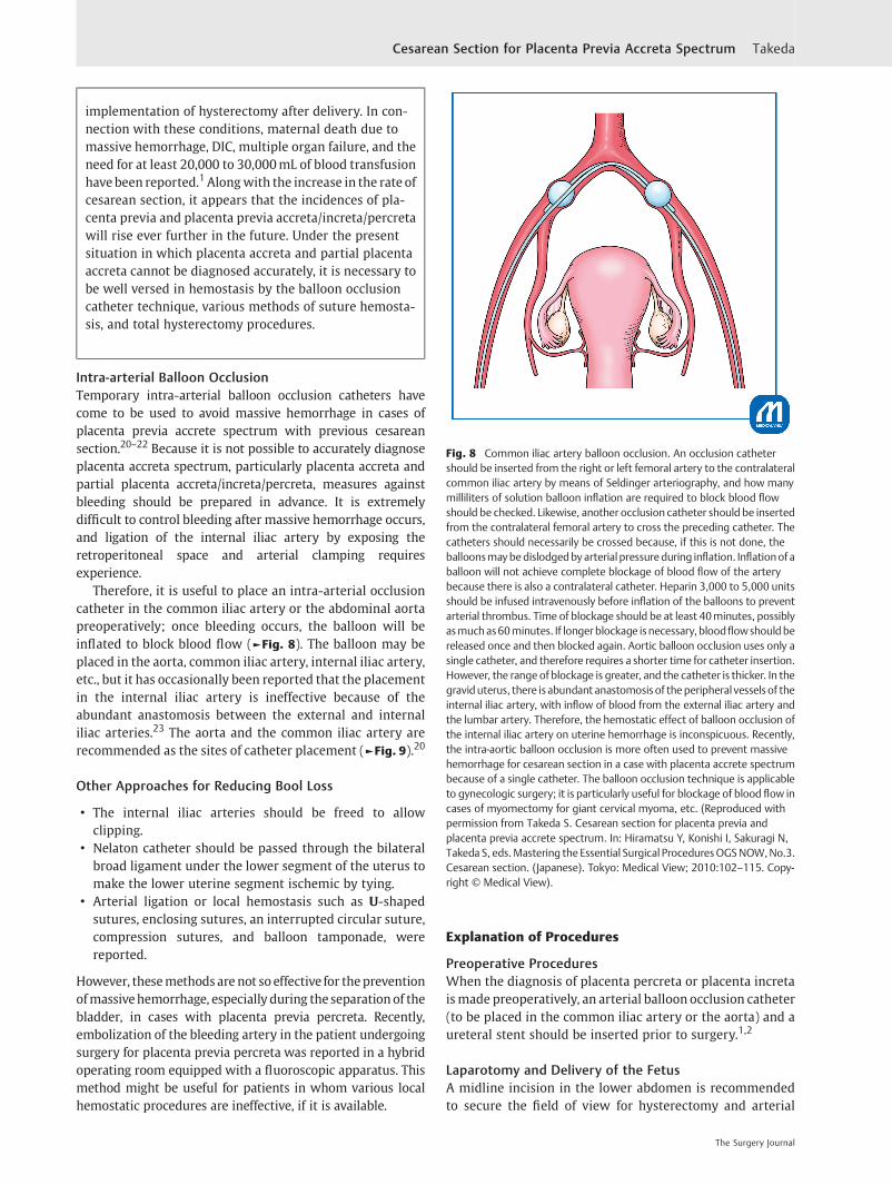
Fig. 7 Intrauterine balloon tamponade. If there is postoperativebleeding from the surface of placenta previa separation or due toatonic bleeding, intrauterine balloon tamponade should beattempted. Bakri, Fuji-metro, etc., balloons are available. After in-fusing physiological saline, in quantities of 80 to 200mL, into theballoon, gauze should be packed in the vagina to prevent the balloonfrom dislodging. (Reproduced with permission from Takeda S. Ce-sarean section for placenta previa and placenta previa accrete spec-trum. In: Hiramatsu Y, Konishi I, Sakuragi N, Takeda S, eds. Masteringthe Essential Surgical Procedures OGS NOW, No.3. Cesarean section.(Japanese). Tokyo: Medical View; 2010:102–115. Copyright © Medi-cal View).
Surgical Steps
The Surgery Journal
Cesarean Section for Placenta Previa Accreta Spectrum Takeda
implementation of hysterectomy after delivery. In con-nection with these conditions, maternal death due tomassive hemorrhage, DIC, multiple organ failure, and theneed for at least 20,000 to 30,000mL of blood transfusionhavebeen reported.1Alongwith the increase in the rate ofcesarean section, it appears that the incidences of pla-centa previa and placenta previa accreta/increta/percretawill rise ever further in the future. Under the presentsituation in which placenta accreta and partial placentaaccreta cannot be diagnosed accurately, it is necessary tobe well versed in hemostasis by the balloon occlusioncatheter technique, various methods of suture hemosta-sis, and total hysterectomy procedures.
Intra-arterial Balloon OcclusionTemporary intra-arterial balloon occlusion catheters havecome to be used to avoid massive hemorrhage in cases ofplacenta previa accrete spectrum with previous cesareansection.20–22 Because it is not possible to accurately diagnoseplacenta accreta spectrum, particularly placenta accreta andpartial placenta accreta/increta/percreta, measures againstbleeding should be prepared in advance. It is extremelydifficult to control bleeding after massive hemorrhage occurs,and ligation of the internal iliac artery by exposing theretroperitoneal space and arterial clamping requiresexperience.
Therefore, it is useful to place an intra-arterial occlusioncatheter in the common iliac artery or the abdominal aortapreoperatively; once bleeding occurs, the balloon will beinflated to block blood flow (►Fig. 8). The balloon may beplaced in the aorta, common iliac artery, internal iliac artery,etc., but it has occasionally been reported that the placementin the internal iliac artery is ineffective because of theabundant anastomosis between the external and internaliliac arteries.23 The aorta and the common iliac artery arerecommended as the sites of catheter placement (►Fig. 9).20
Other Approaches for Reducing Bool Loss
• The internal iliac arteries should be freed to allowclipping.
• Nelaton catheter should be passed through the bilateralbroad ligament under the lower segment of the uterus tomake the lower uterine segment ischemic by tying.
• Arterial ligation or local hemostasis such as U-shapedsutures, enclosing sutures, an interrupted circular suture,compression sutures, and balloon tamponade, werereported.
However, thesemethodsare not so effective for thepreventionofmassivehemorrhage, especially during theseparationof thebladder, in cases with placenta previa percreta. Recently,embolization of the bleeding artery in the patient undergoingsurgery for placenta previa percreta was reported in a hybridoperating room equipped with a fluoroscopic apparatus. Thismethod might be useful for patients in whom various localhemostatic procedures are ineffective, if it is available.
Explanation of Procedures
Preoperative ProceduresWhen the diagnosis of placenta percreta or placenta incretaismade preoperatively, an arterial balloon occlusion catheter(to be placed in the common iliac artery or the aorta) and aureteral stent should be inserted prior to surgery.1,2
Laparotomy and Delivery of the FetusA midline incision in the lower abdomen is recommendedto secure the field of view for hysterectomy and arterial
Fig. 8 Common iliac artery balloon occlusion. An occlusion cathetershould be inserted from the right or left femoral artery to the contralateralcommon iliac artery by means of Seldinger arteriography, and how manymilliliters of solution balloon inflation are required to block blood flowshould be checked. Likewise, another occlusion catheter should be insertedfrom the contralateral femoral artery to cross the preceding catheter. Thecatheters should necessarily be crossed because, if this is not done, theballoonsmaybedislodgedby arterial pressureduring inflation. Inflationof aballoon will not achieve complete blockage of blood flow of the arterybecause there is also a contralateral catheter. Heparin 3,000 to 5,000 unitsshould be infused intravenously before inflation of the balloons to preventarterial thrombus. Time of blockage should be at least 40minutes, possiblyasmuchas60minutes. If longer blockage is necessary, bloodflowshouldbereleased once and then blocked again. Aortic balloon occlusion uses only asingle catheter, and therefore requires a shorter time for catheter insertion.However, the range of blockage is greater, and the catheter is thicker. In thegravid uterus, there is abundant anastomosis of theperipheral vessels of theinternal iliac artery, with inflow of blood from the external iliac artery andthe lumbar artery. Therefore, the hemostatic effect of balloon occlusion ofthe internal iliac artery on uterine hemorrhage is inconspicuous. Recently,the intra-aortic balloon occlusion is more often used to prevent massivehemorrhage for cesarean section in a case with placenta accrete spectrumbecause of a single catheter. The balloon occlusion technique is applicableto gynecologic surgery; it is particularly useful for blockage of blood flow incases of myomectomy for giant cervical myoma, etc. (Reproduced withpermission from Takeda S. Cesarean section for placenta previa andplacenta previa accrete spectrum. In: Hiramatsu Y, Konishi I, Sakuragi N,Takeda S, eds.Mastering the Essential Surgical ProceduresOGSNOW,No.3.Cesarean section. (Japanese). Tokyo: Medical View; 2010:102–115. Copy-right © Medical View).
The Surgery Journal
Cesarean Section for Placenta Previa Accreta Spectrum Takeda

ligation. When there are engorged blood vessels at the siteof placental attachment to the anterior wall of the uterus,allowing placental blood flow to be visualized, or when adiagnosis of placenta percreta or placenta increta hasalready been obtained by diagnostic imaging, a transverseincision in the uterine body or in the uterine fundus(►Fig. 10) should be made to prevent the incision fromreaching the placenta and thereby causing massive hemor-rhage. Rupturing of the membranes, delivery of the fetus,and cutting and ligation of the umbilical cord should beperformed. Then, the myometrial wound should be tempo-rarily closed.
Cesarean HysterectomyIn routine cases, the operation proceeds to total hysterecto-my. However, if there is no bleeding without spontaneousplacental separation, it is also possible to provide conserva-tive treatment with the placenta left in situ or to delayperforming the hysterectomy.
In contrast to cases undergoing a routine cesareanhysterectomy, the placenta adherent to the areas fromthe uterine isthmus to the lower part of the uterine bodyshows a potbelly-like bulging, and many engorged bloodvessels can be seen in areas around the bladder and around
the scar from prior cesarean section. In some cases, theanterior surface of the uterus and the site of placentalattachment are purple to dark red, and the placenta istranslucent (►Fig. 11). The bladder is often elevated be-cause of the prior cesarean section. For total hysterectomy,the operator should avoid vascular injury, ligating veinswhenever possible, and pay attention to intraoperativeuterine hemorrhage from the blood flow via the vaginaand the bladder.
Cutting of the Round Ligament and Incision and Exposure ofthe Broad Ligament of the UterusThe round ligament of the uterus and the ovarian ligamentshould be held bilaterally with a long straight Kocher clampto elevate the uterus. After picking up the round ligamentwith Pean forceps, its medial side and lateral side should beligated with absorbable thread, followed by cutting be-tween the two. The lateral side should be processed byapplying a double ligature. While pulling up the ligature, theperitoneum of the anterior lobe of the broad ligamentshould be incised in the directions toward the infundibu-lopelvic ligament and toward the bladder to expose theretroperitoneal space. At this time, the infundibulopelvicligament should be traced cranially to identify the ureter.
Fig. 9 Abundant pathways of collateral circulation to the uterus. Abdominal aorta. Lumbar artery. Common iliac artery. Iliac branch of theiliolumbar artery. Iliolumbar artery. Internal iliac artery. Superior gluteal artery. Inferior gluteal artery. Inferior rectal artery. Inferior epigastricartery. Ascending branch. Deep femoral artery. Lateral circumflex femoral artery. Ovarian artery. Inferior mesenteric artery. Lateral sacral artery.Superior rectal artery. Middle rectal artery. Uterine artery. Vaginal artery. Obturator artery. Medial circumflex femoral artery. Femoral artery(Reproduced with permission from Takeda S. Cesarean section for placenta previa and placenta previa accrete spectrum. In: Hiramatsu Y, KonishiI, Sakuragi N, Takeda S, eds. Mastering the Essential Surgical Procedures OGS NOW, No.3. Cesarean section. (Japanese). Tokyo: Medical View;2010:102–115. Copyright © Medical View).
The Surgery Journal
Cesarean Section for Placenta Previa Accreta Spectrum Takeda

Cutting of the Ovarian Ligament and the Fallopian TubeThe peritoneum in the vicinity of the cut end of the roundligament should be incised in the direction toward theovarian ligament, and the vascular plexus in areas aroundthe tube and the ovary should be identified. A hole should becreated and expanded with a Kelly clamp in the posteriorlobe of the nonvascular broad ligament located inferior to thevascular plexus. The ovarian ligament and the tube should beclamped with serrated forceps and cut between the forcepson the uterine side. The lateral cut end should be suturedemploying a figure-eight suture or interrupted suture. Be-cause this procedure involves focused ligation, it is necessaryto use the double ligature technique to prevent loosening. Ifthe cut end is close to the suture thread, Kobayashi’s ligation(vertical Z suture)24 should be added. When the cut end isbroad, or when there are engorged blood vessels, the tubeand the ovarian ligament should be ligated and cut separate-ly. Recently tubectomy is performed for prophylaxis of thetubal origin ovarian cancer.
Incision in the Anterior and Posterior Lobe of the BroadLigamentThe course of the ureter should be carefully assessed in allcases. Such assessment is easy if a ureteral stent is in place. Ifadhesion is severe, and the peritoneum of the vesicouterine
pouch is not clearly identifiable, it should be left untouched.The bladder should not be separated until the final stage.
Cutting and Suturing of the Cardinal LigamentThe connective tissue around the cardinal ligament shouldbe separated and cut, followed by identification of uterineblood vessels, confirmation of the route of the ureter, andcutting and ligation of the ascending branches of the uterineartery and vein. Then, the sacrouterine ligament should becut and sutured.
Thelowerpartof theuterusshowsapotbelly-likebulging; thecardinal ligament is broad andelongated, and, therefore, cautionis necessary to avoid excessive cutting (►Figs. 10 and 11).Because the ureter is located extremely close to the uterus,thecourseof theureter shouldbechecked,or thestentshouldbeconfirmed. For cutting of the cardinal ligament (parametrium),the ligament should be clamped on the uterine side as well, toprevent bleeding on the uterine side. The cutting and suturingprocedure of the cardinal ligament should be performed in atleast three steps. Because the bladder is not yet separated, thecutting and suturing of the cardinal ligament should be ad-vancedwhileseparating thebladderandtheconnectivetissue inthe anterior portion of the cardinal ligament.
Separation of the BladderWhen there is bleeding around the bladder or massiveuterine hemorrhage, blood flow should be blocked by theocclusion catheter as needed. If there is no bleeding, blockageof the blood flow is performed before separation of thebladder. Blockage should be maintained for not more than40minutes to 1 hour. If a blockage over an extended timeperiod is necessary, blood flow blockage should be releasedonce followed by reinstitution of the blockage.
If the blood flowof the common iliac artery or aorta (flowto the lower limbs) is to be blocked, unfractionated heparin3,000 to 5,000 units (60–100 units/kg) should be infusedintravenously in advance.
When separating the bladder, the site of placenta percretaand the site of adhesion should be left untouched, and theseparating procedure should begin with a lower part of thebladder (Pelosi method).19 The space between the bladderand the lower uterine segment should be separated. After apenetration passage is formed like a tunnel between the rightand left sides, the vaginal wall and the remaining para-metrium should be held with serrated large curved forcepsto block blood flow from the vagina (►Fig. 12).
When cutting the adhesion between the bladder and theuterus using an electric cautery tool or other equipment, it ishelpful to elevate the bladder to add tension to the site ofincision. In cases of complete placenta percreta with hema-turia, opening the bladder lumen is helpful. Even if theplacenta comes out of the uterine wall, bleeding inside theuterus is predominant, and therefore, the separating proce-dure should be performedwithout hesitation. Bleeding fromthe bladder separation surface is often severe, and thebleeding area should be sutured and reinforced with 3–0absorbable thread in a coveringmanner. If the bladder lumenis open, double layer suture should be used.
Fig. 10 Transverse incision in the uterine fundus (Kotsuji technique).A transverse incision is made in the anterior or posterior wall of theuterine fundus, avoiding the uterine horn and keeping a substantialdistance from the area of placental attachment. When the incisionreaches the uterine cavity, the myometrium is held with two Kellyclamps, and incised between the clamps to extend the incision. TheseKelly clamps should be kept in place at this site until the fetus isdelivered and the myometrium sutured. (Reproduced with permissionfrom Takeda S. Cesarean section for placenta previa and placentaprevia accrete spectrum. In: Hiramatsu Y, Konishi I, Sakuragi N, TakedaS, eds. Mastering the Essential Surgical Procedures OGS NOW, No.3.Cesarean section. (Japanese). Tokyo: Medical View; 2010:102–115.Copyright © Medical View).
The Surgery Journal
Cesarean Section for Placenta Previa Accreta Spectrum Takeda

Cutting and Closure of the Vaginal CanalAfter the bladder is separated, the anterior vaginal wallshould be cut, and the uterus then excised. The vaginalwall should be closed by Z suture or simple ligation. Becauseblood flowof the external iliac artery system via the cardinalligament is extremely abundant, strict hemostasis isnecessary.
Drain Insertion and Abdominal ClosureAfter intraperitoneal irrigation, a drain should be placed inthe Douglas pouch. The pelvic peritoneum may be left open.An adhesion preventingmaterial such as Seprafilm should beapplied to the pelvic floor and under the abdominalwall, andthe abdominal incision wound should then be closed.
Special Occasions
Delayed Hysterectomy (The Two-Stage Hysterectomy)In cesarean section cases, if the placenta does not separatespontaneously, and there is no bleeding, the surgery can becompleted with the placenta left in situ without tryingvigorously to separate it. The placenta may be separated atthe second stage after blood flow into the placenta isreduced, or the amount of bleeding may be decreased byscheduled delayed hysterectomy.1,2 In these cases, emboli-zation of the internal iliac artery or the uterine artery prior tothe two-stage operation reportedly leads to a decrease in theamount of bleeding.5 Delayed hysterectomy has a drawbacksince, even if scheduled, it cannot be performed in cases in
which the placenta has been separated, and hemorrhage hasoccurred, during cesarean section.
Conservative Management of Placenta Increta/PercretaThe uterus can be preserved under the conditions that theplacenta is not separated, and there is no hemorrhage,during the cesarean section. After abdominal closure isperformed with the placenta left in situ, spontaneousseparation of the placenta can be anticipated.25,26 In anyevent, patients should be informed in advance of the risk ofmassive hemorrhage and complications such as sepsis dueto infection.
Obstetric Damage ControlHemostasis often cannot be achieved promptly in cases ofmassive hemorrhage during cesarean section, accompaniedby DIC. In such obstetric cases with DIC, particularly in thepresence of hypothermia, acidosis, and vasopressor require-ment, damage control surgery (DCS) and resuscitation,which represent the therapeutic concept of life-savingintervention for severe trauma should be performed be-cause ordinary hemostatic procedures such as sutures,ligation, and coagulation, etc., are not effective and bleedingpersists.17,18 To treat this condition, first, towel packing ofthe abdomen or the whole pelvis should be performed toprovide pressure hemostasis as a part of the DCS, stoppingsurgical procedures, and pressing on the abdominal aortatemporarily by hands. Second, the patient should bewarmed up to keep in appropriate body temperature and
Fig. 11 Surgical specimen of the uterus with placenta percreta. The fetus was delivered by transverse incision in the uterine fundus. The lowerpart of the uterus was enlarged, showing a potbelly-like protrusion, in a broad range due to placental attachment. The ureter was locatedextremely close to the uterus. From the time of separation of the bladder, blood flow was blocked twice for 30minutes with a balloon occlusioncatheter to perform total hysterectomy. The placenta was exposed in the area of bladder separation, leading to a pathological diagnosis ofplacenta percreta. The amount of intraoperative bleeding was 1,500mL. Autologous blood alone, a quantity of 500mL, was transfused, and thesurgery was completed. (a) Anterior uterine view; (b) Posterior uterine view. The posterior uterine walls were cut to open the uterine cavity. Seethe placenta to invade into the anterior uterine lower segment around the cesarean section scar. (Reproduced with permission from Takeda S.Cesarean section for placenta previa and placenta previa accrete spectrum. In: Hiramatsu Y, Konishi I, Sakuragi N, Takeda S, eds. Mastering theEssential Surgical Procedures OGS NOW, No.3. Cesarean section. (Japanese). Tokyo: Medical View; 2010:102–115. Copyright © Medical View).
The Surgery Journal
Cesarean Section for Placenta Previa Accreta Spectrum Takeda

be treated with blood transfusion and “triple C supplement”such as combined administration of concentrated coagula-tion factors and FFP promptly to obtain a blood fibrinogenlevel of at least 150 to 200mg/dL and to resuscitate thepatient from shock and DIC and to prevent maternaldeath.17
If possible, intraoperative insertion of intra-aortic balloonocclusion catheter could be another choice. If coagulopathy iseliminated, the conventional hemostatic procedures becomeeffective. Thus, implementation of resuscitation while thesurgical procedure is suspended allows avoidance of hemor-rhagic death.16–18
Treatment for Massive Hemorrhage and DICMassive hemorrhage during delivery and total hysterectomyto control bleeding occur far more frequently during cesare-an section than during vaginal delivery. Therefore, it isextremely important for obstetricians to learn appropriateand prompt responses to massive hemorrhage andDIC.14,16–18
It is necessary to make arrangements for securing ofstaff, division of roles, access to the venous system, bloodtransfusion, testing, cross-matching, whole body manage-ment, hemostatic procedures, recording data, etc., all to beconducted in parallel. Communication among doctors, nurs-ing staff, and clerical workers on a regular basis is impor-
tant. The results of blood typing and irregular antibodytesting (typing and screening) at the time of health check-ups for pregnant women are useful for emergency transfu-sion. If there are no irregular antibodies, the physiologicalsaline method is usually used for cross-matching tests.However, in critical massive hemorrhage cases, blood ofthe same ABO group is used, omitting the cross-matchingtest. If the blood of the same ABO group is insufficient, noncrossmatched compatible RBC such as type O RBC and typeAB FFP may be used. Infusion of approximately 15 units ofFFP is required for raising the fibrinogen level by 100mg/dL.Transfusion of RBC and FFP should be performed to achievethe goals of hemoglobin 7 to 8 g/dL or more, prothrombintime 70% or more, fibrinogen 150mg/dL or more, and totalprotein 4.0 g/dL or more. The platelet concentrate should bemaintained at 100,000/mm3 or more.
Simulation Training for Obstetric EmergencyIt is important to be familiar with emergency care andtransfusion for obstetrical critical hemorrhage and to run asimulation of the preparation and actions to be taken inemergency settings. Such simulation training as the JapanCouncil for Implementation of Maternal Emergency Life-Saving System (J-CIMELS) training should involve the wholehospital, including not only the obstetrical team consisting ofmedical and paramedical staff members but also the clericalpersonnel in charge of the arrangement of blood transfusion,human resources, transfer of patients, and so on. Implemen-tation of training and realistic simulations on a routine basisin the actual clinical setting is extremely important forensuring prompt and appropriate responses to emergencycases.27
References1 Takeda S. Cesarean section for placenta previa and placenta previa
accrete spectrum. In: Takeda S, Hiramatsu Y, Konishi I, SakuragiN, eds. OGS NOW,No.3. Cesarean Section. Mastering the Essentialand Practical Surgical Procedures. Tokyo: Medical View; 2010:102–115
2 Takeda S, Murayama Y. Cesarean hysterectomy for placentaprevia accrete spectrum. In: Konishi I, Hiramatsu Y, SakuragiN, Takeda S, eds. OGS NOW,No.9. Surgery for Pregnancywith Placenta Previa and Placenta Accrete: Careful Preparationand Critical Management. Tokyo: Medical View; 2012:122–133
3 Committee on Obstetric Practice. ACOG committee opinion:placenta accrete. Number 266, January 2002. American Collegeof Obstetricians and Gynecologists. Int J Gynaecol Obstet 2002;77(01):77–78
4 Oppenheimer L; Maternal Fetal Medicine Committee. Diagnosisand management of placenta previa. J Obstet Gynaecol Can 2007;29(03):261–266
5 Sumigama S, Itakura A, Ota T, et al. Placenta previa increta/percreta in Japan: a retrospective study of ultrasound findings,management and clinical course. J Obstet Gynaecol Res 2007;33(05):606–611
6 Jauniaux ERM, Alfirevic Z, Bhide AG, et al. Placenta praevia andplacenta accreta: diagnosis and management. RCOG Green-topGuideline No. 27a. BJOG 2018. Available at: https://www.rcog.org.uk/en/guidelines-research-services/guidelines/gtg27a. AccessedDecember 7, 2019
Fig. 12 Pelosi method. When blood flow cannot be blocked after amajor part of the cardinal ligament is cut or when adhesions areextensive in placenta percreta cases, separation of the bladder shouldbe performed in the last step. First, manual separation between thenonadherent vaginal wall under the placenta percreta and the lowerpart of the bladder should be performed to make a tunnel. The vaginalwall should be held with large curved forceps from the right and leftsides, and the blood flow from the vagina should be blocked. Finally,the bladder is separated from the uterus. The area of adhesion.Bladder. (Reproduced with permission from Takeda S, Murayama Y.Cesarean hysterectomy for placenta previa accrete spectrum. In:Hiramatsu Y, Konishi I, Sakuragi N, Takeda S, eds. Mastering theEssential Surgical Procedures OGS NOW, No.9. Surgery for pregnancywith placenta previa and placenta accrete: Careful preparation andcritical management. (Japanese). Tokyo: Medical View;2012:122–133. Copyright © Medical View).
The Surgery Journal
Cesarean Section for Placenta Previa Accreta Spectrum Takeda

7 Oyelese Y, Smulian JC. Placenta previa, placenta accreta, and vasaprevia. Obstet Gynecol 2006;107(04):927–941
8 Finberg HJ, Williams JW. Placenta accreta: prospective sono-graphic diagnosis in patients with placenta previa and priorcesarean section. J Ultrasound Med 1992;11(07):333–343
9 Thia EWH, Lee SL, Tan HK, Tan LK. Ultrasonographical features ofmorbidly-adherent placentas. Singapore Med J 2007;48(09):799–802, quiz 803
10 Chou MM, Ho ESC, Lee YH. Prenatal diagnosis of placenta previaaccreta by transabdominal color Doppler ultrasound. UltrasoundObstet Gynecol 2000;15(01):28–35
11 Palacios Jaraquemada JM, Bruno CH. Magnetic resonance imagingin 300 cases of placenta accreta: surgical correlation of newfindings. Acta Obstet Gynecol Scand 2005;84(08):716–724
12 Berkley EM, Abuhamad A. Imaging of placenta accreta spectrum.Clin Obstet Gynecol 2018;61(04):755–765
13 Kuromaki K, Takeda S, Seki H, Kinoshita K, Maeda H. Indicationand efficacy of autologous blood transfusion for pregnant women.J Obstet Gynaecol Res 2002;28(03):182–183
14 Takeda S,Makino S, Takeda J, et al. Japanese clinical practice guidefor critical obstetrical hemorrhage (2017 revision). J ObstetGynaecol Res 2017;43(10):1517–1521
15 Makino S, Tanaka T, Yorifuji T, Koshiishi T, Sugimura M, Takeda S.Double vertical compression sutures: a novel conservative ap-proach to managing post-partum haemorrhage due to placentapraevia and atonic bleeding. Aust N Z J Obstet Gynaecol 2012;52(03):290–292
16 Takeda S, Takeda J, Makino S. A minimally invasive hemostaticstrategy in obstetrics aiming to preserve uterine function andenhance the safety of subsequent pregnancies. Hypertens ResPregnancy 2019;7(01):9–15
17 Takeda J, Makino S, Takeda S. Hemostasis for massive hemorrhageduring cesarean section. In: Schmolzer G, ed. Cesarean Delivery.IntechOpen; 2019. In press. DOI: 10.5772/intechopen.86394
18 Takeda J, Takeda S. Management of disseminated intravascularcoagulation associated with placental abruption and measures toimprove outcomes. Obstet Gynecol Sci 2019;62(05):299–306
19 Pelosi MA III, Pelosi MA. Modified cesarean hysterectomy forplacenta previa percretawith bladder invasion: retrovesical loweruterine segment bypass. Obstet Gynecol 1999;93(5 Pt 2):830–833
20 Takeda J, Makino S. Temporary arterial balloon occlusion forobstetrical field. In: Takeda S, Kuwatsuru R, eds. Gynecologicand Obstetric Prophylactic Hemostasis by Intra-arterial BalloonOcclusion. Singapore: Springer; 2018:33–39
21 Ono Y, Murayama Y, Era S, et al. Study of the utility and problemsof common iliac artery balloon occlusion for placenta previa withaccreta. J Obstet Gynaecol Res 2018;44(03):456–462
22 Sone M, Nakajima Y, Woodhams R, et al. Interventional radiologyfor critical hemorrhage in obstetrics: Japanese Society of Inter-ventional Radiology (JSIR) procedural guidelines. Jpn J Radiol2015;33(04):233–240
23 Iwata A, Murayama Y, Itakura A, Baba K, Seki H, Takeda S.Limitations of internal iliac artery ligation for the reduction ofintraoperative hemorrhage during cesarean hysterectomy incases of placenta previa accreta. J Obstet Gynaecol Res 2010;36(02):254–259
24 Takeda S, Kikuchi I. Surgery for endometrial cyst. In: Hiramatsu Y,Konishi I, Sakuragi N, Takeda S, eds. OGS NOW,No.1 Incisions,Closures and Operations for Adnexal Lesions. Mastering the BasicSurgical Techniques. Tokyo: Medical View; 2010:126–133
25 Ueda Y, Kondoh E, Kakui K, et al. Serial magnetic resonanceimaging of placenta percreta with bladder involvement duringpregnancy and postpartum: a case report. J Obstet Gynaecol Res2013;39(01):359–363
26 Sentilhes L, Kayem G, Silver RM. Conservative management ofplacentaaccreta spectrum.ClinObstetGynecol2018;61(04):783–794
27 Takeda S. Education and training approaches for reducing mater-nal deaths in Japan. Hypertens Res Pregnancy 2018;6:15–19
The Surgery Journal
Cesarean Section for Placenta Previa Accreta Spectrum Takeda