changing pou dimerization preferences converts...
TRANSCRIPT
Article
Changing POU dimerization preferences convertsOct6 into a pluripotency inducerStepan Jerabek1 , Calista KL Ng2, Guangming Wu1, Marcos J Arauzo-Bravo3,4, Kee-Pyo Kim1,
Daniel Esch1, Vikas Malik5,6,7, Yanpu Chen5,6,7, Sergiy Velychko1, Caitlin M MacCarthy1,
Xiaoxiao Yang5,6,7, Vlad Cojocaru1,8, Hans R Schöler1,9,* & Ralf Jauch5,6,7,**
Abstract
The transcription factor Oct4 is a core component of molecularcocktails inducing pluripotent stem cells (iPSCs), while othermembers of the POU family cannot replace Oct4 with comparableefficiency. Rather, group III POU factors such as Oct6 induce neurallineages. Here, we sought to identify molecular features determin-ing the differential DNA-binding and reprogramming activity ofOct4 and Oct6. In enhancers of pluripotency genes, Oct4 co-operates with Sox2 on heterodimeric SoxOct elements. By re-analyzing ChIP-Seq data and performing dimerization assays, wefound that Oct6 homodimerizes on palindromic OctOct morecooperatively and more stably than Oct4. Using structural andbiochemical analyses, we identified a single amino acid directingbinding to the respective DNA elements. A change in this amino aciddecreases the ability of Oct4 to generate iPSCs, while the reversemutation in Oct6 does not augment its reprogramming activity.Yet, with two additional amino acid exchanges, Oct6 acquires theability to generate iPSCs and maintain pluripotency. Together, wedemonstrate that cell type-specific POU factor function is deter-mined by select residues that affect DNA-dependent dimerization.
Keywords DNA binding; Oct4; POU factors; reprogramming to pluripotency
Subject Category Stem Cells
DOI 10.15252/embr.201642958 | Received 26 June 2016 | Revised 3 November
2016 | Accepted 8 November 2016
Introduction
In 2006, somatic cells were shown to be reprogrammable to induced
pluripotent stem cells (iPSCs) by the overexpression of just four
transcription factors (TFs)—Oct4, Sox2, Klf4, and c-Myc (OSKM)
[1]. Oct4 is considered to be a unique reprogramming factor, as it
could not be replaced by other paralogous members of the POU (Pit-
Unc-POU) protein family, while both Sox2 and Klf4 are replaceable
and c-Myc can be omitted altogether [2]. Exogenous Oct4 is the
most common component of reprogramming mixtures, and activa-
tion of endogenous Oct4 is a crucial step in inducing pluripotency. It
is unknown which molecular features of Oct4 confer its unique
properties, and why other POU factors cannot induce pluripotency
in somatic cells.
Oct4 (encoded by the Pou5f1 gene; reviewed in detail in [3]) is a
member of octamer-binding (Oct) TFs, named after the octamer
DNA motif with a consensus sequence ATGCAAAT [4–8]. The POU
DNA-binding domain has a bipartite structure with two subdomains
—the N-terminal POU-specific domain (POUS) and C-terminal POU
homeodomain (POUHD)—which are connected by a flexible linker
region of variable sequence and length among the POU factors [9].
The cooperation between both POUS and POUHD facilitates proper
DNA binding of POU TFs [10], and the linker region further influ-
ences the specificity and conformation of the POU–DNA complex
[11–13]. The POU factors also possess N- and C-terminal transacti-
vation domains (TADs), which are not conserved among members
of this protein family.
Oct4 and other POU factors can bind DNA in versatile modes.
Early experimental work done in vitro revealed two motifs on which
Oct factors can form homodimers. First, two Oct4 molecules need to
1 Max Planck Institute for Molecular Biomedicine, Münster, Germany2 Institute of Medical Biology, Singapore City, Singapore3 Biodonostia Health Research Institute, San Sebastián, Spain4 IKERBASQUE, Basque Foundation for Science, Bilbao, Spain5 Genome Regulation Laboratory, Drug Discovery Pipeline, South China Institute for Stem Cell Biology and Regenerative Medicine, Guangzhou Institutes of Biomedicine and
Health, Chinese Academy of Sciences, Guangzhou, China6 Key Laboratory of Regenerative Biology, South China Institute for Stem Cell Biology and Regenerative Medicine, Guangzhou Institutes of Biomedicine and Health, Chinese
Academy of Sciences, Guangzhou, China7 Guangdong Provincial Key Laboratory of Stem Cell and Regenerative Medicine, South China Institute for Stem Cell Biology and Regenerative Medicine, Guangzhou
Institutes of Biomedicine and Health, Chinese Academy of Sciences, Guangzhou, China8 Center for Multiscale Theory and Computation, University of Münster, Münster, Germany9 Medical Faculty, University of Münster, Münster, Germany
*Corresponding author. Tel: +49 251 70365 300; E-mail: [email protected]**Corresponding author. Tel: +86 20 32093805; E-mail: [email protected]
ª 2016 The Authors. Published under the terms of the CC BY 4.0 license EMBO reports 1
Published online: December 22, 2016

bind to a “palindromic octamer recognition element” (PORE;
ATTTGAAATGCAAAT) for efficient gene activation [14]. Second,
POU members can also homodimerize on “more palindromic Oct
factor recognition element” (MORE; ATGCATATGCAT) [15–17].
The configuration of the bound dimers is substantially different on
the PORE and MORE DNA elements and influences the recruitment
of specific cofactors [16].
Further, Oct4 heterodimerizes with alternative partners in the
context of different DNA elements. For example, Oct4 dimerizes
with Sox2, and the Oct–Sox interface comprises the POUS of Oct4
and the high-mobility group (HMG) box domain of Sox2 [18–21].
Formation of the Oct4–Sox2 heterodimer is dependent upon the
specific DNA element [22]. Genome-wide TF binding studies in
ESCs have further authenticated the significance of the Sox2–Oct4
interaction and identified a canonical SoxOct element (CATTGTCAT
GCAAAT) in the enhancers of many pluripotency-related genes,
such as Pou5f1, Nanog, and Utf1 [23–25]. We had previously
reported that Sox17 cooperates poorly with Oct4 on the canonical
SoxOct element and does not induce pluripotency [26]. However,
when a single amino acid at the Oct4 interface of Sox17 was modi-
fied, the resultant Sox17EK mutant was found to efficiently cooper-
ate with Oct4 and turns into a powerful iPSC inducer in mouse and
human cells [26–29]. A reciprocal Sox2KE mutation eliminates the
pluripotency inducing activity of Sox2. Biochemical assays and
ChIP-Seq demonstrated that wild-type (WT) Sox17 also cooperates
with Oct4, but on an alternative “compressed” DNA element which
lacks a single base pair between the Sox and Oct half sites (CATTG
TATGCAAAT). The dimer switch from Sox2–Oct4 to Sox17–Oct4
contributes to the differentiation of ESCs into primitive endoderm
[26,28]. The fact that subtle modifications at the molecular inter-
faces of Sox TFs can profoundly swap their lineage-specifying activi-
ties inspired us to ask whether we could identify analogous
structural features that are responsible for the function of Oct4.
Here, we compared Oct4 binding motifs to those of other POU
factors by re-analyzing ChIP-Seq data and by using quantitative
cooperativity assays. We specifically compared Oct4 to Oct6 (en-
coded by the Pou3f1 gene), a member of the POU III group. The
POU III TFs Brn2 and Brn4 have previously been used for the
successful conversion of mouse and human cells into neurons and
neural precursor cells [30–37]. Interestingly, for cells undergoing
lineage conversions triggered by OSKM and BSKM (Brn4 instead of
Oct4), a transient Oct4-positive state was recently described [38]. So
far, Oct6 was not used for any lineage conversion. Moreover, Oct6
does not appear to be capable of generating iPSCs [2]. Here, we
report differences between Oct4 and other POU TFs in dimerizing
on composite DNA motifs as a means to direct specific cell fate
choices and inducing pluripotency.
Results
Oct transcription factors differentially bind enhancersignature motifs
To investigate the basis for reprogramming activity of specific POU
family proteins, we re-analyzed publically available ChIP-Seq data
sets in order to discover enriched DNA motifs de novo in various cell
types [39]. As expected, mouse ESCs showed a marked enrichment
of the SoxOct composite motif (P-value 1e-7,149). In contrast, all
analyzed somatic cells revealed the palindromic MORE among the
top scoring motifs including Oct2 in B cells (1e-218), mouse embry-
onic fibroblasts (MEFs) after 48 h of transfection with Brn2 (1e-
1,120) and Brn2 in unipotent/oligopotent mouse neural progenitor
cells (mNPCs, P-value 1e-1,234) (Fig 1A). To further determine the
abundance of the SoxOct heterodimer and MORE homodimer motifs,
we performed position weight matrix (pwm) scanning as well as
text search using IUPAC strings corresponding to SoxOct and MORE
sequences in the ChIP-Seq-enriched regions. Consistent with de
novo motif discovery, these analyses show that the MORE motif
predominates in somatic Oct binding sites, whereas the SoxOct motif
is strongly enriched in the Oct4 binding sites of pluripotent cells
(Fig EV1A). Structural models illustrate the profound topological
differences in DNA-bound Sox–Oct heterodimers and Oct–Oct
homodimers (Fig 1B and C), and we speculated that these dif-
ferences contribute to the formation of disparate enhanceosomes in
pluripotent and somatic cells.
To study the differential binding preferences of POU TFs in vitro,
we performed quantitative electrophoretic mobility shift assays
(EMSAs) using purified POU domains and compared binding of the
Oct4 POU and the Oct6 POU to MORE DNA (Fig 1D). Homodimeric
Oct4/MORE complexes are prominent only at high protein concentra-
tions when the free DNA becomes limiting. In contrast, Oct6 POU/
MORE dimer bands are clearly visible from the beginning of the titra-
tion series, demonstrating higher cooperativity of Oct6 compared
with Oct4. Next, we extended the analysis and included a total of six
TFs belonging to POU classes I, II, III, V, and VI (Figs 1E and F, and
EV1B). We quantified the cooperativity of the homodimeric Oct–Oct
and heterodimeric Sox–Oct binding of the POU proteins to the MORE
(OctOct) and SoxOct elements using previously derived equations
[27,40]. Cooperativity represents the ratio of the equilibrium binding
constant for the binding of a factor to free DNA to the binding of
DNA in the presence of another protein (Sox2 for heterodimers and
another POU factor for homodimers). The higher the cooperativity,
the more one protein facilitates the binding of a second protein. In
accordance with our in silico analysis, we found that the Oct4 POU
only weakly dimerizes on the MORE, whereas all other POU factors
examined strongly cooperate on this element (Fig 1E). The dif-
ferences in the SoxOct element are less pronounced, but we observed
~twofold preference for the formation of the Sox2–Oct4 complex.
Nevertheless, Pit1, Oct1, Oct6, and Brn2 can also form heterodimers
on the SoxOct element (Fig 1F). Brn5 is the only POU factor that
could not heterodimerize with Sox2.
We further verified the rebalanced binding pattern of Oct4 and
Oct6 in single-tube EMSAs. In these experiments, POU factors
and Sox2 are simultaneously incubated with FAM-labeledMORE and
Cy5-labeled SoxOct DNA elements and the equilibrium abundance of
FAM and Cy5 DNA is quantified using successive fluorescent scans
(Fig EV1C and D). Consistent with the cooperativity measurements,
plotting the ratios of heterodimers (Cy5 scan) and homodimers
(FAM scan) bands revealed a relative preference of Oct4 for the
SoxOct motif and a preference of the other POUs for the MORE motif
(Fig EV1D). The differential propensity of Oct4 and somatic POU
factors to associate with SoxOct and MORE DNA elements could
contribute to their functional uniqueness. One interesting possibility
is that the two dimeric states are part of a cis-regulatory system
helping to establish and/or maintain different cell fates.
EMBO reports ª 2016 The Authors
EMBO reports Reprogramming with an engineered Oct6 Stepan Jerabek et al
2
Published online: December 22, 2016

A
D
E
G
F
B C
Figure 1. Oct transcription factors exhibit differential preferences for composite DNA-binding sites.
A Position weight matrices obtained using de novo motif discovery (HOMER) and ChIP-Seq summits of Oct4 in mouse ESCs [56], Brn2 in MEFs 48 h aftertransfections, and Brn2 in mouse NPCs [23].
B, C Molecular models based on the crystal structures previously published [44] represent configurations of two dimers on DNA: Sox2–Oct4 heterodimer (B) and Oct6–Oct6 homodimer (C). Oct4 POU domain, green; Oct4/Oct6 linker region, magenta; Sox2 HMG, cyan; Oct6 POU domain, orange. Individual helixes of POU domainsare numbered. In both models, the DNA is represented as a light gray surface. Unless indicated otherwise, the representation is kept throughout the manuscript.
D EMSAs with a twofold dilutions series starting at 2 lM of the Oct4 POU (left) or the Oct6 POU (right) in the presence of Cy5-labeled MORE DNA. The free DNA andDNA bound as monomer or dimer are indicated.
E, F Bar plots of EMSA-derived cooperativity factors for Oct–Oct homodimers on the MORE (E) or Sox2–Oct heterodimers on the SoxOct element (F) are shown for apanel of six POU proteins. The mean is shown with standard deviation as error bars (n ≥ 3), and Tukey’s multiple comparison of means was used for assessment ofstatistical significances (***P < 0.001). N.A.: not assessed.
G Properties of Oct4 and Oct6 homodimers from molecular dynamics (MD) simulations. The structural snapshots illustrate Oct6 (first monomer in orange, second indark blue) and the three parameters used: drmsd = the root-mean-square deviation of interatomic distances measured for the backbone of the structured domains(excluding the linker); h = the angle describing the kink in helix a4 (data for both monomers 1 and 2 are shown); d = the distance between the centers of mass ofthe two helices a5; w = the DNA minor groove width in the region between the two monomers.
Source data are available online for this figure.
ª 2016 The Authors EMBO reports
Stepan Jerabek et al Reprogramming with an engineered Oct6 EMBO reports
3
Published online: December 22, 2016

The Oct4 homodimer is unstable and structurally flexible
Besides equilibrium binding, the kinetics of TF–DNA interactions
is important to elicit regulatory responses [41]. In particular, the
residence time on cis-regulatory DNA sequence is critical. We
therefore tested whether the Oct6 and Oct4 POU domains assemble
with MORE and SoxOct DNA with a different dissociation kinetics
using competition EMSAs. Strikingly, the Oct6 homodimer on
MORE DNA is substantially more stable than both Oct6–Sox2 and
Oct4–Sox2 heterodimers on SoxOct elements (Fig EV2A–D). The
Oct4 homodimer dissociates more quickly than the Oct6 homod-
imer from MORE DNA. Therefore, the formation of short-lived
Oct4/MORE versus long-lived Oct6/MORE homodimers likely
contributes to the unique function of the two POU factors. To
investigate the structural basis of the observed differences, classi-
cal molecular dynamics simulations were performed demonstrating
that both Oct4 and Oct6 homodimers stably bound the MORE DNA
during several hundred ns-simulated timescale. However, Oct4
showed more flexibility as measured by the root-mean-square
deviation of the interatomic distances (drmsd) of the backbone of
the structured regions (Fig 1G). Moreover, in Oct4, the kink in
helix a4 (POUS) induced by Pro70 (angle h) was more pronounced
and a concerted motion of the two POUHD domains (distance d)
induces a narrowing of the minor groove (width w) in the region
of the DNA between the two monomers (Fig 1G). These findings
provide a structural-based explanation for the less favorable
homodimer geometry in Oct4.
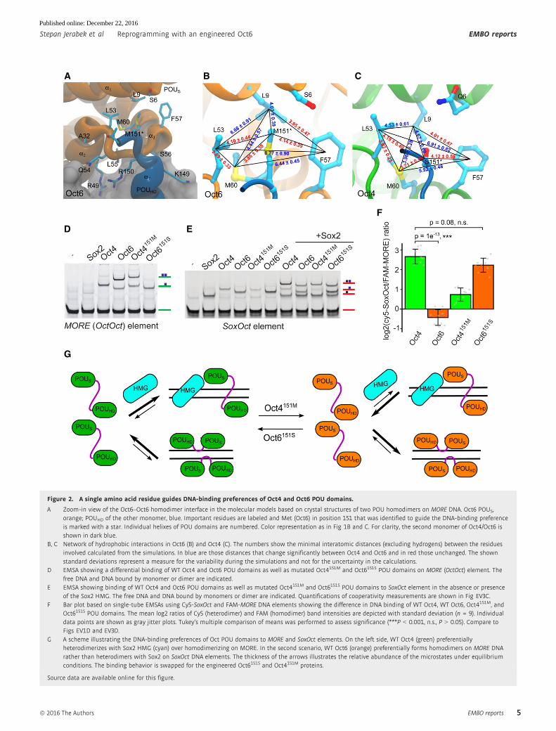
Rebalancing Oct homo- and heterodimerization with a singleamino acid swap
We next sought to identify structural determinants of the differential
homodimerization on MORE DNA using the crystal structures of
Oct1 and Oct6 [17,42] and structural model of Oct4 on MORE DNA.
Residue 151 mediates intermolecular contact between the POUHD
and the POUS [42]. This residue maps to the C-terminal part of the
DNA recognition helix 7 of the POUHD and encodes a Met in the POU
III group or other aliphatic residues (Val or Ile) in the POU I and POU
II groups, respectively. In contrast, Oct4 encodes a polar Ser at posi-
tion 151 (Figs 2A and EV3A). We inspected the chemical environ-
ment of residue 151 in structural models and during molecular
dynamics (MD) simulations. In Oct6 and Oct4S151M, Met151 docks in
a hydrophobic pocket of the POUS domain of the second monomer
(Fig 2A). MD simulations revealed a network of stable hydrophobic
interactions around Met151 in Oct6 as shown by the minimal inter-
atomic distances between the residues involved (Fig 2B). In Oct4,
the hydrophobic pocket is closed by a concerted motion leading to
the more pronounced kink in helix a4 (Fig 1G). Specifically, Leu9 (a1of the POUS) and Phe62 (a4 of the POUS) move closer to the center of
the pocket to compensate for the smaller, hydrophilic side chain of
Ser151 establishing a less optimal network of interactions (Fig 2C).
We therefore reasoned that residue 151 constitutes a key determinant
guiding homodimeric DNA recognition by POU TFs.
To test this hypothesis, we exchanged this amino acid to
construct mutated Oct4151M and Oct6151S proteins (Fig EV3B). To
examine dimerization of mutant POU factors on the MORE, we
performed EMSAs with the different variants (Fig 2D and E). Strik-
ingly, we found that Oct4151M gains the ability to homodimerize on
the MORE with a cooperativity similar to that of the WT Oct6 POU
domain (Fig EV3C). In contrast, the reciprocal Oct6151S mutation
reduced the cooperativity toward the level of WT Oct4. However,
binding to the SoxOct element is not substantially altered, as both
mutant proteins retain the ability to heterodimerize with Sox2
(Figs 2E and EV3C). Consistently, the ratio of heterodimers and
homodimers in single-tube EMSAs shifted toward the MORE for
Oct4151M and toward SoxOct for Oct6151S (Figs 2F and EV3D).
In summary, we identified a single amino acid residue in helix 7
of the POUHD of Oct factors determining the discrimination between
SoxOct and MORE DNA (Fig 2G).
Identification of Oct4 elements critical forpluripotency induction
We next asked whether modifications in MORE and SoxOct DNA
recognition have an impact on global transcriptional programs and,
ultimately, on cellular fate decisions. To address this, we systemati-
cally probed structural elements that might be responsible for the
uniqueness of Oct4 during somatic cell reprogramming. For this
analysis, four structural elements were selected as illustrated by the
structural models in Fig 3A and B. First, the amino acid influencing
homodimerization on the MORE was mutated to generate Oct4151M.
Second, we chose a double mutant in the first alpha helix of the
Oct4 POUS subdomain (Oct47D,22K) previously shown to be required
for maintaining pluripotency [43]. Third, a double mutation that
completely abolishes Oct4 interaction with Sox2 on canonical
SoxOct elements was generated [17] (Oct421Y,29R). Fourth, the POU
domain linker of Oct4 was replaced by that of Oct6 (Oct4LinkO6).
Altogether, we generated 10 Oct4-based constructs (Fig 3C). Except
for the SoxOct mutation designed to abolish the interaction between
Sox2 and Oct4, we modified the selected residues by replacing them
with their Oct6 counterparts. Of note, our structure-based linker
alignment differs from the one published previously [44]. The dif-
ference is caused by a low conservation of the N-terminal part of
the linker, and as a result of the new alignment, Oct6 has one addi-
tional positive charge in its “RK” region following the POU linker
(Fig EV3A, Appendix Fig S1A–C).
After assessment of viral titers by qRT–PCR (Appendix Fig S2A),
we infected mouse embryonic fibroblasts (OG2-MEFs) with the
combination of retroviruses carrying an Oct4 variant together with
three other mouse factors—Sox2, Klf4, and c-Myc. The assay was
evaluated 16 days after the infection. The plates for all combina-
tions of transcription factors were screened for the presence of Oct4-
GFP-positive colonies under fluorescence microscope (Appendix Fig
S2B), and the efficiency was plotted as the number of GFP-positive
colonies for each condition (Fig 3C). Oct4151M and Oct47D,22K gener-
ated GFP-positive colonies (Fig 3D, Appendix Fig S2B) with a
colony yield of about 60% compared to WT Oct4. When the Sox2–
Oct4 interface was completely disrupted (Oct421Y,29R), we observed
no GFP-positive colonies at all (Fig 3C and D, Appendix Fig S2B).
This confirms the importance of Sox2–Oct4 heterodimerization, in
agreement with recent work revealing the relevance of distinct
Sox2–Oct4 heterodimer configurations for induction and mainte-
nance of pluripotency [45]. Interestingly, the Oct4SoxOct/151M mutant
induced a small number of GFP-positive colonies, suggesting that
the 151M mutation rescued some of the detrimental effects intro-
duced by the Oct421Y,29R. When the Oct4 linker region was replaced
EMBO reports ª 2016 The Authors
EMBO reports Reprogramming with an engineered Oct6 Stepan Jerabek et al
4
Published online: December 22, 2016
A B C
D
G
E
F
Figure 2. A single amino acid residue guides DNA-binding preferences of Oct4 and Oct6 POU domains.
A Zoom-in view of the Oct6–Oct6 homodimer interface in the molecular models based on crystal structures of two POU homodimers on MORE DNA. Oct6 POUS,orange; POUHD of the other monomer, blue. Important residues are labeled and Met (Oct6) in position 151 that was identified to guide the DNA-binding preferenceis marked with a star. Individual helixes of POU domains are numbered. Color representation as in Fig 1B and C. For clarity, the second monomer of Oct4/Oct6 isshown in dark blue.
B, C Network of hydrophobic interactions in Oct6 (B) and Oct4 (C). The numbers show the minimal interatomic distances (excluding hydrogens) between the residuesinvolved calculated from the simulations. In blue are those distances that change significantly between Oct4 and Oct6 and in red those unchanged. The shownstandard deviations represent a measure for the variability during the simulations and not for the uncertainty in the calculations.
D EMSA showing a differential binding of WT Oct4 and Oct6 POU domains as well as mutated Oct4151M and Oct6151S POU domains on MORE (OctOct) element. Thefree DNA and DNA bound by monomer or dimer are indicated.
E EMSA showing binding of WT Oct4 and Oct6 POU domains as well as mutated Oct4151M and Oct6151S POU domains to SoxOct element in the absence or presenceof the Sox2 HMG. The free DNA and DNA bound by monomers or dimer are indicated. Quantifications of cooperativity measurements are shown in Fig EV3C.
F Bar plot based on single-tube EMSAs using Cy5-SoxOct and FAM-MORE DNA elements showing the difference in DNA binding of WT Oct4, WT Oct6, Oct4151M, andOct6151S POU domains. The mean log2 ratios of Cy5 (heterodimer) and FAM (homodimer) band intensities are depicted with standard deviation (n = 9). Individualdata points are shown as gray jitter plots. Tukey’s multiple comparison of means was performed to assess significance (***P < 0.001, n.s., P > 0.05). Compare toFigs EV1D and EV3D.
G A scheme illustrating the DNA-binding preferences of Oct POU domains to MORE and SoxOct elements. On the left side, WT Oct4 (green) preferentiallyheterodimerizes with Sox2 HMG (cyan) over homodimerizing on MORE. In the second scenario, WT Oct6 (orange) preferentially forms homodimers on MORE DNArather than heterodimers with Sox2 on SoxOct DNA elements. The thickness of the arrows illustrates the relative abundance of the microstates under equilibriumconditions. The binding behavior is swapped for the engineered Oct6151S and Oct4151M proteins.
Source data are available online for this figure.
ª 2016 The Authors EMBO reports
Stepan Jerabek et al Reprogramming with an engineered Oct6 EMBO reports
5
Published online: December 22, 2016

A
C
D
B
Figure 3. The residue guiding DNA-binding preference is a key Oct4 determinant in pluripotent cell generation.
A, B Molecular models show the position of mutated residues in the structures of two dimers on DNA: Sox2–Oct4 heterodimer (A) and Oct4–Oct4 homodimer (B). Ser inposition 151 (asterisk) was mutated to Met in order to shift the preference of Oct4 for homodimerization. The individual helixes of the POU domains are numbered.
C On the left side, a schematic overview of Oct4 and its mutants used for iPSC generation. POUS and POUHD are shown as green bars connected by the linker. Sitesmutated to the respective Oct6 residues and Oct6 linker are denoted in orange, and the mutation in the Sox–Oct interface is in black. The efficiency of theseconstructs for iPSC generation from MEFs is depicted as the absolute number of GFP-positive colonies on the right. Error bars represent standard deviations ofbiological replicates (n = 3), and differences between selected samples were compared using ANOVA (P-values: Oct4WTxOct47D,22K P = 6.9e-3; Oct4WTxOct4LinkO6
P = 7.3e-4; Oct4WTxOct4151M P = 1.2e-2; Oct4151MxOct4LinkO6,151M P = 1.1e-2; Oct4LinkO6,151M xOct47D,22K,LinkO6,151M P = 7.1e-3) (***P < 0.001, **P < 0.01, *P < 0.05).D GFP-positive colonies of mouse iPSCs generated by Oct4 mutants in combination with Sox2, Klf4, and c-Myc. Colonies were imaged 16 days after second viral
infection, using a fluorescence microscope. Scale bars: 250 lm; 10× objective.
Source data are available online for this figure.
EMBO reports ª 2016 The Authors
EMBO reports Reprogramming with an engineered Oct6 Stepan Jerabek et al
6
Published online: December 22, 2016
by its Oct6 counterpart (Oct4LinkO6), the iPSC generation efficiency
dropped to 30% of the WT level (Fig 3C). Additionally, we
performed reprogramming experiments using two different
Oct4LinkO6 constructs side by side; one construct was based on a
sequence alignment of the linkers and one on a structure-based
alignment (Appendix Fig S1C). This experiment showed that when
the entire Oct4 and Oct6 RK motifs are aligned with the central gap
as the structural alignment suggests, the Oct4LinkO6 devoid of one
positively charged “R” is able to induce pluripotency, albeit with
reduced efficiency (Appendix Fig S1C).
Overall, our reprogramming experiments provided evidence for
important roles of rationally selected Oct4 modifications for the
generation of iPSCs.
An engineered Oct6 is capable of induction and maintenanceof pluripotency
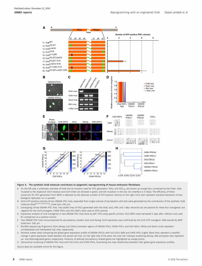
As WT Oct6 cannot induce pluripotency, we asked whether
elements critical for Oct4 function would enable the conversion of
Oct6 into a reprogramming factor (Fig 4A). We first adjusted the
concentration of viruses (Fig EV4A) and confirmed that Oct6 cannot
replace Oct4 in somatic cell reprogramming, as an O6SKM cocktail
did not produce GFP-positive colonies. Moreover, swapping of the
four structural elements one by one was insufficient to convert Oct6
into a reprogramming TF. However, when 151S was combined with
the 7K, 22T double mutant identified by Nishimoto et al to be criti-
cal for pluripotency maintenance [43], we consistently obtained
iPSC colonies (Figs 4A and EV4B). Colony yield further increased
when the Oct6 linker region was replaced with its Oct4 counterpart.
We therefore established two stable iPSC lines generated with this
engineered Oct6 (Oct67K,22T,LinkO4,151S; O6SKM iPSCs1 and O6SKM
iPSCs2) for further characterization (Fig 4B).
Next, we confirmed the absence of Oct4 transgene (Fig 4C) and
verified the efficient silencing of all four viral transgenes in O6SKM
cells (Fig 4D). Moreover, both O6SKM lines were karyotypically
normal (Fig EV4C). We then asked whether the O6SKM iPSCs
possess all hallmarks of pluripotency. First, we confirmed the
expression of both endogenous Sox2 and Nanog in O6SKM iPSCs by
immunochemistry (Fig 4E). Next, we determined the DNA methyla-
tion status of Oct4 and Nanog promoter regions by bisulfate
sequencing using the collagen type I alpha 1 (Col1a1) locus as a
control. CpG methylation present in MEFs was completely removed
from the Oct4 and Nanog regulatory regions in both O6SKM iPSC
lines like in mouse ESCs (Fig 4F). Finally, we compared the global
gene expression profiles of O6SKM iPSCs to O4SK iPSCs, bona fide
OG2-ESCs, as well as previously published O4SKM cells and MEFs
(Figs 4G and EV4D). Overall, all pairwise scatter plots as well as the
hierarchical clustering demonstrated that the global gene expression
profile is highly similar to standard O4SK iPSCs and ESCs, but
substantially different from the profile of MEFs (Figs 4G and H, and
EV4D).
In order to assess the differentiation potential of O6SKM iPSCs,
we performed two assays. In one assay, we generated embryoid
bodies (EBs) via the hanging-drop method. After immunostaining of
the spontaneously differentiated EBs, we verified the expression of
markers typical for all three germ layers (Fig 5A). In the other
assay, O6SKM iPSCs were injected subcutaneously into severe
combined immunodeficiency (SCID) mice to see whether they had
the potential to form teratomas. Indeed, cells from both O6SKM
iPSC lines gave rise to teratomas and cells of all three germ layers
could be detected (Fig EV5A). Finally, the developmental potential
was evaluated by the aggregation assay. For this, we aggregated the
O6SKM iPSCs with diploid mouse embryos. Once the aggregates
developed into blastocysts, they were transferred into recipient
mice. The ability of O6SKM iPSCs to contribute to the germline was
demonstrated after observation of Oct4-GFP signal in the gonads of
E13.5 fetuses and E19.5 pups (Fig EV5B and C). We genotyped the
tails of chimeras (Fig EV5D) and confirmed the presence of the Oct6
transgene (Fig EV5E). To test for germline transmission, we mated
the chimeric mice with foster mothers. In the newborn pups
(Fig 5B), we observed GFP signal in the ovaries and testes (Fig 5C).
We subsequently also tested for the presence of Oct6 transgenes by
PCR and found that the viral transgene could be readily amplified
(Fig EV5F).
Last, we asked whether Oct67K,22T,LinkO4,151S can maintain
pluripotency using a complementation assay and the transgenic
ZHBTc4 ES cell line [43]. In this assay, endogenous Oct4 is
suppressed upon doxycycline addition and pluripotency can only be
rescued if additional exogenous factors are provided. Exogenous
WT Oct4 is able to rescue pluripotency, while WT Oct6 cannot [43].
It was found that even a low level of Oct67K,22T,LinkO4,151S can rescue
pluripotency and sustain the expression of pluripotency genes
(Fig EV5G and H). ESCs rescued with Oct67K,22T,LinkO4,151S exhibit
the typical colony morphology and self-renew, albeit with reduced
proliferation rate when compared to WT Oct4-rescued cells.
In summary, our engineered Oct6 produces iPSCs passing all
in vitro and in vivo assays validating their pluripotency and is also
able to maintain the pluripotency of ESCs.
Discussion
Oct4 has unique DNA-binding preferences
Oct4 is a master regulator of pluripotency, and it is the only factor
that cannot be substituted by any paralogous family member during
iPSC generation [2]. What then are the unique molecular features of
this POU factor endowing it with this capacity? Previous studies
have provided evidence that the DNA-binding POU domain should
be at the center of our search. For example, constructs lacking either
C- or N-terminal TAD still maintained a stem cell phenotype and
yielded high numbers of ESC colonies in the Oct4 complementation
assay [46]. Moreover, an unbiased Ala scan along the Oct4 molecule
and subsequent reprogramming indicated that the POU domain—
but not the N- and C-terminal TADs—is the most critical part of the
protein for iPSC generation [47]. Furthermore, the N- and C-termini
of Oct4 could be replaced with the TAD of the Yes-associated
protein (YAP) without loss of reprogramming activity [47].
However, introduction of only two mutations into helix 1 of the
POUS led to a dramatic decrease in ability of Oct4 to maintain
pluripotency in vitro [43].
After re-analyzing a panel of ChIP-Seq data for the POU domain
protein, we noticed that Oct4 binds the SoxOct motif, whereas
somatic POUs preferentially form homodimers on palindromic bind-
ing sites (Figs 1A and EV1A). Neural POU proteins have previously
been reported to form highly cooperative homodimers in vitro [48].
ª 2016 The Authors EMBO reports
Stepan Jerabek et al Reprogramming with an engineered Oct6 EMBO reports
7
Published online: December 22, 2016
A
B
E
G H
F
C D
Figure 4. The synthetic Oct6 molecule contributes to epigenetic reprogramming of mouse embryonic fibroblasts.
A On the left side, a schematic overview of Oct6 and its mutants used for iPSC generation. POUS and POUHD are shown as orange bars connected by the linker. Sitesmutated to the respective Oct4 residues and Oct4 linker are denoted in green, and the mutation in the Sox–Oct interface is in black. The efficiency of theseconstructs for iPSC generation from MEFs is depicted as the absolute number of GFP-positive colonies on the right. Error bars represent standard deviations of twobiological replicates run in parallel.
B Oct4-GFP-positive colonies of two O6SKM iPSC lines, expanded from single colonies of pluripotent cells that were generated by the contribution of the synthetic Oct6molecule (Oct67K,22T,LinkO4,151S). Scale bars: 250 lm.
C Genotyping of two O6SKM iPSC lines. Two stable lines of iPSCs generated with the Oct6, Sox2, Klf4, and c-Myc retroviral set are positive for these four transgenes, butnegative for the Oct4 transgene. OSKM iPSCs and OG2-MEFs were used as PCR controls.
D Expression analysis of viral transgenes in two O6SKM iPSC lines done by qRT–PCR using specific primers. OG2-MEFs were harvested 5 days after infection and usedfor comparison as a positive control.
E Two O6SKM iPSC lines immunostained for pluripotency markers Sox2 and Nanog. Oct4 expression was confirmed by the Oct4-GFP transgene. DNA stained by DAPI.Scale bars: 100 lm.
F Bisulfite sequencing of genomic Oct4, Nanog, and Col1a1 promoter regions of O6SKM iPSCs, OSKM iPSCs, and OG2-MEFs. White and black circles representunmethylated and methylated CpG sites, respectively.
G Pairwise scatter plots comparing the global gene expression profile of O6SKM iPSCs1 with OG2 ESCs (left) and O4SK iPSCs (right). Black lines represent a twofoldchange in gene expression levels between the paired cell lines. On the right side of the plots, the color bar indicates scattering density. Red and green dots representup- and downregulated genes, respectively. Positions of selected pluripotency-related genes are highlighted as orange points.
H Hierarchical clustering of O6SKM iPSC lines with OG2 ESCs and O4SK iPSCs, illustrating the close relationship between their global gene expression profiles.
Source data are available online for this figure.
EMBO reports ª 2016 The Authors
EMBO reports Reprogramming with an engineered Oct6 Stepan Jerabek et al
8
Published online: December 22, 2016

A recent study has shown that class III POU TFs preferentially target
the MORE sequence in NPCs [49]. The authors used full-length
proteins fused to large fluorescent tags to verify that Oct6 preferen-
tially forms homodimers, whereas Sox2 preferentially heterodimer-
izes with Oct4 on SoxOct elements. However, diminished binding of
Oct4 to the MORE DNA element per se was not reported. We
extended this work by using untagged DNA-binding domains and
quantitative cooperativity measurements of a panel of five POU
proteins. Our results show that Pit1, Oct1, Oct6, Brn2, and Oct4 all
show positive cooperativity on the canonical SoxOct motif (Fig 1F).
In contrast, the binding mode on the MORE DNA element is mark-
edly different, as the cooperativity and the residence time of Oct4
are profoundly reduced (Figs 1E and EV2). We therefore surmise
that the reduced cooperativity on the MORE DNA element contri-
butes to the unique mechanism setting Oct4 apart from other POU
factors (Fig EV1C and D).
Modulating Oct4 and Oct6 DNA recognition and its relevance forthe interaction with cofactors
In our search for the structural basis of binding differences in the
MORE DNA element, we identified candidate elements in the crystal
structures of Oct1 and Oct6 TFs bound to this DNA motif [17,42].
By exchanging a single residue between Oct4 and Oct6, we swapped
their DNA-binding preferences. Our in vitro experiments showed
that the Oct6151S mutant is re-distributed from MORE to SoxOct
elements, while binding of the mutated Oct4151M shifts toward
homodimers on the MORE DNA element (Fig 2D and E). Moreover,
after changing the binding properties of the Oct4 mutant, we
observed a drop in its capability for iPSC generation to about 65%
of the WT protein. Conversely, Oct6 could only generate iPSCs if the
ability to homodimerize on the MORE was decreased with a recipro-
cal mutation (Figs 4A and EV4B). Therefore, rebalancing the forma-
tion of heterodimeric and homodimeric complexes substantially
alters the ability of POU factors to direct cell fate decisions.
A previous study has shown that the class I POU factor Pit1
can either activate the growth hormone gene or cause its effective
suppression in a cell type-dependent manner [50]. This switch of a
biological outcome is triggered by a two-base pair difference in the
promoter DNA. Apparently, allosteric changes to the conformation
of the POU domain induced by the DNA sequence influence the
factor’s interaction with a specific repressor complex [50]. Simi-
larly, another seminal study demonstrated that the B-cell-specific
coactivator OBF1 interacts with Oct factor dimers on PORE DNA.
In contrast, Oct dimers bound to the MORE DNA element do not
recruit OBF1 [16]. We thus surmise that the differential association
of Oct6 and Oct4 with SoxOct versus MORE DNA element not only
affects which genomic loci are targeted, but also which set of
cofactors is recruited and how the expression of nearby genes is
regulated. Testing this hypothesis requires the examination of the
interactome of Oct4 and Oct6 in the context of specific enhancer
signatures.
Multiple functions of the POU linker region
One of surprising observations in our study was that a chimeric
protein in which the Oct4 linker is substituted by the Oct6 linker
(Oct4LinkO6) still retains 30% of the iPSCs inducing efficiency
(Fig 3C and D). This result contrasts former observations reporting
no iPSC colonies for Oct4LinkO6 constructs [43,44]. This discrepancy
can be explained by a structural alignment of the linker sequences,
in which the entire RK motifs of Oct4 and Oct6 are aligned with a
central gap causing an Arg residue to be part of the swapped linker
in one but not the other construct (Appendix Fig S1A). Recently, an
interplay between the linker segment and the RK motif following
the linker has been described and shown to affect not only DNA
binding but also transactivation potential and reprogramming effi-
ciency [13]. However, whether the two different POU linkers inter-
act with distinctive epigenetic modifiers remains unknown.
Cooperativity between exogenous Oct4 and Sox2 during theinitiation of reprogramming?
Mutations in the Sox–Oct interface of Oct4 (Oct47D,22K) led to
considerable drop in reprogramming efficiency, demonstrating that
uncoupling of Oct4 from Sox2 (Oct421Y,29R) has a profound
A B C
Figure 5. Cells reprogrammed using the synthetic Oct6 molecule show pluripotency in vitro and in vivo.
A In vitro differentiation of O6SKM iPSC lines into cells of all three germ layers as shown by immunochemistry: endoderm (a-fetoprotein, AFP), mesoderm (a-smoothmuscle actin, SMA), and ectoderm (b-tubulin, TUJ1). Nuclei (DNA) were stained by Hoechst (blue). Scale bars: 200 lm.
B F1 offspring of chimera male mice with contribution from O6SKM iPSCs and CD1 female mice.C Pictures demonstrating germline transmission; endogenous Oct4-GFP signal was detected in the gonads of F1 pups. Scale bars: 200 lm.
ª 2016 The Authors EMBO reports
Stepan Jerabek et al Reprogramming with an engineered Oct6 EMBO reports
9
Published online: December 22, 2016
detrimental effect on iPSC generation (Fig 3C and D). A study using
single-molecule imaging recently showed a sequential engagement
of TFs to their target sites in ESCs. The authors showed that Sox2
binds to DNA first and subsequently recruits Oct4 to assemble the
Sox–Oct heterodimer [51]. Possibly, this recruitment process is
impaired for the Oct421Y,29R protein. Moreover, Oct4 seems to
possess an intriguing ability to bind a closed chromatin as a pioneer
factor and to initiate chromatin opening [52]. Our results strongly
suggest that cooperation between Oct4 and Sox2—and not Oct4
alone—is involved in not only maintaining but also establishing
pluripotency. Whether and how this cooperation affects opening of
the chromatin remain elusive.
Outlook
Synthetic biology is emerging as a provider of powerful tools for
cellular reprogramming and stem cell biology with a focus on the
CRISPR-Cas-based genome engineering. In addition, new opportuni-
ties arise to design artificial TFs with enhanced biological properties
(reviewed in [53]). Our identification of critical elements underlying
POU function and determining the unique properties of Oct4 can
guide the generation of synthetic TFs that direct cellular identities as
an alternative to CRISPR-Cas-based TF design.
Materials and Methods
ChIP-Seq analysis
In order to compare the occurrence of OctOct (MORE) versus SoxOct
motifs bound by Oct2, Brn2, or Oct4 in different cell types, we down-
loaded ChIP-Seq data from the GEO database. Oct2 peaks in B cells
(GSE21512) [39], Brn2 in MEFs after 48 h of reprogramming
(GSE43916) [54], Brn2 in NPCs (GSE43916) [54], (GSE35496) [55],
and Oct4 in ESCs (GSE11724) [56]. ChIP-Seq reads were aligned to
the mouse genome (mm10 assembly) using bowtie2 [57]. TF binding
peaks were called using MACS [58] with the respective input data as
control. As the SRA files from Oct2 ChIP-Seq in B-cell study were not
available, we used peak coordinates provided by the authors and
used UCSC batch coordinate conversion (liftOver) function to
convert mm8 coordinates to mm10. Motif analysis of Oct2, Brn2, and
Oct4 binding at either the MORE or SoxOct motif was done using
HOMER findMotifsGenome.pl [39]. Motif occurrences were calcu-
lated within 200-bp windows centered on ChIP-Seq summits by using
FindMotifsGenome.pl with options –find and –len 12,14,16 using
PWMs discovered from de novo motif analysis. Motif counting was
also performed using word search in R (https://www.r-project.org/)
where genomic locations were converted to genomicRanges objects
and sequences were retrieved from mm10 genome version using the
BSgenome.Mmusculus.UCSC.mm10 object and the getSeq function
(Biostrings). SoxOct and MORE motif searches were performed using
IUPAC strings (SoxOct = CWTTSTHATGCAAAT and
MORE = ATGMATATKCAT) allowing for one mismatch per 6 bp.
EMSA
EMSAs were performed essentially as described [27] using 100 nM
of 50Cy5-labeled SoxOct or MORE dsDNA mixed with 100–500 nM of
Oct POUs and 20–100 nM of Sox2 HMG (79-aa protein). Reaction
mixtures were incubated for 1 hr on ice in the dark. For single-tube
EMSAs, FAM-labeled MORE DNA was used. Protein–DNA
complexes were separated on native pre-run 1× Tris–glycine native
gels at 200 V for 15–30 min. Gel images were taken using Typhoon
9140 PhosphorImager (GE Healthcare) with 500–700 PMT voltage at
50-lm pixel-size resolution with platen focal plane. The relative
abundance of each possible microstate was quantified using Image-
Quant TL software (Amersham Biosciences) with rubber band
option as background correction with a box size of 80 per lane. At
least three replicates were performed to calculate omega values. To
ensure omega values are estimated under equilibrium conditions,
only lanes where all microstates (dimers, monomers, and free DNA)
were clearly detectable were used for the calculations. Cooperativity
factors (x) were calculated after quantifying the fraction of bound
DNA in EMSAs using the below equations. Equations were derived
using Boltzmann weights and principles of statistical mechanisms
as detailed in [27,40].
Heterodimer cooperativity:
x ¼ Kd1
Kd21¼ Kd2
Kd12¼ f0f3
f1f2;
where Kd1 and Kd2 are the equilibrium binding constants for the
proteins 1 and 2 to the composite DNA element alone and Kd21
and Kd12 the equilibrium binding constants of binding of proteins 1
and 2 to the DNA element in the presence of the respective other
protein. f denotes the bound fraction of the DNA as dimer (f3),
monomer 1 (f1), monomer 2 (f2), and the free DNA (f0).
Homodimer cooperativity:
x ¼ Kd1
Kd11¼ 4f0f2
f 21;
where Kd1 and Kd11 are equilibrium binding constants for the bind-
ing of protein 1 to a DNA element with two of its binding elements
as monomer or dimer, respectively. f denotes the fraction of bound
DNA for the homodimeric state (f2), the dimeric state (f1), and the
free DNA (f0).
Sequences of EMSA oligonucleotides are in Appendix Table S1.
EMSAs to monitor transcription factor–DNA dissociation
The buffer system, proteins, and DNA elements are identical to the
EMSAs performed under equilibrium conditions. The lid of the mini
gel chamber (Bio-Rad) was removed to allow for sample loading
without powering off the gel. The binding reaction of the POU
domains and labeled reporter DNA was prepared in a 220 ll reactionvolume and incubated for 1 h for the reaction to reach equilibrium.
Subsequently, unlabeled competitor DNA was added from a 100 lMstock and 20 ll aliquots were removed from the reaction mix after
different time intervals and loaded onto running gels. 10% native
PAGE gels were used for the experiment and run at 100 V for 1.5 h.
Gels were imaged using a FLA-7000 image reader (FUJIFILM).
Structural modeling of DNA-bound homodimers
We built 1,250 homology models for Oct4 and Oct6 homodimers
using MODELLER 9.17 (https://salilab.org/modeller/) and the
EMBO reports ª 2016 The Authors
EMBO reports Reprogramming with an engineered Oct6 Stepan Jerabek et al
10
Published online: December 22, 2016
following templates (referred by PDB ID): (i) 2xsd—crystal structure
of Oct6 homodimer bound to MORE [42]; (ii) 1e3o—crystal struc-
ture of Oct1 homodimer bound to MORE [17]; (iii) 3l1p—crystal
structure of Oct4 homodimer bound to a palindromic site different
than MORE [44]; (iv) 1gt0—crystal structure of the Oct1–Sox2–DNA
ternary complex [21]. We imposed symmetry restraints on Ca, Cb,and Cc atoms to enforce a similar structure of the two monomers.
We transferred the DNA as a rigid body (using special restraints)
from the 1e3o structure. We used a “slow” optimization protocol
followed by a “slow” molecular dynamics-based refinement proto-
col. The linker region (residues 76–96) was modeled using “loop-
model” without imposing any restraints on its structure. The best
three models of each homodimer were selected based on a normal-
ized energy score. For the Oct4 models, the scores were �0.8621,
�0.8473, and �0.8433, whereas for Oct6, the scores were �0.9331,
�0.9229, and �0.9125 (the score ranging from +2 to �2). Then, we
extended DNA at each end with 5 base pairs to avoid potential simu-
lation artifacts due to a too short DNA length in the crystal structure
and adapted the sequence of DNA in CHIMERA to obtain the final
sequence in the models: 50-CACAGTGCTCATGCATATGCATGAGCCTGGGA-30 (MORE motif highlighted).
MD simulations
Ionizable residues were assigned their standard protonation state at
neutral pH. The 50 and 30 ends of the DNA and the N-termini of the
proteins were methylated, whereas the C-termini of the proteins were
acetylated to avoid potential truncation artifacts. Then, the systems
were (i) solvated in a truncated octahedral box of SPCE water extend-
ing at least 12 A from any protein/DNA atom, (ii) neutralized with
46 Na+ ions and additional 100 mM KCl (135 ions) using the Merz
ions [59] to mimic the experimental ionic strength. We used the
Amber-ff14SB [60] and the Amber-parmbsc1 [61] force fields for
proteins and DNA, respectively. First, the energy of the systems was
minimized in 11 steps as described in Appendix Table S2. Then, the
systems were equilibrated for 10.3 ns in 15 steps as described in
Appendix Table S3. Finally, each system was simulated in NPT
ensemble for 200 ns with a timestep of 2 fs resulting in ensembles of
600 ns (from the three selected models) for each Oct4 and Oct6
homodimers bound to MORE DNA. The temperature was maintained
at 300 K with Langevin dynamics with a damping coefficient of 0.1/
ps. The pressure was maintained at 1 atm with the Nose Hoover
Langevin piston method with the period and decay of 1.2 and 1.0 ps,
respectively. The direct calculation of the non-bonded interactions
was truncated at 10 A. Long-range electrostatics were calculated
using the particle mesh Ewald algorithm [62]. A correction to the
energy and the pressure was also applied to account for the truncated
long-range Lenard-Jones interactions [63]. The length of the atomic
bonds involving hydrogen atoms was constrained with the SHAKE
[64] and the SETTLE [65] algorithms for the macromolecule and
water, respectively. All simulations were performed in NAMD 2.11
[66]. Snapshots were selected for analysis every 4 ps.
Site-directed mutagenesis
Mutations into full-length CDS of the mouse Oct4 and Oct6 proteins
(the Consensus CDS Database IDs are 37600.1 and 57296.1, respec-
tively) were made in the pMX cloning vector using the QuikChange
Site-Directed Mutagenesis Kit according to the manufacturer’s proto-
col (Agilent Technologies). Specific oligos used for each modifi-
cation of DNA sequence are listed in Appendix Table S4. Mutant
plasmids were selected after DNA sequencing.
Cell culture
HEK293T cells, mouse embryonic fibroblasts (MEFs), and OG2
MEFs were cultured in low-glucose (1,000 mg/l) Dulbecco’s modi-
fied Eagle’s medium (DMEM, Sigma-Aldrich), supplemented with
10% fetal bovine serum (FBS, Biochrom), 2 mM L-glutamine and 1×
penicillin/streptomycin (Sigma-Aldrich), 1% non-essential amino
acids (Sigma-Aldrich), and 0.10 mM b-mercaptoethanol. OG2-MEFs
were isolated as described previously [1]. Mouse embryonic stem
cells (mESCs) and iPSCs were maintained in high-glucose
(4,500 mg/l) DMEM (Sigma-Aldrich), supplemented with 10% fetal
bovine serum (Biochrom), 5% serum replacement (Gibco), 2 mM
L-glutamine, 1× penicillin/streptomycin (Sigma-Aldrich), 1% non-
essential amino acids (Sigma-Aldrich), 1% sodium pyruvate (Sigma-
Aldrich), 0.10 mM b-mercaptoethanol (Gibco) with 1,000 units of
leukemia inhibitory factor (LIF; prepared in house) on a feeder layer
of gamma-irradiated MEFs; experiments under feeder-free condi-
tions were performed using mESC medium and 2,000 units of LIF,
as previously described [67].
Virus production, induction of pluripotent stem cells
Prior to transfection, HEK293T cells were seeded on 100-mm dishes.
The following day, the pMX retroviral vectors containing wild-type
Oct4, Oct6, Sox2, Klf4, and c-Myc as well as mutated cDNAs for mouse
Oct4 or Oct6 were co-transfected with packaging helper plasmid pCL-
Eco into 2 × 10⁶ HEK293T cells using the Fugene6 transfection reagent
(Roche). The medium was changed on the next day. 48 hours after the
infection, virus-containing supernatants were collected, filtered
(Millex-HV 0.45 lm; Millipore), supplemented with 6 lg/ml prota-
mine sulfate (Sigma-Aldrich), and used directly for infection. OG2-
MEFs were plated on six-well plates at a density of 2.5 × 10⁴ cells per
well, grown for 24 h, and transduced twice in 24-h intervals with equal
amount of each virus-containing supernatant. The medium was
changed to mESC medium 1 day after the second infection. ESC
medium was changed every second day. Reprogramming experiment
was repeated three times, and the GFP colonies were counted 16 days
after the second infection under a fluorescent microscope.
Oct4 complementation assay
The ZHBTc4 embryonic stem cells were infected with WT Oct4, WT
Oct6, and Oct67K,22T,LinkO4,151S using lentiviral supernatants follow-
ing procedures established by Niwa et al [68]. 24 hours after the
infection, doxycycline was added to the ESC medium (final concen-
tration: 1 lg/1 ml), in order to suppress the endogenous Oct4, as
described in [68]. The self-renewing rescued ES colonies were main-
tained in culture for further analysis by qRT–PCR.
qRT–PCR and microarray
RNA samples to be analyzed by qRT–PCR and microarrays were
prepared using RNeasy Mini Kit (QIAGEN) with on-column DNA
ª 2016 The Authors EMBO reports
Stepan Jerabek et al Reprogramming with an engineered Oct6 EMBO reports
11
Published online: December 22, 2016

digestion. Complementary DNA for qRT–PCR was synthesized with
the M-MLV Reverse Transcriptase (Affymetrix). Transcript levels
were determined using ABI PRISM Sequence Detection System 7900,
and gene expression was normalized to the housekeeping gene
Gapdh. Specific qRT–PCR primers are listed in Appendix Table S5.
For microarray analysis, 300 ng of total RNA per sample was
used as input using a linear amplification protocol (Ambion), which
involved synthesis of T7-linked double-stranded cDNA and 12 h of
in vitro transcription incorporating biotin-labeled nucleotides. Puri-
fied and labeled cDNA was then hybridized for 18 h onto MouseRef-
8 v2 expression BeadChips (Illumina) following the manufacturer’s
instructions. After washing, chips were stained with streptavidin-
Cy3 (GE Healthcare) and scanned using the iScan reader (Illumina).
At least two independent iPSC lines were analyzed.
The data discussed in this publication have been deposited in
NCBI’s Gene Expression Omnibus [69] and are accessible through
GEO Series accession number GSE81908 (https://www.ncbi.nlm.
nih.gov/geo/query/acc.cgi?acc=GSE81908).
Genotyping
Genomic DNA was purified using QIAamp DNA Mini Kit (QIAGEN)
and amplified using the GoTaq DNA Polymerase (Promega), and
specific PCR primers are listed in Appendix Table S5. PCR products
were analyzed using 2% agarose gels.
In vitro differentiation of mouse iPSCs
Embryoid bodies (EBs) were generated from mouse iPSCs via the
hanging-drop method, with 1 × 10³ cells in a 30 ll drop of mouse ESC
medium without LIF. EBs were gently collected after 5 days and plated
on gelatinized 6-well plates at a density of 20 EBs per well. EBs were
cultured for 3 weeks and LIF-negative ESC medium was changed every
3 days. After spontaneous differentiation, structures of all three germ
layers were observed, and immunostaining was performed.
Teratoma formation
About 5 × 10⁶ iPSCs were injected subcutaneously into the flanks of
severe combined immunodeficiency (SCID) mice. After 4–5 weeks,
mice were sacrificed and the teratoma that had formed was excised,
fixed in 4% paraformaldehyde, stained with hematoxylin and eosin,
and subjected to histological examination.
Immunochemistry
The cells were fixed by incubation in 4% (v/v) paraformaldehyde in
phosphate-buffered saline (PBS) for 20 min at room temperature
(RT) and then rinsed three times with PBS. Cells were permeabilized
by incubation in 0.1% Triton X-100 in PBS for 15 min at RT and
then washed three times with PBS. Cells were blocked in 5% BSA
and 1% goat serum in PBS for 1 h at RT. Primary antibodies—goat
polyclonal anti-Sox2 (Y17; Santa Cruz; 1:400), rat monoclonal anti-
Nanog (eBioscience; 1:1,000), mouse monoclonal anti-smooth
muscle actin (SMA) (Sigma-Aldrich; 1:500), goat polyclonal anti-a-fetoprotein (AFP) (C-19; Santa Cruz; 1:400), mouse monoclonal
anti-b-tubulin III (Sigma-Aldrich; 1:800)—were diluted in 1% BSA
in PBS and applied at 4°C for overnight. The cells were washed with
PBS three times for 5 min. Alexa Fluor 568, 647, fluorophore-conju-
gated secondary antibodies (Invitrogen) were diluted 1:1,000 in 1%
BSA/PBS. Secondary antibodies were applied for 2 h at RT in the
dark and cells were subsequently washed with PBS three times
for 5 min. Finally, cells were incubated for 10 min in 300 ng/ml
DAPI/PBS and rinsed thrice with PBS for 1 min. Samples were later
visualized using a fluorescent microscope.
Karyotyping of mouse iPSCs
Mouse iPSCs were cultured in a 3-cm dish and the medium was
changed 12 h before starting the protocol. To obtain single cells,
cells were trypsinized and kept on a gelatinized dish for 40 min to
quickly remove attached MEFs. Any remaining big clumps of MEFs
were removed by using a 40-lm filter. Cells were transferred to a
15-ml tube and incubated in 2 ml DMEM with 0.5 lg/ml nocoda-
zole for 2 h. Next, cells were centrifuged at 1 × 10³ rpm for
4 min, and the cell pellet was resuspended in 100 ll medium.
1 ml of prewarmed 0.56% KCl was slowly added drop by drop to
a total volume of 3 ml. The cell suspension was incubated for
12 min at 37°C in a water bath, and cells were pelleted by
centrifugation (1 × 10³ rpm, 5 min) and resuspended in 100 ll0.56% KCl. After 1 min, a fresh fixative (methanol/acetic acid at a
ratio of 3:1) was slowly added, starting from 5 ll up to 3 ml. The
cells were incubated at RT for 30 min, washed three times with
2 ml of fixative, and resuspended in 50–300 ll of the fixative.
Large cell clusters were allowed to disintegrate for 1 min and one
drop was placed on a dry cover slide and left for at least 30 min
to air-dry. Samples were stained with DAPI and examined for
metaphase plates.
Bisulfite sequencing
To determine DNA methylation status at regulatory regions of Oct4,
Nanog, and Col1a1, bisulfite conversion was carried out on 2 lg of
isolated genomic DNA from cells with the EZ DNA methylation kit
(Zymo research) according to the manufacturer’s protocol. The
bisulfite-converted DNA was amplified by PCR with previously
described primers [70,71]. The PCR products were cloned into the
pCRII TOPO vector (Invitrogen), and plasmids extracted from indi-
vidual clones were sequenced by GATC-biotech (http://www.gatc-
biotech.com/en/index.html). Sequences were analyzed using the
Quantification Tool for Methylation Analysis (QUMA; http://
quma.cdb.riken.jp).
Chimera generation and germline transmission
All (C57BL/6 × C3H) F1 female mice for embryo collection were
treated with 7.5 IU pregnant mare’s serum gonadotropin (PMSG)
and 7.5 IU human chorionic gonadotropin (HCG) in 48 h apart, and
then crossed with CD1 male mice. Eight-cell embryos were flushed
from female mice at E2.5 and placed in M2 medium [70]. Trypsin-
digested iPSCs (8–12 cells and one embryo per aggregate) were
transferred into a depression in the microdrop of KSOM. Meanwhile,
batches of 30–50 embryos were briefly incubated in acidified
Tyrode’s solution [70] until dissolution of their zona pellucida. A
single 2n embryo was placed in each depression. All aggregates
were cultured overnight at 37°C and 5% CO2. After 24 h of culture,
EMBO reports ª 2016 The Authors
EMBO reports Reprogramming with an engineered Oct6 Stepan Jerabek et al
12
Published online: December 22, 2016
the majority of aggregates had formed blastocysts. 10–14 embryos
were transferred into one uterine horn of an E2.5 pseudopregnant
recipient. 11 days after embryo transfer, E13.5 fetuses were
collected and their gonads were checked for the presence of
Oct4-GFP. 34 full-term chimera pups were delivered by natural birth
or Cesarean section at E19.5. Of all 34 pups, seven pups died at birth
and seven female pups were sacrificed. Thus, 20 male pups were set
up with foster mothers to check for germline transmission after
sexual maturation.
Data availability
Primary data
Stepan Jerabek, Calista K. L. Ng, Guangming Wu, Marcos J. Arauzo-
Bravo, Kee-Pyo Kim, Daniel Esch, Vikas Malik, Yanpu Chen, Sergiy
Velychko, Caitlin M. MacCarthy, Xiaoxiao Yang, Vlad Cojocaru,
Hans R. Scholer and Ralf Jauch (2016) Changing POU dimerization
preferences converts Oct6 into a pluripotency inducer. Gene Expres-
sion Omnibus GSE81908.
Referenced data
Heinz S, Benner C, Spann N, Bertolino E, Lin YC, Laslo P, Cheng JX,
Murre C, Singh H, Glass CK (2010) Simple combinations of lineage-
determining transcription factors prime cis-regulatory elements
required for macrophage and B-cell identities. Gene Expression
Omnibus GSE21512.
Wapinski OL, Vierbuchen T, Qu K, Lee QY, Chanda S, Fuentes
DR, Giresi PG, Ng YH, Marro S, Neff NF, Drechsel D, Martynoga B,
Castro DS, Webb AE, Sudhof TC, Brunet A, Guillemot F, Chang HY,
Wernig M (2013) Hierarchical mechanisms for direct reprogramming
of fibroblasts to neurons. Gene Expression Omnibus GSE43916.
Lodato MA, Ng CW, Wamstad JA, Cheng AW, Thai KK, Fraenkel
E, Jaenisch R, Boyer LA (2013) SOX2 co-occupies distal enhancer
elements with distinct POU factors in ESCs and NPCs to specify cell
state. Gene Expression Omnibus GSE35496.
Marson A, Levine SS, Cole MF, Frampton GM, Brambrink T,
Johnstone S, Guenther MG, Johnston WK, Wernig M, Newman J,
Calabrese JM, Dennis LM, Volkert TL, Gupta S, Love J, Hannett N,
Sharp PA, Bartel DP, Jaenisch R, Young RA (2008) Connecting
microRNA genes to the core transcriptional regulatory circuitry of
embryonic stem cells. Gene Expression Omnibus GSE11724.
Remenyi A, Tomilin A, Pohl E, Lins K, Philippsen A, Reinbold R,
Scholer HR, Wilmanns M (2001) Differential dimer activities of the
transcription factor Oct-1 by DNA-induced interface swapping.
Protein Data Bank 1E3O.
Remenyi A, Lins K, Nissen LJ, Reinbold R, Scholer HR,
Wilmanns M (2003) Crystal structure of a POU/HMG/DNA ternary
complex suggests differential assembly of Oct4 and Sox2 on two
enhancers. Protein Data Bank 1GT0.
Jauch R, Choo SH, Ng CKL, Kolatkar PR (2011) Crystal structure
of the dimeric Oct6 (Pou3f1) POU domain bound to palindromic
MORE DNA. Protein Data Bank 2XSD.
Esch D, Vahokoski J, Groves MR, Pogenberg V, Cojocaru V, Vom
Bruch H, Han D, Drexler HC, Arauzo-Bravo MJ, Ng CK, Jauch R,
Wilmanns M, Scholer HR (2013) A unique Oct4 interface is crucial
for reprogramming to pluripotency. Protein Data Bank 3L1P.
Expanded View for this article is available online.
AcknowledgementsR.J. is supported by a 2013 MOST China-EU Science and Technology Coopera-
tion Program (Grant No. 2013DFE33080), MOST grant 2016YFA0100700, the
National Natural Science Foundation of China (Grant No. 31471238), a 100
talent award of the Chinese Academy of Sciences, and a Science and Technol-
ogy Planning Project of Guangdong Province, China (2014B030301058,
2016A050503038). V.C. is supported with funding and computer resources by
the Max Planck Society and the German Research Foundation (Grant Number
CO 975/1-1). S.J. thanks the Cells in Motion (CiM) Graduate School and the
International Max Planck Research School—Molecular Biomedicine (IMPRS-
MBM) Joint Graduate Program for support. V.M. is supported by a CAS-TWAS
President’s Fellowship of the University of the Chinese Academy of Sciences
(UCAS) and The World Academy of Science (TWAS).
Author contributionsSJ and CKLN designed and performed the experiments and analyzed the data.
GW performed in vivo assays. MJA-B analyzed the microarray data. K-PK
performed the DNA methylation analysis. DE helped with experimental design,
cloning, and reprogramming. VM and RJ performed the ChIP-Seq data analysis.
YC performed the kinetic EMSAs. CMM helped with preparation of material for
the kinetic EMSAs. SV contributed to the reprogramming data set. XY validated
reprogramming experiments and analyzed data. VC performed and interpreted
the structural models and simulations and contributed to the design of the
study. HRS and RJ obtained funding and designed the study. SJ and RJ wrote
the manuscript.
Conflict of interestThe authors declare that they have no conflict of interest.
References
1. Takahashi K, Yamanaka S (2006) Induction of pluripotent stem cells
from mouse embryonic and adult fibroblast cultures by defined factors.
Cell 126: 663 – 676
2. Nakagawa M, Koyanagi M, Tanabe K, Takahashi K, Ichisaka T, Aoi T,
Okita K, Mochiduki Y, Takizawa N, Yamanaka S (2008) Generation of
induced pluripotent stem cells without Myc from mouse and human
fibroblasts. Nat Biotechnol 26: 101 – 106
3. Jerabek S, Merino F, Scholer HR, Cojocaru V (2014) OCT4: dynamic DNA
binding pioneers stem cell pluripotency. Biochim Biophys Acta 1839:
138 – 154
4. Parslow TG, Blair DL, Murphy WJ, Granner DK (1984) Structure of the 50
ends of immunoglobulin genes: a novel conserved sequence. Proc Natl
Acad Sci USA 81: 2650 – 2654
5. Falkner FG, Zachau HG (1984) Correct transcription of an immunoglobu-
lin kappa gene requires an upstream fragment containing conserved
sequence elements. Nature 310: 71 – 74
6. Lenardo MJ, Staudt L, Robbins P, Kuang A, Mulligan RC, Baltimore D
(1989) Repression of the IgH enhancer in teratocarcinoma cells associ-
ated with a novel octamer factor. Science 243: 544 – 546
7. Scholer HR, Hatzopoulos AK, Balling R, Suzuki N, Gruss P (1989) A family
of octamer-specific proteins present during mouse embryogenesis:
evidence for germline-specific expression of an Oct factor. EMBO J 8:
2543 – 2550
8. Okamoto K, Okazawa H, Okuda A, Sakai M, Muramatsu M, Hamada H
(1990) A novel octamer binding transcription factor is differentially
expressed in mouse embryonic cells. Cell 60: 461 – 472
ª 2016 The Authors EMBO reports
Stepan Jerabek et al Reprogramming with an engineered Oct6 EMBO reports
13
Published online: December 22, 2016
9. Herr W, Sturm RA, Clerc RG, Corcoran LM, Baltimore D, Sharp PA, Ingra-
ham HA, Rosenfeld MG, Finney M, Ruvkun G et al (1988) The POU
domain: a large conserved region in the mammalian pit-1, oct-1, oct-2,
and Caenorhabditis elegans unc-86 gene products. Genes Dev 2:
1513 – 1516
10. Verrijzer CP, Alkema MJ, van Weperen WW, Van Leeuwen HC, Strating
MJ, van der Vliet PC (1992) The DNA binding specificity of the bipartite
POU domain and its subdomains. EMBO J 11: 4993 – 5003
11. Aurora R, Herr W (1992) Segments of the POU domain influence one
another’s DNA-binding specificity. Mol Cell Biol 12: 455 – 467
12. van Leeuwen HC, Strating MJ, Rensen M, de Laat W, van der Vliet PC
(1997) Linker length and composition influence the flexibility of Oct-1
DNA binding. EMBO J 16: 2043 – 2053
13. Kong X, Liu J, Li L, Yue L, Zhang L, Jiang H, Xie X, Luo C (2015) Functional
interplay between the RK motif and linker segment dictates Oct4-DNA
recognition. Nucleic Acids Res 43: 4381 – 4392
14. Botquin V, Hess H, Fuhrmann G, Anastassiadis C, Gross MK, Vriend G,
Scholer HR (1998) New POU dimer configuration mediates antagonistic
control of an osteopontin preimplantation enhancer by Oct-4 and Sox-
2. Genes Dev 12: 2073 – 2090
15. Jacobson EM, Li P, Leon-del-Rio A, Rosenfeld MG, Aggarwal AK (1997)
Structure of Pit-1 POU domain bound to DNA as a dimer: unexpected
arrangement and flexibility. Genes Dev 11: 198 – 212
16. Tomilin A, Remenyi A, Lins K, Bak H, Leidel S, Vriend G, Wilmanns M,
Scholer HR (2000) Synergism with the coactivator OBF-1 (OCA-B, BOB-
1) is mediated by a specific POU dimer configuration. Cell 103:
853 – 864
17. Remenyi A, Tomilin A, Pohl E, Lins K, Philippsen A, Reinbold R, Scholer
HR, Wilmanns M (2001) Differential dimer activities of the transcription
factor Oct-1 by DNA-induced interface swapping. Mol Cell 8: 569 – 580
18. Leger H, Sock E, Renner K, Grummt F, Wegner M (1995) Functional inter-
action between the pou domain protein Tst-1/Oct-6 and the high-mobi-
lity-group protein Hmg-I/Y. Mol Cell Biol 15: 3738 – 3747
19. Ambrosetti DC, Basilico C, Dailey L (1997) Synergistic activation of the
fibroblast growth factor 4 enhancer by Sox2 and Oct-3 depends on
protein-protein interactions facilitated by a specific spatial arrangement
of factor binding sites. Mol Cell Biol 17: 6321 – 6329
20. Nishimoto M, Fukushima A, Okuda A, Muramatsu M (1999) The gene for
the embryonic stem cell coactivator UTF1 carries a regulatory element
which selectively interacts with a complex composed of Oct-3/4 and
Sox-2. Mol Cell Biol 19: 5453 – 5465
21. Remenyi A, Lins K, Nissen LJ, Reinbold R, Scholer HR, Wilmanns M
(2003) Crystal structure of a POU/HMG/DNA ternary complex suggests
differential assembly of Oct4 and Sox2 on two enhancers. Genes Dev 17:
2048 – 2059
22. Lam CS, Mistri TK, Foo YH, Sudhaharan T, Gan HT, Rodda D, Lim LH,
Chou C, Robson P, Wohland T et al (2012) DNA-dependent Oct4-Sox2
interaction and diffusion properties characteristic of the pluripotent cell
state revealed by fluorescence spectroscopy. Biochem J 448: 21 – 33
23. Boyer LA, Lee TI, Cole MF, Johnstone SE, Levine SS, Zucker JR, Guenther
MG, Kumar RM, Murray HL, Jenner RG et al (2005) Core transcriptional
regulatory circuitry in human embryonic stem cells. Cell 122: 947 – 956
24. Loh YH, Wu Q, Chew JL, Vega VB, Zhang W, Chen X, Bourque G, George
J, Leong B, Liu J et al (2006) The Oct4 and Nanog transcription network
regulates pluripotency in mouse embryonic stem cells. Nat Genet 38:
431 – 440
25. Chen X, Xu H, Yuan P, Fang F, Huss M, Vega VB, Wong E, Orlov YL,
Zhang WW, Jiang JM et al (2008) Integration of external signaling
pathways with the core transcriptional network in embryonic stem cells.
Cell 133: 1106 – 1117
26. Jauch R, Aksoy I, Hutchins AP, Ng CKL, Tian XF, Chen JX, Palasingam P,
Robson P, Stanton LW, Kolatkar PR (2011) Conversion of Sox17 into a
pluripotency reprogramming factor by reengineering its association with
Oct4 on DNA. Stem Cells 29: 940 – 951
27. Ng CK, Li NX, Chee S, Prabhakar S, Kolatkar PR, Jauch R (2012) Decipher-
ing the Sox-Oct partner code by quantitative cooperativity measure-
ments. Nucleic Acids Res 40: 4933 – 4941
28. Aksoy I, Jauch R, Chen J, Dyla M, Divakar U, Bogu GK, Teo R, Leng Ng CK,
Herath W, Lili S et al (2013) Oct4 switches partnering from Sox2 to
Sox17 to reinterpret the enhancer code and specify endoderm. EMBO J
32: 938 – 953
29. Aksoy I, Jauch R, Eras V, Chng WB, Chen J, Divakar U, Ng CK, Kolatkar
PR, Stanton LW (2013) Sox transcription factors require selective interac-
tions with Oct4 and specific transactivation functions to mediate repro-
gramming. Stem Cells 31: 2632 – 2646
30. Vierbuchen T, Ostermeier A, Pang ZP, Kokubu Y, Sudhof TC, Wernig M
(2010) Direct conversion of fibroblasts to functional neurons by defined
factors. Nature 463: 1035 – 1041
31. Pang ZP, Yang N, Vierbuchen T, Ostermeier A, Fuentes DR, Yang TQ, Citri
A, Sebastiano V, Marro S, Sudhof TC et al (2011) Induction of human
neuronal cells by defined transcription factors. Nature 476: 220 – 223
32. Pfisterer U, Kirkeby A, Torper O, Wood J, Nelander J, Dufour A, Bjorklund
A, Lindvall O, Jakobsson J, Parmar M (2011) Direct conversion of human
fibroblasts to dopaminergic neurons. Proc Natl Acad Sci USA 108:
10343 – 10348
33. Ambasudhan R, Talantova M, Coleman R, Yuan X, Zhu S, Lipton SA, Ding
S (2011) Direct reprogramming of adult human fibroblasts to functional
neurons under defined conditions. Cell Stem Cell 9: 113 – 118
34. Son EY, Ichida JK, Wainger BJ, Toma JS, Rafuse VF, Woolf CJ, Eggan K
(2011) Conversion of mouse and human fibroblasts into functional
spinal motor neurons. Cell Stem Cell 9: 205 – 218
35. Marro S, Pang ZP, Yang N, Tsai MC, Qu K, Chang HY, Sudhof TC, Wernig
M (2011) Direct lineage conversion of terminally differentiated hepato-
cytes to functional neurons. Cell Stem Cell 9: 374 – 382
36. Lujan E, Chanda S, Ahlenius H, Sudhof TC, Wernig M (2012) Direct
conversion of mouse fibroblasts to self-renewing, tripotent neural
precursor cells. Proc Natl Acad Sci USA 109: 2527 – 2532
37. Han DW, Tapia N, Hermann A, Hemmer K, Hoing S, Arauzo-Bravo MJ,
Zaehres H, Wu G, Frank S, Moritz S et al (2012) Direct reprogramming
of fibroblasts into neural stem cells by defined factors. Cell Stem Cell 10:
465 – 472
38. Bar-Nur O, Verheul C, Sommer AG, Brumbaugh J, Schwarz BA, Lipchina I,
Huebner AJ, Mostoslavsky G, Hochedlinger K (2015) Lineage conversion
induced by pluripotency factors involves transient passage through an
iPSC stage. Nat Biotechnol 33: 761 – 768
39. Heinz S, Benner C, Spann N, Bertolino E, Lin YC, Laslo P, Cheng JX, Murre
C, Singh H, Glass CK (2010) Simple combinations of lineage-determining
transcription factors prime cis-regulatory elements required for macro-
phage and B cell identities. Mol Cell 38: 576 – 589
40. BabuRajendran N, Palasingam P, Narasimhan K, Sun W, Prabhakar S,
Jauch R, Kolatkar PR (2010) Structure of Smad1 MH1/DNA complex
reveals distinctive rearrangements of BMP and TGF-beta effectors.
Nucleic Acids Res 38: 3477 – 3488
41. Lickwar CR, Mueller F, Hanlon SE, McNally JG, Lieb JD (2012) Genome-
wide protein-DNA binding dynamics suggest a molecular clutch for
transcription factor function. Nature 484: 251 – 255
EMBO reports ª 2016 The Authors
EMBO reports Reprogramming with an engineered Oct6 Stepan Jerabek et al
14
Published online: December 22, 2016
42. Jauch R, Choo SH, Ng CKL, Kolatkar PR (2011) Crystal structure of the
dimeric Oct6 (Pou3f1) POU domain bound to palindromic MORE DNA.
Proteins 79: 674 – 677
43. Nishimoto M, Miyagi S, Yamagishi T, Sakaguchi T, Niwa H, Muramatsu
M, Okuda A (2005) Oct-3/4 maintains the proliferative embryonic
stem cell state via specific binding to a variant octamer sequence
in the regulatory region of the UTF1 locus. Mol Cell Biol 25:
5084 – 5094
44. Esch D, Vahokoski J, Groves MR, Pogenberg V, Cojocaru V, Vom Bruch H,
Han D, Drexler HC, Arauzo-Bravo MJ, Ng CK et al (2013) A unique Oct4
interface is crucial for reprogramming to pluripotency. Nat Cell Biol 15:
295 – 301
45. Tapia N, MacCarthy C, Esch D, Gabriele Marthaler A, Tiemann U,
Arauzo-Bravo MJ, Jauch R, Cojocaru V, Scholer HR (2015) Dissecting the
role of distinct OCT4-SOX2 heterodimer configurations in pluripotency.
Sci Rep 5: 13533
46. Niwa H, Masui S, Chambers I, Smith AG, Miyazaki J (2002) Phenotypic
complementation establishes requirements for specific POU domain and
generic transactivation function of Oct-3/4 in embryonic stem cells. Mol
Cell Biol 22: 1526 – 1536
47. Jin W, Wang L, Zhu F, Tan W, Lin W, Chen D, Sun Q, Xia Z (2016) Critical
POU domain residues confer Oct4 uniqueness in somatic cell repro-
gramming. Sci Rep 6: 20818
48. Rhee JM, Gruber CA, Brodie TB, Trieu M, Turner EE (1998) Highly cooper-
ative homodimerization is a conserved property of neural POU proteins.
J Biol Chem 273: 34196 – 34205
49. Mistri TK, Devasia AG, Chu LT, Ng WP, Halbritter F, Colby D, Martynoga
B, Tomlinson SR, Chambers I, Robson P et al (2015) Selective influence
of Sox2 on POU transcription factor binding in embryonic and neural
stem cells. EMBO Rep 16: 1177 – 1191
50. Scully KM, Jacobson EM, Jepsen K, Lunyak V, Viadiu H, Carriere C, Rose
DW, Hooshmand F, Aggarwal AK, Rosenfeld MG (2000) Allosteric effects
of Pit-1 DNA sites on long-term repression in cell type specification.
Science 290: 1127 – 1131
51. Chen J, Zhang Z, Li L, Chen BC, Revyakin A, Hajj B, Legant W, Dahan M,
Lionnet T, Betzig E et al (2014) Single-molecule dynamics of enhanceo-
some assembly in embryonic stem cells. Cell 156: 1274 – 1285
52. Soufi A, Donahue G, Zaret KS (2012) Facilitators and impediments of the
pluripotency reprogramming factors’ initial engagement with the
genome. Cell 151: 994 – 1004
53. Yang X, Malik V, Jauch R (2015) Reprogramming cells with synthetic
proteins. Asian J Androl 17: 394 – 402
54. Wapinski OL, Vierbuchen T, Qu K, Lee QY, Chanda S, Fuentes DR,
Giresi PG, Ng YH, Marro S, Neff NF et al (2013) Hierarchical mecha-
nisms for direct reprogramming of fibroblasts to neurons. Cell 155:
621 – 635
55. Lodato MA, Ng CW, Wamstad JA, Cheng AW, Thai KK, Fraenkel E,
Jaenisch R, Boyer LA (2013) SOX2 co-occupies distal enhancer elements
with distinct POU factors in ESCs and NPCs to specify cell state. PLoS
Genet 9: e1003288
56. Marson A, Levine SS, Cole MF, Frampton GM, Brambrink T, Johnstone S,
Guenther MG, Johnston WK, Wernig M, Newman J et al (2008) Connect-
ing microRNA genes to the core transcriptional regulatory circuitry of
embryonic stem cells. Cell 134: 521 – 533
57. Langmead B, Trapnell C, Pop M, Salzberg SL (2009) Ultrafast and
memory-efficient alignment of short DNA sequences to the human
genome. Genome Biol 10: R25
58. Zhang Y, Liu T, Meyer CA, Eeckhoute J, Johnson DS, Bernstein BE,
Nusbaum C, Myers RM, Brown M, Li W et al (2008) Model-based analy-
sis of ChIP-Seq (MACS). Genome Biol 9: R137
59. Li P, Song LF, Merz KM Jr (2015) Systematic parameterization of mono-
valent ions employing the nonbonded model. J Chem Theory Comput 11:
1645 – 1657
60. Maier JA, Martinez C, Kasavajhala K, Wickstrom L, Hauser KE, Simmer-
ling C (2015) ff14SB: improving the accuracy of protein side chain and
backbone parameters from ff99SB. J Chem Theory Comput 11:
3696 – 3713
61. Ivani I, Dans PD, Noy A, Perez A, Faustino I, Hospital A, Walther J, Andrio
P, Goni R, Balaceanu A et al (2016) Parmbsc1: a refined force field for
DNA simulations. Nat Methods 13: 55 – 58
62. Essmann U, Perera L, Berkowitz ML, Darden T, Lee H, Pedersen LG (1995)
A smooth particle mesh Ewald method. J Chem Phys 103: 8577 – 8593
63. Shirts MR, Mobley DL, Chodera JD, Pande VS (2007) Accurate and effi-
cient corrections for missing dispersion interactions in molecular simu-
lations. J Phys Chem B 111: 13052 – 13063
64. Leimkuhler BJ, Skeel RD (1994) Symplectic numerical integrators in
constrained Hamiltonian systems. J Comput Phys 112: 117 – 125
65. Miyamoto S, Kollman PA (1992) Settle: an analytical version of the SHAKE
and RATTLE algorithm for rigid water models. J Comput Chem 13: 952 – 962
66. Phillips JC, Braun R, Wang W, Gumbart J, Tajkhorshid E, Villa E, Chipot
C, Skeel RD, Kale L, Schulten K (2005) Scalable molecular dynamics with
NAMD. J Comput Chem 26: 1781 – 1802
67. Kim JB, Zaehres H, Wu GM, Gentile L, Ko K, Sebastiano V, Arauzo-Bravo
MJ, Ruau D, Han DW, Zenke M et al (2008) Pluripotent stem cells
induced from adult neural stem cells by reprogramming with two
factors. Nature 454: 646 – 650
68. Niwa H, Miyazaki J, Smith AG (2000) Quantitative expression of Oct-3/4
defines differentiation, dedifferentiation or self-renewal of ES cells. Nat
Genet 24: 372 – 376
69. Edgar R, Domrachev M, Lash AE (2002) Gene expression omnibus: NCBI
gene expression and hybridization array data repository. Nucleic Acids
Res 30: 207 – 210
70. Han DW, Tapia N, Joo JY, Greber B, Arauzo-Bravo MJ, Bernemann C, Ko
K, Wu G, Stehling M, Do JT et al (2010) Epiblast stem cell subpopula-
tions represent mouse embryos of distinct pregastrulation stages. Cell
143: 617 – 627
71. Hong JY, Lee SH, Lee SC, Kim JW, Kim KP, Kim SM, Tapia N, Lim KT, Kim
J, Ahn HS et al (2014) Therapeutic potential of induced neural stem cells
for spinal cord injury. J Biol Chem 289: 32512 – 32525
72. Whyte WA, Orlando DA, Hnisz D, Abraham BJ, Lin CY, Kagey MH, Rahl
PB, Lee TI, Young RA (2013) Master transcription factors and mediator
establish super-enhancers at key cell identity genes. Cell 153: 307 – 319
License: This is an open access article under the
terms of the Creative Commons Attribution 4.0
License, which permits use, distribution and reproduc-
tion in any medium, provided the original work is
properly cited.
ª 2016 The Authors EMBO reports
Stepan Jerabek et al Reprogramming with an engineered Oct6 EMBO reports
15
Published online: December 22, 2016