chapter 22 | prokaryotes: bacteria and archaea 589 22 ... · alkaliphiles ph 9 or above...
TRANSCRIPT

22 | PROKARYOTES:BACTERIA ANDARCHAEA
Figure 22.1 Certain prokaryotes can live in extreme environments such as the Morning Glory pool, a hot spring inYellowstone National Park. The spring’s vivid blue color is from the prokaryotes that thrive in its very hot waters. (credit:modification of work by Jon Sullivan)
Chapter Outline
22.1: Prokaryotic Diversity
22.2: Structure of Prokaryotes: Bacteria and Archaea
22.3: Prokaryotic Metabolism
22.4: Bacterial Diseases in Humans
22.5: Beneficial Prokaryotes
Introduction
In the recent past, scientists grouped living things into five kingdoms—animals, plants, fungi, protists, andprokaryotes—based on several criteria, such as the absence or presence of a nucleus and other membrane-
bound organelles, the absence or presence of cell walls, multicellularity, and so on. In the late 20th century, thepioneering work of Carl Woese and others compared sequences of small-subunit ribosomal RNA (SSU rRNA),which resulted in a more fundamental way to group organisms on Earth. Based on differences in the structureof cell membranes and in rRNA, Woese and his colleagues proposed that all life on Earth evolved along threelineages, called domains. The domain Bacteria comprises all organisms in the kingdom Bacteria, the domainArchaea comprises the rest of the prokaryotes, and the domain Eukarya comprises all eukaryotes—includingorganisms in the kingdoms Animalia, Plantae, Fungi, and Protista.
Two of the three domains—Bacteria and Archaea—are prokaryotic. Prokaryotes were the first inhabitants on
Chapter 22 | Prokaryotes: Bacteria and Archaea 589
Earth, appearing 3.5 to 3.8 billion years ago. These organisms are abundant and ubiquitous; that is, they arepresent everywhere. In addition to inhabiting moderate environments, they are found in extreme conditions: fromboiling springs to permanently frozen environments in Antarctica; from salty environments like the Dead Sea toenvironments under tremendous pressure, such as the depths of the ocean; and from areas without oxygen,such as a waste management plant, to radioactively contaminated regions, such as Chernobyl. Prokaryotesreside in the human digestive system and on the skin, are responsible for certain illnesses, and serve animportant role in the preparation of many foods.
22.1 | Prokaryotic Diversity
By the end of this section, you will be able to do the following:
• Describe the evolutionary history of prokaryotes
• Discuss the distinguishing features of extremophiles
• Explain why it is difficult to culture prokaryotes
Prokaryotes are ubiquitous. They cover every imaginable surface where there is sufficient moisture, andthey also live on and inside virtually all other living things. In the typical human body, prokaryotic cellsoutnumber human body cells by about ten to one. They comprise the majority of living things in all ecosystems.Some prokaryotes thrive in environments that are inhospitable for most living things. Prokaryotes recyclenutrients—essential substances (such as carbon and nitrogen)—and they drive the evolution of newecosystems, some of which are natural and others man-made. Prokaryotes have been on Earth since longbefore multicellular life appeared. Indeed, eukaryotic cells are thought to be the descendants of ancientprokaryotic communities.
Prokaryotes, the First Inhabitants of Earth
When and where did cellular life begin? What were the conditions on Earth when life began? We now knowthat prokaryotes were likely the first forms of cellular life on Earth, and they existed for billions of years beforeplants and animals appeared. The Earth and its moon are dated at about 4.54 billion years in age. This estimateis based on evidence from radiometric dating of meteorite material together with other substrate material fromEarth and the moon. Early Earth had a very different atmosphere (contained less molecular oxygen) than it doestoday and was subjected to strong solar radiation; thus, the first organisms probably would have flourished wherethey were more protected, such as in the deep ocean or far beneath the surface of the Earth. Strong volcanicactivity was common on Earth at this time, so it is likely that these first organisms—the first prokaryotes—wereadapted to very high temperatures. Because early Earth was prone to geological upheaval and volcanic eruption,and was subject to bombardment by mutagenic radiation from the sun, the first organisms were prokaryotes thatmust have withstood these harsh conditions.
Microbial Mats
Microbial mats or large biofilms may represent the earliest forms of prokaryotic life on Earth; there is fossilevidence of their presence starting about 3.5 billion years ago. It is remarkable that cellular life appeared onEarth only a billion years after the Earth itself formed, suggesting that pre-cellular “life” that could replicate itselfhad evolved much earlier. A microbial mat is a multi-layered sheet of prokaryotes (Figure 22.2) that includesmostly bacteria, but also archaeans. Microbial mats are only a few centimeters thick, and they typically growwhere different types of materials interface, mostly on moist surfaces. The various types of prokaryotes thatcomprise them carry out different metabolic pathways, and that is the reason for their various colors. Prokaryotesin a microbial mat are held together by a glue-like sticky substance that they secrete called extracellular matrix.
The first microbial mats likely obtained their energy from chemicals found near hydrothermal vents. Ahydrothermal vent is a breakage or fissure in the Earth’s surface that releases geothermally heated water.With the evolution of photosynthesis about three billion years ago, some prokaryotes in microbial mats came touse a more widely available energy source—sunlight—whereas others were still dependent on chemicals fromhydrothermal vents for energy and food.
590 Chapter 22 | Prokaryotes: Bacteria and Archaea
This OpenStax book is available for free at http://cnx.org/content/col24361/1.8

Figure 22.2 A microbial mat. (a) This microbial mat, about one meter in diameter, is growing over a hydrothermal ventin the Pacific Ocean in a region known as the “Pacific Ring of Fire.” The mat’s colony of bacteria helps retain microbialnutrients. Chimneys such as the one indicated by the arrow allow gases to escape. (b) In this micrograph, bacteriaare visualized using fluorescence microscopy. (credit a: modification of work by Dr. Bob Embley, NOAA PMEL, ChiefScientist; credit b: modification of work by Ricardo Murga, Rodney Donlan, CDC; scale-bar data from Matt Russell)
Stromatolites
Fossilized microbial mats represent the earliest record of life on Earth. A stromatolite is a sedimentarystructure formed when minerals are precipitated out of water by prokaryotes in a microbial mat (Figure 22.3).Stromatolites form layered rocks made of carbonate or silicate. Although most stromatolites are artifacts from thepast, there are places on Earth where stromatolites are still forming. For example, growing stromatolites havebeen found in the Anza-Borrego Desert State Park in San Diego County, California.
Figure 22.3 Stromatolites. (a) These living stromatolites are located in Shark Bay, Australia. (b) These fossilizedstromatolites, found in Glacier National Park, Montana, are nearly 1.5 billion years old. (credit a: Robert Young; creditb: P. Carrara, NPS)
The Ancient Atmosphere
Evidence indicates that during the first two billion years of Earth’s existence, the atmosphere was anoxic,meaning that there was no molecular oxygen. Therefore, only those organisms that can grow withoutoxygen—anaerobic organisms—were able to live. Autotrophic organisms that convert solar energy into chemicalenergy are called phototrophs, and they appeared within one billion years of the formation of Earth. Then,cyanobacteria, also known as “blue-green algae,” evolved from these simple phototrophs at least one billionyears later. It was the ancestral cyanobacteria (Figure 22.4) that began the “oxygenation” of the atmosphere:Increased atmospheric oxygen allowed the evolution of more efficient O2-utilizing catabolic pathways. It alsoopened up the land to increased colonization, because some O2 is converted into O3 (ozone) and ozoneeffectively absorbs the ultraviolet light that could have otherwise caused lethal mutations in DNA. The currentevidence suggests that the increase in O2 concentrations allowed the evolution of other life forms.
Chapter 22 | Prokaryotes: Bacteria and Archaea 591

Figure 22.4 Cyanobacteria. This hot spring in Yellowstone National Park flows toward the foreground. Cyanobacteriain the spring are green, and as water flows down the gradient, the intensity of the color increases as cell densityincreases. The water is cooler at the edges of the stream than in the center, causing the edges to appear greener.(credit: Graciela Brelles-Mariño)
Microbes Are Adaptable: Life in Moderate and Extreme Environments
Some organisms have developed strategies that allow them to survive harsh conditions. Almost all prokaryoteshave a cell wall, a protective structure that allows them to survive in both hypertonic and hypotonic aqueousconditions. Some soil bacteria are able to form endospores that resist heat and drought, thereby allowing theorganism to survive until favorable conditions recur. These adaptations, along with others, allow bacteria toremain the most abundant life form in all terrestrial and aquatic ecosystems.
Prokaryotes thrive in a vast array of environments: Some grow in conditions that would seem very normal tous, whereas others are able to thrive and grow under conditions that would kill a plant or an animal. Bacteriaand archaea that are adapted to grow under extreme conditions are called extremophiles, meaning “lovers ofextremes.” Extremophiles have been found in all kinds of environments: the depths of the oceans, hot springs,the Arctic and the Antarctic, in very dry places, deep inside Earth, in harsh chemical environments, and in highradiation environments (Figure 22.5), just to mention a few. Because they have specialized adaptations thatallow them to live in extreme conditions, many extremophiles cannot survive in moderate environments. Thereare many different groups of extremophiles: They are identified based on the conditions in which they growbest, and several habitats are extreme in multiple ways. For example, a soda lake is both salty and alkaline, soorganisms that live in a soda lake must be both alkaliphiles and halophiles (Table 22.1). Other extremophiles,like radioresistant organisms, do not prefer an extreme environment (in this case, one with high levels ofradiation), but have adapted to survive in it (Figure 22.5). Organisms like these give us a better understanding ofprokaryotic diversity and open up the possibility of finding new prokaryotic species that may lead to the discoveryof new therapeutic drugs or have industrial applications.
Extremophiles and Their Preferred Conditions
Extremophile Conditions for Optimal Growth
Acidophiles pH 3 or below
Alkaliphiles pH 9 or above
Thermophiles Temperature 60–80 °C (140–176 °F)
Hyperthermophiles Temperature 80–122 °C (176–250 °F)
Psychrophiles Temperature of -15-10 °C (5-50 °F) or lower
Halophiles Salt concentration of at least 0.2 M
Osmophiles High sugar concentration
Table 22.1
592 Chapter 22 | Prokaryotes: Bacteria and Archaea
This OpenStax book is available for free at http://cnx.org/content/col24361/1.8

Figure 22.5 Radiation-tolerant prokaryotes. Deinococcus radiodurans, visualized in this false color transmissionelectron micrograph, is a prokaryote that can tolerate very high doses of ionizing radiation. It has developed DNA repairmechanisms that allow it to reconstruct its chromosome even if it has been broken into hundreds of pieces by radiationor heat. (credit: modification of work by Michael Daly; scale-bar data from Matt Russell)
Prokaryotes in the Dead Sea
One example of a very harsh environment is the Dead Sea, a hypersaline basin that is located between Jordanand Israel. Hypersaline environments are essentially concentrated seawater. In the Dead Sea, the sodiumconcentration is 10 times higher than that of seawater, and the water contains high levels of magnesium (about40 times higher than in seawater) that would be toxic to most living things. Iron, calcium, and magnesium,
elements that form divalent ions (Fe2+, Ca2+, and Mg2+), produce what is commonly referred to as “hard” water.Taken together, the high concentration of divalent cations, the acidic pH (6.0), and the intense solar radiation flux
make the Dead Sea a unique, and uniquely hostile, ecosystem[1]
(Figure 22.6).
What sort of prokaryotes do we find in the Dead Sea? The extremely salt-tolerant bacterial mats includeHalobacterium, Haloferax volcanii (which is found in other locations, not only the Dead Sea), Halorubrumsodomense, and Halobaculum gomorrense, and the archaean Haloarcula marismortui, among others.
Figure 22.6 Halophilic prokaryotes. (a) The Dead Sea is hypersaline. Nevertheless, salt-tolerant bacteria thrive in thissea. (b) These halobacteria cells can form salt-tolerant bacterial mats. (credit a: Julien Menichini; credit b: NASA;scale-bar data from Matt Russell)
Unculturable Prokaryotes and the Viable-but-Non-Culturable State
The process of culturing bacteria is complex and is one of the greatest discoveries of modern science. Germanphysician Robert Koch is credited with discovering the techniques for pure culture, including staining and using
1. Bodaker, I, Itai, S, Suzuki, MT, Feingersch, R, Rosenberg, M, Maguire, ME, Shimshon, B, and others. Comparative community genomicsin the Dead Sea: An increasingly extreme environment. The ISME Journal 4 (2010): 399–407, doi:10.1038/ismej.2009.141. published online24 December 2009.
Chapter 22 | Prokaryotes: Bacteria and Archaea 593

growth media. Microbiologists typically grow prokaryotes in the laboratory using an appropriate culture mediumcontaining all the nutrients needed by the target organism. The medium can be liquid, broth, or solid. After anincubation time at the right temperature, there should be evidence of microbial growth (Figure 22.7). Koch'sassistant Julius Petri invented the Petri dish, whose use persists in today’s laboratories. Koch worked primarilywith the Mycobacterium tuberculosis bacterium that causes tuberculosis and developed guidelines, calledKoch's postulates, to identify the organisms responsible for specific diseases. Koch's postulates continue tobe widely used in the medical community. Koch’s postulates include that an organism can be identified as thecause of disease when it is present in all infected samples and absent in all healthy samples, and it is able toreproduce the infection after being cultured multiple times. Today, cultures remain a primary diagnostic tool inmedicine and other areas of molecular biology.
Figure 22.7 Bacteria growing on blood agar plates. In these agar plates, the growth medium is supplemented withred blood cells. Blood agar becomes transparent in the presence of hemolytic Streptococcus, which destroys redblood cells and is used to diagnose Streptococcus infections. The plate on the left is inoculated with non-hemolyticStaphylococcus (large white colonies), and the plate on the right is inoculated with hemolytic Streptococcus (tiny clearcolonies). If you look closely at the right plate, you can see that the agar surrounding the bacteria has turned clear.(credit: Bill Branson, NCI)
Koch's postulates can be fully applied only to organisms that can be isolated and cultured. Some prokaryotes,however, cannot grow in a laboratory setting. In fact, over 99 percent of bacteria and archaea are unculturable.For the most part, this is due to a lack of knowledge as to what to feed these organisms and how to grow them;they may have special requirements for growth that remain unknown to scientists, such as needing specificmicronutrients, pH, temperature, pressure, co-factors, or co-metabolites. Some bacteria cannot be culturedbecause they are obligate intracellular parasites and cannot be grown outside a host cell.
In other cases, culturable organisms become unculturable under stressful conditions, even though the sameorganism could be cultured previously. Those organisms that cannot be cultured but are not dead are in aviable-but-non-culturable (VBNC) state. The VBNC state occurs when prokaryotes respond to environmentalstressors by entering a dormant state that allows their survival. The criteria for entering into the VBNC state arenot completely understood. In a process called resuscitation, the prokaryote can go back to “normal” life whenenvironmental conditions improve.
Is the VBNC state an unusual way of living for prokaryotes? In fact, most of the prokaryotes living in the soilor in oceanic waters are non-culturable. It has been said that only a small fraction, perhaps one percent, ofprokaryotes can be cultured under laboratory conditions. If these organisms are non-culturable, then how is itknown whether they are present and alive? Microbiologists use molecular techniques, such as the polymerasechain reaction (PCR), to amplify selected portions of DNA of prokaryotes, e.g., 16S rRNA genes, demonstratingtheir existence. (Recall that PCR can make billions of copies of a DNA segment in a process calledamplification.)
The Ecology of Biofilms
Some prokaryotes may be unculturable because they require the presence of other prokaryotic species. Until acouple of decades ago, microbiologists used to think of prokaryotes as isolated entities living apart. This model,however, does not reflect the true ecology of prokaryotes, most of which prefer to live in communities wherethey can interact. As we have seen, a biofilm is a microbial community (Figure 22.8) held together in a gummy-textured matrix that consists primarily of polysaccharides secreted by the organisms, together with some proteinsand nucleic acids. Biofilms typically grow attached to surfaces. Some of the best-studied biofilms are composedof prokaryotes, although fungal biofilms have also been described, as well as some composed of a mixture of
594 Chapter 22 | Prokaryotes: Bacteria and Archaea
This OpenStax book is available for free at http://cnx.org/content/col24361/1.8

fungi and bacteria.
Biofilms are present almost everywhere: they can cause the clogging of pipes and readily colonize surfaces inindustrial settings. In recent, large-scale outbreaks of bacterial contamination of food, biofilms have played amajor role. They also colonize household surfaces, such as kitchen counters, cutting boards, sinks, and toilets,as well as places on the human body, such as the surfaces of our teeth.
Interactions among the organisms that populate a biofilm, together with their protective exopolysaccharidic(EPS) environment, make these communities more robust than free-living, or planktonic, prokaryotes. The stickysubstance that holds bacteria together also excludes most antibiotics and disinfectants, making biofilm bacteriahardier than their planktonic counterparts. Overall, biofilms are very difficult to destroy because they are resistantto many common forms of sterilization.
Figure 22.8 Development of a biofilm. Five stages of biofilm development are shown. During stage 1, initialattachment, bacteria adhere to a solid surface via weak van der Waals interactions (forces produced by inducedelectrical interactions between atoms). During stage 2, irreversible attachment, hairlike appendages called pilipermanently anchor the bacteria to the surface. During stage 3, maturation I, the biofilm grows through celldivision and recruitment of other bacteria. An extracellular matrix composed primarily of polysaccharides holds thebiofilm together. During stage 4, maturation II, the biofilm continues to grow and takes on a more complex shape.During stage 5, dispersal, the biofilm matrix is partly broken down, allowing some bacteria to escape and colonizeanother surface. Micrographs of a Pseudomonas aeruginosa biofilm in each of the stages of development areshown. (credit: D. Davis, Don Monroe, PLoS)
Compared to free-floating bacteria, bacteria in biofilms often show increased resistance to antibiotics anddetergents. Why do you think this might be the case?
22.2 | Structure of Prokaryotes: Bacteria and Archaea
By the end of this section, you will be able to do the following:
• Describe the basic structure of a typical prokaryote
• Describe important differences in structure between Archaea and Bacteria
There are many differences between prokaryotic and eukaryotic cells. The name "prokaryote" suggests thatprokaryotes are defined by exclusion—they are not eukaryotes, or organisms whose cells contain a nucleusand other internal membrane-bound organelles. However, all cells have four common structures: the plasmamembrane, which functions as a barrier for the cell and separates the cell from its environment; the cytoplasm,a complex solution of organic molecules and salts inside the cell; a double-stranded DNA genome, theinformational archive of the cell; and ribosomes, where protein synthesis takes place. Prokaryotes come in
Chapter 22 | Prokaryotes: Bacteria and Archaea 595

various shapes, but many fall into three categories: cocci (spherical), bacilli (rod-shaped), and spirilli (spiral-shaped) (Figure 22.9).
Figure 22.9 Common prokaryotic cell types. Prokaryotes fall into three basic categories based on their shape,visualized here using scanning electron microscopy: (a) cocci, or spherical (a pair is shown); (b) bacilli, or rod-shaped;and (c) spirilli, or spiral-shaped. (credit a: modification of work by Janice Haney Carr, Dr. Richard Facklam, CDC; creditc: modification of work by Dr. David Cox; scale-bar data from Matt Russell)
The Prokaryotic Cell
Recall that prokaryotes are unicellular organisms that lack membrane-bound organelles or other internalmembrane-bound structures (Figure 22.10). Their chromosome—usually single—consists of a piece of circular,double-stranded DNA located in an area of the cell called the nucleoid. Most prokaryotes have a cell walloutside the plasma membrane. The cell wall functions as a protective layer, and it is responsible for theorganism’s shape. Some bacterial species have a capsule outside the cell wall. The capsule enables theorganism to attach to surfaces, protects it from dehydration and attack by phagocytic cells, and makespathogens more resistant to our immune responses. Some species also have flagella (singular, flagellum) usedfor locomotion, and pili (singular, pilus) used for attachment to surfaces including the surfaces of other cells.Plasmids, which consist of extra-chromosomal DNA, are also present in many species of bacteria and archaea.
Figure 22.10 The features of a typical prokaryotic cell. Flagella, capsules, and pili are not found in all prokaryotes.
Recall that prokaryotes are divided into two different domains, Bacteria and Archaea, which together withEukarya, comprise the three domains of life (Figure 22.11).
596 Chapter 22 | Prokaryotes: Bacteria and Archaea
This OpenStax book is available for free at http://cnx.org/content/col24361/1.8

Figure 22.11 The three domains of living organisms. Bacteria and Archaea are both prokaryotes but differ enough tobe placed in separate domains. An ancestor of modern Archaea is believed to have given rise to Eukarya, the thirddomain of life. Major groups of Archaea and Bacteria are shown.
Characteristics of bacterial phyla are described in Figure 22.12 and Figure 22.13. Major bacterial phylainclude the Proteobacteria, the Chlamydias, the Spirochaetes, the photosynthetic Cyanobacteria, and theGram-positive bacteria. The Proteobacteria are in turn subdivided into several classes, from the Alpha- to theEpsilon proteobacteria. Eukaryotic mitochondria are thought to be the descendants of alphaproteobacteria, whileeukaryotic chloroplasts are derived from cyanobacteria. Archaeal phyla are described in Figure 22.14.
Chapter 22 | Prokaryotes: Bacteria and Archaea 597
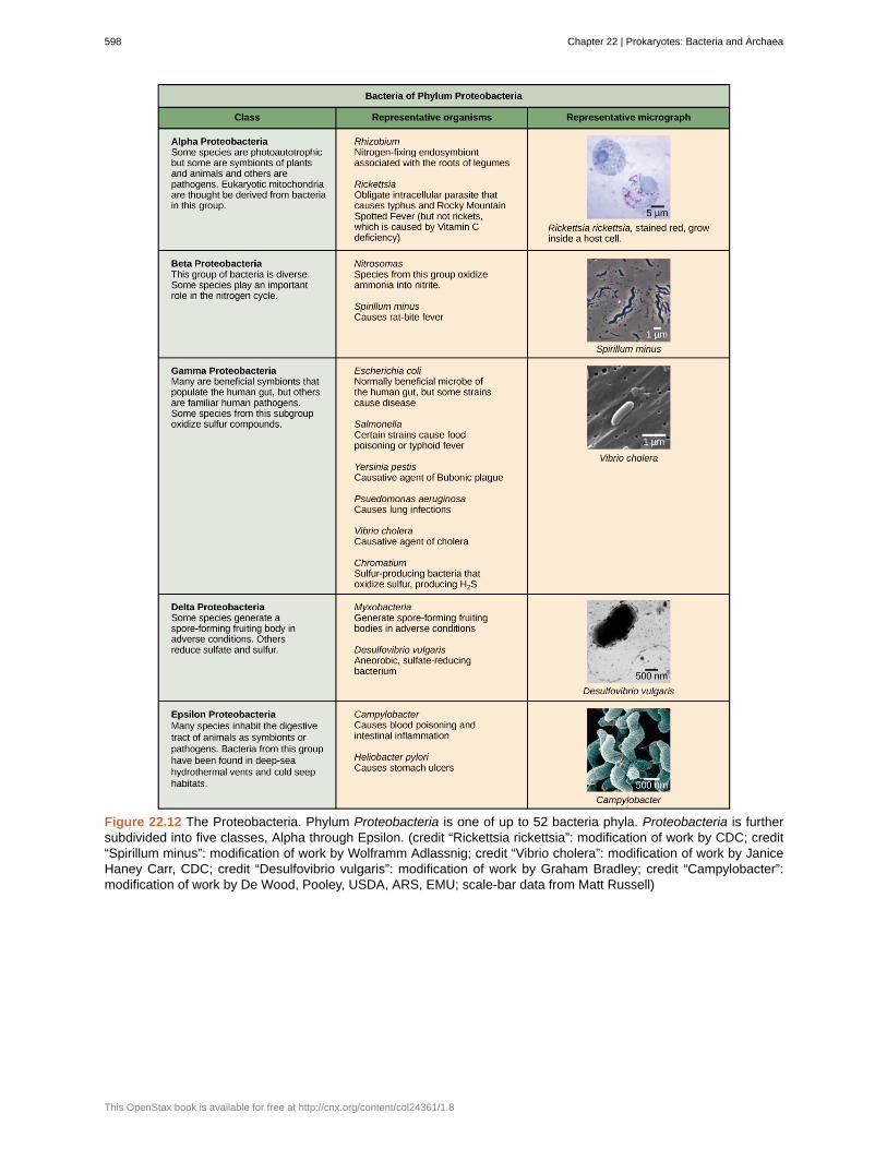
Figure 22.12 The Proteobacteria. Phylum Proteobacteria is one of up to 52 bacteria phyla. Proteobacteria is furthersubdivided into five classes, Alpha through Epsilon. (credit “Rickettsia rickettsia”: modification of work by CDC; credit“Spirillum minus”: modification of work by Wolframm Adlassnig; credit “Vibrio cholera”: modification of work by JaniceHaney Carr, CDC; credit “Desulfovibrio vulgaris”: modification of work by Graham Bradley; credit “Campylobacter”:modification of work by De Wood, Pooley, USDA, ARS, EMU; scale-bar data from Matt Russell)
598 Chapter 22 | Prokaryotes: Bacteria and Archaea
This OpenStax book is available for free at http://cnx.org/content/col24361/1.8

Figure 22.13 Other bacterial phyla. Chlamydia, Spirochetes, Cyanobacteria, and Gram-positive bacteria are describedin this table. Note that bacterial shape is not phylum-dependent; bacteria within a phylum may be cocci, rod-shaped,or spiral. (credit “Chlamydia trachomatis”: modification of work by Dr. Lance Liotta Laboratory, NCI; credit “Treponemapallidum”: modification of work by Dr. David Cox, CDC; credit “Phormidium”: modification of work by USGS; credit“Clostridium difficile”: modification of work by Lois S. Wiggs, CDC; scale-bar data from Matt Russell)
Chapter 22 | Prokaryotes: Bacteria and Archaea 599

Figure 22.14 Archaeal phyla. Archaea are separated into four phyla: the Korarchaeota, Euryarchaeota,Crenarchaeota, and Nanoarchaeota. (credit “Halobacterium”: modification of work by NASA; credit “Nanoarchaeotumequitans”: modification of work by Karl O. Stetter; credit “Korarchaeota”: modification of work by Office of Science ofthe U.S. Dept. of Energy; scale-bar data from Matt Russell)
The Plasma Membrane of Prokaryotes
The prokaryotic plasma membrane is a thin lipid bilayer (6 to 8 nanometers) that completely surrounds thecell and separates the inside from the outside. Its selectively permeable nature keeps ions, proteins, andother molecules within the cell and prevents them from diffusing into the extracellular environment, whileother molecules may move through the membrane. Recall that the general structure of a cell membrane is aphospholipid bilayer composed of two layers of lipid molecules. In archaeal cell membranes, isoprene (phytanyl)chains linked to glycerol replace the fatty acids linked to glycerol in bacterial membranes. Some archaealmembranes are lipid monolayers instead of bilayers (Figure 22.15).
600 Chapter 22 | Prokaryotes: Bacteria and Archaea
This OpenStax book is available for free at http://cnx.org/content/col24361/1.8

Figure 22.15 Bacterial and archaeal phospholipids. Archaeal phospholipids differ from those found in Bacteria andEukarya in two ways. First, they have branched phytanyl sidechains instead of linear ones. Second, an ether bondinstead of an ester bond connects the lipid to the glycerol.
The Cell Wall of Prokaryotes
The cytoplasm of prokaryotic cells has a high concentration of dissolved solutes. Therefore, the osmoticpressure within the cell is relatively high. The cell wall is a protective layer that surrounds some cells and givesthem shape and rigidity. It is located outside the cell membrane and prevents osmotic lysis (bursting due toincreasing volume). The chemical composition of the cell wall varies between Archaea and Bacteria, and alsovaries between bacterial species.
Bacterial cell walls contain peptidoglycan, composed of polysaccharide chains that are cross-linked by unusualpeptides containing both L- and D-amino acids including D-glutamic acid and D-alanine. (Proteins normallyhave only L-amino acids; as a consequence, many of our antibiotics work by mimicking D-amino acids andtherefore have specific effects on bacterial cell-wall development.) There are more than 100 different forms ofpeptidoglycan. S-layer (surface layer) proteins are also present on the outside of cell walls of both Archaea andBacteria.
Bacteria are divided into two major groups: Gram positive and Gram negative, based on their reaction to Gramstaining. Note that all Gram-positive bacteria belong to one phylum; bacteria in the other phyla (Proteobacteria,Chlamydias, Spirochetes, Cyanobacteria, and others) are Gram-negative. The Gram staining method is namedafter its inventor, Danish scientist Hans Christian Gram (1853–1938). The different bacterial responses to thestaining procedure are ultimately due to cell wall structure. Gram-positive organisms typically lack the outermembrane found in Gram-negative organisms (Figure 22.16). Up to 90 percent of the cell-wall in Gram-positivebacteria is composed of peptidoglycan, and most of the rest is composed of acidic substances called teichoicacids. Teichoic acids may be covalently linked to lipids in the plasma membrane to form lipoteichoic acids.Lipoteichoic acids anchor the cell wall to the cell membrane. Gram-negative bacteria have a relatively thin cellwall composed of a few layers of peptidoglycan (only 10 percent of the total cell wall), surrounded by an outerenvelope containing lipopolysaccharides (LPS) and lipoproteins. This outer envelope is sometimes referred toas a second lipid bilayer. The chemistry of this outer envelope is very different, however, from that of the typicallipid bilayer that forms plasma membranes.
Chapter 22 | Prokaryotes: Bacteria and Archaea 601

Figure 22.16 Cell walls in Gram-positive and Gram-negative bacteria. Bacteria are divided into two major groups:Gram positive and Gram negative. Both groups have a cell wall composed of peptidoglycan: in Gram-positivebacteria, the wall is thick, whereas in Gram-negative bacteria, the wall is thin. In Gram-negative bacteria, the cellwall is surrounded by an outer membrane that contains lipopolysaccharides and lipoproteins. Porins are proteinsin this cell membrane that allow substances to pass through the outer membrane of Gram-negative bacteria. InGram-positive bacteria, lipoteichoic acid anchors the cell wall to the cell membrane. (credit: modification of workby "Franciscosp2"/Wikimedia Commons)
Which of the following statements is true?
a. Gram-positive bacteria have a single cell wall anchored to the cell membrane by lipoteichoic acid.
b. Porins allow entry of substances into both Gram-positive and Gram-negative bacteria.
c. The cell wall of Gram-negative bacteria is thick, and the cell wall of Gram-positive bacteria is thin.
d. Gram-negative bacteria have a cell wall made of peptidoglycan, whereas Gram-positive bacteria havea cell wall made of lipoteichoic acid.
Archaean cell walls do not have peptidoglycan. There are four different types of archaean cell walls. One typeis composed of pseudopeptidoglycan, which is similar to peptidoglycan in morphology but contains differentsugars in the polysaccharide chain. The other three types of cell walls are composed of polysaccharides,glycoproteins, or pure protein. Other differences between Bacteria and Archaea are seen in Table 22.2. Notethat features related to DNA replication, transcription and translation in Archaea are similar to those seen ineukaryotes.
Differences and Similarities between Bacteria and Archaea
Structural Characteristic Bacteria Archaea
Cell type Prokaryotic Prokaryotic
Cell morphology Variable Variable
Cell wall Contains peptidoglycan Does not contain peptidoglycan
Cell membrane type Lipid bilayer Lipid bilayer or lipid monolayer
Plasma membrane lipids Fatty acids-glycerol ester Phytanyl-glycerol ethers
Chromosome Typically circular Typically circular
Replication origins Single Multiple
Table 22.2
602 Chapter 22 | Prokaryotes: Bacteria and Archaea
This OpenStax book is available for free at http://cnx.org/content/col24361/1.8

Differences and Similarities between Bacteria and Archaea
Structural Characteristic Bacteria Archaea
RNA polymerase Single Multiple
Initiator tRNA Formyl-methionine Methionine
Streptomycin inhibition Sensitive Resistant
Calvin cycle Yes No
Table 22.2
Reproduction
Reproduction in prokaryotes is asexual and usually takes place by binary fission. (Recall that the DNA of aprokaryote is a single, circular chromosome.) Prokaryotes do not undergo mitosis; instead, the chromosome isreplicated and the two resulting copies separate from one another, due to the growth of the cell. The prokaryote,now enlarged, is pinched inward at its equator and the two resulting cells, which are clones, separate. Binaryfission does not provide an opportunity for genetic recombination or genetic diversity, but prokaryotes can sharegenes by three other mechanisms.
In transformation, the prokaryote takes in DNA shed by other prokaryotes into its environment. If anonpathogenic bacterium takes up DNA for a toxin gene from a pathogen and incorporates the new DNA intoits own chromosome, it too may become pathogenic. In transduction, bacteriophages, the viruses that infectbacteria, may move short pieces of chromosomal DNA from one bacterium to another. Transduction results in arecombinant organism. Archaea also have viruses that may translocate genetic material from one individual toanother. In conjugation, DNA is transferred from one prokaryote to another by means of a pilus, which bringsthe organisms into contact with one another, and provides a channel for transfer of DNA. The DNA transferredcan be in the form of a plasmid or as a composite molecule, containing both plasmid and chromosomal DNA.These three processes of DNA exchange are shown in Figure 22.17.
Reproduction can be very rapid: a few minutes for some species. This short generation time coupled withmechanisms of genetic recombination and high rates of mutation result in the rapid evolution of prokaryotes,allowing them to respond to environmental changes (such as the introduction of an antibiotic) very quickly.
Figure 22.17 Gene transfer mechanisms in prokaryotes. There are three mechanisms by which prokaryotes canexchange DNA. In (a) transformation, the cell takes up prokaryotic DNA directly from the environment. The DNAmay remain separate as plasmid DNA or be incorporated into the host genome. In (b) transduction, a bacteriophageinjects DNA into the cell that contains a small fragment of DNA from a different prokaryote. In (c) conjugation, DNA istransferred from one cell to another via a mating bridge, or pilus, that connects the two cells after the sex pilus drawsthe two bacteria close enough to form the bridge.
Chapter 22 | Prokaryotes: Bacteria and Archaea 603
The Evolution of ProkaryotesHow do scientists answer questions about the evolution of prokaryotes? Unlike with animals, artifacts inthe fossil record of prokaryotes offer very little information. Fossils of ancient prokaryotes look like tinybubbles in rock. Some scientists turn to genetics and to the principle of the molecular clock, which holdsthat the more recently two species have diverged, the more similar their genes (and thus proteins) will be.Conversely, species that diverged long ago will have more genes that are dissimilar.
Scientists at the NASA Astrobiology Institute and at the European Molecular Biology Laboratorycollaborated to analyze the molecular evolution of 32 specific proteins common to 72 species of
prokaryotes.[2]
The model they derived from their data indicates that three important groups ofbacteria—Actinobacteria, Deinococcus, and Cyanobacteria (collectively called Terrabacteria by theauthors)—were the first to colonize land. Actinobacteria are a group of very common Gram-positive bacteriathat produce branched structures like fungal mycelia, and include species important in decomposition oforganic wastes. You will recall that Deinococcus is a genus of bacterium that is highly resistant to ionizingradiation. It has a thick peptidoglycan layer in addition to a second external membrane, so it has features ofboth Gram-positive and Gram-negative bacteria.
Cyanobacteria are photosynthesizers, and were probably responsible for the production of oxygen on theancient earth. The timelines of divergence suggest that bacteria (members of the domain Bacteria) divergedfrom common ancestral species between 2.5 and 3.2 billion years ago, whereas the Archaea divergedearlier: between 3.1 and 4.1 billion years ago. Eukarya later diverged from the archaean line. The workfurther suggests that stromatolites that formed prior to the advent of cyanobacteria (about 2.6 billion yearsago) photosynthesized in an anoxic environment and that because of the modifications of the Terrabacteriafor land (resistance to drying and the possession of compounds that protect the organism from excess light),photosynthesis using oxygen may be closely linked to adaptations to survive on land.
22.3 | Prokaryotic Metabolism
By the end of this section, you will be able to do the following:
• Identify the macronutrients needed by prokaryotes, and explain their importance
• Describe the ways in which prokaryotes get energy and carbon for life processes
• Describe the roles of prokaryotes in the carbon and nitrogen cycles
Prokaryotes are metabolically diverse organisms. In many cases, a prokaryote may be placed into a speciesclade by its defining metabolic features: Can it metabolize lactose? Can it grow on citrate? Does it produceH2S? Does it ferment carbohydrates to produce acid and gas? Can it grow under anaerobic conditions? Sincemetabolism and metabolites are the product of enzyme pathways, and enzymes are encoded in genes, themetabolic capabilities of a prokaryote are a reflection of its genome. There are many different environmentson Earth with various energy and carbon sources, and variable conditions to which prokaryotes may beable to adapt. Prokaryotes have been able to live in every environment from deep-water volcanic vents toAntarctic ice by using whatever energy and carbon sources are available. Prokaryotes fill many niches on Earth,including involvement in nitrogen and carbon cycles, photosynthetic production of oxygen, decomposition ofdead organisms, and thriving as parasitic, commensal, or mutualistic organisms inside multicellular organisms,including humans. The very broad range of environments that prokaryotes occupy is possible because they havediverse metabolic processes.
2. Battistuzzi, FU, Feijao, A, and Hedges, SB. A genomic timescale of prokaryote evolution: Insights into the origin of methanogenesis,phototrophy, and the colonization of land. BioMed Central: Evolutionary Biology 4 (2004): 44, doi:10.1186/1471-2148-4-44.
604 Chapter 22 | Prokaryotes: Bacteria and Archaea
This OpenStax book is available for free at http://cnx.org/content/col24361/1.8
Needs of Prokaryotes
The diverse environments and ecosystems on Earth have a wide range of conditions in terms of temperature,available nutrients, acidity, salinity, oxygen availability, and energy sources. Prokaryotes are very well equippedto make their living out of a vast array of nutrients and environmental conditions. To live, prokaryotes need asource of energy, a source of carbon, and some additional nutrients.
Macronutrients
Cells are essentially a well-organized assemblage of macromolecules and water. Recall that macromoleculesare produced by the polymerization of smaller units called monomers. For cells to build all of the moleculesrequired to sustain life, they need certain substances, collectively called nutrients. When prokaryotes growin nature, they must obtain their nutrients from the environment. Nutrients that are required in large amountsare called macronutrients, whereas those required in smaller or trace amounts are called micronutrients. Justa handful of elements are considered macronutrients—carbon, hydrogen, oxygen, nitrogen, phosphorus, andsulfur. (A mnemonic for remembering these elements is the acronym CHONPS.)
Why are these macronutrients needed in large amounts? They are the components of organic compounds incells, including water. Carbon is the major element in all macromolecules: carbohydrates, proteins, nucleic acids,lipids, and many other compounds. Carbon accounts for about 50 percent of the composition of the cell. Incontrast, nitrogen represents only 12 percent of the total dry weight of a typical cell. Nitrogen is a component ofproteins, nucleic acids, and other cell constituents. Most of the nitrogen available in nature is either atmosphericnitrogen (N2) or another inorganic form. Diatomic (N2) nitrogen, however, can be converted into an organic formonly by certain microorganisms, called nitrogen-fixing organisms. Both hydrogen and oxygen are part of manyorganic compounds and of water. Phosphorus is required by all organisms for the synthesis of nucleotides andphospholipids. Sulfur is part of the structure of some amino acids such as cysteine and methionine, and isalso present in several vitamins and coenzymes. Other important macronutrients are potassium (K), magnesium(Mg), calcium (Ca), and sodium (Na). Although these elements are required in smaller amounts, they are veryimportant for the structure and function of the prokaryotic cell.
Micronutrients
In addition to these macronutrients, prokaryotes require various metallic elements in small amounts. Theseare referred to as micronutrients or trace elements. For example, iron is necessary for the function of thecytochromes involved in electron-transport reactions. Some prokaryotes require other elements—such as boron(B), chromium (Cr), and manganese (Mn)—primarily as enzyme cofactors.
The Ways in Which Prokaryotes Obtain Energy
Prokaryotes are classified both by the way they obtain energy, and by the carbon source they use for producingorganic molecules. These categories are summarized in Table 22.3. Prokaryotes can use different sources ofenergy to generate the ATP needed for biosynthesis and other cellular activities. Phototrophs (or phototrophicorganisms) obtain their energy from sunlight. Phototrophs trap the energy of light using chlorophylls, or in afew cases, bacterial rhodopsin. (Rhodopsin-using phototrophs, oddly, are phototrophic, but not photosynthetic,since they do not fix carbon.) Chemotrophs (or chemosynthetic organisms) obtain their energy from chemicalcompounds. Chemotrophs that can use organic compounds as energy sources are called chemoorganotrophs.Those that can use inorganic compounds, like sulfur or iron compounds, as energy sources are calledchemolithotrophs.
Energy-producing pathways may be either aerobic, using oxygen as the terminal electron acceptor, oranaerobic, using either simple inorganic compounds or organic molecules as the terminal electron acceptor.Since prokaryotes lived on Earth for nearly a billion years before photosynthesis produced significant amountsof oxygen for aerobic respiration, many species of both Bacteria and Archaea are anaerobic and their metabolicactivities are important in the carbon and nitrogen cycles discussed below.
The Ways in Which Prokaryotes Obtain Carbon
Prokaryotes not only can use different sources of energy, but also different sources of carbon compounds.Autotrophic prokaryotes synthesize organic molecules from carbon dioxide. In contrast, heterotrophicprokaryotes obtain carbon from organic compounds. To make the picture more complex, the terms that describehow prokaryotes obtain energy and carbon can be combined. Thus, photoautotrophs use energy from sunlight,and carbon from carbon dioxide and water, whereas chemoheterotrophs obtain both energy and carbon froman organic chemical source. Chemolithoautotrophs obtain their energy from inorganic compounds, and theybuild their complex molecules from carbon dioxide. Finally, prokaryotes that get their energy from light, but theircarbon from organic compounds, are photoheterotrophs. The table below (Table 22.3) summarizes carbon and
Chapter 22 | Prokaryotes: Bacteria and Archaea 605

energy sources in prokaryotes.
Carbon and Energy Sources in Prokaryotes
Energy Sources Carbon Sources
Light Chemicals Carbon dioxide Organic compounds
Phototrophs Chemotrophs Autotrophs Heterotrophs
Organic chemicals Inorganic chemicals
Chemo-organotrophs Chemolithotrophs
Table 22.3
Role of Prokaryotes in Ecosystems
Prokaryotes are ubiquitous: There is no niche or ecosystem in which they are not present. Prokaryotes playmany roles in the environments they occupy. The roles they play in the carbon and nitrogen cycles are vitalto life on Earth. In addition, the current scientific consensus suggests that metabolically interactive prokaryoticcommunities may have been the basis for the emergence of eukaryotic cells.
Prokaryotes and the Carbon Cycle
Carbon is one of the most important macronutrients, and prokaryotes play an important role in the carbon cycle(Figure 22.18). The carbon cycle traces the movement of carbon from inorganic to organic compounds andback again. Carbon is cycled through Earth’s major reservoirs: land, the atmosphere, aquatic environments,sediments and rocks, and biomass. In a way, the carbon cycle echoes the role of the “four elements” firstproposed by the ancient Greek philosopher, Empedocles: fire, water, earth, and air. Carbon dioxide is removedfrom the atmosphere by land plants and marine prokaryotes, and is returned to the atmosphere via therespiration of chemoorganotrophic organisms, including prokaryotes, fungi, and animals. Although the largestcarbon reservoir in terrestrial ecosystems is in rocks and sediments, that carbon is not readily available.
Participants in the carbon cycle are roughly divided among producers, consumers, and decomposers of organiccarbon compounds. The primary producers of organic carbon compounds from CO2 are land plants andphotosynthetic bacteria. A large amount of available carbon is found in living land plants. A related source ofcarbon compounds is humus, which is a mixture of organic materials from dead plants and prokaryotes thathave resisted decomposition. (The term "humus," by the way, is the root of the word "human.") Consumerssuch as animals and other heterotrophs use organic compounds generated by producers and release carbondioxide to the atmosphere. Other bacteria and fungi, collectively called decomposers, carry out the breakdown(decomposition) of plants and animals and their organic compounds. Most carbon dioxide in the atmosphere isderived from the respiration of microorganisms that decompose dead animals, plants, and humus.
In aqueous environments and their anoxic sediments, there is another carbon cycle taking place. In this case, thecycle is based on one-carbon compounds. In anoxic sediments, prokaryotes, mostly archaea, produce methane(CH4). This methane moves into the zone above the sediment, which is richer in oxygen and supports bacteriacalled methane oxidizers that oxidize methane to carbon dioxide, which then returns to the atmosphere.
606 Chapter 22 | Prokaryotes: Bacteria and Archaea
This OpenStax book is available for free at http://cnx.org/content/col24361/1.8

Figure 22.18 The carbon cycle. Prokaryotes play a significant role in continuously moving carbon through thebiosphere. (credit: modification of work by John M. Evans and Howard Perlman, USGS)
Prokaryotes and the Nitrogen Cycle
Nitrogen is a very important element for life because it is a major constituent of proteins and nucleic acids.It is a macronutrient, and in nature, it is recycled from organic compounds to ammonia, ammonium ions,nitrate, nitrite, and nitrogen gas by many processes, many of which are carried out only by prokaryotes. Asillustrated in Figure 22.19, prokaryotes are key to the nitrogen cycle. The largest pool of nitrogen available inthe terrestrial ecosystem is gaseous nitrogen (N2) from the air, but this nitrogen is not usable by plants, whichare primary producers. Gaseous nitrogen is transformed, or “fixed” into more readily available forms, such asammonia (NH3), through the process of nitrogen fixation. Nitrogen-fixing bacteria include Azotobacter in soiland the ubiquitous photosynthetic cyanobacteria. Some nitrogen fixing bacteria, like Rhizobium, live in symbioticrelationships in the roots of legumes. Another source of ammonia is ammonification, the process by whichammonia is released during the decomposition of nitrogen-containing organic compounds. The ammonium ionis progressively oxidized by different species of bacteria in a process called nitrification. The nitrification process
begins with the conversion of ammonium to nitrite (NO2-), and continues with the conversion of nitrite to nitrate.
Nitrification in soils is carried out by bacteria belonging to the genera Nitrosomas, Nitrobacter, and Nitrospira.
Most nitrogen in soil is in the form of ammonium (NH4+) or nitrate (NO3
-). Ammonia and nitrate can be used byplants or converted to other forms.
Ammonia released into the atmosphere, however, represents only 15 percent of the total nitrogen released; therest is as N2 and N2O (nitrous oxide). Ammonia is catabolized anaerobically by some prokaryotes, yielding N2as the final product. Denitrifying bacteria reverse the process of nitrification, reducing the nitrate from soils togaseous compounds such as N2O, NO, and N2.
Chapter 22 | Prokaryotes: Bacteria and Archaea 607
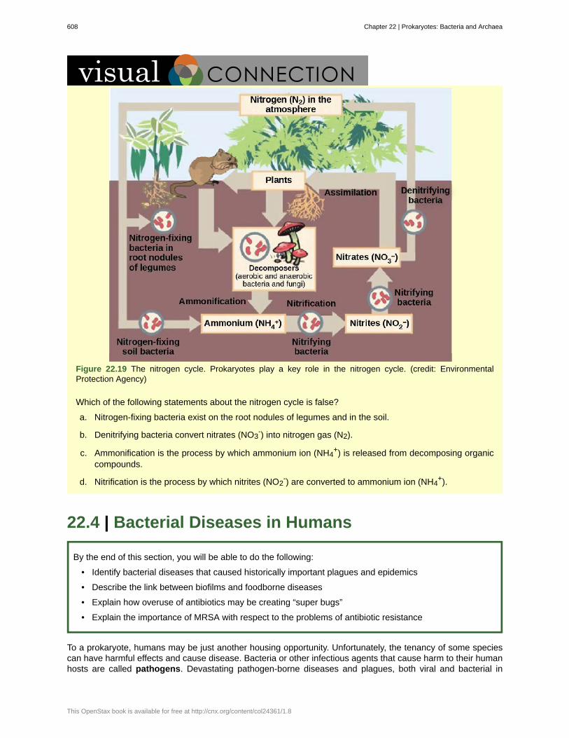
Figure 22.19 The nitrogen cycle. Prokaryotes play a key role in the nitrogen cycle. (credit: EnvironmentalProtection Agency)
Which of the following statements about the nitrogen cycle is false?
a. Nitrogen-fixing bacteria exist on the root nodules of legumes and in the soil.
b. Denitrifying bacteria convert nitrates (NO3-) into nitrogen gas (N2).
c. Ammonification is the process by which ammonium ion (NH4+) is released from decomposing organic
compounds.
d. Nitrification is the process by which nitrites (NO2-) are converted to ammonium ion (NH4
+).
22.4 | Bacterial Diseases in Humans
By the end of this section, you will be able to do the following:
• Identify bacterial diseases that caused historically important plagues and epidemics
• Describe the link between biofilms and foodborne diseases
• Explain how overuse of antibiotics may be creating “super bugs”
• Explain the importance of MRSA with respect to the problems of antibiotic resistance
To a prokaryote, humans may be just another housing opportunity. Unfortunately, the tenancy of some speciescan have harmful effects and cause disease. Bacteria or other infectious agents that cause harm to their humanhosts are called pathogens. Devastating pathogen-borne diseases and plagues, both viral and bacterial in
608 Chapter 22 | Prokaryotes: Bacteria and Archaea
This OpenStax book is available for free at http://cnx.org/content/col24361/1.8

nature, have affected humans and their ancestors for millions of years. The true cause of these diseases was notunderstood until modern scientific thought developed, and many people thought that diseases were a “spiritualpunishment.” Only within the past several centuries have people understood that staying away from afflictedpersons, disposing of the corpses and personal belongings of victims of illness, and sanitation practices reducedtheir own chances of getting sick.
Epidemiologists study how diseases are transmitted and how they affect a population. Often, they must followingthe course of an epidemic—a disease that occurs in an unusually high number of individuals in a population atthe same time. In contrast, a pandemic is a widespread, and usually worldwide, epidemic. An endemic diseaseis a disease that is always present, usually at low incidence, in a population.
Long History of Bacterial Disease
There are records about infectious diseases as far back as 3000 B.C. A number of significant pandemics causedby bacteria have been documented over several hundred years. Some of the most memorable pandemics led tothe decline of cities and entire nations.
In the 21st century, infectious diseases remain among the leading causes of death worldwide, despite advancesmade in medical research and treatments in recent decades. A disease spreads when the pathogen that causesit is passed from one person to another. For a pathogen to cause disease, it must be able to reproduce in thehost’s body and damage the host in some way.
The Plague of Athens
In 430 B.C., the Plague of Athens killed one-quarter of the Athenian troops who were fighting in the greatPeloponnesian War and weakened Athens’s dominance and power. The plague impacted people living inovercrowded Athens as well as troops aboard ships that had to return to Athens. The source of the plaguemay have been identified recently when researchers from the University of Athens were able to use DNA fromteeth recovered from a mass grave. The scientists identified nucleotide sequences from a pathogenic bacterium,
Salmonella enterica serovar Typhi (Figure 22.20), which causes typhoid fever.[3]
This disease is commonly seenin overcrowded areas and has caused epidemics throughout recorded history.
3. Papagrigorakis MJ, Synodinos PN, and Yapijakis C. Ancient typhoid epidemic reveals possible ancestral strain of Salmonella entericaserovar Typhi. Infect Genet Evol 7 (2007): 126–7, Epub 2006 Jun.
Chapter 22 | Prokaryotes: Bacteria and Archaea 609

Figure 22.20 Salmonella enterica. Salmonella enterica serovar Typhi, the causative agent of Typhoid fever, is aGram-negative, rod-shaped gamma proteobacterium. Typhoid fever, which is spread through feces, causes intestinalhemorrhage, high fever, delirium, and dehydration. Today, between 16 and 33 million cases of this re-emerging diseaseoccur annually, resulting in over 200,000 deaths. Carriers of the disease can be asymptomatic. In a famous case in theearly 1900s, a cook named Mary Mallon (“Typhoid Mary”) unknowingly spread the disease to over fifty people, three ofwhom died. Other serotypes of Salmonella cause food poisoning. (credit: modification of work by NCI, CDC)
Bubonic Plagues
From 541 to 750, the Plague of Justinian, an outbreak of what was likely bubonic plague, eliminated one-quarterto one-half of the human population in the eastern Mediterranean region. The population in Europe dropped by50 percent during this outbreak. Astoundingly, bubonic plague would strike Europe more than once!
Bubonic plague is caused by the bacterium Yersinia pestis. One of the most devastating pandemics attributedto bubonic plague was the Black Death (1346 to 1361). It is thought to have originated in China and spreadalong the Silk Road, a network of land and sea trade routes, to the Mediterranean region and Europe, carriedby fleas living on black rats that were always present on ships. The Black Death was probably named for thetissue necrosis (Figure 22.21c) that can be one of the symptoms. The "buboes" of bubonic plague were painfullyswollen areas of lymphatic tissue. A pneumonic form of the plague, spread by the coughing and sneezing ofinfected individuals, spreads directly from human to human and can cause death within a week. The pneumonicform was responsible for the rapid spread of the Black Death in Europe. The Black Death reduced the world’spopulation from an estimated 450 million to about 350 to 375 million. Bubonic plague struck London yet againin the mid-1600s (Figure 22.21). In modern times, approximately 1,000 to 3,000 cases of plague arise globallyeach year, and a “sylvatic” form of plague, carried by fleas living on rodents such as prairie dogs and blackfooted ferrets, infects 10 to 20 people annually in the American Southwest. Although contracting bubonic plaguebefore antibiotics meant almost certain death, the bacterium responds to several types of modern antibiotics,and mortality rates from plague are now very low.
610 Chapter 22 | Prokaryotes: Bacteria and Archaea
This OpenStax book is available for free at http://cnx.org/content/col24361/1.8

Figure 22.21 The Black Death. The (a) Great Plague of London killed an estimated 200,000 people, or about 20percent of the city’s population. The causative agent, the (b) bacterium Yersinia pestis, is a Gram-negative, rod-shapedbacterium from the class Gammaproteobacteria. The disease is transmitted through the bite of an infected flea, whichis carried on a rodent. Symptoms include swollen lymph nodes, fever, seizure, vomiting of blood, and (c) gangrene.(credit b: Rocky Mountain Laboratories, NIAID, NIH; scale-bar data from Matt Russell; credit c: Textbook of MilitaryMedicine, Washington, D.C., U.S. Dept. of the Army, Office of the Surgeon General, Borden Institute)
Watch a video (http://openstaxcollege.org/l/black_death) on the modern understanding of the Black
Death—bubonic plague in Europe during the 14th century.
Migration of Diseases to New Populations
One of the negative consequences of human exploration was the accidental “biological warfare” that resultedfrom the transport of a pathogen into a population that had not previously been exposed to it. Over the centuries,Europeans tended to develop genetic immunity to endemic infectious diseases, but when European conquerorsreached the western hemisphere, they brought with them disease-causing bacteria and viruses, which triggeredepidemics that completely devastated many diverse populations of Native Americans, who had no naturalresistance to many European diseases. It has been estimated that up to 90 percent of Native Americansdied from infectious diseases after the arrival of Europeans, making conquest of the New World a foregoneconclusion.
Emerging and Re-emerging Diseases
The distribution of a particular disease is dynamic. Changes in the environment, the pathogen, or the hostpopulation can dramatically impact the spread of a disease. According to the World Health Organization (WHO),an emerging disease (Figure 22.22) is one that has appeared in a population for the first time, or that may haveexisted previously but is rapidly increasing in incidence or geographic range. This definition also includes re-emerging diseases that were previously under control. Approximately 75 percent of recently emerging infectiousdiseases affecting humans are zoonotic diseases. Zoonoses are diseases that primarily infect animals but canbe transmitted to humans; some are of viral origin and some are of bacterial origin. Brucellosis is an example ofa prokaryotic zoonosis that is re-emerging in some regions, and necrotizing fasciitis (commonly known as flesh-eating bacteria) has been increasing in virulence for the last 80 years for unknown reasons.
Chapter 22 | Prokaryotes: Bacteria and Archaea 611

Figure 22.22 Emerging diseases. The map shows regions where bacterial diseases are emerging or re-emerging.(credit: modification of work by NIH)
Some of the present emerging diseases are not actually new, but are diseases that were catastrophic in the past(Figure 22.23). They devastated populations and became dormant for a while, just to come back, sometimesmore virulent than before, as was the case with bubonic plague. Other diseases, like tuberculosis, were nevereradicated but were under control in some regions of the world until coming back, mostly in urban centers withhigh concentrations of immunocompromised people. WHO has identified certain diseases whose worldwide re-emergence should be monitored. Among these are three viral diseases (dengue fever, yellow fever, and zika),and three bacterial diseases (diphtheria, cholera, and bubonic plague). The war against infectious diseases hasno foreseeable end.
Figure 22.23 Lyme Disease. Lyme disease often, but not always, results in (a) a characteristic bullseye rash. Thedisease is caused by a (b) Gram-negative spirochete bacterium of the genus Borrelia. The bacteria (c) infect ticks,which in turn infect mice. Deer are the preferred secondary host, but the ticks also may feed on humans. Untreated,the disease causes chronic disorders in the nervous system, eyes, joints, and heart. The disease is named afterLyme, Connecticut, where an outbreak occurred in 1995 and has subsequently spread. The disease is not new,however. Genetic evidence suggests that Ötzi the Iceman, a 5,300-year-old mummy found in the Alps, was infectedwith Borrelia. (credit a: James Gathany, CDC; credit b: CDC; scale-bar data from Matt Russell)
Foodborne Diseases
Prokaryotes are everywhere: They readily colonize the surface of any type of material, and food is not anexception. Most of the time, prokaryotes colonize food and food-processing equipment in the form of a biofilm,as we have discussed earlier. Outbreaks of bacterial infection related to food consumption are common.A foodborne disease (commonly called “food poisoning”) is an illness resulting from the consumption thepathogenic bacteria, viruses, or other parasites that contaminate food. Although the United States has one ofthe safest food supplies in the world, the U.S. Centers for Disease Control and Prevention (CDC) has reported
612 Chapter 22 | Prokaryotes: Bacteria and Archaea
This OpenStax book is available for free at http://cnx.org/content/col24361/1.8
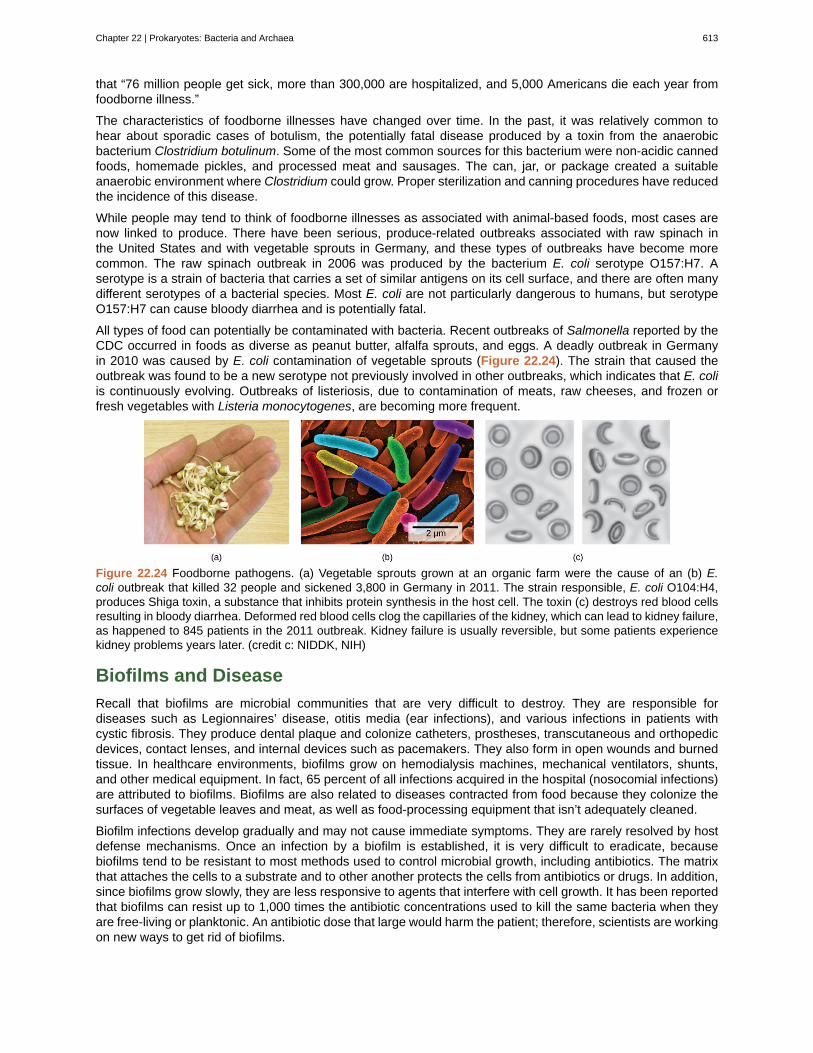
that “76 million people get sick, more than 300,000 are hospitalized, and 5,000 Americans die each year fromfoodborne illness.”
The characteristics of foodborne illnesses have changed over time. In the past, it was relatively common tohear about sporadic cases of botulism, the potentially fatal disease produced by a toxin from the anaerobicbacterium Clostridium botulinum. Some of the most common sources for this bacterium were non-acidic cannedfoods, homemade pickles, and processed meat and sausages. The can, jar, or package created a suitableanaerobic environment where Clostridium could grow. Proper sterilization and canning procedures have reducedthe incidence of this disease.
While people may tend to think of foodborne illnesses as associated with animal-based foods, most cases arenow linked to produce. There have been serious, produce-related outbreaks associated with raw spinach inthe United States and with vegetable sprouts in Germany, and these types of outbreaks have become morecommon. The raw spinach outbreak in 2006 was produced by the bacterium E. coli serotype O157:H7. Aserotype is a strain of bacteria that carries a set of similar antigens on its cell surface, and there are often manydifferent serotypes of a bacterial species. Most E. coli are not particularly dangerous to humans, but serotypeO157:H7 can cause bloody diarrhea and is potentially fatal.
All types of food can potentially be contaminated with bacteria. Recent outbreaks of Salmonella reported by theCDC occurred in foods as diverse as peanut butter, alfalfa sprouts, and eggs. A deadly outbreak in Germanyin 2010 was caused by E. coli contamination of vegetable sprouts (Figure 22.24). The strain that caused theoutbreak was found to be a new serotype not previously involved in other outbreaks, which indicates that E. coliis continuously evolving. Outbreaks of listeriosis, due to contamination of meats, raw cheeses, and frozen orfresh vegetables with Listeria monocytogenes, are becoming more frequent.
Figure 22.24 Foodborne pathogens. (a) Vegetable sprouts grown at an organic farm were the cause of an (b) E.coli outbreak that killed 32 people and sickened 3,800 in Germany in 2011. The strain responsible, E. coli O104:H4,produces Shiga toxin, a substance that inhibits protein synthesis in the host cell. The toxin (c) destroys red blood cellsresulting in bloody diarrhea. Deformed red blood cells clog the capillaries of the kidney, which can lead to kidney failure,as happened to 845 patients in the 2011 outbreak. Kidney failure is usually reversible, but some patients experiencekidney problems years later. (credit c: NIDDK, NIH)
Biofilms and Disease
Recall that biofilms are microbial communities that are very difficult to destroy. They are responsible fordiseases such as Legionnaires’ disease, otitis media (ear infections), and various infections in patients withcystic fibrosis. They produce dental plaque and colonize catheters, prostheses, transcutaneous and orthopedicdevices, contact lenses, and internal devices such as pacemakers. They also form in open wounds and burnedtissue. In healthcare environments, biofilms grow on hemodialysis machines, mechanical ventilators, shunts,and other medical equipment. In fact, 65 percent of all infections acquired in the hospital (nosocomial infections)are attributed to biofilms. Biofilms are also related to diseases contracted from food because they colonize thesurfaces of vegetable leaves and meat, as well as food-processing equipment that isn’t adequately cleaned.
Biofilm infections develop gradually and may not cause immediate symptoms. They are rarely resolved by hostdefense mechanisms. Once an infection by a biofilm is established, it is very difficult to eradicate, becausebiofilms tend to be resistant to most methods used to control microbial growth, including antibiotics. The matrixthat attaches the cells to a substrate and to other another protects the cells from antibiotics or drugs. In addition,since biofilms grow slowly, they are less responsive to agents that interfere with cell growth. It has been reportedthat biofilms can resist up to 1,000 times the antibiotic concentrations used to kill the same bacteria when theyare free-living or planktonic. An antibiotic dose that large would harm the patient; therefore, scientists are workingon new ways to get rid of biofilms.
Chapter 22 | Prokaryotes: Bacteria and Archaea 613
Antibiotics: Are We Facing a Crisis?
The word antibiotic comes from the Greek anti meaning “against” and bios meaning “life.” An antibiotic isa chemical, produced either by microbes or synthetically, that is hostile to or prevents the growth of otherorganisms. Today’s media often address concerns about an antibiotic crisis. Are the antibiotics that easily treatedbacterial infections in the past becoming obsolete? Are there new “superbugs”—bacteria that have evolved tobecome more resistant to our arsenal of antibiotics? Is this the beginning of the end of antibiotics? All thesequestions challenge the healthcare community.
One of the main causes of antibiotic resistance in bacteria is overexposure to antibiotics. The imprudent andexcessive use of antibiotics has resulted in the natural selection of resistant forms of bacteria. The antibiotic killsmost of the infecting bacteria, and therefore only the resistant forms remain. These resistant forms reproduce,resulting in an increase in the proportion of resistant forms over non-resistant ones. In addition to transmissionof resistance genes to progeny, lateral transfer of resistance genes on plasmids can rapidly spread these genesthrough a bacterial population. A major misuse of antibiotics is in patients with viral infections like colds or theflu, against which antibiotics are useless. Another problem is the excessive use of antibiotics in livestock. Theroutine use of antibiotics in animal feed promotes bacterial resistance as well. In the United States, 70 percent ofthe antibiotics produced are fed to animals. These antibiotics are given to livestock in low doses, which maximizethe probability of resistance developing, and these resistant bacteria are readily transferred to humans.
Watch a recent news report (http://openstaxcollege.org/l/antibiotics) on the problem of routine antibioticadministration to livestock and antibiotic-resistant bacteria.
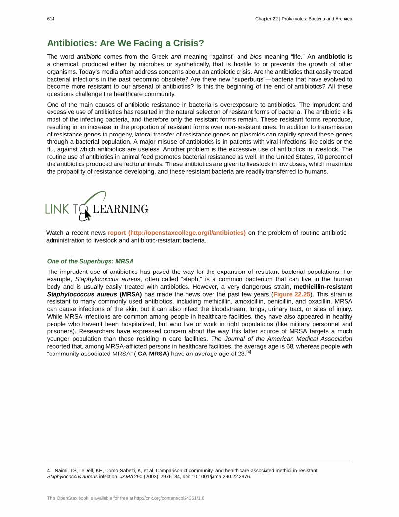
One of the Superbugs: MRSA
The imprudent use of antibiotics has paved the way for the expansion of resistant bacterial populations. Forexample, Staphylococcus aureus, often called “staph,” is a common bacterium that can live in the humanbody and is usually easily treated with antibiotics. However, a very dangerous strain, methicillin-resistantStaphylococcus aureus (MRSA) has made the news over the past few years (Figure 22.25). This strain isresistant to many commonly used antibiotics, including methicillin, amoxicillin, penicillin, and oxacillin. MRSAcan cause infections of the skin, but it can also infect the bloodstream, lungs, urinary tract, or sites of injury.While MRSA infections are common among people in healthcare facilities, they have also appeared in healthypeople who haven’t been hospitalized, but who live or work in tight populations (like military personnel andprisoners). Researchers have expressed concern about the way this latter source of MRSA targets a muchyounger population than those residing in care facilities. The Journal of the American Medical Associationreported that, among MRSA-afflicted persons in healthcare facilities, the average age is 68, whereas people with“community-associated MRSA” ( CA-MRSA) have an average age of 23.[4]
4. Naimi, TS, LeDell, KH, Como-Sabetti, K, et al. Comparison of community- and health care-associated methicillin-resistantStaphylococcus aureus infection. JAMA 290 (2003): 2976–84, doi: 10.1001/jama.290.22.2976.
614 Chapter 22 | Prokaryotes: Bacteria and Archaea
This OpenStax book is available for free at http://cnx.org/content/col24361/1.8

Figure 22.25 MRSA. This scanning electron micrograph shows methicillin-resistant Staphylococcus aureus bacteria,commonly known as MRSA. S. aureus is not always pathogenic, but can cause diseases such as food poisoning andskin and respiratory infections. (credit: modification of work by Janice Haney Carr; scale-bar data from Matt Russell)
In summary, the medical community is facing an antibiotic crisis. Some scientists believe that after years ofbeing protected from bacterial infections by antibiotics, we may be returning to a time in which a simple bacterialinfection could again devastate the human population. Researchers are developing new antibiotics, but it takesmany years of research and clinical trials, plus financial investments in the millions of dollars, to generate aneffective and approved drug.
Chapter 22 | Prokaryotes: Bacteria and Archaea 615

EpidemiologistEpidemiology is the study of the occurrence, distribution, and determinants of health and disease in apopulation. It is, therefore, part of public health. An epidemiologist studies the frequency and distribution ofdiseases within human populations and environments.
Epidemiologists collect data about a particular disease and track its spread to identify the original modeof transmission. They sometimes work in close collaboration with historians to try to understand the waya disease evolved geographically and over time, tracking the natural history of pathogens. They gatherinformation from clinical records, patient interviews, surveillance, and any other available means. Thatinformation is used to develop strategies, such as vaccinations (Figure 22.26), and design public healthpolicies to reduce the incidence of a disease or to prevent its spread. Epidemiologists also conduct rapidinvestigations in case of an outbreak to recommend immediate measures to control it.
An epidemiologist has a bachelor’s degree, plus a master’s degree in public health (MPH). Manyepidemiologists are also physicians (and have an M.D. or D.O degree), or they have a Ph.D. in anassociated field, such as biology or microbiology.
Figure 22.26 Vaccination. Vaccinations can slow the spread of communicable diseases. (credit: modification ofwork by Daniel Paquet)
22.5 | Beneficial Prokaryotes
By the end of this section, you will be able to do the following:
• Explain the need for nitrogen fixation and how it is accomplished
• Describe the beneficial effects of bacteria that colonize our skin and digestive tracts
• Identify prokaryotes used during the processing of food
• Describe the use of prokaryotes in bioremediation
Fortunately, only a few species of prokaryotes are pathogenic! Prokaryotes also interact with humans and otherorganisms in a number of ways that are beneficial. For example, prokaryotes are major participants in the carbonand nitrogen cycles. They produce or process nutrients in the digestive tracts of humans and other animals.Prokaryotes are used in the production of some human foods, and also have been recruited for the degradationof hazardous materials. In fact, our life would not be possible without prokaryotes!
616 Chapter 22 | Prokaryotes: Bacteria and Archaea
This OpenStax book is available for free at http://cnx.org/content/col24361/1.8

Cooperation between Bacteria and Eukaryotes: Nitrogen Fixation
Nitrogen is a very important element to living things, because it is part of nucleotides and amino acids that arethe building blocks of nucleic acids and proteins, respectively. Nitrogen is usually the most limiting element interrestrial ecosystems, with atmospheric nitrogen, N2, providing the largest pool of available nitrogen. However,eukaryotes cannot use atmospheric, gaseous nitrogen to synthesize macromolecules. Fortunately, nitrogencan be “fixed,” meaning it is converted into a more accessible form—ammonia (NH3)—either biologically orabiotically.
Abiotic nitrogen fixation occurs as a result of physical processes such as lightning or by industrial processes.Biological nitrogen fixation (BNF) is exclusively carried out by prokaryotes: soil bacteria, cyanobacteria,and Frankia spp. (filamentous bacteria interacting with actinorhizal plants such as alder, bayberry, and sweetfern). After photosynthesis, BNF is the most important biological process on Earth. The overall nitrogen fixationequation below represents a series of redox reactions (Pi stands for inorganic phosphate).
N2 + 16ATP + 8e− + 8H+ → 2NH3 + 16ADP + 16Pi + H2
The total fixed nitrogen through BNF is about 100 to 180 million metric tons per year, which contributes about 65percent of the nitrogen used in agriculture.
Cyanobacteria are the most important nitrogen fixers in aquatic environments. In soil, members of the generaClostridium and Azotobacter are examples of free-living, nitrogen-fixing bacteria. Other bacteria livesymbiotically with legume plants, providing the most important source of fixed nitrogen. Symbionts may fixmore nitrogen in soils than free-living organisms by a factor of 10. Soil bacteria, collectively called rhizobia,are able to symbiotically interact with legumes to form nodules, specialized structures where nitrogen fixationoccurs (Figure 22.27). Nitrogenase, the enzyme that fixes nitrogen, is inactivated by oxygen, so the noduleprovides an oxygen-free area for nitrogen fixation to take place. The oxygen is sequestered by a form ofplant hemoglobin called leghemoglobin, which protects the nitrogenase, but releases enough oxygen to supportrespiratory activity.
Symbiotic nitrogen fixation provides a natural and inexpensive plant fertilizer: It reduces atmospheric nitrogento ammonia, which is easily usable by plants. The use of legumes is an excellent alternative to chemicalfertilization and is of special interest to sustainable agriculture, which seeks to minimize the use of chemicals andconserve natural resources. Through symbiotic nitrogen fixation, the plant benefits from using an endless sourceof nitrogen: the atmosphere. The bacteria benefit from using photosynthates (carbohydrates produced duringphotosynthesis) from the plant and having a protected niche. In addition, the soil benefits from being naturallyfertilized. Therefore, the use of rhizobia as biofertilizers is a sustainable practice.
Why are legumes so important? Some, like soybeans, are key sources of agricultural protein. Some of the mostimportant legumes consumed by humans are soybeans, peanuts, peas, chickpeas, and beans. Other legumes,such as alfalfa, are used to feed cattle.
Figure 22.27 Nitrogen-fixation nodules on soybean roots. Soybean (Glycine max) is a legume that interactssymbiotically with the soil bacterium Bradyrhizobium japonicum to form specialized structures on the roots callednodules where nitrogen fixation occurs. (credit: USDA)
Chapter 22 | Prokaryotes: Bacteria and Archaea 617

Microbes on the Human BodyThe commensal bacteria that inhabit our skin and gastrointestinal tract do a host of good things for us. Theyprotect us from pathogens, help us digest our food, and produce some of our vitamins and other nutrients.These activities have been known for a long time. More recently, scientists have gathered evidence thatthese bacteria may also help regulate our moods, influence our activity levels, and even help control weightby affecting our food choices and absorption patterns. The Human Microbiome Project has begun theprocess of cataloging our normal bacteria (and archaea) so we can better understand these functions.
A particularly fascinating example of our normal flora relates to our digestive systems. People who takehigh doses of antibiotics tend to lose many of their normal gut bacteria, allowing a naturally antibiotic-resistant species called Clostridium difficile to overgrow and cause severe gastric problems, especiallychronic diarrhea (Figure 22.28). Obviously, trying to treat this problem with antibiotics only makes it worse.However, it has been successfully treated by giving the patients fecal transplants from healthy donors toreestablish the normal intestinal microbial community. Clinical trials are underway to ensure the safety andeffectiveness of this technique.
Figure 22.28 Clostridium difficile. This scanning electron micrograph shows Clostridium difficile, a Gram-positive,rod-shaped bacterium that causes severe diarrhea. Infection commonly occurs after the normal gut fauna areeradicated by antibiotics, and in the hospital can be deadly to seriously ill patients. (credit: modification of work byCDC, HHS; scale-bar data from Matt Russell)
Scientists are also discovering that the absence of certain key microbes from our intestinal tract may setus up for a variety of problems. This seems to be particularly true regarding the appropriate functioning ofthe immune system. There are intriguing findings that suggest that the absence of these microbes is animportant contributor to the development of allergies and some autoimmune disorders. Research is currentlyunderway to test whether adding certain microbes to our internal ecosystem may help in the treatment ofthese problems, as well as in treating some forms of autism.
Early Biotechnology: Cheese, Bread, Wine, Beer, and Yogurt
According to the United Nations Convention on Biological Diversity, biotechnology is “any technologicalapplication that uses biological systems, living organisms, or derivatives thereof, to make or modify products
or processes for specific use."[5]
The concept of “specific use” involves some sort of commercial application.Genetic engineering, artificial selection, antibiotic production, and cell culture are current topics of study inbiotechnology and will be described in later chapters. However, humans were using prokaryotes before the termbiotechnology was even coined. Some of the products of this early biotechnology are as familiar as cheese,bread, wine, beer, and yogurt, which employ both bacteria and other microbes, such as yeast, a fungus (Figure22.29).
5. http://www.cbd.int/convention/articles/?a=cbd-02 (http://openstax.org/l/UN_convention) , United Nations Convention on BiologicalDiversity: Article 2: Use of Terms.
618 Chapter 22 | Prokaryotes: Bacteria and Archaea
This OpenStax book is available for free at http://cnx.org/content/col24361/1.8

Figure 22.29 Some foods produced by microorganisms. Some of the products derived from the use of prokaryotesin early biotechnology include (a) cheese, (b) wine, (c) beer and bread, and (d) yogurt. (credit bread: modification ofwork by F. Rodrigo/Wikimedia Commons; credit wine: modification of work by Jon Sullivan; credit beer and bread:modification of work by Kris Miller; credit yogurt: modification of work by Jon Sullivan)
Cheese production began around 4,000 to 7,000 years ago when humans began to breed animals and processtheir milk. Fermentation in this case preserves nutrients: Milk will spoil relatively quickly, but when processedas cheese, it is more stable. As for beer, the oldest records of brewing are about 6,000 years old and werean integral part of the Sumerian culture. Evidence indicates that the Sumerians discovered fermentation bychance. Wine has been produced for about 4,500 years, and evidence suggests that cultured milk products, likeyogurt, have existed for at least 4,000 years.
Using Prokaryotes to Clean up Our Planet: Bioremediation
Microbial bioremediation is the use of prokaryotes (or microbial metabolism) to remove pollutants.Bioremediation has been used to remove agricultural chemicals (e.g., pesticides, fertilizers) that leach fromsoil into groundwater and the subsurface. Certain toxic metals and oxides, such as selenium and arsenic
compounds, can also be removed from water by bioremediation. The reduction of SeO4-2 to SeO3
-2 and to Se0
(metallic selenium) is a method used to remove selenium ions from water. Mercury (Hg) is an example of a toxicmetal that can be removed from an environment by bioremediation. As an active ingredient of some pesticides,mercury is used in industry and is also a by-product of certain processes, such as battery production. Methylmercury is usually present in very low concentrations in natural environments, but it is highly toxic because itaccumulates in living tissues. Several species of bacteria can carry out the biotransformation of toxic mercury
into nontoxic forms. These bacteria, such as Pseudomonas aeruginosa, can convert Hg+2 into Hg0, which isnontoxic to humans.
One of the most useful and interesting examples of the use of prokaryotes for bioremediation purposes is thecleanup of oil spills. The significance of prokaryotes to petroleum bioremediation has been demonstrated inseveral oil spills in recent years, such as the Exxon Valdez spill in Alaska (1989) (Figure 22.30), the Prestigeoil spill in Spain (2002), the spill into the Mediterranean from a Lebanon power plant (2006), and more recently,the BP oil spill in the Gulf of Mexico (2010). In the case of oil spills in the ocean, ongoing natural bioremediationtends to occur, since there are oil-consuming bacteria in the ocean prior to the spill. In addition to these naturally
Chapter 22 | Prokaryotes: Bacteria and Archaea 619

occurring oil-degrading bacteria, humans select and engineer bacteria that possess the same capability withincreased efficacy and spectrum of hydrocarbon compounds that can be processed. Bioremediation is enhancedby the addition of inorganic nutrients that help bacteria to grow.
Some hydrocarbon-degrading bacteria feed on hydrocarbons in the oil droplet, breaking down the hydrocarbonsinto smaller subunits. Some species, such as Alcanivorax borkumensis, produce surfactants that solubilizethe oil (making it soluble in water), whereas other bacteria degrade the oil into carbon dioxide. Under idealconditions, it has been reported that up to 80 percent of the nonvolatile components in oil can be degraded withinone year of the spill. Other oil fractions containing aromatic and highly branched hydrocarbon chains are moredifficult to remove and remain in the environment for longer periods of time.
Figure 22.30 Prokaryotes and bioremediation. (a) Cleaning up oil after the Exxon Valdez spill in Alaska, workers hosedoil from beaches and then used a floating boom to corral the oil, which was finally skimmed from the water surface.Some species of bacteria are able to solubilize and degrade the oil. (b) One of the most catastrophic consequencesof oil spills is the damage to fauna. (credit a: modification of work by NOAA; credit b: modification of work byGOLUBENKOV, NGO: Saving Taman)
620 Chapter 22 | Prokaryotes: Bacteria and Archaea
This OpenStax book is available for free at http://cnx.org/content/col24361/1.8

acidophile
alkaliphile
ammonification
anaerobic
anoxic
antibiotic
biofilm
biological nitrogen fixation
bioremediation
biotechnology
Black Death
botulism
CA-MRSA
capsule
chemotroph
conjugation
cyanobacteria
decomposer
denitrification
emerging disease
endemic disease
epidemic
extremophile
foodborne disease
Gram negative
Gram positive
halophile
KEY TERMS
organism with optimal growth pH of three or below
organism with optimal growth pH of nine or above
process by which ammonia is released during the decomposition of nitrogen-containingorganic compounds
refers to organisms that grow without oxygen
without oxygen
biological substance that, in low concentration, is antagonistic to the growth of prokaryotes
microbial community that is held together by a gummy-textured matrix
conversion of atmospheric nitrogen into ammonia exclusively carried out byprokaryotes
use of microbial metabolism to remove pollutants
any technological application that uses living organisms, biological systems, or their derivativesto produce or modify other products
devastating pandemic that is believed to have been an outbreak of bubonic plague caused by thebacterium Yersinia pestis
disease produced by the toxin of the anaerobic bacterium Clostridium botulinum
MRSA acquired in the community rather than in a hospital
external structure that enables a prokaryote to attach to surfaces and protects it from dehydration
organism that obtains energy from chemical compounds
process by which prokaryotes move DNA from one individual to another using a pilus
bacteria that evolved from early phototrophs and oxygenated the atmosphere; also known asblue-green algae
organism that carries out the decomposition of dead organisms
transformation of nitrate from soil to gaseous nitrogen compounds such as N2O, NO, and N2
disease making an initial appearance in a population or that is increasing in incidence orgeographic range
disease that is constantly present, usually at low incidence, in a population
disease that occurs in an unusually high number of individuals in a population at the same time
organism that grows under extreme or harsh conditions
any illness resulting from the consumption of contaminated food, or of the pathogenicbacteria, viruses, or other parasites that contaminate food
bacterium whose cell wall contains little peptidoglycan but has an outer membrane
bacterium that contains mainly peptidoglycan in its cell walls
organism that require a salt concentration of at least 0.2 M
Chapter 22 | Prokaryotes: Bacteria and Archaea 621

hydrothermal vent
hyperthermophile
microbial mat
MRSA
nitrification
nitrogen fixation
nodule
nutrient
osmophile
pandemic
peptidoglycan
phototroph
pilus
pseudopeptidoglycan
psychrophile
radioresistant
resuscitation
S-layer
serotype
stromatolite
teichoic acid
thermophile
transduction
transformation
viable-but-non-culturable (VBNC) state
zoonosis
fissure in Earth’s surface that releases geothermally heated water
organism that grows at temperatures between 80–122 °C
multi-layered sheet of prokaryotes that may include bacteria and archaea
(methicillin-resistant Staphylococcus aureus) very dangerous Staphylococcus aureus strain resistant tomultiple antibiotics
conversion of ammonium into nitrite and nitrate in soils
process by which gaseous nitrogen is transformed, or “fixed” into more readily available formssuch as ammonia
novel structure on the roots of certain plants (legumes) that results from the symbiotic interactionbetween the plant and soil bacteria, and is the site of nitrogen fixation
essential substances for growth, such as carbon and nitrogen
organism that grows in a high sugar concentration
widespread, usually worldwide, epidemic disease
material composed of polysaccharide chains cross-linked to unusual peptides
organism that is able to make its own food by converting solar energy to chemical energy
surface appendage of some prokaryotes used for attachment to surfaces including other prokaryotes
component of archaea cell walls that is similar to peptidoglycan in morphology butcontains different sugars
organism that grows at temperatures of -15 °C or lower
organism that grows in high levels of radiation
process by which prokaryotes that are in the VBNC state return to viability
surface-layer protein present on the outside of cell walls of archaea and bacteria
strain of bacterium that carries a set of similar antigens on its cell surface, often many in a bacterialspecies
layered sedimentary structure formed by precipitation of minerals by prokaryotes in microbial mats
polymer associated with the cell wall of Gram-positive bacteria
organism that lives at temperatures between 60–80 °C
process by which a bacteriophage moves DNA from one prokaryote to another
process by which a prokaryote takes in DNA found in its environment that is shed by otherprokaryotes
survival mechanism of bacteria facing environmental stressconditions
disease that primarily infects animals that is transmitted to humans
622 Chapter 22 | Prokaryotes: Bacteria and Archaea
This OpenStax book is available for free at http://cnx.org/content/col24361/1.8

CHAPTER SUMMARY
22.1 Prokaryotic Diversity
Prokaryotes existed for billions of years before plants and animals appeared. Hot springs and hydrothermalvents may have been the environments in which life began. Microbial mats are thought to represent the earliestforms of life on Earth. A microbial mat is a multi-layered sheet of prokaryotes that grows at interfaces betweendifferent types of material, mostly on moist surfaces. Fossilized microbial mats are called stromatolites andconsist of laminated organo-sedimentary structures formed by precipitation of minerals by prokaryotes. Theyrepresent the earliest fossil record of life on Earth.
During the first two billion years, the atmosphere was anoxic and only anaerobic organisms were able to live.Cyanobacteria evolved from early phototrophs and began the oxygenation o the atmosphere. The increase inoxygen concentration allowed the evolution of other life forms.
Bacteria and archaea grow in virtually every environment. Those that survive under extreme conditions arecalled extremophiles (extreme lovers). Some prokaryotes cannot grow in a laboratory setting, but they are notdead. They are in the viable-but-non-culturable (VBNC) state. The VBNC state occurs when prokaryotes entera dormant state in response to environmental stressors. Most prokaryotes are colonial and prefer to live incommunities where interactions take place. A biofilm is a microbial community held together in a gummy-textured matrix.
22.2 Structure of Prokaryotes: Bacteria and Archaea
Prokaryotes (domains Archaea and Bacteria) are single-celled organisms that lack a nucleus. They have asingle piece of circular DNA in the nucleoid area of the cell. Most prokaryotes have a cell wall that lies outsidethe boundary of the plasma membrane. Some prokaryotes may have additional structures such as a capsule,flagella, and pili. Bacteria and Archaea differ in the lipid composition of their cell membranes and thecharacteristics of the cell wall. In archaeal membranes, phytanyl units, rather than fatty acids, are linked toglycerol. Some archaeal membranes are lipid monolayers instead of bilayers.
The cell wall is located outside the cell membrane and prevents osmotic lysis. The chemical composition of cellwalls varies between species. Bacterial cell walls contain peptidoglycan. Archaean cell walls do not havepeptidoglycan, but they may have pseudopeptidoglycan, polysaccharides, glycoproteins, or protein-based cellwalls. Bacteria can be divided into two major groups: Gram positive and Gram negative, based on the Gramstain reaction. Gram-positive organisms have a thick peptidoglycan layer fortified with teichoic acids. Gram-negative organisms have a thin cell wall and an outer envelope containing lipopolysaccharides andlipoproteins.
Prokaryotes can transfer DNA from one cell to another by three mechanisms: transformation (uptake ofenvironmental DNA), transduction (transfer of genomic DNA via viruses), and conjugation (transfer of DNA bydirect cell contact).
22.3 Prokaryotic Metabolism
As the oldest living inhabitants of Earth, prokaryotes are also the most metabolically diverse; they flourish inmany different environments with various energy and carbon sources, variable temperature, pH, pressure,oxygen and water availability. Nutrients required in large amounts are called macronutrients, whereas thoserequired in trace amounts are called micronutrients or trace elements. Macronutrients include C, H, O, N, P, S,K, Mg, Ca, and Na. In addition to these macronutrients, prokaryotes require various metallic elements forgrowth and enzyme function. Prokaryotes use different sources of energy to assemble macromolecules fromsmaller molecules. Phototrophs obtain their energy from sunlight, whereas chemotrophs obtain energy fromchemical compounds. Energy-producing pathways may be either aerobic or anaerobic.
Prokaryotes play roles in the carbon and nitrogen cycles. Producers capture carbon dioxide from theatmosphere and convert it to organic compounds. Consumers (animals and other chemoorganotrophicorganisms) use organic compounds generated by producers and release carbon dioxide into the atmosphereby respiration. Carbon dioxide is also returned to the atmosphere by the microbial decomposers of deadorganisms. Nitrogen also cycles in and out of living organisms, from organic compounds to ammonia,ammonium ions, nitrite, nitrate, and nitrogen gas. Prokaryotes are essential for most of these conversions.Gaseous nitrogen is transformed into ammonia through nitrogen fixation. Ammonia is anaerobically catabolizedby some prokaryotes, yielding N2 as the final product. Nitrification is the conversion of ammonium into nitrite.
Chapter 22 | Prokaryotes: Bacteria and Archaea 623

Nitrification in soils is carried out by bacteria. Denitrification is also performed by bacteria and transforms nitratefrom soils into gaseous nitrogen compounds, such as N2O, NO, and N2.
22.4 Bacterial Diseases in Humans
Some prokaryotes are human pathogens. Devastating diseases and plagues have been among us since earlytimes and remain among the leading causes of death worldwide. Emerging diseases are those rapidlyincreasing in incidence or geographic range. They can be new or re-emerging diseases (previously undercontrol). Many emerging diseases affecting humans originate in animals (zoonoses), such as brucellosis. Agroup of re-emerging bacterial diseases recently identified by WHO for monitoring include bubonic plague,diphtheria, and cholera. Foodborne diseases result from the consumption of food contaminated with food,pathogenic bacteria, viruses, or parasites.
Some bacterial infections have been associated with biofilms: Legionnaires’ disease, otitis media, and infectionof patients with cystic fibrosis. Biofilms can grow on human tissues, like dental plaque; colonize medicaldevices; and cause infection or produce foodborne disease by growing on the surfaces of food and food-processing equipment. Biofilms are resistant to most of the methods used to control microbial growth. Theexcessive use of antibiotics has resulted in a major global problem, since resistant forms of bacteria have beenselected over time. A very dangerous strain, methicillin-resistant Staphylococcus aureus (MRSA), has wreakedhavoc recently across the world.
22.5 Beneficial Prokaryotes
Pathogens are only a small percentage of all prokaryotes. In fact, prokaryotes provide essential services tohumans and other organisms. Nitrogen, which is not usable by eukaryotes in its plentiful atmospheric form, canbe “fixed,” or converted into ammonia (NH3) either biologically or abiotically. Biological nitrogen fixation (BNF) isexclusively carried out by prokaryotes, and constitutes the second most important biological process on Earth.Although some terrestrial nitrogen is fixed by free-living bacteria, most BNF comes from the symbioticinteraction between soil rhizobia and the roots of legume plants.
Human life is only possible due to the action of microbes, both those in the environment and those species thatcall us home. Internally, they help us digest our food, produce vital nutrients for us, protect us from pathogenicmicrobes, and help train our immune systems to function properly.
Microbial bioremediation is the use of microbial metabolism to remove pollutants. Bioremediation has beenused to remove agricultural chemicals that leach from soil into groundwater and the subsurface. Toxic metalsand oxides, such as selenium and arsenic compounds, can also be removed by bioremediation. Probably oneof the most useful and interesting examples of the use of prokaryotes for bioremediation purposes is thecleanup of oil spills.
VISUAL CONNECTION QUESTIONS
1. Figure 22.8 Compared to free-floating bacteria,bacteria in biofilms often show increased resistanceto antibiotics and detergents. Why do you think thismight be the case?
2. Figure 22.16 Which of the following statements istrue?
a. Gram-positive bacteria have a single cellwall anchored to the cell membrane bylipoteichoic acid.
b. Porins allow entry of substances into bothGram-positive and Gram-negative bacteria.
c. The cell wall of Gram-negative bacteria isthick, and the cell wall of Gram-positivebacteria is thin.
d. Gram-negative bacteria have a cell wallmade of peptidoglycan, whereas Gram-positive bacteria have a cell wall made oflipoteichoic acid.
3. Figure 22.19 Which of the following statementsabout the nitrogen cycle is false?
a. Nitrogen fixing bacteria exist on the rootnodules of legumes and in the soil.
b. Denitrifying bacteria convert nitrates (NO3-)
into nitrogen gas (N2).c. Ammonification is the process by which
ammonium ion (NH4+) is released from
decomposing organic compounds.d. Nitrification is the process by which nitrites
(NO2-) are converted to ammonium ion
(NH4+).
624 Chapter 22 | Prokaryotes: Bacteria and Archaea
This OpenStax book is available for free at http://cnx.org/content/col24361/1.8

REVIEW QUESTIONS
4. The first forms of life on Earth were thought tobe_________.
a. single-celled plantsb. prokaryotesc. insectsd. large animals such as dinosaurs
5. Microbial mats __________.a. are the earliest forms of life on Earthb. obtained their energy and food from
hydrothermal ventsc. are multi-layered sheets of prokaryotes
including mostly bacteria but also archaead. all of the above
6. The first organisms that oxygenated theatmosphere were
a. cyanobacteriab. phototrophic organismsc. anaerobic organismsd. all of the above
7. Halophiles are organisms that require________.
a. a salt concentration of at least 0.2 Mb. high sugar concentrationc. the addition of halogensd. all of the above
8. Many of the first prokaryotes to be cultured in ascientific lab were human or animal pathogens. Whywould these species be more readily cultured thannon-pathogenic prokaryotes?
a. Pathogenic prokaryotes are hardier thannon-pathogenic prokaryotes.
b. Non-pathogenic prokaryotes require moresupplements in their growth media.
c. Most of the necessary culture conditionscould be inferred for pathogenicprokaryotes.
d. Pathogenic bacteria can grow as freebacteria, but non-pathogenic bacteria onlygrow as parts of large colonies.
9. The presence of a membrane-enclosed nucleus isa characteristic of ________.
a. prokaryotic cellsb. eukaryotic cellsc. all cellsd. viruses
10. Which of the following consist of prokaryoticcells?
a. bacteria and fungib. archaea and fungic. protists and animalsd. bacteria and archaea
11. The cell wall is ________.
a. interior to the cell membraneb. exterior to the cell membranec. a part of the cell membraned. interior or exterior, depending on the
particular cell
12. Organisms most likely to be found in extremeenvironments are ________.
a. fungib. bacteriac. virusesd. archaea
13. Prokaryotes stain as Gram-positive or Gram-negative because of differences in the cell _______.
a. wallb. cytoplasmc. nucleusd. chromosome
14. Pseudopeptidoglycan is a characteristic of thewalls of ________.
a. eukaryotic cellsb. bacterial prokaryotic cellsc. archaean prokaryotic cellsd. bacterial and archaean prokaryotic cells
15. The lipopolysaccharide layer (LPS) is acharacteristic of the wall of ________.
a. archaean cellsb. Gram-negative bacteriac. bacterial prokaryotic cellsd. eukaryotic cells
16. Which of the following elements is not amicronutrient?
a. boronb. calciumc. chromiumd. manganese
17. Prokaryotes that obtain their energy fromchemical compounds are called _____.
a. phototrophsb. auxotrophsc. chemotrophsd. lithotrophs
18. Ammonification is the process by which _____.
a. ammonia is released during thedecomposition of nitrogen-containingorganic compounds
b. ammonium is converted to nitrite and nitratein soils
c. nitrate from soil is transformed to gaseousnitrogen compounds such as NO, N2O, andN2
d. gaseous nitrogen is fixed to yield ammonia
Chapter 22 | Prokaryotes: Bacteria and Archaea 625
19. Plants use carbon dioxide from the air and aretherefore called _____.
a. consumersb. producersc. decomposerd. carbon fixers
20. Cyanobacteria harness energy from the sunthrough photosynthesis, and oxidize water to provideelectrons for energy generation. Thus, we classifycyanobacteria as _________.
a. photolithotrophsb. photoautotrophsc. chemolithoautotrophsd. chemo-organotrophs
21. A disease that is constantly present in apopulation is called _____.
a. pandemicb. epidemicc. endemicd. re-emerging
22. Which of the statements about biofilms isincorrect?
a. Biofilms are considered responsible fordiseases such as cystic fibrosis.
b. Biofilms produce dental plaque, andcolonize catheters and prostheses.
c. Biofilms colonize open wounds and burnedtissue.
d. All statements are incorrect.
23. Which of these statements is true?a. An antibiotic is any substance produced by
a organism that is antagonistic to the growthof prokaryotes.
b. An antibiotic is any substance produced bya prokaryote that is antagonistic to thegrowth of other viruses.
c. An antibiotic is any substance produced bya prokaryote that is antagonistic to thegrowth of eukaryotic cells.
d. An antibiotic is any substance produced bya prokaryote that prevents growth of thesame prokaryote.
24. A person in England arrives at a medical clinicwith a fever and swollen lymph nodes shortly afterreturning from a visit to New Mexico. For whichbacteria should the doctor test the patient?
a. Salmonella entericab. Borrelia burgdorferic. Clostridium botulinumd. Yersinia pestis
25. MRSA has emerged as a serious infectiousdisease, with the first case of methicillin-resistant S.aureus being detected in 1961. Why are medicalprofessionals so concerned when antibiotics existthat can kill MRSA?
a. MRSA can transfer methicillin-resistance toother bacteria.
b. Patients are not treated with correctantibiotics rapidly enough to prevent seriousillness.
c. MRSA could acquire additional antibioticresistance genes from other bacteria tobecome a “super bug.”
d. All of the above.
26. Which of these occurs through symbiotic nitrogenfixation?
a. The plant benefits from using an endlesssource of nitrogen.
b. The soil benefits from being naturallyfertilized.
c. Bacteria benefit from using photosynthatesfrom the plant.
d. All of the above occur.
27. Synthetic compounds found in an organism butnot normally produced or expected to be present inthat organism are called _____.
a. pesticidesb. bioremediatorsc. recalcitrant compoundsd. xenobiotics
28. Bioremediation includes _____.a. the use of prokaryotes that can fix nitrogenb. the use of prokaryotes to clean up pollutantsc. the use of prokaryotes as natural fertilizersd. All of the above
29. In addition to providing yogurt with its uniqueflavor and texture, lactic acid-producing bacteria alsoprovide which additional benefit during foodproduction?
a. Providing xenobioticsb. Lowering the pH to kill pathogenic bacteriac. Pasteurizing milk productsd. Breaking down lactose for lactose-intolerant
individuals
CRITICAL THINKING QUESTIONS
30. Describe briefly how you would detect thepresence of a non-culturable prokaryote in anenvironmental sample.
31. Why do scientists believe that the first organismson Earth were extremophiles?
32. A new bacterial species is discovered andclassified as an endolith, an extremophile that livesinside rock. If the bacteria were discovered in thepermafrost of Antarctica, describe two extremophilefeatures the bacteria must possess.
626 Chapter 22 | Prokaryotes: Bacteria and Archaea
This OpenStax book is available for free at http://cnx.org/content/col24361/1.8

33. Mention three differences between bacteria andarchaea.
34. Explain the statement that both types, bacteriaand archaea, have the same basic structures, butbuilt from different chemical components.
35. A scientist isolates a new species of prokaryote.He notes that the specimen is a bacillus with a lipidbilayer and cell wall that stains positive forpeptidoglycan. Its circular chromosome replicatesfrom a single origin of replication. Is the specimenmost likely an Archaea, a Gram-positive bacterium,or a Gram-negative bacterium? How do you know?
36. Think about the conditions (temperature, light,pressure, and organic and inorganic materials) thatyou may find in a deep-sea hydrothermal vent. Whattype of prokaryotes, in terms of their metabolic needs(autotrophs, phototrophs, chemotrophs, etc.), wouldyou expect to find there?
37. Farmers continually rotate the crops grown indifferent fields to maintain nutrients in the soil. Howwould planting soybeans in a field the year after thefield was used to grow carrots help maintain nitrogen
in the soil?
38. Imagine a region of soil became contaminated,killing bacteria that decompose dead plants andanimals. How would this effect the carbon cycle inthe area? Be specific in stating where carbon wouldaccumulate in the cycle.
39. Explain the reason why the imprudent andexcessive use of antibiotics has resulted in a majorglobal problem.
40. Researchers have discovered that washingspinach with water several times does not preventfoodborne diseases due to E. coli. How can youexplain this fact?
41. Your friend believes that prokaryotes are alwaysdetrimental and pathogenic. How would you explainto them that they are wrong?
42. Many people use antimicrobial soap to killbacteria on their hands. However, overuse mayactually increase the risk of infection. How could thisoccur?
Chapter 22 | Prokaryotes: Bacteria and Archaea 627

628 Chapter 22 | Prokaryotes: Bacteria and Archaea
This OpenStax book is available for free at http://cnx.org/content/col24361/1.8