chapter 27- reproduction and embryonic development
DESCRIPTION
Acrosome Allantois Amnion Apoptosis Asexual Bartholin’s gland Blastocoel Blastocyst Blastula Budding Bulbourethral glands Cervix Chorion Chorionic villi Cleavage Clitoris Coelom Copulation Corpus luteum Ectoderm Ectopic pregnancy Ejaculation Ejaculatory duct Embryo Endoderm - PowerPoint PPT PresentationTRANSCRIPT

Chapter 27- Reproduction and
Embryonic Development
• Acrosome• Allantois• Amnion• Apoptosis• Asexual• Bartholin’s gland• Blastocoel• Blastocyst• Blastula• Budding• Bulbourethral glands• Cervix• Chorion• Chorionic villi• Cleavage• Clitoris• Coelom• Copulation• Corpus luteum• Ectoderm• Ectopic pregnancy• Ejaculation• Ejaculatory duct• Embryo• Endoderm• Endometrium• Epididymis• External fertilization• Extraembryonic membranes• Fertilization• Fertilization envelope• Fetus• Fission• Follicles• Fragmentation• Gametes• Gastrula• Gastrulation• Gestation• Glans• Hermaphroditism• Human chorionic gonadotropin (HCG)• Hymen
• In vitro fertilization (IVF)• Induction• Internal fertilization• Labia majora• Labia minora• Menstrual cycle• Menstruation• Mesoderm• Neural tube• Notochord• Oogenesis• Orgasm• Ovarian cycle• Ovaries• Oviduct• Ovulation• Ovum• Pattern formation• Penis• Placenta• Prepuce• Primary oocyte• Primary spermatocytes• Prostate gland• Regeneration• Reproduction• Scrotum• Secondary spermatocytes• Semen• Seminal vesicles• Seminiferous tubules• Sexual• Sperm• Spermatogenesis• Testes• Trimesters• Trophoblast• Tubal ligation• Uterus• Vagina• Vas deferens• Vasectomy• Yolk sac• Zygote
Reproduction
• Creation of new organisms from existing ones
• 2 types– Asexual– sexual

Asexual reproduction
• Creation of offspring whose genes come from 1 parent– Budding- splitting off new individuals from existing one– Fission- 1 indiv splits into 2 of relatively equal size– Fragmentation- parent body breaks into pieces
• Must be accompanied by regeneration- regrowth of body parts from the pieces
• Advantages: don’t have to find a mate, or move, produces many offspring quickly
• Disadvantages: populations are genetically the same

Sexual reproduction
• Creation of offspring by fusion of 2 haploid gametes– Sperm (n) + ovum (n) =
zygote (2n)
• Advantages: increase genetic variety
• Disadvantages: need to find a mate

Hermaphroditism
• Organism has male and female reproductive systems
• Can do both sexual and asexual repro.

2 types of fertilization
• External fertilization- gametes in water, then fertilization happens– Aquatic invertebrates, fish, reptiles– Timing is important – Courtship rituals enhance simultaneous release of gametes
• Internal fertilization- sperm deposited on or near female reproductive tract

Female anatomy

Female anatomy
• Ovaries- female gonad, production and release of egg cells and hormones– Contains follicles- developing egg surrounded by
layers of cells that nourish and protect, produces estrogen
– Egg released every 28 days- ovulation– Empty follicle- corpus luteum- secretes progesterone
(maintains uterine lining)– If egg isn’t fertilized corpus luteum breaks down
and cycle starts again
Female anatomy• Oviduct- fallopian tube- passage for egg from ovary to
uterus– Fingerlike projections sweep egg into tube, cilia move
egg down tube– Fertilization happens in upper 3rd and starts to divide
• Uterus- site of development– Thick muscular wall- allows stretch– Inner lining- endometrium- filled with blood vessels– Embryo digests into lining and develops there– Embryo 0-9weeks, fetus 10-birth

Female anatomy
• Cervix- narrow opening of uterus into vagina• Vagina- birth canal, thin walls, strong muscle, site of
copulation• Labia majora and minora- protect genetalia• Bartholin’s gland- secretes lubrication along with vaginal
wall
Male anatomy

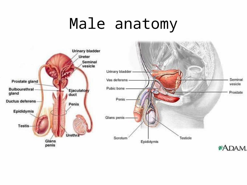
Male anatomy• Testes- male gonad, produces sperm and hormones
– Held in scrotum maintains cooler temp• Epididymis- sperm develops, leaves during ejaculation-
expulsion of sperm with other fluids• Seminal vesicles- secrete fluid to nourish and protect
sperm• Prostate gland- secretes alkaline fluid, protects sperm
from urine in urethra and vagina• Bulbourethral gland- secretes lubricating fluid • Sperm + glandular secretions = semen• Penis- allows for copulation, consists of shaft, glans,
prepuce

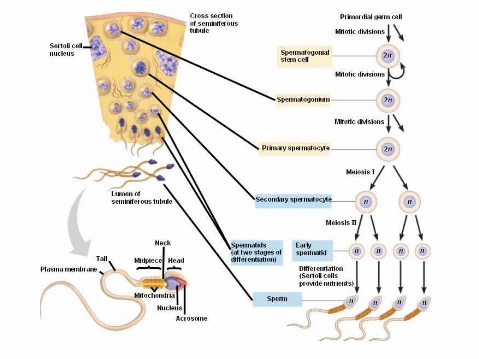
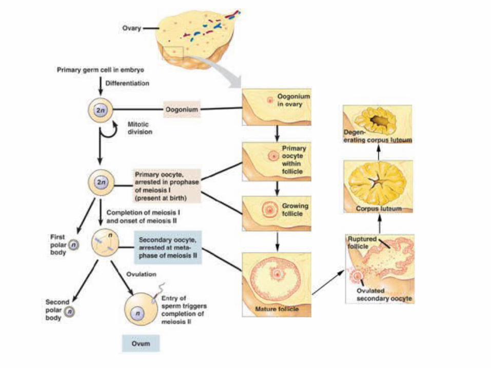
Sperm and ova formation
• Spermatogenesis- in seminiferous tubules in testes
• Oogenesis- begins prior to birth when diploid cell in follicle begins meiosis– At birth each follicle contains dormant primary oocyte– Hormonally triggered to further develop- after puberty
every 28 days– Unequal division of cytoplasm creates an egg and 3
polar bodies– LH- lutenizing hormone- triggers ovulation

Menstrual cycle
• Menstruation- shedding of uterine lining
• Hormones before ovulation- – Releasing hormone from hypothalamus stimulates
anterior pituitary to increase output of FSH and LH– FSH- follicle stimulating hormone- stimulates growth,
in turn follicle produces estrogen peaks before ovulation, triggering burst of FSH and LH

Menstrual cycle
• Hormones at and after ovulation-– LH peak- stimulates meiosis completion, follicle
ruptures and egg is released, corpus luteum develops promotes secretion of estrogen and progesterone by corpus luteum
– Estrogen and progesterone (high levels)- triggers a stop in FSH and LH release- corpus luteum breaks down
– When all levels are low (and if embryo isn’t implanted)- new cycle begins


STI’s
• Figure in section 27.7 is a good overview
Contraception
• Deliberate prevention of pregnancy– Only abstinence is totally effective– 3 types of contraception
• Prevent release of gametes • Prevent fertilization • Prevent implantation

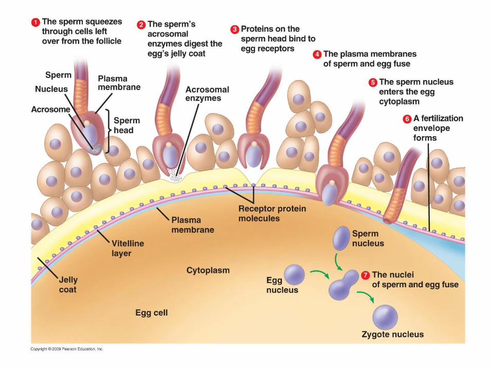
Fertilization • Only 1 sperm enters egg• Sperm cell
– Head contains nucleus and membrane bound sac- acrosome- containing enzymes that help sperm penetrate egg
– Neck and midpiece- contain mitochondria- sperm gets nutrients from semen and makes ATP to help getting to the egg
– Tail- flagellum- for movement

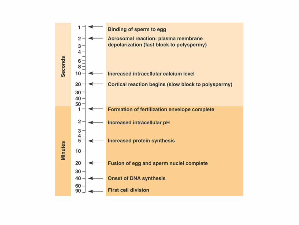
Fertilization
• Sperm meets egg:– Acrosomal enzymes digest outer layer of egg– Species specific proteins bind to surface of 2nd layer-
sperm then moves through layer– Sperm’s plasma membrane fuses with egg’s
• Triggers changes in egg cell• 1- membrane becomes impermeable to sperm• 2- 2nd layer hardens and separates from plasma
membrane- becomes fertilization envelop• 3- burst of metabolic activity in egg
– Sperm nucleus enters egg

After fertilization:
• Cleavage- rapid mitosis of zygote to produce ball of cells, partitions embryo into developmental regions– Blastula- hollow ball of cells

Then:• Gastrulation- sorts cells
into layers and adds more cells – Gastrula- 3 layered ball of
cells– Ectoderm- skin, nervous
system– Mesoderm- internal organ
systems– Endoderm- digestive tract

Then:• Organ formation (after
gastrulation)– Once layers form- cells
start to differentiate into tissues and organs
– Notochord- cartilage-like substance- provides support for developing tissues and later form backbone
– Neural plate and fold- rolls up to produce neural tube- which becomes brain and SC

What else is happening:
• Changes in cell shape- form new structures
• Cell migration- follows chemical trails
• Programmed cell death- apoptosis- Ex: between fingers and toes

Development
• Progresses as signals pass between cells– Induction- 1 cell group influences
development of adjacent cells• Switches on genes to differentiate into
tissues, allows for specialization– Pattern formation- shapes body plan- master
control genes respond to signals that tell where they are in relation to others

Gestation • Carrying young internally while they develop
– Cleavage after 24hrs– 6th/7th day- reaches uterus (100 cells)– Human blastocyst- l layer forms fetus, other- tropoblast- secretes
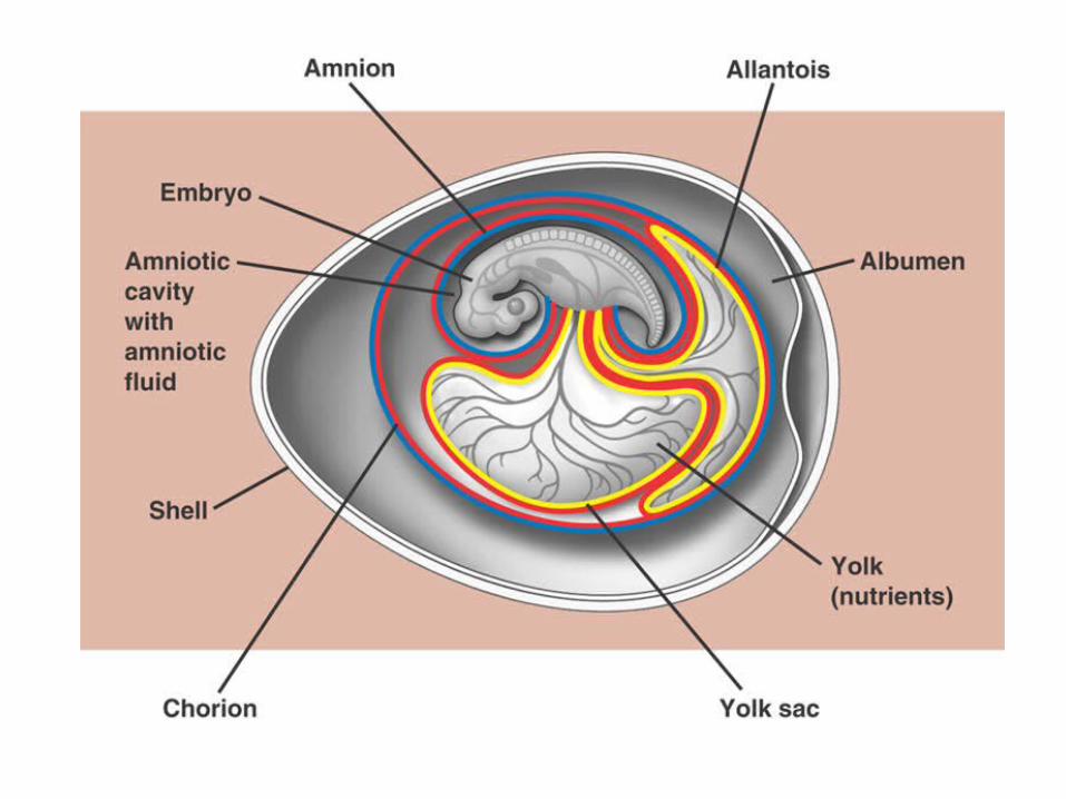
enzymes to allow for implantation, then forms part of placenta– Placenta- organ of nourishment, gas exchange and waste
removal– Day 9-gastrulation– Amnion- fluid filled, protects embryo– Yolk sac- (no yolk)- produces 1st blood cells, and germ cells
(cells that give rise to gamete forming cells)– Allantois- forms part of umbilical cord and bladder– Chorion- becomes embryo’s part of placenta, secretes HCG-
maintains corpus luteum (stops menstruation)
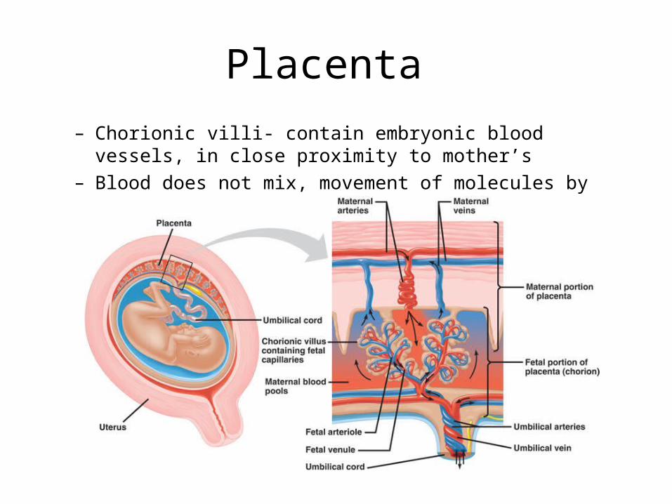
Placenta
– Chorionic villi- contain embryonic blood vessels, in close proximity to mother’s
– Blood does not mix, movement of molecules by diffusion
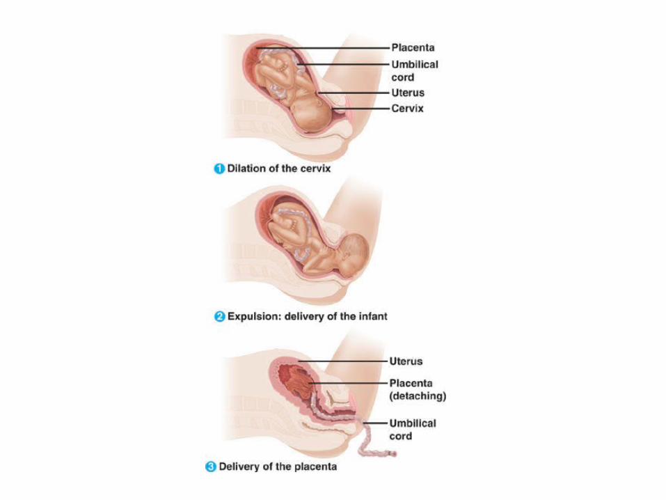
Birth
• Estrogen- triggers oxytocin receptors to form on uterus• Oxytocin- stimulates smooth muscle contractions of
uterus– ** positive feedback- intensifies production and effect
• Cervix needs to dialate, baby is birthed, placenta is delivered
• After birth levels of estrogen and progesterone decrease, allows pit gland to secrete prolactin- milk production


Reproductive technology
• IVF is one example