reproduction and embryonic development of the sand tiger shark, odontaspis taurus (rafinesque)
TRANSCRIPT

REPRODUCTION AND EMBRYONIC DEVELOPMENT OFTHE SAND TIGER SHARK, ODONTASPIS TAURUS (RAFINESQUE)1
R. GRANT GILMORE" JON W. DODRILL', AND PATRICIA A. LINLEY'
ABSTRACT
The capture of one ripe male, 191.5 cm TL, and 26 pregnant female, 236.6-274.3 cm TL, sand tiger sharks,Odontaspis taurus, from the east-central coast of Florida from 1946 to 1980 has permitted examination ofearly reproductive activity and embryonic development in this species.
Variations in ovulation rates and oviducal gland activity produce six distinct egg capsule types at varyingtimes during gestation. Some egg capsules produced during early gestation contain only ovalbumin and/ormucus while others contain several fertilized ova. As gestation proceeds, more capsules contain unfertilizedova and ovulation rates increase. These latter capsules serve principally as food for the survivingembryo.
Sixty-two embryos, 13-1,060 mm TL, provided information on intrauterine development which allowedclassification ofseven developmental periods based on gestation time, embryonic anatomy, posture, activity,and source of nutrition. Initially, embryos 13-18.5 mm TL obtain nutrition from internal coelomic yolk supplies during a period of early tissue differentiation. In embryos between 18.5 and 51 mm TL, consumption ofencapsulated yolk supplies occurs until hatching, between 49 and 63 mm TL. After hatching, the embryo absorbs yolk-sac nutritive supplies and may also consume uterine fluid. At about 100 mm TL, the embryobegins to hunt and consume other intrauterine embryos. Seven to nine months into gestation, ova are no longer fertilized. In each uterus, the single remaining embryo, 334-1,060 mm TL, consumes enlarged yolk capsules containing 7-23 unfertilized ova. Just prior to parturition the maternal ovary is greatly reduced in size.few egg capsules are found within the uteri, and in each uterus the remaining embryo exhibits reduced yolkconsumption and an enlarged liver. Parturition observed in captivity typically takes place from Decemberthrough March, after 9-12 months of gestation. Newborn juveniles are about 100 cm long.
The sand tiger shark, Odontaspis taurus (Rafinesque,1810), is a cosmopolitan species distributed in subtropical and temperate waters at depths <60 m (Basset a1. 1975). In the western Atlantic, adult sand tigersharks occur from the Gulf of Maine to Brazil(Bigelow and Schroeder 1948). Although sand tigersharks have been captured on both coasts of Florida(Springer 1938, 1948, 1963; Clark and von Schmidt1965), captures have been more common along theFlorida east coast (Dodrill4).
Unlike the adults, free-swimming juvenile O. taurusin the western Atlantic are restricted only to temperate (Bigelow and Schroeder 1953) and warmtemperate waters, extending as far south as northernFlorida. Juveniles 109.3-157.7 cm in total length(TL) have been recorded in neritic waters from the
'Contribution No. 305, Harbor Branch Foundation, Inc., FortPierce, Fla.'Harbor Ifranch Foundation, Inc., R.R. 1, Box 196, Fort Pierce,
FL 33450.'District V Naturalist, Division of Recreation and Parks, Florida
Department of Natural Resources, Rt. 1, Box 107-AA, Clermont,FL32711.
'Dodrill, J. W. 1977. A hook and line survey of the sharks foundwithin five hundred meters off shore along Melbourne Beach,Brevard County, Florida. UnpubJ. M.S. Thesis, 304 p. Fla. Inst.Techno!., Melbourne, FL 32901.
Manuscript accepted September 1982.FISHERY BULLETIN: VOL. 81, NO.2, 1983.
vicinity of Fernandina Beach (lat. 30°40'N, NassauCounty) on the Florida Atlantic coast, from CedarKey (Iat. 29°15'N, Levy County) in the northeasternGulf of Mexico (Don Hoyt5), and from the northernGulf of Mexico (Branstetter 1981),
In the western Atlantic, females with near-term embryos have been captured off eastern Florida and inthe northern Gulf of Mexico (Springer 1948; Hoytfootnote 5; Robert Jenkins6). At parturition, twoyoung are born (95-110 cm TL), one developing ineach uterus (Springer 1948; Cadenat 1956; Sadowsky1970; Bass et a1. 1975).Published observations on the early intrauterine
development of O. taurus are limited to the accountsof Coles (1915), Springer (1948), Cadenat (1956),and Bass eta1. (1975), Springer (1948) was the first toobserve embryonic oviphagy in O. taurus. He foundlarge quantities of yolk in the stomachs of embryosdissected from females from the northern Gulf ofMexico and east-central Florida. Bass et a1. (1975)described an intact 40 mm embryo found in the
'Don Hoyt, Florida Shark Club, Inc., Jacksonville, FL :12211, pers.commun.1967-77.'Robert Jenkins, Marineland Inc., St. Augustine, FL 32084, pers.
commun. 1977.
201

stomach ofa 170 mm embryo dissected from a femalefrom Natal, South Africa. These were the smallestembryos yet recorded from O. taurus and providedthe first description of embryonic cannibalism inthis species.The capture of 28 pregnant O. taurus from various
locations on the east coast of Florida (1946-80) provided 62 embryos, 13-1,060 mm TL (Table 1, Fig. 1).These specimens have allowed a more detailed description of early embryonic development in thisspecies than was possible previously. This study describes the various developmental stages in O. taurus based principally on embryonic anatomical development and changes in maternal gonadal morphology.
METHODS
All adult O. taurus specimens examined were captured either on rod and reel sport fishing gear or onstatic 10-30 hook set lines. Fourteen specimens werecaptured 200 m to 19 km from shore in neritic watersoff Melbourne Beach, Brevard County, Fla. (lat.
FISHERY BULLETIN: VOL. 81. NO.2
28°00'N, long. 80° 33'W). All specimens came fromdepths of 5-12 m. A 15th specimen was caught at lat.27°25'N, long. 80° 12'W, east of Fort Pierce Inlet, St.Lucie County, Fla. A 16th specimen, a 240 emfemale, gave birth to two pups at Sea World of Orlando, Fla., and all three were examined. This latteradult female was captured on 21 August 1980 at PortCanaveral, Brevard County (lat. 28°24.5'N). Elevenother specimens were captured prior to our study;these data and, in some cases, embryos from thesespecimens were included (Table 1).
Embryos and adult reproductive tracts were preserved in 10% Formalin7 and stored in 10% bufferedFormalin or 70% ethanol, or were frozen. All of thesespecimens were entered and catalogued into the Indian River Coastal Zone Museum (IRCZM). Eggdiameters and embryos <130 mm TL were measuredusing vernier calipers to the nearest 0.1 mm. Alllength measurements including total length (TL)follow Bass et al. (1975).
'Reference to trade names does not imply endorsement by theNational Marine Fisheries Service, NOAA.
TABLE I.-Uterine embryo and egg capsule data for Odontaspis taurus, from the Florida east coast, arranged chronologically by month ofexamination of embryos, 1947-81.
Adult No. of egg Encapsulated Damaged (8) or consumed (b) Hatched embryosTotal
sIze capsules in uteri embryos (mm. TLI embryos (mm. TL) (mm. TLIno. of
Date (em. TL) Left Right Left Right Left Right Left Right embryos
15 May 1977 254.5 20 20 0 0 0 0 0 0 016May1977 254.9 18 19 0 0 0 0 0 0 0
28 May 1977 260.3 26 27 0 0 0 0 0 0 0
5 June 1978 264.2 8 8 0 0 0 0 0 0 0
5 June 1976 258.1 35±2 34±2 41 42 0 0 57 1 3
5 June 1976 262.5 20±2 20±2 38' 38' 0 0 0 0 2+1
5 June 1976 263.2 20-35 20·35 38' 38' 0 0 0 0 2+1
6 June 1978 274.3 8 8 0 0 0 0 0 0 0
9 June 1976 249.5 29±4 29±4 27 31 0 0 0 0 2
28 June 1976 254.1 47 53 27.34 27.38.46 0 0 63 62 7
8 July 1978 274.2 66" 69" 13.18 1 49(a) 45(a).49(a) 131 131 7
18 July 1976 271.5 78 81 34 0 0 51 (a) 127 100 4
27 July 1975 263.0 7 7 / 7 0 0 317 317±10 2
29 July 1977 254.0 77" 77" 0 0 0 0 271 227 2
5 Aug. 1976 236.6 68 65 0 0 9(b). n(b). 30(b) 41(b) 334 320 9
35(a). 36(b). 41 (al4 Sept. 1970 282.5 17.5 18.5 7 / 7 2+/
4 Sept. 1970 269.2 / 7 / / 330±10 330±10 2
3 Nov. 19621 7 7 7 / / / 650 1
8 Nov. 1954' 7 / 7 7 / / / 830 890 2
24 Nov. 19472 273.0 0 0 0 0 0 0 970 960 2
24 Nov. 1947' 239.0 0 0 0 0 0 0 825 0 1
12 Dec. 1976' 266.7 7 / 7 / 7 1 1,000±10 1.000±10 2
30 Dec. 1958' 261.6 / 7 7 / 7 / 1.025 1.033 2
22 Jan. 1947' / / / 1 / / / l,OOO±10 1.000±10 222 Jan. 1947'2 1 / / / / 7 / 0 0 7
15 Feb. 1959' 261.5 7 / 7 7 7 / 1,080 >1,060 2
9 Mar. 19472 •4 272.0 7 7 7 7 1 1,050 1.03~, 2
22 Mar, 1981 '240.0 / 7 7 1 7 910 2
-length given 8& 1.5 inches, therefore not accurately determined.••Blastodiscs were observed on some eggs.1 Egg capsules and embryos could have been present but were not recorded.'F. G. Wood, formerly of Marineland Inc.. 51. Augustine. FL 32084, pers. commun. 1976~77.
2Springer 1948.'E. Herbe", Florida Shark Club. JeckaonYille, FL 32211, pera. commun. 1976·77."A. McBride. Curator. Marineland Inc .. St. Augustine. FL 32084, unpubl. data. 1947.'Specimens were Btill living in captivity April t 983 at Sea World of Orlando. Fla.
202

GILMORE ET AL.: REPRODUCTION AND EMBRYO DEVELOPMENT OF SAND TIGER SHARKS
The entire reproductive tract was removed and examined as fresh, frozen, or preserved material.Uterine fluid volume was determined by tying offboth ends of the uterus in a fresh specimen, removingthe uterus, making a small incision in the uterine wall,and allowing the contained fluid to drain into agraduated flask. Selected preserved ovaries were cutinto sections which were weighed to the nearest 0.1 g.Ovarian egg counts were made by counting all macroscopic eggs in two preserved sections from anovary of known weight. These ova counts were thenmultiplied by the ratio oftotal ovarian weight/section
weight, to predict the total number ofova in the entireovary.
A 13.0 mm TL embryo taken from an egg capsulefrom a shark caught on 8 July 1978 was embedded inparaffin, cut on a rotary microtome at 6 /.lm on a sagittal plane, and stained with a Cason modification ofthe Mallory-Heidenhain stain (Humason 1972).
Fresh sperm samples were fixed in 2.5% glutaraldehyde, prepared for scanning electron microscopy, and examined on a Zeiss Novascan. SeveralPolaroid electron micrographs were taken forsperm descriptions.
1,100
1,000
900
800
700
E.s.l: 6000.cCD..J0 500>-...cEw
400
300
200
100
------
.-----~--:--........, /./
//
'//
I'I
//
//
I/
/I
I/
//
I/
/~ /
/. //
//
//'
//
/'
-_ ....... '. !.
Apr. I May I June I July I Aug. I Sept. I Oct. I Nov. I Dec. Jan. Feb. I Mar.
18,...-----------------,
16
14
12
10
8
6
2
oJ.,.UI..,L~LI,~~I,_!'I_r_~J.!AH'r;:d~~
FIGURE l.-Recorded lengths of embryos taken from Odolltaspistaurus females captured 1946-80 along the Florida east coast. withhypothetical growth curve (above) of embryos and monthly numbersof adult males and females (bar graph below) captured during thesame 34-yr period.
203

Drawings and Kodachrome transparencies weremade of various embryos, egg capsules, and reproductive organs.
OBSERVATIONS ANDDESCRIPTIONS
Mating Activity
(Mating Period, Location, and Spermatozoa)
The occurrence of similar-size males or females inunisexual groups has been documented on severaloccasions (records of the Florida Shark Club show107 0. taurus landings, Burton 19328 ; Sadowsky1970; Bass et a1. 1975; Hoyt 1976-7'7 see footnote 5;Wood 1976-779). These observations show thatfemale groups of O. taurus make coordinatedseasonal coastal movements possibly for breeding,gestation, and eventually parturition (Fig. 1).Females captured at the same time and location tended to have embryos in the same state of development, suggesting coordinated breeding activity andpostbreeding migrations. Observations of many annual cycles from 1947 to 1981 established winterspring as a breeding period off the Florida east coastand provided comparisons of data on gestation (i.e.,embryonic development rates and seasonality).
A 191.5 em TL ripe male O. taurus, captured 8 February 1980 in shallow water (10 m depth) in thevicinity of Fort Pierce Inlet, St. Lucie County, Fla.(lat. 27°25.TN, long. 800 12.5'W), showed evidence ofrecent mating activity. His claspers were turgid andhematose, with sperm and seminal fluid activelyflowing from the clasper tip. The testes were also enlarged (22.5 X 3.5 em, 0.68 kg). A larger 203 em TLmale examined from Fort Macon, 1.5 km west ofBeaufort Inlet, N.C. (lat. 34°40'N, 10 January 1978),contained testes which were considerably smaller(8.0 X 5.0 em, 0.064 kg). Several scanning electronmicrographs were made of the sperm from the 8 February 1980 male specimen. A single sperm had a typical chondrichthian helical head structure 31 fJ.m longand a tail 40.3 fJ.m long (Fig. 2B). The entire length ofthe sperm was 69-71.5 IJom. Living sperm were observed to rotate about their long axes, propelled by thecircular motion of the extended tails.
Mating scars resulting from copulatory activity
"E. M. Burton, The Charleston Museum, Charleston, S.C., pers.commun. 24 Oct. 1932 to J. T. Nichols, American Museum ofNatural History, N.Y. (made available by Stewart Springer, MoteMarine Lab., Sarasota, FL 33577).'F. G. Wood, Marineland Inc., St. Augustine, FL 32084, pers. com·
roun. 1976-1977.
204
FISHERY BULLETIN: VOL. 81, NO.2
have been commonly observed in female "galeoid"sharks; however, it appears that courtship scars onmales are rare (Springer 1967; Stevens 1974; Pratt1979). Springer (1960) had noted the presence offresh cuts on female Eulamia milberti (= Carcharhinus plumgeus) in correlation with the presenceof early embryos. Springer (1963) found that most ofthe O. taurus taken in a shark fishery operating in theAtlantic off east-central Florida were females with ahigh incidence of courtship scars; but no dates weregiven for these observations. Odontaspis taurusfemales we captured on 9 June and 5 August 1976(Table 1) had tooth puncture wounds between the1st and 2d dorsal fins. The 191.5 cm male, taken on 8February 1930 offFort Pierce Inlet, had been recently raked by another shark along the upper left side ofthe body behind and above the gill openings (Fig.2A). This wound consisted of eight incisions, createdby a narrow, long tooth rather than a flat, wide bladetooth, typical of many carcharhinid sharks. As 0.taurus has a long narrow tooth cusp, it is possible thatthe wound was the result of either an attack by, orcopulation with, another sand tiger shark. These observations indicate that copulatory activity may takeplace off the Florida east coast and therefore accountfor the following observations of the earliest embryonic development in specimens from this geographical region.
Early Gonadal andEmbryonic Developmental Period
(January-September; 0-60 mm TL)
General Female Anatomy
The female reproductive tract of O. tauru, may bedivided into the ovary, ostium, anterior oviduct,oviducal gland, isthmus, uterus, andvagina, typical ofmost galeoid sharks. Only the right ovary isfunctional and enlarged. Above the ovary and attached to it via membranous connective tissue(mesovarium) is the ostium which collects ovulatedova and distributes them to the oviducts. The twooviducts (paired, right and left) bifurcate from the ostium. The anterior oviducts are about 9 mm indiameter and 300 mm in length from ostium to oviducal glands in a 254 cm female. The heart-shapedoviducal glands (53 X 93 mm in the same female)function in egg capsule formation. Much larger thanthe anterior oviduct, the portion of the oviductfollowing the oviducal gland known as the isthmus is20-34 mm in diameter, allowing for the passage ofmultiple encapsulated ova. The isthmus opens into

GILMORE ET AL.: REPRODUCTION AND EMBRYO DEVELOPMENT OF SAND TIGER SHARKS
FIGUIlE 2.-A 191.5 cm TL ripe male Odonlaspis taurus captured 8 Februll/)' 1980 off Fort Pierce Inlet, Fla. (A)Tooth rake scars along upper leftside ofbody behind and above gill openings. (B) Scanning electron micrograph of a69-71.5 J.Lm sperm, with head structure 31 J.LIn long and tail 40.3 J.Lm long (l,950X magnification).
205

FISHERY BULLETIN: VOl;. 81. NO.2
the uterus which is heavily folded and vascularizednear its opening. The 7-8 mm uterine wall in 0. taurusdoes not function in placentation as in carcharhinidsharks. The paired uteri unite posteriorly to form acommon vagina.
Ovarian Activity
This period begins from January to April with insemination of the female O. taurus and extends intothe following September as exhibited by the prolonged fertilization of ova via stored sperm. Ova fertilization apparently occurs in the anterior oviduct oroviducal gland (Fig. 3Ba, b) prior to egg capsule formation. The oviducal gland then produces a varietyof collagen egg capsules, some of which contain fertilized ova (Figs. 4, 5). Egg capsules are thendeposited in the uterus. Although encapsulated embryos are present in the uterus for 5-6 mo, thedevelopment of a single embryo from fertilization tohatching in utero takes about 3-4 mo.The number of ova and the general overall size of
the ovary increased during early pregnancy. Duringthis period, ova diameters ranged from 2.0 to 10.2mm and weights ranged from 1.6 to 410 mg. A 254.5cm TL female O. taurus captured 15 May 1977 contained a 4.6 kg ovary with 22,180 ova 1.3-10.0 mm indiameter (Table 2). Encapsulated fertilized ova (Le.,blastodiscs were evident) were present in the uterus,but no embryos. All 11 sand tiger sharks examined
between June and August possessed greatly hypertrophied right ovaries (left ovaries are atrophic andnonfunctional) weighing between 3.7 and 8.5 kg andtaking up considerable space (360-455 mm in length)in the body cavity (Fig. 3, Table 2). The largest ovary(8.5 kg) came from an 8 July 1978 sand tiger shark,which also had two embryos that were past the "early" uterine developmental stages and in the "posthatch" cannibalistic stage during which consumption of ova would be their primary means ofnutrition.
Oviducal Gland Activity
The paired oviducts ofO. taurus may be divided intofour basic sections (Fig. 3B). The anterior portion (a)is a narrow tube lined with ciliated columnar epithelial cells, extending between the ostium and theoviducal or nidamental gland. This anterior tube is310 mm long in a 254 cm sand tiger shark and about 9mm in diameter. The oviducal gland (b) secretesmucus, ovalbumin, and the major elasmobranch eggcase component, collagen (Wourms 1977). Neitherthe anterior portion of the oviduct nor the oviducalglands were sectioned and examined in detail forsperm storage; therefore, the exact site of fertilization in O. taurus remains unknown. However, fertilization must occur prior to encapsulation of the ovain the shell membrane or collagen egg capsule. Encapsulation takes place within the 'oviducal gland.
TABLE 2.-Comparative reproductive data for female Odontaspis taurus arranged chronologically by month ofcapture. 1947-78.
Ovary
3.7
Weight (kg) Length (em)
40
4.8 36
2
9
2
oo
Embryosin both
uteri
3-10
2·9,5
>1-10
2.5·10.2
Ove
13.200 >5-10
No. Size (mm)
12.810
24.000
24.290
>1.000 >10
22.180 1.3-10.0
>1.000
>1.000
31.0
45.5
41.4
45.7
4.5
8.5
249.5
236.6
312.5
274.2
254.1
296
254.5
254.0
Shark
(em. TL)Date Location
24 Feb. 1960 Gulf of Mexico1
15 Mey 1977 West AtlanticBrevard Co:. Fla.Melbourne Beach
9 June 1976 West Atlantic
Brevard Co.. Fla.
Melbourne Beach
28 June 1976 West Atlantic
Brevard Co.. Fla.Melbourne Beach
8 July 1978 West AtlanticBrevard Co., Fla.
Melbourne Beach
27 July 1947 Gulf of Mexico2
Chandleur Is.. La.
29 July 1977 West AtlanticBrevard Co.. Fla.Floridans Beach
5 Aug. 1976 West AtlanticBrevard Co.• Fla.Melbourne Beach
24 Nov. 1947 West Atlantic 1
Brevard Co.. Fla.
Off Cape Canaveral
273 >100 10
1 Clark and Von Schm'.dt 1965.2Springer 1948.
206

GILMORE ET AL.: REPRODUCTION AND EMBRYO DEVELOPMENT OF SAND TIGER SHARKS
FIGURE 3.-A 254 cm female Odontaspis taurus captured 29 July 1976 at Melbourne Beach, Fla. (A) Enlarged ovary with ova (llandb) extending tlU'ough damaged portions of ovarian membrane. (B) Oviduct consisting of (a) thin tube Icading from ostium to (b)oviducal gland; (c) isthmus, and (d) uterus containing cmbryos and egg capsules.
207

FISHERY BULLETIN: VOL. 81. NO.2
FIGURE 4.-Dissection from a female Odontaspis taurus of (A) oviducal gland (right) and isthmus containing a Type I egg capsulewhich had 16 ova; (B) two Type V "short tail" gel capsules leaving the oviducal gland.
This bulbous organ varies in size with reproductiveactivity and produces a wide variety of egg capsules(Figs. 4, 5). Eggcapsules leave the oviducal gland andproceed down the elastic narrow isthmus (Fig. 3Bc),250-350 mm in length, 20-34 mm in diameter, connecting the gland with the expanded uterus (Fig.3Bd). There is an increase in vascularization and
208
folding of the inner epithelial lining where theisthmus joins the enlarged uterus.The size of the uterus, the volume of the uterine
fluid, and the length of the isthmus increased duringearly gestation (June to July). The volume offluid in asingle uterus increased from 260 ml, to 325 ml, to1,600 ml in specimens from 15 May, 28 May, and

GILMORE ET AL.: REPRODUCTION AND EMBRYO DEVELOPMENT OF SAND TIGER SHARKS
FIGUHE 5.-(A) Type V "short tail" gel capsules from Odolliaspis laurus, containing ovalbumin and/or mucus; (B) Type IV "long tail" gel cap·sules; (e) Type I blastodisc capsule, containing 16 ova; (D) Type II ovoid yolk capsules, containing 18 ova each. (Photo courtesy MarinelandInc., t. Augustine, Fla.)
29 July, respectively. The uterine fluid also increasedin relative cloudiness and contained numerous ruptured egg capsules and yolk fragments.During early gestation the oviducal gland produced
at least six distinct types of egg capsules (Figs. 4,5):
Type I blastodisc capsules (Figs. 4A, 5C)-contain 718 ova, 1-14 of which have visible blastodiscs.This capsule type was more prevalent during earlygestation, as 83% of the intact capsules, examinedin a 16 May 1977 specimen, contained blastodiscs, 25% in a 28 May 1977 specimen, 22% in a28 June 1976 specimen, and none in a 5 August1976 specimen. The overall capsule number wasgenerally low, 15 or fewer per uterus.
Type II ovoid yolk capsules (Fig. 5D)-consist of alight amber shell membrane enclosing a roundedbulbous head containing the ova and a flattenedtransparent amber tail 40-58 mm long. We foundthese capsules to contain a large yolk volume con-
sisting of 7-18 ova (mean = 11), 10 mm indiameter, with no sign of fertilization (i.e., no blastodisc). Springer (1948) found 16-23 ova (mean =19) per capsule of this type in a female containing260.4-266.7 mm emblYos. Ovoid yolk capsules increased in numbers as Type I blastodisc capsulesdeclined. Dimen ions of Type II cap ules rangedfrom 21 to 29 X 78 to 118 mm and weight from 8.6to 19.4 g. Ova in the egg mass during the first 2-3mo of pregnancy comprised only 60-80% of thecapsule volume, while the remainder consisted ofovalbumin and/or mucus adjacent to the tail. Agelatinous ovalbumin/mucoid substance al 0
lined the inner walls of the egg capsule.Type ill reduced yolk capsules-have the same
dimensions as Type II ovoid yolk capsules butcontain only 1-3 ova. Type III capsules were observed only during the first 3 mo of gestation.
Type IV "long tail" gel capsules-contain amber,green, or white gelatinous material, fluid, and noova. Although not determined, the variably
209

colored gelatinoun material probably containsovalbumin and mucus, in different proportions.The dimensions are usually similar to those of theType II capsules but may vary, as total lengths ofup to 170 mm were observed in capsules with verylong tails (Fig. 5B). These capsules were mostcommon during the first 3 mo of gestation.
Type V "short tail" gel capsules (Figs. 4B, 5A)- arethe smallest capsules, are generally flattened, andcontain only gelatinous ovalbumin/mucoid material. These capsules were also most commonduring the first 3 mo of gestation.
Type VI embryo capsules-contain an embryo and areduced volume of yolk. Despite the presence ofmultiple ova and several blastodiscs in embryocapsules, dissection of all Type VI capsules failedto show more than one embryo developing withina single capsule.
Prior to entering the uterus, egg capsules of thesame type were found in similar positions in bothoviducts of a particular adult. No matter how manyeggs were ovulated, encapsulation of albumin wouldoccur synchronously in each oviducal gland, thus producing egg capsules of the same type at the same
C
/ .,.".".
.
" ,"i-'". ,
FISHERY BULLETIN: VOL. 81. NO.2
time. Calculation of egg capsule production rates,based on changes in uterine capsule numbers, indicates that capsule formation takes place at 24-36 hintervals. Initial egg capsules contain ovalbumin and/or mucus derived from the oviducal gland. As theovulation rate and the number and volume of ova increased during later stages of gestation, more ovawere present in the oviducal gland when encapsulation occurred. At this time only ovoid yolk capsules,Type II, were found in the oviduct and uterus.
Embryonic Development
Multiple embryos from O. taurus develop in eachuterus during the early stages of gestation. However,the maximum number of capsules containing macroscopic embryos is low (no more than 9% or 2-7 of allcapsules in both uteri combined at any given time).Encapsulated embryos were found from June to September. The maximum number of embryos in a singleuterus was seven, ranging in size from 19 to 334 mmTL. Four of these seven were found in the mouth andstomach of the largest embryo. After June, the number of undamaged encapsulated embryos and thepercentage of capsules with blastodiscs declined.
8
FIGURE 6.-Threeviewsofa 13 mm embryo (IRCZM 103179) taken from an adultOdontaspis taurus, 274.2 em long, eaptured8 July 1978. (A)Left side; (Bl dorsal; (el ventral.
210

GILMORE ET AL.: REPRODUCTION AND EMBRYO DEVELOPMENT OF SAND TIGER SHARKS
13 MM EMBRYO (IRCZM 103179, Figs. 6-8).The 13 mm embryo is described from one of four embryos, 13-131 mm, taken from the left uterus of a274.2 em sand tiger shark caught 8 July 1978 (Table1). This and an 18 rom embryo were undamaged andencapsulated, while three other embryos partiallyencapsulated or free within the same sand tiger sharkwere damaged by attacks from two larger 131 mmembryos, one in each uterus. The 13 mm embryo wasthe smallest examined. It contained yolk both internally and in a yolk sac. The embryo was obviously restricted in mobility appearing as little more than ayolk mass with a head, notochord, and minute pectoral fin buds. The 13 mm embryo resembles anamphibian embryo after gastrulation and formationof primary organ rudiments. It does not resemble theearly embryos described for other elasmobranchs[e.g., Heterodontusjaponicus (Smith 1942); Chlamydoselachus anguineus (Gudger 1940); Mustelus canis(TeWinkeI1950, 1963)1. Histological sections showedan incomplete connection between internal yolk supplies and an external yolk sac (Fig. 8A). A membraneat the junction of the yolk stalk and the yolk sac ap-
O.1mm-
pears to isolate the yolk-sac yolk from the yolk stalkand coelomic yolk supplies in the 13 mm embryo. Thecoelomic cavity, cardiac stomach, valvular intestine,and pericardial cavity all contained yolk. The maximum horizontal diameter of the embryo was 9 mm,due principally to the contained yolk. This diameterwas greater than that of the yolk sac (6.0 mm). The gillarches and mouth cavity were open, but the latter waslacking dentition. No retinal tissue was seen andgonadal tissue was undifferentiated.
18.5 MM EMBRYO (IRCZM 103134, Fig. 9).The 18.5 mm embryo was from the right uterus of a282.5 em TL female O. taurus captured 4 September1970. Although encapsulated, the embryo and thecapsule hac! been greatly damaged. This embryo wassimilar to the 13 mm embryo but differed in havingless internal yolk and greater differentiation of external features. A spiracle was present as were first andsecond dorsal, caudal, anal, and pelvic fin buds in addition to the pectoral fin buds which had developedearlier. The yolk sac was 6.0 mm in diameter as in the13 mm embryo.
FIGUIlE 7.-Angle horizontal sagittal view of a 13 mm Odonlasp; taurus embryo (IRCZM 103179). head and branchial region: (b) brain;(0) orbit; (ga) gill arches; (ysy) yolk sac yolk.
211
A
•
~7'O.1mm-
FISHERY BULLETIN: VOL. 81. NO.2
B
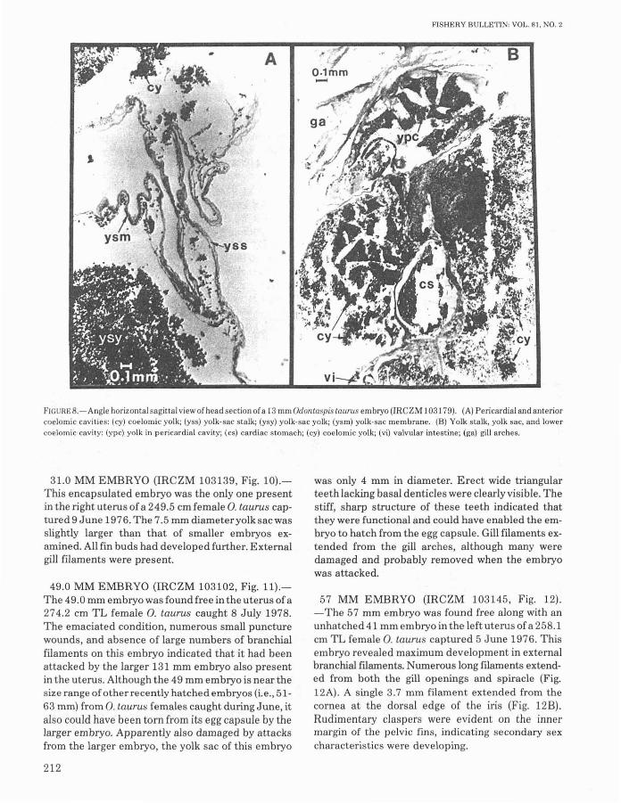
F'IGUHE B.-Angle horizontal sagittal view of head section ofa 13 mm Odontaspis taurus embryo (IRCZM 103179). (A) Perical'dial and anteriorcoelomic cavities: (cy) coelomic yolk; (yss) yolk-sac stalk; (ysy) yolk-sac yolk; (ysm) yolk-sac membrane. (B) Yolk stalk, yolk sac, and lowercoelomic cavity: (ypc) yolk in pericardial cavity; (cs) cardiac stomach; (cy) coelomic yolk; (vi) valvular intestine; (ga) gill arches.
31.0 MM EMBRYO (IRCZM 103139, Fig. 10).This encapsulated embryo was the only one presentin the right uterus of a 249.5 cm female 0. taurus captured 9 June 1976. The 7.5 mID diameter yolk sac wasslightly larger than that of smaller embryos examined. All fin buds had developed further. Externalgill filaments were present.
49.0 MM EMBRYO (IRCZM 103102, Fig. 11).The 49.0 mm embryo was found free in the uterus of a274.2 cm TL female 0_ taurus caught 8 July 1978.The emaciated condition, numerous small puncturewounds, and absence of large numbers of branchialfilaments on this embryo indicated that it had beenattacked by the larger 131 mm embryo also presentin the uterus. Although the 49 mm embryo is near thesize range of other recently hatched embryos (i.e., 5163 mm) from 0. taurus females caught during June, italso could have been torn from its egg capsule by thelarger embryo. Apparently also damaged by attacksfrom the larger embryo, the yolk sac of this embryo
212
was only 4 mm in diameter. Erect wide triangularteeth lacking basal denticles were clearly visible. Thestiff, sharp structure of these teeth indicated thatthey were functional and could have enabled the embryo to hatch from the egg capsule. Gill filaments extended from the gill arches, although many weredamaged and probably removed when the embryowas attacked.
57 MM EMBRYO (IRCZM 103145, Fig. 12).-The 57 mm embryo was found free along with anunhatched 41 mm embryo in the left uterus ofa 258.1cm TL female 0. taurus captured 5 June 1976. Thisembryo revealed maximum development in externalbranchial filaments. Numerous long fliaments extended from both the gill openings and spiracle (Fig.12A). A single 3.7 mm filament extended from thecornea at the dorsal edge of the iris (Fig. 12B)_Rudimentary claspers were evident on the innermargin of the pelvic fins, indicating secondary sexcharacteristics were developing.

GlLMORE ET AL.: REPRODUCTION AND EMBRYO DEVELOPMENT OF SAND TIGER SHARKS
1mm>----<
" .. :../.'. ;":' .....
1mm
FIGURE 9.-Two views of an 18.5 mm Odolliaspis taurus embryo (lRCZM 103134) taken from the right uterus of a 282.5 mm TL female cap·tured 4 September 1970, showing damage by intrauterine attacks from larger embryo.
Posthatch and IntrauterineCannibalistic Period
(June-September; 60-334 mm)
This period is characterized by hatching of thelargest encapsulated embryos, consumption of yolksac yolk supplies, and active cannibalism by thelargest hatched embryo upon other intrauterine encapsulated or small hatched embryos until only oneembryo remains. These events occur simultaneouslyin each uterus. From June to September thisdevelopmental period overlaps the latter part of theearly gestation phases of other sibling embryos.Two hatched embryos, 62 and 63 mm, (Fig. 13) from
each uterus of a late June sand tiger shark werenoticably more robust than five 27-46 mm embryosstill encapsulated in these uteri. However, there wasno evidence that the larger embryos had begun tofeed upon other egg capsules, encapsulated embryos, or other free embryos. The 62 and 63 mmspecimens still possessed 5.5-6.0 mm diameter yolksacs and branchial filaments.At about 100 mm, the embryo has consumed the
contents of the yolk sac and begins obtainingnourishment through adelphophagy and oophagy.Evidence of intrauterine cannibalism was found inthe uterus of a 271.5 cm female 0. taurus, caught 18July 1976, which contained a large hatched embryo(100 mm) that had attacked and badly damaged
213

FISHERY BULLETIN: VOL. 81, NO.2
1mm
FIGURE 10.-View of a 31.0 mm Odonta~pis taurus embryo (IRCZM 103139) taken from the right uterus of a 249.5 cm female captured 9June 1976.
.".11;"•.
FIGURE 1I.-Two views ofa 49 mm Odontaspis taurus embryo (IRCZM 103102) taken from a 274.2 cm TL female captured 8 July 1978, showing emaciation and injuries from intrauterine attacks by a larger 131 mm embryo.
214

GILMORE ET AL.: REPRODUCTION AND EMBRYO DEVELOPMENT OF SAND TIGER SHARKS
FIGURE 12.-(A) A 57 mmOdontaspis taurus embryo (IRCZM 103145) taken from a 258.1 em TL female captured 5 June 1976; (B) enlargement of orbit and spiracle showing associated filaments.
2mm.......
FIGURE 13.-Hatched 62 mm Odolltaspis taurus embryo with 6 mm yolk sac taken from right uterus of a female caught 28 June 1976.Melbourne Beach. Fla.
215

(puncture wounds and torn gut) a 51 mm embryo(drawn to scale; Fig. 14A). Having already developedteeth, the 51 mm embryo (see Figure 11 of a 49 mmembryo) had a potential for competitive interactionwith the larger 100 mID embryo, although at adecided size disadvantage. It is possible that the 51mm embryo had not hatched prior to the attack.However, empty and broken egg capsules were notfound in the uterus. There is no evidence that the 100mm embryo had tried to consume any of the other 81egg capsules in the uterus, nor were there broken ordamaged capsules in the opposite uterus which contained a 127 mm hatched embryo.
We obtained further evidence that hatched embryos and/or encapsulated embryos are selectivelypreyed upon by their larger siblings within the uterus.Two embryos (45 and 49 mm) in the right uterus of an8 July 1978 female O. taurus were badly damaged bythe attack of a 131 mm male embryo. Six empty eggcapsules were found within the same uterus. None ofthe other 63 egg capsules were damaged (some ofwhich contained fertilized ova). In the left uterus, a49 mm embryo had been mutilated by a 131 mm embryo and two of the 66 egg capsules were empty. A334 mm embryo from the left uterus of a 5 August1976 adult 0. taurus had four embryos 9-36 mm TLwithin its pharynx. Two damaged capsules still con-
FISHERY BULLETIN: VOL. 81. NO.2
tained two embryos (35 and 41 mm), both of whichhad been punctured numerous times through thecapsule membrane. Sixty-eight undamaged capsulesdid not contain embryos. None of the 65 undamagedcapsules in the right uterus contained embryos.However, this uterus contained an intact 41 mm embryo with an egg capsule fragment within thestomach of the largest embryo (320 mm).
100 MM EMBRYO (IRCZM 103137, Fig. 14B).This male embryo was found in the right uterus of a271.5 cm adult 0. taurus captured 18 July 1976. Ithad well-developed fin rudiments and a particularlywell-developed caudal fin. The gill slits were largeand without external filaments. Both upper and lowerlabial furrows were prominent. The yolk sac wasabsent although an attachment scar was present.Erect teeth, more slender than in previous embryos,were present in multiple rows. The teeth lackedlateral secondary basal cusps (basal dentides) typical of adult O. taurus. The teeth of this embryo wereobviously functional because punctured and torn eggcapsules and a damaged (tooth-marked) 51 mm embryo were found in the same uterus.
131 MM EMBRYO (IRCZM 103103, Fig. 14C).A male embryo, from an 8 July 1978 sand tiger shark,
B
(ur (.c
1cm
FIGURE 14.-(A) A 51 mm Odontaspis taurus embryo attacked and damaged by (B) a 100 mm male embryo inside the uterus of a 271.5 emfemale captured 18 July 1976 (both IRCZM 103137). (C) A 131 mm male embryo (lRCZM 103103) taken from the uterus ofa female captured8 July 1978. This embryo had attacked and damaged the 49 mm embryo shown in Figure 11.
216

GILMORE ET AL.: REPRODUCTION AND EMBRYO DEVELOPMENT OF SAND TIGER SHARKS
resembled the 100 mm embryo, except that all finsbut the pectorals were similar to those of the adultand the gut was more distended with yolk. This embryo had attacked the 49 mm (Fig. 11) and 45 mmembryos present in the same uterus.
227 AND 271 MM EMBRYOS (IRCZM 103101,Fig. 15A, B).-The 227 mm female and larger 271mm male embryo came from a 29 July 1977 sandtiger shark. The snout was narrow and had lengthened,resembling that of the adult as did other anatomicalfeatures, including the fins. In both embryos the entire digestive tract and abdominal wall were distended from the consumption of yolk. Many brokenegg capsules were also found within the uteri.
334 MM EMBRYO (IRCZM 103135, Fig. 15C).This was a female embryo from a 5 August 1976 sandtiger shark. The stomach was distended with yolk.Many "adultlike" features were apparent. This embryo contained four smaller embryos (9-36 mm) inits pharynx.
• coo
c
Late Gestation, Postcannibalistic,Oophagous, Preparturition Period
(September-March; 334-1,000 mm)
After fertilization of O. taurus ova has ceased and allother developing embryos have been consumed bythe surviving embryo, unfertilized ova become theprimary source of nutrition. This transitional periodbegins in August-September when embryo lengthsreach 330-340 mm.
Embryonic growth and development rates are rapidduring this period (Fig. 15C, Table 3). A 330 mm embryo in September may attain 650-890 mm by lateOctober or early November and 830-970 mm by lateNovember. During this period the embryo consumeslarge quantities of yolk and a length of 1.0 m may bereached in December (Figs. 15D, 16). Embryosreaching 1.0 m are near parturition which may takeplace between December and March, after a gestation period of 9-12 mo. A maximum size of 1.2 m TLmay be reached before birth (Cadenat 1956). A 272
B
FIGURE 15.-Four specimens of embryonic Odontaspis taurus showing progressive abdominal distention from consumed yolk: (A) A 227 mmfemale embryo (IRCZM 103101) from the right uterus and (B) a 271 mm male embryo (lR 2M 103101) from the leftutel'us, ofa female captured 29 July 1977; (C) a 33'1 mm female embryo (JRCZM 103135) taken from a female captured 5 August 1976; and' (D) an 80-100 cmembryo.
217

TABLE 3.-Postparturition growth [total length (TL) and totalweightl oftwo captive juvenile Odontaspis taurus from observationsmade by F. G. Wood at Marineland Inc., St. Augustine, Fla. NR =
not recorded.Male Female
TL Weight TL WeightDate (em) (kg) (em) (kg)
Born 15 Feb. 1959 NA NA NA NA17 Feb. 106 6.2 NA NA
9 Oct. 126 12.6 NA NA29 Dec. 137.5 19.1 139 19.130 Aug. 1960 NA NA 145 NA12 Dec. NA NA NA 37.5280ee. NA NA 167 NA16June1961 NA NA 175.5 40.7 died17 Mar. 1962 167.5 NA died'
Mean growth rate (TL) 1.62 em/rna 2.03 em/rna19.44 em/yr 24.36 em/yr
137 rna old, claspers extended 7.5 em past pelvic fin tip.
cm O. taurus female was captured 10 April 1946 andkept in an aquarium for 11 mo; it died on 9 March1947. Her autopsy revealed two decomposing nearterm embryos 103-105 cm TL (6.1 and 6.4 kg)(McBride 19471°; Springer 1948).
The oophagous stage in development is precededby an increase in ovary size, ovulation rate, number ofova per capsule, and number of Type II capsules produced. The number of ova per capsule increased to amaximum of 23 ovalcapsule during the fall and winter (Fig. 5D). During late gestation the embryosswallowed such great quantities of yolk that theirstomachs became greatly distended. Cadenat (1956)found 1.5 kg of yolk (18.8% total body weight) in anear-term 0. taurus embryo weighing 8 kg. This distention of the abdomen has precipitated the term"yolk stomach" used by earlier authors, particularlyfor the oophagous embryos of Lamna nasus ("Dottermagen" of Lohberger 1910).The distention of the embryonic stomach declines
in the final days near parturition. At birth the youngO. taurus do not have excessive amounts of yolkwithin the digestive tract. We examined a 91.0 cm,3.75 kg dead female pup (Fig. 17) from a 240 cmfemale O. taurus held captive since 21 August 1980,in a display tank ("Shark Encounter") at Sea Worldof Orlando. The pup died immediately after birth on22 March 1981. The stomach and intestine of thenewborn shark were not distended with yolk,although yolk was present. Another pup, born simultaneously with the other uterus, lived and is presentlyon display (April 1983).
Simultaneous to the decline in yolk consumption isan increase in the size of the embryo's liver. The leftand right lobes of the liver of the specimen from Sea
JO A. F. McBride, formerly with Marineland Inc., St. Augustine, Fla.,pers. commun, 8 Nov. 1947 to Stewart Springer, Mote Marine Lab.,Sarasota, FL 33577.
218
FISHERY BULLETIN: VOL. 81, NO.2
World of Orlando measured 20.3 and 23.7 cm, respectively, with a total liver weight of 372 g (9.9% of total body weight). Cadenat (1956) found the liver of anear-term embryo to be relatively large, contributing6.43% ofthe total body weight, in a 110 cm specimen.The large liver in the near-term embryo comparesfavorably with the largest liver recorded in adults at7.54% total body weight (Cadenat 1956). A similarcondition of large liver size and reduced yolk consumption has been observed in a near-term oophagousembryo (97 cm TL) of Isurus paucus (Gilmore inpress).The increase in size of the embryo's liver corre
sponds to an observed decline in maternal ovarian activity and ovary size near the end of gestation(Springer 1948). The liver of the pregnant near-termfemale sand tiger shark also reaches a minimum sizeat this time (2.88 % total body weight, Cadenat 1956),revealing the maximum uilization of the adult's nutritive materials to support the two large, ravenousembryos.
Nutritional supplies stored within the embryo'sliver can then be utilized during the last few days ofgestation and after birth preceding the first captureof prey. The surviving newborn female 0. taurus fromSea World of Orlando did not eat until 25 dafterbirth. She first ate (two pieces of clam) a day after sheattacked and killed another small shark (Triakissemifasciata, Frank Murru ll
). After the initial feedingthe young sand tiger shark ate dead clams, squid, andfish (blue runner, Caranx crysos, sardines, herrings,"smelt", and mackerel) during daily feedingperiods.
Fortunately 0. taurus has been kept in captivity forextended periods (up to 10 yr, 2 mo; R. van derElst12). Several births have taken place both in aSouth African aquarium (van der Elst footnote 12)and American aquaria (Wood footnote 9; Murru footnote 11). Wood (footnote 9) made the following observations of the birth of O. taurus pups in anaquarium at Marineland, St. Augustine, Fla., on 15February 1959 from a female captured 11 November1958 (Fig. 18):
"The head of the first pup was first observedabout 0945 extending 3 to 4 inches [7.6 to 10.2 cm)from the cloaca. The head came out a little furtherduring the next 30 minutes. The pup was bornc. 1015.
"F. Murru, Curator of Fishes, Sea World of Orlando, FL 32809,pers. commun. 1981."R. van der Elst, S. Afr. Assoc. Mar. BioI. Res., Durban, South
Africa, pers. commun. 1977.

GILMORE ET AL.: REPRODUCTION AND EMBRYO DEVELOPMENT OF SAND TIGER SHARKS
FIGURE 16.-Two views of an Odolltaspis taurus embryo (80-100 em) dissected from a dead female, showing extentofpreparturition yolk consumption. Note adultlike color pattern on embryo. Measurements not taken. (Photos courtesy of Marine
land Inc., St. Augustine, Fla.)
219

FISHERY BULLETIN: VOL. 81. NO.2
FIGURE 17.-(Upper) Lateral view of a 91.0 em female Odontaspis taurus (IRCZM 103182) born 22 March 1981 at Sea World of Orlando,Fla.; (lower) view of dentition of same empryo.
"The female had been swimming between 5 and8 feet 11.5 to 2.4 m) off the bottom in the centersection. The pup was born c. 6 ft [1.8 ml above thebottom. It immediately swam off. The mothershark did not alter course or speed at the time thepup fell free."Within less than a minute after the first pup was
born, about 3 inches 17.6 cml of tail appeared. Theend of the tail disappeared 10 to 12 minutes later.Approximately 10 minutes later the tip ofthe sec-
220
ond pup's snout emerged following 3 to 4 inches17.6 to 10.2 cm] ofthe head. The head disappeareda few minutes later. It appeared from this and thedistortions ofthe female shark's belly that the pupturned several times inside of her in the course ofhalf an hour or so.
"The tip of the tail appeared and disappearedagain, then the snout began to emerge about anhour after the first pup had been born. This wasfollowed by gradual emergence to lo~ the head to

GILMORE ET AL.: REPRODUCTION AND EMBRYO DEVELOPMENT OF SAND TIGER SHARKS
c
FIGURE lB.-Aquarium birth of Odonlaspis laurus embryo, 15 February 1959. at Marineland Inc.. St. Augustine, Fla. (A) Adult female withdistended abdomen; (B) initial emergence of emblYo snout; (C. 0) inverted emergence of head to gill openings prior to completingbirth. (Photos courtesy of Marineland Inc., SL Augustine, Fla.)
about the second gill slit. For about 40 minutesthe pup came no farther, then it gradually movedout to the origin of its pectorals. Five to 8 minuteslater the mother abruptly speeded up and bankedin the water with her belly outward. The puppopped out at 1233, rose to the surface, then cameback to the bottom."Both pups swam rapidly and rather erratically
until caught ...."
Other births observed by Wood (footnote 9) werenot so prolonged and were more difficult to analyze,e.g., a birth occurred on 30 December 1958, within 7min following a cloacal discharge. Complete emergence of the embryo took 2-3 s. Regardless of thelength of birthing time, embryos have been consistently observed to emerge headfirst. This is in contrast to recent observations of tail-first births ofcarcharhinoids [e.g., Carcharhinus milberti (Wass1973); Sphyrna mokarran (Mooney 1975); Galeocerdo cuuieri (Bravo 1980)J.
Increase in length and weight after birth in captivitycan be seen in Table 3. Newborn O. taurus gain considerable weight during the first few months. A 106
em, 6.2 kg pup born on 15 February 1959 was 137.5em and 19.1 kg by 29 December 1959. This same pupsurvived in captivity until 17 March 1962. otestaken by Wood (footnote 9) point out that thisspecimen, a male, appeared to be nearing sexualmaturation. Atanage of37 mo and lengthofl67.5 em(Table 3) the shark's claspers extended 75 mm pastthe pelvic tips and the "general appearance" of thetestes indicated the shark was becoming sexually mature. Our observations indicate males are maturewhen at least 191.5 em (see Observations and Descriptions section). These data indicate that westernAtlantic O. taurus may mature earlier than South African specimens which were found to first mature atlengths of 220 em (Bass et al. 1975). South Africanobservations of captive O. taurus indicate that"maturity is attained after about 8 years in thefemales ... although the five year old male that wehave is not far from maturity" (van del' Elst footnote12). Our pregnant females from the east coast ofFlorida ranged in size between 236.6 and 274.3 emTL. These sizes are within the range of 240-272 emfor pregnant South African female 0. taurus (Bass etal. 1975).
221

FISHERY BULLETIN: VOL. 81, NO.2
INTERNAL YOLK PHASE
FIGURE I9.-Embryonic growth curve and nutritional phases indevelopment of Odontaspis taurus.
2 3 4 5 6 7 a 9 10 11
MONTH
CANNa8ALISTICPHASE
PREPARTURITION
PHASE
OOPHAGOUS
PHASE
1100
1000
900
800
700E~ 800
:z:I-Cl 500ZW... .00...0(I- 3000I-
200
The staggered development of the O. taurus embryos indicates that sperm had been stored for 2-4mo, and either fertilization of some ova took place aslate as July and August or development of fertilizedcapsules was somehow delayed.
Embryonic development may be divided into several phases within the developmental periods alreadydiscussed, based on anatomical characteristicsand nutritive strategies (Fig. 19) Encapsulatedearly embryos derive nutrition from internal coelomicyolk supplies, although a yolk sac and stalk are present. The presence of yolk sacs 6.0 mm in diameter orlarger in embryos 13-57 mm demonstrates little apparent change in the external yolk supply during aperiod of extensive growth and differentiation withinthe egg capsule. In the 13 mm embryo, external consumption of other encapsulated ova is improbable,
have been shown to directly affect the secretory activity of the oviduct in Squalus caniculus (Hisaw andAbramowitz 1938; Dodd et a!. 1960; Simpson et a1.1963). Mobilization of egg capsule production in theoviducal gland may take less time than ova maturation, therefore producing egg capsules without ova.
3) Sperm arriving at the oviducal gland may stimulate the gland to secrete ovalbumin and collagen capsules preceding pituitary hormone release. However,pituitary hormones and/or luteal hormones maymaintain ovarian and oviducal gland activity throughgestation.
100 ENCAPSUL.ATED60 YOLK PHASE
DISCUSSION AND SUMMARY
Reproduction in Odontaspis taurus is typified bythe occurrence of both synchronous group and synchronous individual physiological activities. Unisexual male and female groups converge on a matingground, and intersexual behavioral activities such asbiting (i.e., typically male biting female) may serve asa precopulatory release mechanism (Springer 1967;Stevens 1974). Over several years some variation isapparent, but the simultaneous presence of severalfemales in a similar reproductive state off theFlorida east coast indicates a definite seasonality forreproductive activity.
After mating, the oviducal glands produce six basictypes of egg capsules. Capsules without ova are produced initially, suggesting that oviducal gland activity precedes ovulation. Ova-laden egg capsules areproduced during the latter half of gestation, principally as a food source for the remaining embryo ineach uterus.
The synchronous occurrence of egg capsules of thesame type in the oviduct and the variation in ovanumbers per capsule could be partially explained bythree hypothetical physiological mechanisms, portions of which have been documented in variouselasmobranchs:
1) Extrinsic stimuli may cause the pituitary glandto secrete hormones which eventually cause ovarianova to maturate. (Removal of the pituitary inScliorhinus caniculus prevents ovulation, Dodd et al.1960.) During the period of ova maturation, lutealtissue may form (TeWinkel1950; Chieffi 1967) andcould possibly secrete hormones which initiateoviducal gland activity preceding ovulation. Egg capsules would then be produced initially without ova.
TeWinkel (1950) similarly deduced that in Musteluscanis, "... it is not unlikely, therefore, that ovarianhormones present at the time of ovulation or slightlypreceding it, stimulate the secretion of a single eggcase by each oviducal gland irrespective of the number of ova discharged." Sperm would have to bestored if mating activity were the extrinsic stimuli affecting the pituitary and if ova maturation took sometime. Although we have not documented if or wheresperm is stored in 0. taurus, the most likely locationwould be the oviducal gland which has been shown tobe the site for sperm storage in other elasmobranchs(Metten 1939; Prasad 1945; Pratt 1979).
2) Extrinsic stimuli may cause the pituitary to secrete hormones which eventually cause ovarian ova tomaturate and, in addition, directly affect oviducalgland activity. Steroid sex hormones (e.g., estrogen)
222

GILMORE ET AL.: REPRODUCTION AND EMBRYO DEVELOPMENT OF SAND TIGER SHARKS
because cellular differentiation and organ formationwere still in a primitive phase of development. Whenthey have developed sufficiently to consume externalfood,larger early embryos (20-63 mm) may consumeother ova contained within their own capsule.Therefore, following the consumption of internal, endocoelomic yolk, the embryo may enter another nutritional phase while still encapsulated. These observations suggest that initial internal coelomic yolksupplies and other encapsulated ova and albumincontribute more to initial embryonic growth and differentiation in embryos 49-57 mm TL than does theyolk of their own yolk sac. Although several blastodiscs and ova are observed in a single capsule, onlyone embryo develops indicating that the activity ofone blastodisc somehow reduces or arrests the activity of other blastodiscs.After developing functional teeth and hatching at
49-63 mm, the embryo may utilize a variety of nutritive sources. It is possible that intrauterine fluid, aswell as the yolk remaining in the yolk sac, may be afood source. The 62 and 63 mm specimens stillpossessed a 5.5 mm diameter yolk sac and welldeveloped branchial filaments. Uterine fluid wasfound to increase in volume after hatched embryoswere found. It is possible that this fluid may be absorbed through the extensive branchial filamentsfound in these embryos. However, these filamentsalso may have a respiratory function. Of the manyanatomical features observed in the developing embryos, the presence of a filament attached to the cornea of the 57 mm embryo was among the most interesting. Its presence on the cornea suggests a respiratory rather than a nutritive function. The normally high metabolic demand of retinal tissue suggeststhat there may be a need for such a filament.
After the embryo hatches, the yolk sac eventuallydeclines in size demonstrating the utilization of thisnutritive source. Uterine fluids were observed to increase in volume when newly hatched embryos werepresent. This fluid could also be consumed by theembryo. Activity of the hatched embryo within theuterus may cause uterine hormones to induce increased ovarian activity, since ovulation rates anduterine yolk capsules increase after the first embryohatches. Other embryos also developing in some ofthese capsules were not attacked when hatched embryos were only 17-40 mm larger than encapsulatedembryos. The size advantage of a hatched 63 mm embryo over a 46 mm encapsulated embryo may not begreat enough for an active attack, even though thepotential prey is restricted in movement due to its encapsulation. The first embryo to hatch apparentlydoes not begin to hunt for and detect other encap-
sulated embryos until it reaches about 100 mm inlength. Initially only those capsules containing embryos are attacked, while up to 81 capsules withoutembryos are undamaged. Attacks are made bypuncturing and cutting the capsule membrane withteeth. These attacks may also puncture and tear theembryo within the capsule, as we found punctured,dead embryos still encapsulated. The encapsulatedembryo that was attacked is probably consumedlater after the capsule is eventually opened byrepeated attacks from the larger embryo.It is apparent from these data that the first embryo
to hatch and reach a length approximating 100 mmwould be most likely to survive. By the time the embryo reaches a length of 227 -340 mm, during Augustand September, it will have consumed its intrauterine competitors. If the embryo first to developdies in utero before consuming all other embryos, thenext largest embryo will probably become the dominant predator and continue the developmental pattern. The two 320 and 334 mm embryos from 5August 1976 had consumed other embryos and alsocontained 7.5-9.0 g of yolk in their stomachs. Afterreaching 300-400 mm and having consumed allsmaller embryos, the embryo begins attacking eggcapsules which contain 7-23 unfertilized ova. In mostcases the capsules were not consumed but were tornopen near the posterior portion ofthe capsule and theova or gelatinous material had been removed. Embryos 131 mm or greater in length were found to contain varying quantities of yolk in both their stomachsand valvular intestines.
The embryo increases significantly in size (Le., from334 to 1,060 mm) by consuming uterine yolk suppliesand uterine fluid. After the embryos reach a length ofabout 1.0 m and weights of 3.8-10.0 kg, parentalovarian activity is reduced, stomach yolk content ofthe embryo declines, and its liver increases in size.After 9-12 mo of gestation, birth occurs.Teeth in the newborn O. taurus are well developed,
extending beyond the gums (Fig. 17B). The teeth inthe newborn 91 cm female pup we examined hadwell-developed lateral tooth denticles typical ofadult specimens. However, Taniuchi (1970) reportedno O. taurus <100 cm with lateral tooth denticles.
Although only two young are produced at the end ofa lengthy gestation period, they have several selective advantages as top predators in marine foodwebs. The newborn sand tiger sharks are large atbirth and are comparable in size to many commonadult neritic predators (e.g., scombrids and carangids). They are also larger than the young of mostother galeoid sharks (45-60 cm,Wourms 1977).Their larger size as a top predator also allows a
223

greater range of available prey for consumption. Thepredation rate on young 0. taurus will be lower as fewfish are larger. A similar argument has been made byWourms (1977) for the selective advantages ofviviparity in chondrichthyan fishes in general.However in O. taurus, not only is the near-term embryo quite large but also it is conditioned in utero tohunt, attack, and consume prey. At birth they are"experienced young" (Springer 1948). The young sandtiger sharks, one from each uterus, having alreadykilled for survival before birth, may have a selectiveadvantage during competitive interactions withother interspecific predators of similar age or size(except possibly other lamnoid and some galeoidsharks). The advantage in interspecific competitionmay have been demonstrated, although under captive conditions, in the lethal attack of a 25 d-old O.taurus pup on Triakis semifasciata.
ACKNOWLEDGMENTS
We would like to thank Stewart Springer, CharlesRichardson, Kenneth Moore, Frank Murru, RolandoCavazos, and John Collins for donating specimens. F.G. Wood of the Naval Ocean Systems Center, SanDiego, provided valuable notes on his 0 bservations of0. taurus held in aquaria at Marineland Inc., St.Augustine, Fla., while he was employed there.Stewart Springer graciously made available his correspondence on reproduction of the sand tiger sharksand also made comments on the manuscript. RobertJenkins, Curator of the Marineland ResearchLaboratory, provided photographs taken at Marineland Inc. of the sand tiger shark embryos and parturition. Frank Murru, Curator of Fishes, Sea World atOrlando, kindly provided notes on the condition,feeding, and general activity of captive O. taurusspecimens in the Shark Encounter exhibit. Rudy vander E 1st of the South African Association for MarineBiological Research, Ocean Research Institute, Durban, provided information on captive specimens andphotos of embryos. Don Hoyt of the Florida SharkClub, Inc., Jacksonville, Fla., made available theclub's landing records for O. taurus. Robert Jones ofthe Harbor Branch Foundation and two anonymousreviewers made creative and helpful comments onthe manuscript.
LITERATURE CITED
BASS, A. J., J. D. D'AUBREY, AND N. KISTNASAMY.
1975. Sharks of the east coast of southern Africa. IV. Thefamilies Odontaspididae, Scapanorhynchidae, lsuridae,Cetorhinidae, Alopiidae, Orectolobidae and Rhiniodon-
224
FISHERY BULLETIN: VOL. 81, NO.2
tidae. Oceanogr. Res. lnst. (Durban), Invest. Rep. 39,102 p.
BIGELOW, H. B., AND W. C. SCHROEDER.
1948. Lancelets, Cyclostomes, and Sharks. In A. E. Parr(editor), Fishes of the western north Atlantic, Part I,576 p. Mem. Sears Found Mar. Res., Yale Univ. 1.
1953. Fishes of the Gulf of Maine. U.S. Fish Wildl Serv.,Fish. Bull 53, 577 p.
BRANSTETTER, S.1981. Biological notes on the sharks of the north central Gulf
of Mexico. Contrib. Mar. Sci. 24:13-34.BRAVO, R.
1980. Upstaging the film stars: tiger shark gives birth. SeaFront. 26:170-171.
CADENAT, J.1956. Note d'ichtyologie ouest-africaine. XIV.-Remarques
biologiques sur Ie Requin-sable Carcharias (Odontaspis)taurus Rafinesque 1810. Bull. lnst. Fr. Afr. Noire18:1249-1256.
CHIEFFI, G.
1967. The reproductive system of elasmobranchs: Developmental and endocrinological aspects. In P. W. Gilbert, R.F. Mathewson, and D. P. Rail (editors), Sharks, skates andrays, p. 553-580. Johns Hopkins Press, Bait.
CLARK, E., AND K. VON SCHMIDT.
1965. Sharks of the central gulf coast of Florida. Bull. Mar.Sci. 15:13-83.
COLES, R. J.1915. Notes on the sharks and rays of Cape Lookout,
N.C. Proc. BioI. Soc. Wash. 28:89-94.DODD, J. M., P. J. EVENNETT, AND C. K. GODDARD.
1960. Reproductive endocrinology in cyclostomes and elasmobranchs. Symp. Zool. Soc. Lond. 1:77-103.
GILMORE, R. G.In press. Observations on the embryos of the longfin mako,
Isurus paucus, .and the bigeye thresher, Alopias superci/iosus. Copeia 1983:
GUDGER, E. W.1940. The breeding habits, reproductive organs, and external
embryonic development of Chlamydoselachus based onnotes and drawings left by Bashford Dean. In E. W.Gudger (editor), Bashford Dean memorial volume archaic fishes, Art. 7, p. 521-646. Am. Mus. Nat. Hist.,N.Y.
HISAW, F. L., AND A. A. ABRAMOWITZ.
1938. Physiology of reproduction in the dogfishes, Musteliscanis and Squalus acanthius. Woods Hole Oceanogr.lnst. Rep. 1938, p. 22.
HUMASON, G. L.1972. Animal tissue techniques. 3d ed. W. H. Freeman
Co., San Franc., 641 p.LOHBERGER, J.
1910. Uberzwei reisuge embryonen von Lamma (Beitrii zurNaturgeschichte Ostasiens). Abh. Bayer, Akad. Wiss.4(Suppl. 2):1-45.
METTEN, H.1939. Studies on the reproduction of the dogfish. Philos.
Trans. R. Soc. Lond., 230 (Ser. B):217-238.MOONEY, M. J.
1975. Hammerheads born in captivity. Sea Front. 21:359361.
PRASAD, R. R.1945. The structure, phylogenetic significance, and function
of the nidamental glands of some elasmobranchs of theMadras coast. Proc. lnst. Sci. India 11:282-302.

GILMORE ET AL.: REPRODUCTION AND EMBRYO DEVELOPMENT OF SAND TIGER SHARKS
PRATI, H. L., JR.1979. Reproduction in the blue shark, Prionace glauca. Fish.
Bull., U.S. 77:445-470.SADOWSKY, V.
197O. On the dentition of the sand shark, Odontaspis taurus,from the vicinity of Cananeia, Brazil. Bolm lnst. Oceanogr. S. Paulo. 18(1):37-44.
SIMPSON, T. H., R. S. WRIGHT, AND S. V. HUNT.1963. Sex hormones in fish. Part II. The oestrogens of
Scyliorhinus caniculus. J. Endocrinol. 26:499-507.SMITH, B. G.
1942. The heterodontid sharks: Their natural history and theexternal development ofHeterodontus (Cestracion)japonicus based on notes and drawings by Bashford Dean. In E.W. Gudger (editor), Bashford Dean memorial volume archaic fishes, Art. 8, p. 651-770. Am. Mus. Nat. Hist.,N.Y.
SPRINGER, S.1938. Notes on the sharks of Florida. Proc. Fla. Acad. Sci.
3:9-41.1948. Oviphagous embryos of the sand shark, Carcharias
taurus. Copeia 1948:153-157.1960. Natural history of the sandbar shark, Eulamia milber
ti. U.S. Fish Wildl. Serv., Fish. Bull. 61:1-38.1963. Field observations on large sharks of the Florida
Caribbean region. In P. W. Gilbert (editor), Sharks andsurvival, p. 95-113. D. C. Heath and Co., Boston.
1967. Social organization of shark populations. In P. W.Gilbert, R. F. Mathewson, and D. P. Rall (editors), Sharks,skates and rays, p. 149-174. Johns Hopkins Press,Bait.
STEVENS, J. D.1974. The occurrence and significance of tooth cuts on the
blue shark (Prionace glauca L.) from British waters. J.Mar. BioI. Assoc. U. K. 54:373-378.
TANIUCHI, T.
1970. Variation in the teeth of the sand shark, Odontaspistaurus (Rafinesque) taken from the East China Sea. Jpn.J. lchthyol. 17:37-44.
TEWINKEL, L. E.1950. Notes on ovulation, ova, and early development in the
smooth dogfish, Mustelus canis. BioI. Bull. (Woods Hole)99:474-486.
1963. Notes on the smooth dogfish, Mustelus canL., duringthe first three months of gestation. II. Structural modifications of yolk-sacs and yolk-stalks correlated with increasing absorptive function. J. Exp. Zool. 152:123137.
WASS, R. C.
1973. Size, growth, and reproduction of the sandbar shark,Carcharhinus milberti, in Hawaii. Pac. Sci. 27:305-318.
WOURMS, J. P.
1977. Reproduction and development in chondrichthyanfishes. Am. Zool. 17:379-410.
225