chapter 28 the origins of eukaryotic diversity by henry hsieh perry huang kevin kim joon park period...
TRANSCRIPT
Chapter 28The Origins of Eukaryotic Diversity
ByHenry HsiehPerry HuangKevin KimJoon Park
Period 6


Introduction to the Protists
Protists existed at least a billion years ago, before the origin of plants, fungi, and animals
They were the earliest descendants of prokaryotes
Oldest fossils called acritarchs – Precambrian objects – about 2.1 billion years old

Protists are the most diverse of all eukaryotes
All protists are eukaryotes About 60,000 known species of unicellular
protists, few colonial and multicellular species
Incredibly complex at cellular level because a single cell would have to perform many functions

Protists are the most diverse of all eukaryotes (cont.)
Protists are metabolically diverse Most are aerobic in their metabolism and use
mitochondria for cellular respiration 3 categories of nutritional diversity:
1. protozoa (animal-like protists)2. absorptive (funguslike) protists3. algae (photosynthetic plantlike protists)
Protists are the most diverse of all eukaryotes (cont.)
Most protists are motile, can have flagella or cilia- Eukaryotic flagella and cilia are extensions of the cytoplasm, with bundles of microtubules covered by the plasma membrane
Some protists are asexual, others can reproduce sexually with meiosis and syngamy (the union of 2 gametes)

Protists are the most diverse of all eukaryotes (cont.)
Resistant cells called cysts are formed at some point in life cycle
Protists can be found almost anywhere with water
Important constituents of plankton – the communities of organisms that drift near water surface that are the bases of most marine and freshwater food webs
Symbiosis was involved in the genesis of eukaryotes from prokaryotes
The endomembrane system of eukaryotic cells: the nuclear envelope, endoplasmic reticulum, Golgi apparatus, and related structures-- may have evolved from infoldings of the plasma membrane of an ancestral prokaryote
Endosymbiosis led to mitochondria, chloroplasts, and other features of eukaryotic cells

Symbiosis was involved in the genesis of eukaryotes from prokaryotes (cont.)
Serial endosymbiosis proposes that mitochondria and chloroplasts were formerly small prokaryotes living within larger cells.
All eukaryotes have mitochrondria Only photosynthetic eukaryotes have
chloroplasts Mitochondria is proposed to have evolved
before chloroplasts

Protist Systematics and PhylogenyMonophyletic taxa are emerging from modern research in protist
schematics
Unicellular eukaryotes assigned to Kingdom Protista
Later included some multicellular organisms, such as seaweeds
Kingdom Protista is polyphyletic (members derived from two or more ancestral forms not common to all members)
For now, divide into 5 groups of Protists: Archaezoa, Euglenozoa, Alveolata, Stramenopila, Rhodophyta.

Fig 28-1. Too diverse for one kingdom: a small sample of
protists.

Members of candidate kingdom Archaezoa lack mitochondria and may represent early
eukaryotic lineages
Several protists lack mitrochondria, leads to the hypothesis that the lineages of these organisms diverged before the endosymbiotic event that gave rise to mitochondria
Protists that lack mitochondria located in Kingdom Archaezoa
Diplomonads – subgroup of archaezoans have flagella, two separate nuclei, no mitochondria, no plastids, and a simple cytoskeleton

Members of candidate kingdom Archaezoa lack mitochondria and may represent early
eukaryotic lineages (cont.)
A diplomonad parasite called Giardia lamblia infects human intestines
Giardia and others are living relics of an early lineage of eukaryotes – evidence indicates that their genes once coded for mitochondria. This indicates that their ancestors once possessed mitrochondria, but have lost them over evolutionary history.

Giardia lamblia

Candidate kingdom Euglenozoa includes both autotrophic and
heterotrophic flagellatesFlagellates- A term that is not used in formal taxonomy.
-Molecular indicates that two groups of flagllateseuglenoids and kinetoplastids make up the monophyletic candidate kingdom
Euglenozoa.Euglenoids-(Englena and its close relatives) are characterized by an anterior
pocket, or chamber, from which one or two flagella emerge.Paramylum, a glucose ploymer that functions as a storage molecule, is also
characteristic of euglenoids.
Euglena is chiefy autotrophic, absorbing organic molecules from their surroundings or engulfing prey by phagocytosis.
The kinetoplastids have a single large mitochondrion associated with unique organelle, the kinetoplast, that houses extranulear DNA.
Subsurface cavities(alveoli) are diagnostic of candidate kingdom Alveolata
Another monophyletic candidate kingdom that is emerging from molecular systematics,
-The Alveolata,
-draws together a group of photosynthetic flagellates ( the dinoflagellates),
-a group of parasites (apicomplexans),
-and a a distinctive group of eukaryotes that move by means of cilia( the cilates).
-Alveolates have a small membrane-bounded cavities (alveoli) under their cell surfaces,
functions are unknown; may help stabilize the cell surface and regulate the cell’s water and ion content

Dinoflagellata (Dinoflagellates)& Apicomplexa (Apicomplexans)
Dinoflagellates are abundant components of the vast aquatic pastures of phytoplankton that are suspended near the water surface and provide the
foundation of most marine and many freshwater food webs. There are also heterotrophic species of dinoflagellates.
Apicomplexans are parasites of animals, Some cause serious human diseases
The parasites disseminate as tiny infectious cells called sporozoite

Fig 28-13. The two-host life history of Plasmodium , the apicomplexan that causes malaria. (Colors are not true to life.)
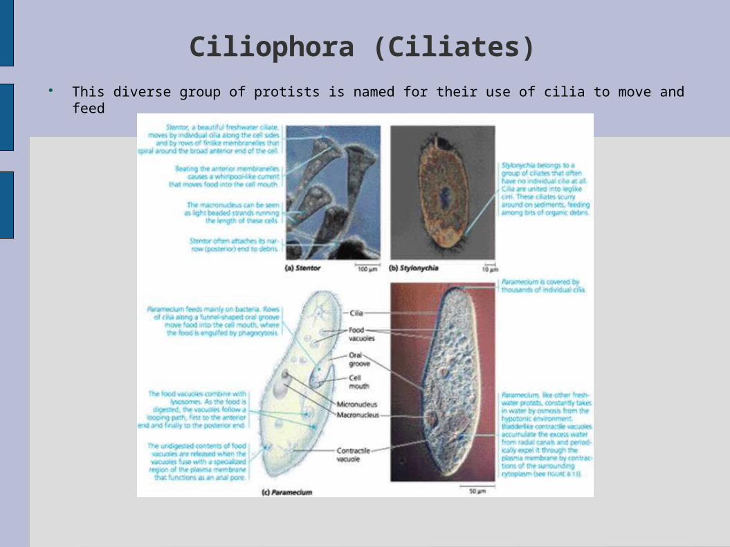
Ciliophora (Ciliates) This diverse group of protists is named for their use of cilia to move and feed
Ciliophora (Ciliates) Con.
Most ciliates live as solitary cells in fresh water. In contrast to most flagella, cilia are relatively short. They are associated with a submembrane system of microtubules that may coordinate the movement of the thousands of cilia.Some ciliates are completely covered by rows of cilia, whereas others have their cilia clustered into fewer rows or tufts.The specific arrangements adapt the ciliates for their diverse lifestyles. Some species, for instance, scurry about on leglike structures constructed from many cilia bonded together. A unique feature of ciliate genetics is the presence of two types of nuclei,a large macronucleus and usually several tiny micronuclei.The genes are not distributed in typical chromosomes but are instead packaged into a much larger number of small units, each with hundreds of copies of just a few genes.The macronucleus controls the everyday functions of the cell by synthesizing RNA and is also necessary for asexual reproduction.Ciliates generally reproduce by binary fission,The sexual shuffling of genes occurs during the process known as conjugation

Fig 28-15. Conjugation and genetic recombination in Paramecium caudatum .

A diverse assemblage of unicellular eukaryotes move by means of pseudodia
The three gropus we discuss in this section represent some of the immense diversity of unicellular eukaryotes that move and often feed by means of cellular extensions called pseudpopdia.
Most of these organisms are heterotrophs that actively seek and consume bacteria, other protists, and detritus (dead organic matter). There are also symbiotic species, including some parasites cause human diseases.
Little is known about their phylogeny.
Rhizopods (Amoebas)
Are all unicellular and use pseudopodia to move & to feed.The cytoskeleton, consisting of microtibules and microfilamentsFunctions in amoeboid movement
Meiosis & sex are NOT known to occur in amoebas.Reproduce asexually by various mechanisms of cell division.
Inhabit both freshwater & marine environments and are also abundant in soilsThe majority of amoebas are free-living, but some are important parasites, including Entamoeba histolytica Cause amoebic dysentery in humans(These organisms spread via contaminated drinking water, food, or eating utensils)

Actinopods (Heliozoans & Radiolarians)
Means “ray foot” A reference to the slender pseudopodia called axopodia that
radiate from these exceptionally beautiful protists.
Each axopodium is reinforced by a bundle of microtubules cover by thin layer of cytoplasm
Most actinpods are planktonic
Their projections place an extensive area of cellular surface in contact with the surrounding water, help the organism float and feed
Actinopods (Heliozoans & Radiolarians) con.
Heliozoan(“sun animals”)- living in freshwater. Skeletons consist of siliceous(glassy) or chitnous unfused
plates.
Radiolarian(not used in formal taxonomy)-refers to several groups of mostly marine actinopods w/skeleton fused into one delcate piece-commonly made of silica
After actinopods die, their skeletons settle settle to the seafloor, where they have accumlated as an ooze that is hundreds of meters thick in some locations
Foraminiferans (Forams)
Are almost all marine. Most live in sand or attach themselves to rocks &
algae, but some are abundant in plankton Named for their poruous shells The shells generally multichambered and consist of
organic material hardened w/calcium carbonate. Strands of cytoplasm(pseudopodia) extend through the
pores Functioning-swimming, shell formation, and feeding.(Many also derive nourishment from the photosynthesis of
symbiotic algae that live w/in the shells)
Slime molds have structural adaptations and life cycles that enhance their ecological role as decomposers
2 groups of protists called slime molds
Resemble fungi in appearance and lifestyle Similarities are convergence
In their cellular organization, reproduction and life cycles
Slime molds depart from the true fungi is partly due to convergent evolution of filamentous body structure
A morphological adaptation that increase exposure to the environment and enhances the ecological role of these organisms as decomposers
Slime molds have complex life cycles that are adaptations that contribute to survival in changing habitats and facilitate dispersat to new food sources
Plasmodial Slime Molds (Myxomycota)
Are more attractive than their name implies.
Many brightly pigmented, usually yellow or orange, but all are heterotrophic.
The feeding stage of the cycle is an amoeboid mass called a plasmodium
Plasmodium are not multicellular
The cytoplasmic streaming apparently helps distribute nutrients & oxygen
The plasmodium engulfs food particles by phagocytosis as it grows by extending pseudopodia through moist soil, leaf mulch, or rotting logs.
If the habitat of slime mold dried out or no food left, the plasmodium ceases growth and differentiates into a stage of the life cycle that function in sexual reproduction

Cellular Slime Molds(Acrasiomycota)
Pose a semantic question what it means to be an individual organism.
Although the feeding stage of the life cycle consists of solitary cells that function individually
When food is depleted the cells form an aggregate that function as a unit
Though the mass of cells resembles a plasmodial slime mold The distinction is that the cells of a cellular slime mold maintain their
identity and remain separated by their membranes.
Cellular Slime molds also differ from Plas. Slime molds in being haploid organisms
Cellular Slime molds have fruiting bodies that function in asexual reproduction.
Most of Cell. Slime mold have no flagellated stages