chapter 4 · pdf filedecreased deaths from bacterial infections. © 2013 pearson...
TRANSCRIPT
© 2013 Pearson Education, Inc.Lectures by Edward J. Zalisko
PowerPoint® Lectures forCampbell Essential Biology, Fifth Edition, and
Campbell Essential Biology with Physiology,
Fourth Edition
– Eric J. Simon, Jean L. Dickey, and Jane B. Reece
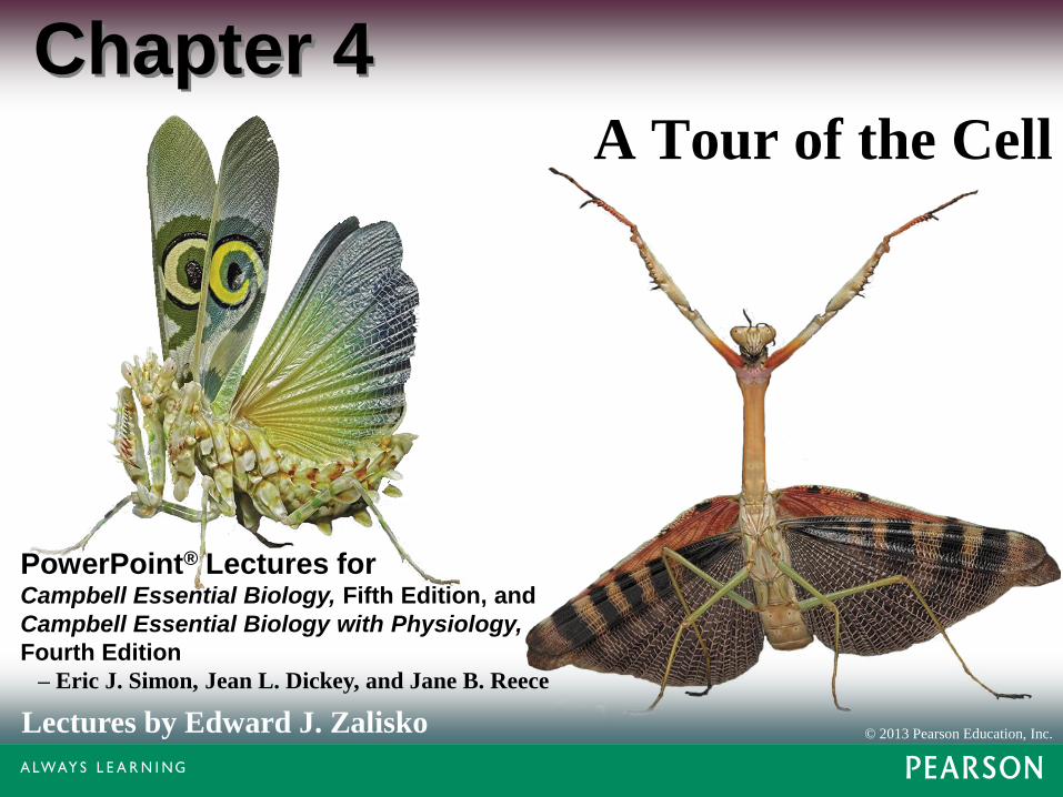
Chapter 4A Tour of the Cell

Biology and Society:
Antibiotics: Drugs that Target Bacterial Cells
• Antibiotics were first isolated from mold in 1928.
• The widespread use of antibiotics drastically
decreased deaths from bacterial infections.
© 2013 Pearson Education, Inc.

Figure 4.0

• Most antibiotics kill bacteria while minimally
harming the human host by binding to structures
found only on bacterial cells.
• Some antibiotics bind to the bacterial ribosome,
leaving human ribosomes unaffected.
• Other antibiotics target enzymes found only in the
bacterial cells.
Biology and Society:
Antibiotics: Drugs that Target Bacterial Cells
© 2013 Pearson Education, Inc.
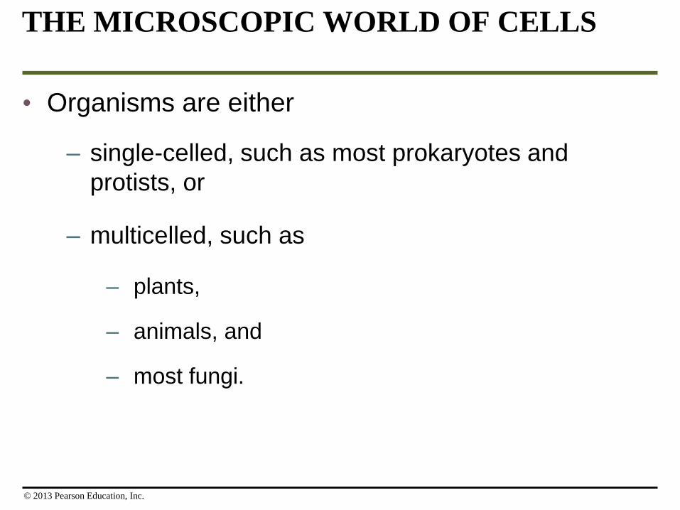
THE MICROSCOPIC WORLD OF CELLS
• Organisms are either
– single-celled, such as most prokaryotes and
protists, or
– multicelled, such as
– plants,
– animals, and
– most fungi.
© 2013 Pearson Education, Inc.

Microscopes as Windows on the World of Cells
• Light microscopes can be used to explore the
structures and functions of cells.
• When scientists examine a specimen on a
microscope slide,
– light passes through the specimen and
– lenses enlarge, or magnify, the image.
© 2013 Pearson Education, Inc.

• Magnification is an increase in the object’s image
size compared to its actual size.
• Resolving power is the ability of an optical
instrument to show two objects as separate.
Microscopes as Windows on the World of Cells
© 2013 Pearson Education, Inc.

• Cells were first described in 1665 by Robert
Hooke.
• By the mid-1800s, the accumulation of scientific
evidence led to the cell theory, which states that
– all living things are composed of cells and
– all cells come from other cells.
Microscopes as Windows on the World of Cells
© 2013 Pearson Education, Inc.

• The electron microscope (EM) uses a beam of
electrons, which results in 100-fold better
resolution than light microscope.
• Two kinds of electron microscopes reveal different
parts of cells.
• Scanning electron microscopes (SEMs)
examine cell surfaces.
• Transmission electron microscopes (TEMs) are
useful for studying the internal structure of a cell.
Microscopes as Windows on the World of Cells
© 2013 Pearson Education, Inc.

Figure 4.1
Light Micrograph (LM)Scanning Electron
Micrograph (SEM)
TYPES OF MICROGRAPHS
Transmission Electron
Micrograph (TEM)

• The most powerful electron microscopes can
– magnify up to 100,000 times and
– distinguish between objects 0.2 nanometers apart.
• Light microscopes are still very useful for studying
living cells.
Microscopes as Windows on the World of Cells
© 2013 Pearson Education, Inc.

Figure 4.2

Figure 4.310 m
1 m
10 cm
1 cm
1 mm
100 µm
10 µm
Human height
Chicken egg
Frog eggs
Length of somenerve andmuscle cells
Un
aid
ed
eye
Lig
ht
mic
ros
co
pe
Plant andanimal cells
Most bacteria
Nuclei
Mitochondria1 µm
100 nm
10 nm
1 nm
0.1 nm
Smallest bacteria
Viruses
Ribosomes
Proteins
Lipids
Small molecules
Atoms
Ele
ctr
on
mic
ros
co
pe

The Two Major Categories of Cells
• The countless cells on Earth fall into two basic categories:
1. Prokaryotic cells — Bacteria and Archaea and
2. Eukaryotic cells — protists, plants, fungi, and animals.
© 2013 Pearson Education, Inc.

The Two Major Categories of Cells
• All cells have several basic features.
– They are all bounded by a thin plasma membrane.
– Inside all cells is a thick, jelly-like fluid called the cytosol, in which cellular components are suspended.
– All cells have one or more chromosomes carrying genes made of DNA.
– All cells have ribosomes, tiny structures that build proteins according to the instructions from the DNA.
© 2013 Pearson Education, Inc.
• Prokaryotic cells are older than eukaryotic cells.
– Prokaryotes appeared about 3.5 billion years ago.
– Eukaryotes appeared about 2.1 billion years ago.
• Prokaryotic cells are
– usually smaller than eukaryotic cells and
– simpler in structure.
The Two Major Categories of Cells
© 2013 Pearson Education, Inc.

• Eukaryotes
– Only eukaryotic cells have organelles, membrane-enclosed structures that perform specific functions.
– The most important organelle is the nucleus, which
– houses most of a eukaryotic cell’s DNA and
– is surrounded by a double membrane.
The Two Major Categories of Cells
© 2013 Pearson Education, Inc.

• A prokaryotic cell lacks a nucleus. Its DNA is coiled into a nucleus-like region called the nucleoid, which is not partitioned from the rest of the cell by membranes.
The Two Major Categories of Cells
© 2013 Pearson Education, Inc.

Figure 4.4
Plasma membrane
Cell wall
Capsule
Prokaryotic
flagellum
Ribosomes
Nucleoid
Pili
Co
lori
ze
d T
EM

Figure 4.UN11
Prokaryotic Cells Eukaryotic Cells
• Smaller
• Simpler
• Most do not have organelles
• Found in bacteria and archaea
• Larger
• More complex
• Have organelles
• Found in protists, plants, fungi,
animals
CATEGORIES OF CELLS

An Overview of Eukaryotic Cells
• Eukaryotic cells are fundamentally similar.
• The region between the nucleus and plasma
membrane is the cytoplasm.
• The cytoplasm consists of various organelles
suspended in the liquid cytosol.
© 2013 Pearson Education, Inc.

• Unlike animal cells, plant cells have
– chloroplasts, which convert light energy to the
chemical energy of food in the process of
photosynthesis, and
– protective cell walls.
An Overview of Eukaryotic Cells
© 2013 Pearson Education, Inc.
• Only animal cells have lysosomes, bubbles of
digestive enzymes surrounded by membranes.
An Overview of Eukaryotic Cells
© 2013 Pearson Education, Inc.
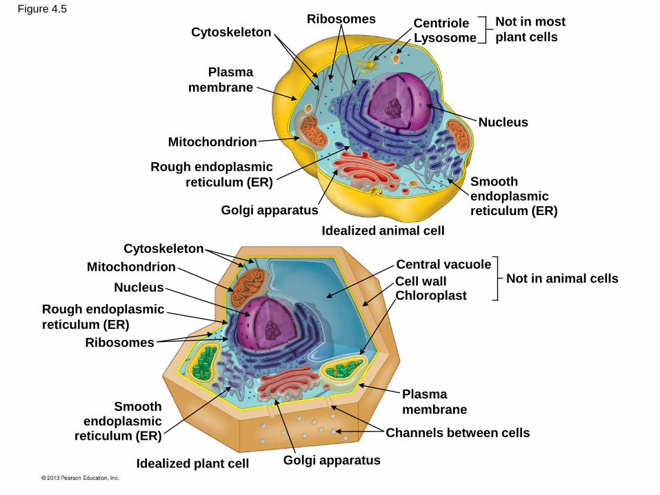
Figure 4.5
CytoskeletonRibosomes Centriole
Lysosome
Nucleus
Plasma
membrane
Mitochondrion
Rough endoplasmic
reticulum (ER)
Golgi apparatus
Smoothendoplasmicreticulum (ER)
Idealized animal cell
Idealized plant cell
Cytoskeleton
Mitochondrion
Nucleus
Rough endoplasmic
reticulum (ER)
Ribosomes
Smoothendoplasmic
reticulum (ER)
Golgi apparatus
Plasma
membrane
Channels between cells
Not in most
plant cells
Central vacuole
Cell wallChloroplast
Not in animal cells

MEMBRANE STRUCTURE
• The plasma membrane separates the living cell
from its nonliving surroundings.
© 2013 Pearson Education, Inc.

• The remarkably thin membranes of cells are composed mostly of
– lipids and
– proteins.
• The lipids belong to a special category called phospholipids.
• Phospholipids form a two-layered membrane, the phospholipid bilayer.
The Plasma Membrane: A Fluid Mosaic of Lipids and Proteins
© 2013 Pearson Education, Inc.

• Most membranes have specific proteins embedded
in the phospholipid bilayer.
• These proteins help regulate traffic across the
membrane and perform other functions.
The Plasma Membrane: A Fluid Mosaic of Lipids and Proteins
© 2013 Pearson Education, Inc.

• The plasma membrane is a fluid mosaic.
– Fluid because molecules can move freely past one
another.
– A mosaic because of the diversity of proteins in the
membrane.
The Plasma Membrane: A Fluid Mosaic of Lipids and Proteins
© 2013 Pearson Education, Inc.

Figure 4.6
(a) Phospholipid bilayer of membrane
(b) Fluid mosaic model of membrane
Outside of cell Outside of cell
Hydrophilic
head
Hydrophobic
tail
Hydrophilic
region of
protein
Hydrophilic
head
Hydrophobic
tail
Hydrophobic
regions of
protein
Phospholipidbilayer
Phospholipid
Proteins
Cytoplasm (inside of cell)
Cytoplasm (inside of cell)

Figure 4.UN12
Outside of cell
Cytoplasm (inside of cell)
Protein
Phospholipid
Hydrophilic
Hydrophilic
Hydrophobic

The Process of Science:
What Makes a Superbug?
• Particularly dangerous strains of bacteria, known as MRSA, are unaffected by several common antibiotics.
• Observation: Some bacteria use a protein called PSM to disable human immune cells by forming holes that rip apart the plasma membrane.
© 2013 Pearson Education, Inc.

The Process of Science:
What Makes a Superbug?
• Question: Does PSM play a role in MRSA infections?
• Hypothesis: MRSA bacteria lacking the ability to produce PSM would be less deadly than normal MRSA strains.
© 2013 Pearson Education, Inc.

• Experiment: Researchers infected
– seven mice with normal MRSA and
– eight mice with MRSA that does not produce PSM.
• Results:
– All seven mice infected with normal MRSA died.
– Five of the eight mice infected with MRSA that
does not produce PSM survived.
The Process of Science:
What Makes a Superbug?
© 2013 Pearson Education, Inc.

• Conclusions:
– MRSA strains appear to use the membrane-
destroying PSM protein, but
– factors other than PSM protein contributed to the
death of mice (possibly other membrane-
destroying proteins).
The Process of Science:
What Makes a Superbug?
© 2013 Pearson Education, Inc.

Figure 4.7a-3
MRSA bacterium
producing PSM
proteins
Methicillin-resistant
Staphylococcus
aureus (MRSA)
Co
lori
zed
SE
M
PSM proteins
forming hole in
human immune
cell plasma
membrane
Plasma
membrane
PSM
protein
Pore
Cell bursting,
losing its
contents through
the holes
1
2
3
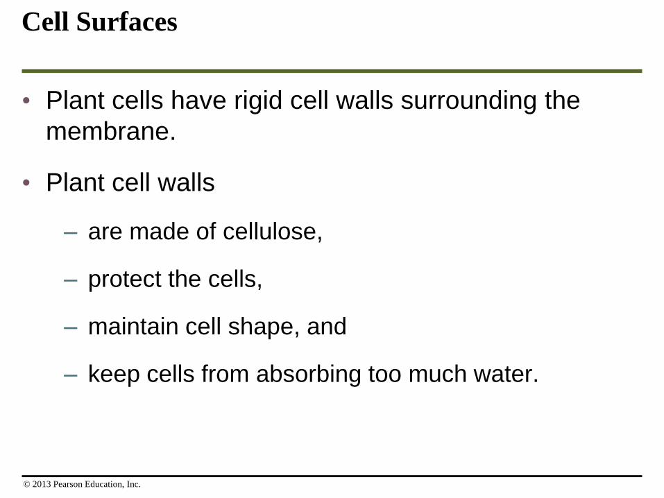
Cell Surfaces
• Plant cells have rigid cell walls surrounding the
membrane.
• Plant cell walls
– are made of cellulose,
– protect the cells,
– maintain cell shape, and
– keep cells from absorbing too much water.
© 2013 Pearson Education, Inc.

• Animal cells
– lack cell walls and
– typically have an extracellular matrix, which
– helps hold cells together in tissues and
– protects and supports them.
Cell Surfaces
© 2013 Pearson Education, Inc.

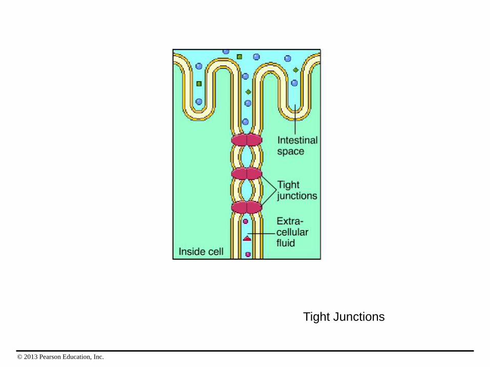
• The surfaces of most animal cells contain cell
junctions, structures that connect cells together
into tissues, allowing them to function in a
coordinated way.
Cell Surfaces
© 2013 Pearson Education, Inc.

© 2013 Pearson Education, Inc.
Desmosomes

© 2013 Pearson Education, Inc.
Gap Junctions
© 2013 Pearson Education, Inc.
Tight Junctions

THE NUCLEUS AND RIBOSOMES:GENETIC CONTROL OF THE CELL
• The nucleus is the chief executive of the cell.
– Genes in the nucleus store information necessary
to produce proteins.
– Proteins do most of the work of the cell.
© 2013 Pearson Education, Inc.

Structure and Function of the Nucleus
• The nucleus is separated from the cytoplasm by a
double membrane called the nuclear envelope.
• Pores in the envelope allow materials to move
between the nucleus and cytoplasm.
• The nucleus contains a nucleolus where
ribosomes are made.
© 2013 Pearson Education, Inc.

Figure 4.8
RibosomesChromatin
fiber Nucleolus Nuclear poreNuclear
envelope
Surface of nuclear envelope Nuclear pores
TE
M
TE
M

• Stored in the nucleus are long DNA molecules and
associated proteins that form fibers called
chromatin.
• Each long chromatin fiber constitutes one
chromosome.
• The number of chromosomes in a cell depends on
the species.
Structure and Function of the Nucleus
© 2013 Pearson Education, Inc.

Figure 4.9
DNA molecule
Chromosome
Proteins
Chromatinfiber
Ribosomes
• Ribosomes are responsible for protein synthesis.
• Ribosome components are made in the nucleolus
but assembled in the cytoplasm.
© 2013 Pearson Education, Inc.

Figure 4.10
Ribosome
Protein
mRNA

• Ribosomes may assemble proteins while the
ribosomes are
– suspended in the fluid of the cytoplasm or
– attached to the outside of the nucleus or an
organelle called the endoplasmic reticulum.
Ribosomes
© 2013 Pearson Education, Inc.
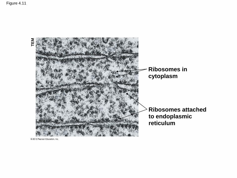
Figure 4.11
Ribosomes incytoplasm
Ribosomes attachedto endoplasmicreticulum
TE
M

How DNA Directs Protein Production
• DNA programs protein production in the cytoplasm
by transferring its coded information into
messenger RNA (mRNA).
• Messenger RNA exits the nucleus through pores in
the nuclear envelope.
• A ribosome moves along the mRNA, translating
the genetic message into a protein with a specific
amino acid sequence.
© 2013 Pearson Education, Inc.

Figure 4.12-3
Synthesis of
mRNA in the
nucleus
Nucleus
DNA
mRNA
Cytoplasm
mRNAMovement of
mRNA into
cytoplasm via
nuclear pore
Ribosome
Protein
Synthesis of
protein in the
cytoplasm
1
2
3
THE ENDOMEMBRANE SYSTEM:
MANUFACTURING AND DISTRIBUTING
CELLULAR PRODUCTS
• Many membranous organelles forming the
endomembrane system in a cell are
interconnected either
– directly by their membranes or
– by transfer of membrane segments between them.
© 2013 Pearson Education, Inc.

The Endoplasmic Reticulum
• The endoplasmic reticulum (ER) is one of the
main manufacturing facilities in a cell.
• The ER
– produces an enormous variety of molecules,
– is connected to the nuclear envelope, and
– is composed of smooth and rough ER.
© 2013 Pearson Education, Inc.
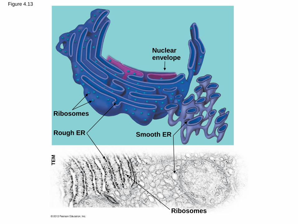
Figure 4.13
Nuclearenvelope
Smooth ERRough ER
Ribosomes
Ribosomes
TE
M

Rough ER
• The ―rough‖ in rough ER refers to ribosomes that
stud the outside of this portion of the ER
membrane.
• These ribosomes produce membrane proteins and
secretory proteins.
• Some products manufactured by rough ER are
dispatched to other locations in the cell by
transport vesicles, sacs made of membrane that
bud off from the rough ER.
© 2013 Pearson Education, Inc.

Figure 4.14
Proteins aremodified inthe ER.
Secretoryproteins depart. Vesicles bud off from the ER.
A ribosomelinks amino acids.
RibosomeTransportvesicle
Polypeptide
Protein
Rough ER

Smooth ER
• The smooth ER
– lacks surface ribosomes,
– produces lipids, including steroids, and
– helps liver cells detoxify circulating drugs.
© 2013 Pearson Education, Inc.

The Golgi Apparatus
• The Golgi apparatus
– works in partnership with the ER and
– receives, refines, stores, and distributes chemical
products of the cell.
© 2013 Pearson Education, Inc.

Figure 4.15
1
2
3
“Receiving” side of the
Golgi apparatus
New vesicle forming
Transport vesicle
from rough ER
“Receiving” side of
the Golgi apparatus New
vesicle
forming
Transport
vesicle
from the
Golgi
apparatus
Plasma
membrane
“Shipping” side of
the Golgi apparatus

Lysosomes
• A lysosome is a membrane-bound sac of
digestive enzymes found in animal cells.
• Lysosomes are absent from most plant cells.
• Enzymes in a lysosome can break down large
molecules such as
– proteins,
– polysaccharides,
– fats, and
– nucleic acids.
© 2013 Pearson Education, Inc.
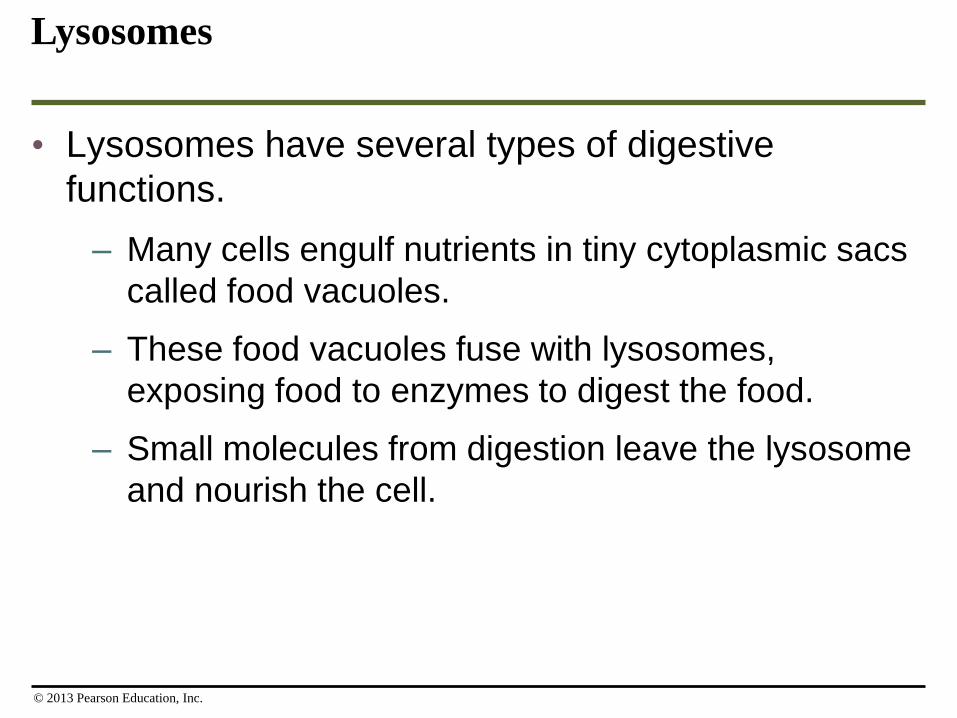
• Lysosomes have several types of digestive
functions.
– Many cells engulf nutrients in tiny cytoplasmic sacs
called food vacuoles.
– These food vacuoles fuse with lysosomes,
exposing food to enzymes to digest the food.
– Small molecules from digestion leave the lysosome
and nourish the cell.
Lysosomes
© 2013 Pearson Education, Inc.

© 2013 Pearson Education, Inc.
Lysosome Formation

Figure 4.16
Plasma membrane Digestive enzymes
Lysosome
Digestion
Food vacuole
Lysosome
Digestion
(a) A lysosome digesting food (b) A lysosome breaking down the molecules of
damaged organelles
Vesicle containing
damaged organelle
Vesicle containing two
damaged organelles
Organelle
fragment
Organelle fragment
TE
M

• Lysosomes can also
– destroy harmful bacteria,
– break down damaged organelles, and
– sculpt tissues during embryonic development,
helping to form structures such as fingers.
Lysosomes
© 2013 Pearson Education, Inc.

Vacuoles
• Vacuoles are large sacs of membrane that bud
from the
– ER,
– Golgi apparatus, or
– plasma membrane.
• Contractile vacuoles of protists pump out excess
water in the cell.
© 2013 Pearson Education, Inc.
• Central vacuoles of plants
– store organic nutrients,
– absorb water, and
– may contain pigments or poisons.
Vacuoles
© 2013 Pearson Education, Inc.

Figure 4.17A vacuole filling with water
A vacuole contracting
(a) Contractile vacuole in Paramecium
(b) Central vacuole in a plant cell
Central
vacuole
Co
lori
zed
TE
M
LM
LM

© 2013 Pearson Education, Inc.
Blast Animation: Vesicle Transport Along Microtubules
Select ―Play‖

Figure 4.18
Golgi
apparatus
Transport
vesicle
Plasma
membrane
Secretory
protein
New vesicle forming
Transport vesicle from
the Golgi apparatus
Vacuoles store some
cell products.
Lysosomes carrying digestive
enzymes can fuse
with other vesicles.
Transport vesicles carry enzymes and
other proteins from the rough ER to the
Golgi for processing.
Some products
are secreted from
the cell.
Golgi apparatus
Rough ER
TE
M
Plasma membrane

CHLOROPLASTS AND MITOCHONDRIA: ENERGY CONVERSION
• Cells require a continuous energy supply to
perform the work of life.
• Two organelles act as cellular power stations:
1. chloroplasts and
2. mitochondria.
© 2013 Pearson Education, Inc.
Chloroplasts
• Most of the living world runs on the energy
provided by photosynthesis.
• Photosynthesis is the conversion of light energy
from the sun to the chemical energy of sugar and
other organic molecules.
• Chloroplasts are
– unique to the photosynthetic cells of plants and
algae and
– the organelles that perform photosynthesis.
© 2013 Pearson Education, Inc.

• Chloroplasts are divided into three major
compartments by internal membranes:
1. the space between the two membranes,
2. the stroma, a thick fluid within the chloroplast, and
3. the space within grana, membrane-enclosed discs
and tubes that trap light energy and convert it to
chemical energy.
Chloroplasts
© 2013 Pearson Education, Inc.
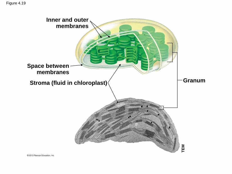
Figure 4.19
Inner and outermembranes
Space betweenmembranes
Stroma (fluid in chloroplast) Granum
TE
M
Mitochondria
• Mitochondria
– are the organelles of cellular respiration,
– are found in almost all eukaryotic cells, and
– produce ATP from the energy of food molecules.
© 2013 Pearson Education, Inc.

• An envelope of two membranes encloses the
mitochondrion:
1. an outer smooth membrane and
2. an inner membrane that
– has numerous infoldings called cristae and
– encloses a thick fluid called the matrix.
Mitochondria
© 2013 Pearson Education, Inc.

Figure 4.20
Outer
membrane
Innermembrane
Cristae
Matrix
Space betweenmembranes
TE
M

• Mitochondria and chloroplasts contain their own
DNA, which encodes some of their proteins.
• This DNA is evidence that mitochondria and
chloroplasts evolved from free-living prokaryotes in
the distant past.
Mitochondria
© 2013 Pearson Education, Inc.

Figure 4.UN13
Light energy
PHOTOSYNTHESIS
Chloroplast
Mitochondrion
ATPCELLULAR
RESPIRATION
Chemical
energy
(food)

THE CYTOSKELETON: CELL SHAPE AND MOVEMENT
• The cytoskeleton is a network of fibers extending
throughout the cytoplasm.
© 2013 Pearson Education, Inc.

Maintaining Cell Shape
• The cytoskeleton
– provides mechanical support to the cell and
– helps a cell maintain its shape.
© 2013 Pearson Education, Inc.

• The cytoskeleton contains several types of fibers
made from different proteins:
– Microtubules are straight and hollow tubes that
guide the movement of organelles and
chromosomes.
– Intermediate filaments and microfilaments are
thinner and solid.
• The cytoskeleton provides anchorage and
reinforcement for many organelles.
Maintaining Cell Shape
© 2013 Pearson Education, Inc.
• The cytoskeleton is dynamic.
• Changes in the cytoskeleton contribute to the
amoeboid (crawling) movements of
– the protist Amoeba and
– some of our white blood cells.
Maintaining Cell Shape
© 2013 Pearson Education, Inc.

Figure 4.21
(a) Microtubules in the cytoskeleton
(b) Microtubulesand movementL
M
LM

Cilia and Flagella
• Cilia and flagella are motile appendages that aid in
movement.
– Flagella propel the cell through their undulating,
whiplike motion.
– Cilia move in a coordinated back-and-forth motion.
– Cilia and flagella have the same basic architecture,
but cilia are generally shorter and more numerous
than flagella.
© 2013 Pearson Education, Inc.

• Cilia may extend from nonmoving cells.
• On cells lining the human trachea, cilia help sweep
mucus with trapped debris out of the lungs.
Cilia and Flagella
© 2013 Pearson Education, Inc.

© 2013 Pearson Education, Inc.
Cilia and Flagella

Figure 4.22
(a) Flagellum
of a human
sperm cell
(b) Cilia on a protist (c) Cilia lining the
respiratory tractC
olo
rized
SE
M
Co
lori
zed
SE
M
Co
lori
zed
SE
M

Evolution Connection:The Evolution of Antibiotic Resistance
• Many antibiotics disrupt cellular structures of
invading microorganisms.
• Introduced in the 1940s, penicillin worked well
against such infections.
• But over time, bacteria that were resistant to
antibiotics, such as the MRSA strain, were favored.
• The widespread use and abuse of antibiotics
continue to favor bacteria that resist antibiotics.
© 2013 Pearson Education, Inc.

Figure 4.23