chapter 42 circulation and gas exchange gas exchange in animals (continued) part 2
TRANSCRIPT
CHAPTER 42 CIRCULATION AND GAS EXCHANGE
Gas Exchange in Animals (continued)
Part 2
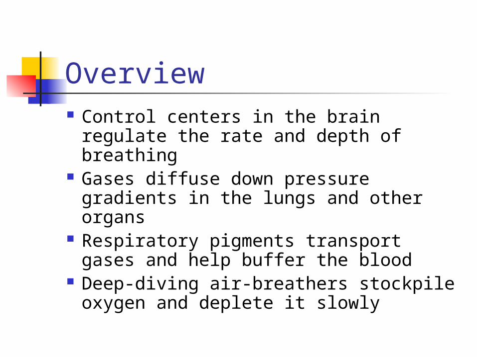
Overview Control centers in the brain regulate
the rate and depth of breathing Gases diffuse down pressure
gradients in the lungs and other organs
Respiratory pigments transport gases and help buffer the blood
Deep-diving air-breathers stockpile oxygen and deplete it slowly
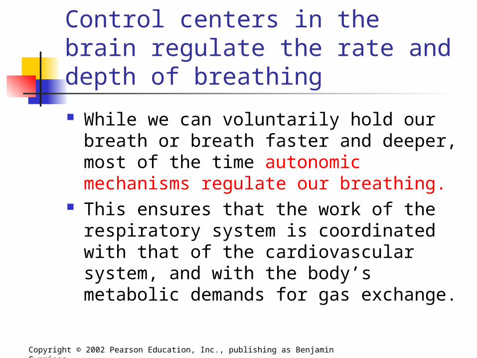
Control centers in the brain regulate the rate and depth of breathing
While we can voluntarily hold our breath or breath faster and deeper, most of the time autonomic mechanisms regulate our breathing.
This ensures that the work of the respiratory system is coordinated with that of the cardiovascular system, and with the body’s metabolic demands for gas exchange.
Copyright © 2002 Pearson Education, Inc., publishing as Benjamin Cummings
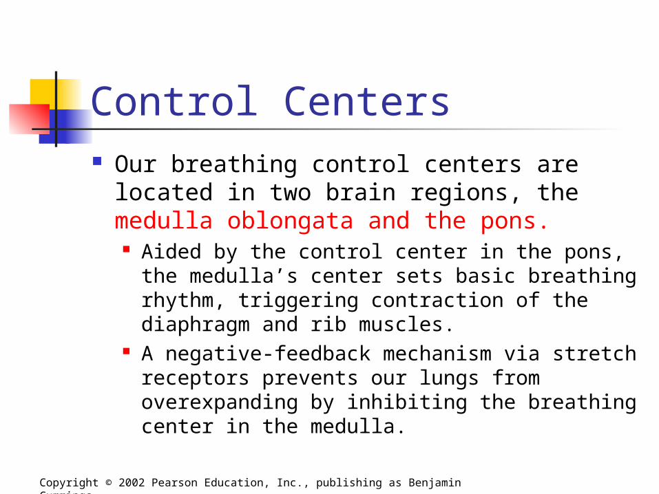
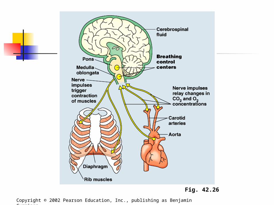
Control Centers Our breathing control centers are located
in two brain regions, the medulla oblongata and the pons. Aided by the control center in the pons, the
medulla’s center sets basic breathing rhythm, triggering contraction of the diaphragm and rib muscles.
A negative-feedback mechanism via stretch receptors prevents our lungs from overexpanding by inhibiting the breathing center in the medulla.
Copyright © 2002 Pearson Education, Inc., publishing as Benjamin Cummings
Copyright © 2002 Pearson Education, Inc., publishing as Benjamin Cummings
Fig. 42.26

Control Centers The medulla’s control center monitors the
CO2 level of the blood and regulated breathing activity appropriately. Its main cues about CO2 concentration come
from slight changes in the pH of the blood and cerebrospinal fluid bathing the brain.
Carbon dioxide reacts with water to form carbonic acid, which lowers the pH.
When the control center registers a slight drop in pH, it increases the depth and rate of breathing, and the excess CO2 is eliminated in exhaled air.
Copyright © 2002 Pearson Education, Inc., publishing as Benjamin Cummings
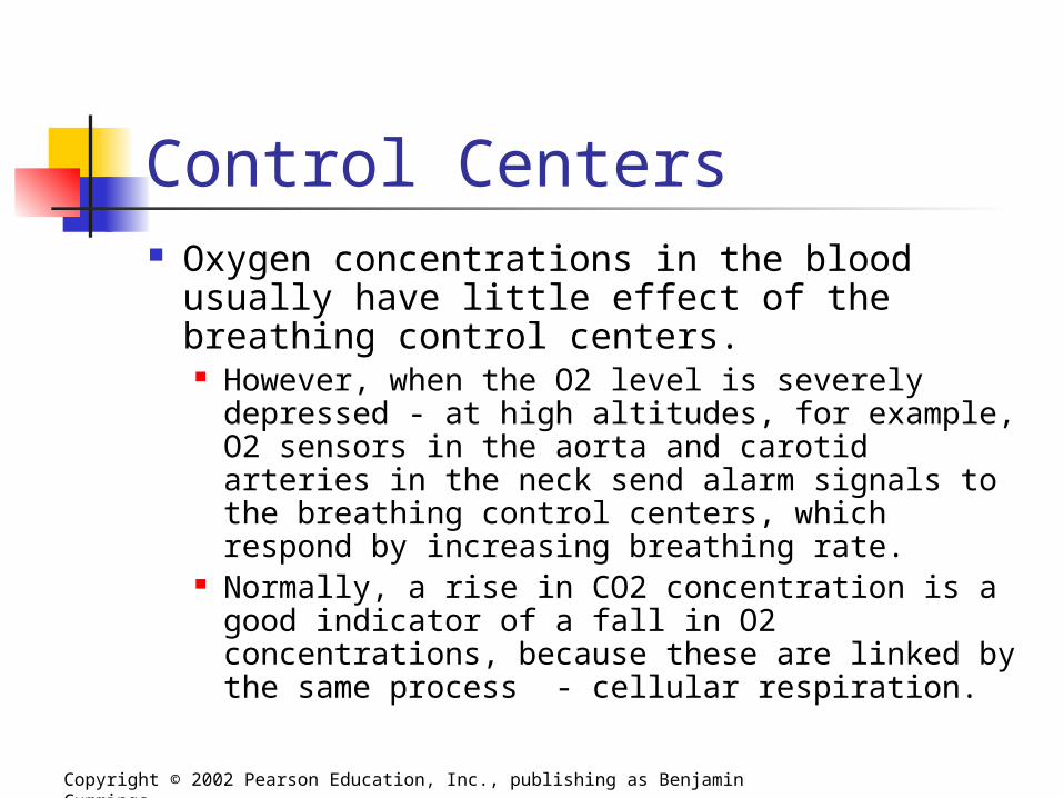
Control Centers Oxygen concentrations in the blood usually
have little effect of the breathing control centers. However, when the O2 level is severely
depressed - at high altitudes, for example, O2 sensors in the aorta and carotid arteries in the neck send alarm signals to the breathing control centers, which respond by increasing breathing rate.
Normally, a rise in CO2 concentration is a good indicator of a fall in O2 concentrations, because these are linked by the same process - cellular respiration.
Copyright © 2002 Pearson Education, Inc., publishing as Benjamin Cummings
Control Centers The breathing center responds to a variety of
nervous and chemical signals and adjusts the rate and depth of breathing to meet the changing demands of the body. However, breathing control is only effective if it is
coordinated with control of the circulatory system, so that there is a good match between lung ventilation and the amount of blood flowing through alveolar capillaries.
For example, during exercise, cardiac output is matched to the increased breathing rate, which enhances O2 uptake and CO2 removal as blood flows through the lungs.
Gases diffuse down pressure gradients in the lungs and other organs For a gas, whether present in air or
dissolved in water, diffusion depends on differences in a quantity called partial pressure, the contribution of a particular gas to the overall total. At sea level, the atmosphere exerts a total
pressure of 760 mm Hg. Since the atmosphere is 21% oxygen (by
volume), the partial pressure of oxygen (abbreviated PO2) is 0.21 x 760, or about 160 mm Hg.
The partial pressure of CO2 is only 0.23 mm Hg.
Copyright © 2002 Pearson Education, Inc., publishing as Benjamin Cummings
Partial Pressure of Gases When water is exposed to air, the amount of
a gas that dissolves in water is proportional to its partial pressure in the air and its solubility in water. An equilibrium is eventually reached when gas
molecules enter and leave the solution at the same rate.
At this point, the gas is said to have the same partial pressure in the solution as it does in the air.
Thus, in a glass of water exposed to air at sea-level air pressure, the PO2 is 160 mm Hg and the PCO2 is 0.23 mm Hg.
Copyright © 2002 Pearson Education, Inc., publishing as Benjamin Cummings
Partial Pressure of Gases A gas will always diffuse from a
region of higher partial pressure to a region of lower partial pressure.
Partial Pressure of Gases Blood arriving at the lungs via the
pulmonary arteries has a lower PO2 and a higher PCO2 than the air in the alveoli. As blood enters the alveolar capillaries, CO2
diffuses from blood to the air within the alveoli, and oxygen in the alveolar air dissolves in the fluid that coats the epithelium and diffuses across the surface into the blood.
By the time blood leaves the lungs in the pulmonary veins, its PO2 have been raised and its PCO2 has been lowered.
Copyright © 2002 Pearson Education, Inc., publishing as Benjamin Cummings
Partial Pressure of Gases In the tissue capillaries, gradients of
partial pressure favor the diffusion of oxygen out of the blood and carbon dioxide into the blood. Cellular respiration removes oxygen from and
adds carbon dioxide to the interstitial fluid by diffusion, and from the mitochondria in nearby cells.
After the blood unloads oxygen and loads carbon dioxide, it is returned to the heart and pumped to the lungs again, where it exchanges gases with air in the alveoli.
Copyright © 2002 Pearson Education, Inc., publishing as Benjamin Cummings
Respiratory pigments transport gases and help buffer the blood The low solubility of oxygen in water is a
fundamental problem for animals that rely on the circulatory systems for oxygen delivery. For example, a person exercising consumes
almost 2 L of O2 per minute, but at normal body temperature and air pressure, only 4.5 mL of O2 can dissolve in a liter of blood in the lungs.
If 80% of the dissolved O2 were delivered to the tissues (an unrealistically high percentage), the heart would need to pump 500 L of blood per minute - a ton every 2 minutes.
Copyright © 2002 Pearson Education, Inc., publishing as Benjamin Cummings
Respiratory pigments In fact, most animals transport most of the O2
bound to special proteins called respiratory pigments instead of dissolved in solution. Respiratory pigments, often contained within
specialized cells, circulate with the blood. The presence of respiratory pigments increases the
amount of oxygen in the blood to about 200 mL of O2 per liter of blood.
For our exercising individual, the cardiac output wold need to be a manageable 20-25 L of blood per minute to meet the oxygen demands of the systemic system.
Respiratory pigments A diversity of respiratory pigments
have evolved in various animal taxa to support their normal energy metabolism. One example, hemocyanin, found in
the hemolymph of arthropods and many mollusks, has copper as its oxygen-binding component, coloring the blood bluish.
Copyright © 2002 Pearson Education, Inc., publishing as Benjamin Cummings
Respiratory pigments The respiratory pigment of almost
all vertebrates is the protein hemoglobin, contained within red blood cells.
Hemoglobin consists of four subunits, each with a cofactor called a heme group that has an iron atom at its center.
Because iron actually binds to O2, each hemoglobin molecule can carry four molecules of O2.
Respiratory pigments Like all respiratory pigments, hemoglobin
must bind oxygen reversibly, loading oxygen at the lungs or gills and unloading it in other parts of the body. Loading and unloading depends on cooperation
among the subunits of the hemoglobin molecule. The binding of O2 to one subunit induces the
remaining subunits to change their shape slightly such that their affinity for oxygen increases.
When one subunit releases O2, the other three quickly follow suit as a conformational change lowers their affinity for oxygen.
Copyright © 2002 Pearson Education, Inc., publishing as Benjamin Cummings
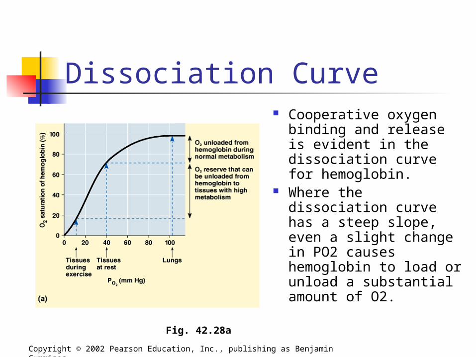
Dissociation Curve Cooperative oxygen
binding and release is evident in the dissociation curve for hemoglobin.
Where the dissociation curve has a steep slope, even a slight change in PO2 causes hemoglobin to load or unload a substantial amount of O2.
Copyright © 2002 Pearson Education, Inc., publishing as Benjamin Cummings
Fig. 42.28a
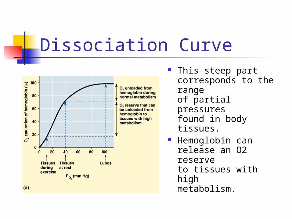
Dissociation Curve This steep part
corresponds to the range of partial pressures found in body tissues.
Hemoglobin can release an O2 reserve to tissues with high metabolism.
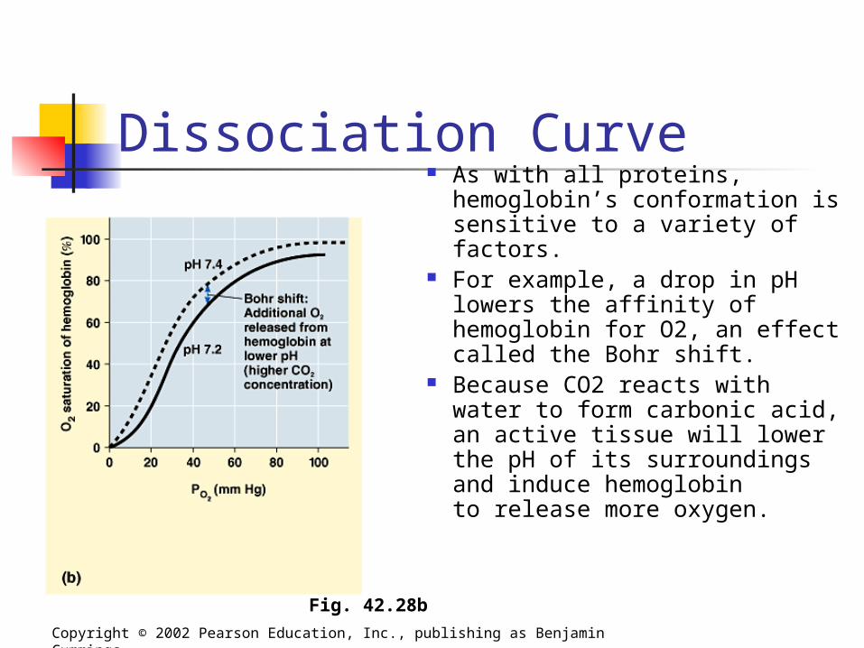
Dissociation Curve As with all proteins,
hemoglobin’s conformation is sensitive to a variety of factors.
For example, a drop in pH lowers the affinity of hemoglobin for O2, an effect called the Bohr shift.
Because CO2 reacts with water to form carbonic acid, an active tissue will lower the pH of its surroundingsand induce hemoglobinto release more oxygen.
Copyright © 2002 Pearson Education, Inc., publishing as Benjamin Cummings
Fig. 42.28b
Carbon Dioxide Transport In addition to oxygen transport,
hemoglobin also helps transport carbon dioxide and assists in buffering blood pH. About 7% of the CO2 released by respiring
cells is transported in solution. Another 23% binds to amino groups of
hemoglobin. About 70% is transported as bicarbonate
ions.
Copyright © 2002 Pearson Education, Inc., publishing as Benjamin Cummings
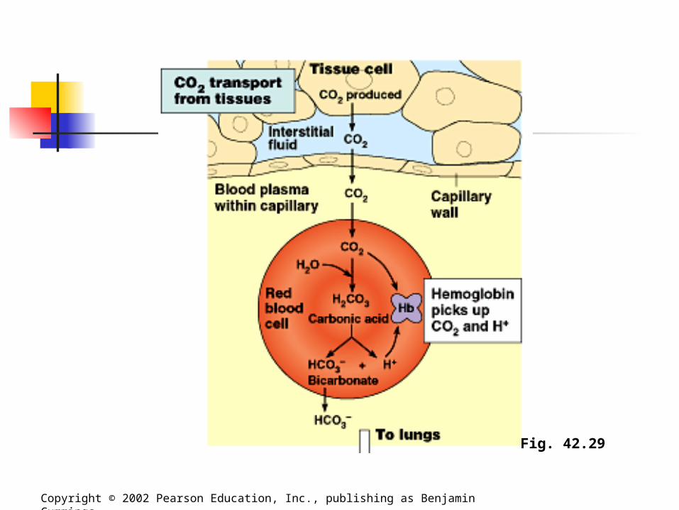
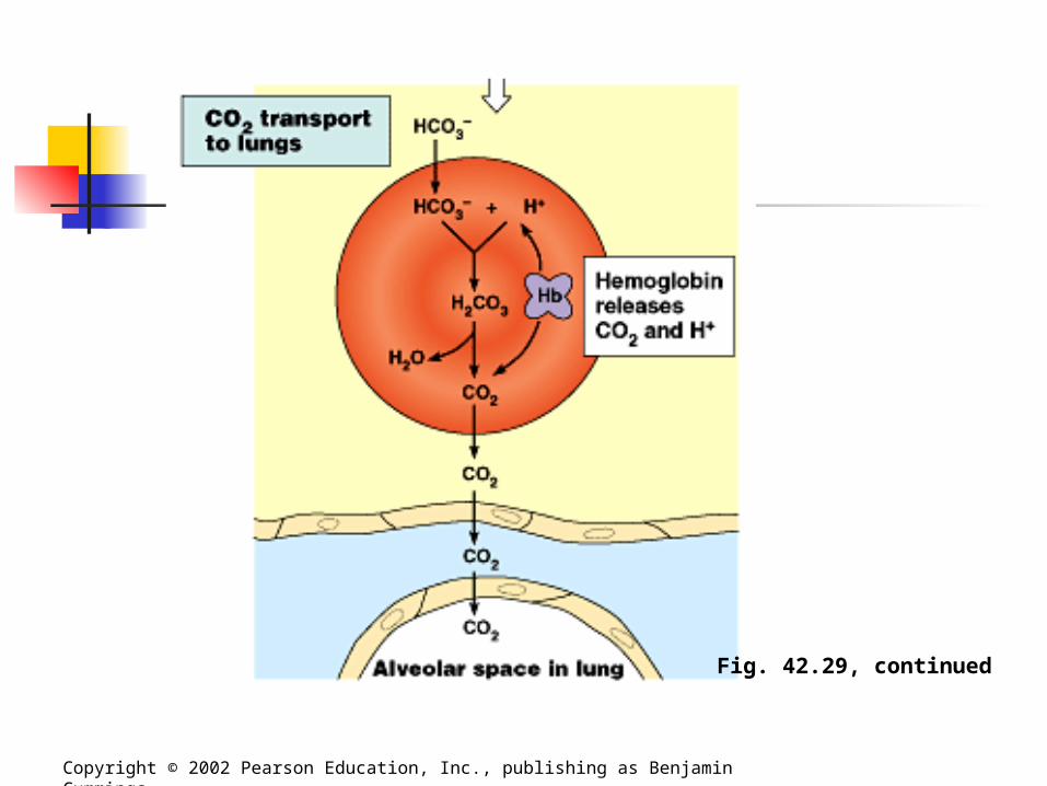
Carbon Dioxide Transport Carbon dioxide from respiring cells
diffuses into the blood plasma and then into red blood cells, where some is converted to bicarbonate, assisted by the enzyme carbonic anhydrase. At the lungs, the equilibrium shifts in
favor of conversion of bicarbonate to CO2.
Copyright © 2002 Pearson Education, Inc., publishing as Benjamin Cummings
Copyright © 2002 Pearson Education, Inc., publishing as Benjamin Cummings
Fig. 42.29
Copyright © 2002 Pearson Education, Inc., publishing as Benjamin Cummings
Fig. 42.29, continued
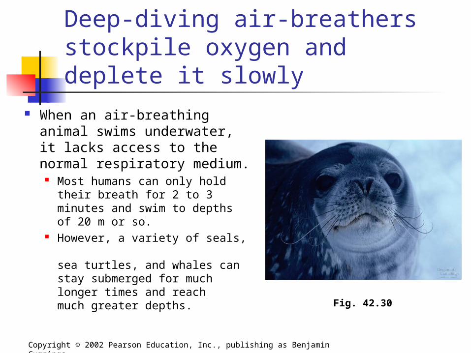
Deep-diving air-breathers stockpile oxygen and deplete it slowly
When an air-breathing animal swims underwater, it lacks access to the normal respiratory medium.
Most humans can only hold their breath for 2 to 3 minutes and swim to depths of 20 m or so.
However, a variety of seals, sea turtles, and whales can stay submerged for much longer times and reach much greater depths.
Copyright © 2002 Pearson Education, Inc., publishing as Benjamin Cummings
Fig. 42.30
Deep-Divers One adaptation of these deep-divers,
such as the Weddell seal, is an ability to store large amounts of O2 in the tissues. Compared to a human, a seal can store
about twice as much O2 per kilogram of body weight, mostly in the blood and muscles.
About 36% of our total O2 is in our lungs and 51% in our blood.
In contrast, the Weddell seal holds only about 5% of its O2 in its small lungs and stockpiles 70% in the blood.
Copyright © 2002 Pearson Education, Inc., publishing as Benjamin Cummings
Adaptations of Deep Divers Several adaptations create these physiological
differences between the seal and other deep-divers in comparison to humans. First, the seal has about twice the volume of blood
per kilogram of body weight as a human. Second, the seal can store a large quantity of
oxygenated blood in its huge spleen, releasing this blood after the dive begins.
Third, diving mammals have a high concentration of an oxygen-storing protein called myoglobin in their muscles.
This enables a Weddell seal to store about 25% of its O2 in muscle, compared to only 13% in humans.
Adaptations of Deep Divers
Diving vertebrates not only start a dive with a relatively large O2 stockpile, but they also have adaptations that conserve O2.
They swim with little muscular effort and often use buoyancy changes to glide passively upward or downward.
Their heart rate and O2 consumption rate decreases during the dive and most blood is routed to the brain, spinal cord, eyes, adrenal glands, and placenta (in pregnant seals).
Blood supply is restricted or even shut off to the muscles, and the muscles can continue to derive ATP from fermentation after their internal O2 stores are depleted.
Copyright © 2002 Pearson Education, Inc., publishing as Benjamin Cummings