chapter immunogenetics 13
TRANSCRIPT

184
C H A P T E R
13Immunogenetics
‘Medicinal discovery,It moves in mighty leaps,It leapt straight past the common coldAnd gave it us for keeps.’
Pam Ayers
IMMUNITY
Microorganisms, insects and other infectious agents are far more numerous than members of the human race, and without effective defense mechanisms against their activity humankind would rapidly succumb. The immune system in all its forms is our defense mechanism, and in order to understand the inherited disorders of immunity, we must fi rst understand the fundamentals of the genetic basis of immunity.
Immune defense mechanisms can be divided into two main types: innate immunity, which includes a number of non-specifi c systems that do not require or involve prior contact with the infectious agent, and specific acquired or adaptive immunity, which involves a tailor-made immune response that occurs after exposure to an infectious agent. Both types can involve either humoral immunity, which combats extracellular infections, or cell-mediated immunity, which fi ghts intracellular infections.
INNATE IMMUNITY
The fi rst simple type of defense against infection is a mechanical barrier. The skin functions most of the time as an impermeable barrier but, in addition, the acidic pH of sweat is inhibitory to bacterial growth. The membranes lining the respiratory and gastrointestinal tracts are protected by mucus. In the case of the respiratory tract, further protection is provided by ciliary movement, whereas other bodily fluids contain a variety of bactericidal agents, such as lysozymes in tears. If an organism succeeds in invading the body, phagocytosis and bactericidal agents come into effect.
HUMORAL INNATE IMMUNITY
A number of soluble factors are involved in innate immunity; they help to minimize tissue injury by limiting the spread of infectious microorganisms. These are often called the acute-phase proteins and include C-reactive protein, mannose-binding protein and serum amyloid P component. The fi rst two act by facilitating the attachment of one of the components of complement, C3b, to the surface of the microorganism, which becomes opsonized (made ready) for adherence to phagocytes, whereas the latter binds lysosomal enzymes to connective tissues. In addition, cells when infected by a virus, synthesize and secrete interferon, which interferes with viral replication by reducing messenger RNA (mRNA) stability and interfering with translation.
Complement
Complement is a complex series of 20 or so interacting plasma proteins that can be activated by the cell membranes of invading microorganisms, in what is termed the alternative pathway. The various components of complement interact in a specifi c cascade sequence, resulting in a localized acute infl ammatory response through the action of mediators released from mast cells and tissue macrophages. These result in increased vascular permeability and the attraction of phagocytes in the process known as chemotaxis. In addition, the later components of the complement cascade generate a membrane attack complex, which induces defects in the cell membrane, resulting in the lysis of microorganisms (Fig. 13.1).
Complement can also be activated through the classic pathway, by the binding of antibody with antigen (see below, p. 185).
CELL-MEDIATED INNATE IMMUNITY
Phagocytosis
Microorganisms are engulfed and digested by two major types of cell: polymorphonuclear neutrophils or macrophages. Polymorphonuclear neutrophils are found mainly in the bloodstream, whereas macrophages occur primarily in tissues around the basement membrane of blood vessels in connective tissue, lung, liver and the lining of the sinusoids of the spleen and the medullary sinuses of the lymph nodes. Surface antigens on microorganisms result in their being engulfed and fusing with the
Ch13-S2917.indd 184Ch13-S2917.indd 184 4/30/07 10:21:06 AM4/30/07 10:21:06 AM

IMMUNOGENETICS
185
13
granules of the phagocyte, subjecting them to the action of the bactericidal agents of the intracellular granules, which contain hydrogen peroxide, hydroxyl radicals and nitrous oxide, and leading to their destruction.
Extracellular killing
Virally infected cells can be killed by large granular lymphocytes, known as natural killer cells. These have carbohydrate-binding receptors on their cell surface that recognize high molecular weight glycoproteins expressed on the surface of the infected cell as a result of the virus taking over the cellular replicative functions. Attachment of the natural killer cells to the infected cells results in the release of a number of agents; this, in turn, results in damage to the membrane of the infected cell, leading to cell death.
SPECIFIC ACQUIRED IMMUNITY
Many infective microorganisms have, through mutation and selective pressures, developed strategies to overcome or evade the mechanisms associated with innate immunity. There is a need, therefore, to be able to generate specifi c acquired or adaptive immunity. This can, like innate immunity, be separated into both humoral and cell-mediated processes.
HUMORAL SPECIFIC ACQUIRED IMMUNITY
The main mediators of humoral specifi c acquired immunity are immunoglobulins or antibodies. Antibodies are able to recognize and bind to antigens of infecting microorganisms, leading to
the activation of phagocytes and the initiation of the classic pathway of complement, resulting in the generation of the membrane attack complex (see Fig. 13.1). Exposure to a specifi c antigen results in the clonal proliferation of a small lymphocyte derived from the bone marrow (hence ‘B’ lymphocytes), resulting in mature antibody-producing cells or plasma cells.
Lymphocytes capable of producing antibodies express on their surface copies of the immunoglobulin (Ig) for which they code, which acts as a surface receptor for antigen. Binding of the antigen results, in conjunction with other membrane-associated proteins, in signal transduction leading to the clonal expansion and production of antibody. In the fi rst instance this results in the primary response with production of IgM and subsequently IgG. Re-exposure to the same antigen results in enhanced antibody levels in a shorter period of time, known as the secondary response, refl ecting what is known as antigen-specifi c immunological memory.
Immunoglobulins
The immunoglobulins, or antibodies, are one of the major classes of serum protein. Their function, both in the recognition of antigenic variability and in effector activities, was initially revealed by protein, and more recently DNA, studies of their structure.
Immunoglobulin structure
Papaine, a proteolytic enzyme, splits the immunoglobulin molecule into three fragments. Two of the fragments are similar, each containing an antibody site capable of combining with a specifi c antigen and therefore referred to as the antigen-binding fragment or Fab. The third fragment can be crystalized and was therefore called Fc. The Fc fragment determines the secondary biological functions of antibody molecules, binding complement and Fc receptors on a number of different cell types involved in the immune response.
The immunoglobulin molecule is made up of four polypeptide chains: two ‘light’ (L) and two ‘heavy’ (H) chains of approximately 220 and 440 amino acids in length, respectively. They are held together in a Y shape by disulfide bonds and non-covalent interactions. Each Fab fragment is composed of L chains linked to the amino-terminal portion of the H chains, whereas each Fc fragment is composed only of the carboxy-terminal portion of the H chains (Fig. 13.2).
Immunoglobulin isotypes, subclasses and idiotypes
There are fi ve different types of heavy chain, designated respectively as g, m, a, d and e, one each respectively for the fi ve major antibody classes, or what are known as isotypes: IgG, IgM, IgA, IgD and IgE. Further analysis has revealed that the L chains are one of two types, either kappa (k) or lambda (l), the two types of L chain occurring in all fi ve classes of antibody, but with only one type of light chain occurring in each individual antibody. Thus the molecular formula for IgG is l2g2 or k2g2. The characteristics of the various classes of antibody are outlined in brief in Table 13.1.
Lysis
Membraneattackcomplex
C5C6C7C8C9
Mast cell degranulationChemotaxis
Opsonization
C3 C3a + C3b
C1qrs
C4C2
'Classical' pathway
Recognition ofantigen–antibody
complexes
D
C3B
'Alternative' pathway
Recognitionof
bacteria
Fig. 13.1Classic and alternative pathways of complement activation.
(Adapted from Paul W E (ed.) 1993 Fundamental immunology.
Raven Press, New York.)
Ch13-S2917.indd 185Ch13-S2917.indd 185 4/30/07 10:21:06 AM4/30/07 10:21:06 AM

IMMUNOGENETICS13
186
In addition, there are four IgG subclasses, IgG1, IgG2, IgG3 and IgG4, and two IgA subclasses, IgA1 and IgA2, which differ in their amino-acid sequence and interchain disulfi de bonds. Individual antibody molecules that recognize specifi c antigens are known as idiotypes.
Immunoglobulin allotypes
The fi ve classes of immunoglobulin occur in all normal individuals, but allelic variants, or what are known as antibody allotypes of these
classes, have also been identifi ed. These are the Gm system associated with the heavy chain of IgG, the Am system associated with the IgA heavy chain, the Km and Inv systems associated with the k light chain, the Oz system for the l light chain and the Em allotype for the IgE heavy chain. The Gm and Km systems are independent of each other and are polymorphic (p. 128), the frequencies of the different alleles varying in different ethnic groups.
Generation of antibody diversity
It could seem paradoxical for a single protein molecule to exhibit suffi cient structural heterogeneity to have specifi city for a large number of different antigens. Different combinations of heavy and light chains could, to some extent, account for this diversity. It would, however, require thousands of structural genes for each chain type to provide suffi cient variability for the large number of antibodies produced in response to the equally large number of antigens to which individuals can be exposed. Our initial understanding of how this could occur came from persons with a malignancy of antibody-producing cells, or what is known as multiple myeloma.
Multiple myeloma
Persons with multiple myeloma make a single or monoclonal antibody species in large abundance, which is excreted in large quantities in their urine. This protein, known as Bence Jones protein, consists of antibody light chains. Comparisons of this protein from different patients with myeloma revealed the amino-terminal ends of the molecule to be quite variable in their amino-acid sequence whereas the carboxy-terminal ends were relatively constant. These are called the variable, or V, and constant, or C, regions, respectively. Further detailed analysis of the amino-acid sequence of the V regions of different myeloma proteins showed four regions that varied little from one antibody to another, known as framework regions (FR 1–4), and three markedly variable regions interspersed between these, known as hypervariable regions (HV I–III) (see Fig. 13.2).
Antigen-bindingsites
Hypervariableregions I–III
Frameworkregions 1–4
Carboxy terminal
Disulphidebonds
Fc
Fab
Somatic recombination
V D J C
Amino terminal
Heav
y ch
ainLig
ht ch
ain
Table 13.1 Classes of human immunoglobulin
Class Mol. wt (Da) Serum concentration Antibody activity Complement Placental
(mg/ml) fi xation transfer
IgG 150 000 8–16 Binds to microorganisms and neutralizes bacterial toxins � �
IgM 900 000 0.5–2 Produced in early immune response, especially in � �
bacteremia
IgA 160 000 1.4–4 Guards mucosal surfaces � �
IgD 185 000 0–0.4 On lymphocyte cell surface, involved in control of � �
activation and suppression
IgE 200 000 Trace In parasitic and allergic reactions � �
Fig. 13.2Model of antibody molecule structure.
Ch13-S2917.indd 186Ch13-S2917.indd 186 4/30/07 10:21:07 AM4/30/07 10:21:07 AM

IMMUNOGENETICS
187
13DNA studies of antibody diversity
As long ago as 1965, Dreyer and Bennett proposed that an antibody could be encoded by separate ‘genes’ in germline cells that undergo rearrangement, or, as they termed it, ‘scrambling’, in lymphocyte development. Comparison of the restriction maps of the DNA segments coding for the C and V regions of the immunoglobulin l light chains in embryonic and antibody-producing cells revealed that they were far apart in the former but close together in the latter. More detailed analysis revealed that the DNA segments coding for the V and C regions of the light chain are separated by some 1500 base pairs (bp) in antibody-producing cells. The intervening DNA segment was found to code for a joining, or J, region immediately adjacent to the V region of the light chain. The k light chain was shown to have the same structure. Cloning and DNA sequencing of heavy-chain genes in germline cells revealed that they have a fourth region, called diversity, or D, between the V and J regions.
There are estimated to be some 60 different DNA segments coding for the V region of the heavy chain, approximately 40 DNA segments coding for the V region of the k light chain and 30 DNA segments coding for the l light chain V region. Six functional DNA segments code for the J region of the heavy chain, fi ve for the J region of the k light chain and four for the J region of the l light chain. A single DNA segment codes for the C region of the k light chain, seven DNA segments code for the C region of the l light chain and 11 functional DNA segments code for the C region of the different classes of heavy chain. There are also 27 functional DNA segments coding for the D region of the heavy chain (Fig. 13.3).
Estimation of the number of DNA segments coding for these various portions of the antibody molecule is confounded by the presence of a large number of unexpressed DNA sequences or pseudogenes (p. 16). Although the coding DNA segments for the various regions of the antibody molecule can be referred to as genes, use of this term in regard to antibodies has deliberately been avoided because they could be considered an exception to the general rule of ‘one gene–one enzyme (or protein)’ (p. 158).
Antibody gene rearrangement
The genes for the k and l light chains and the heavy chains in humans have been assigned to chromosomes 2, 22 and 14, respectively. Only one of each of the relevant types of DNA segment is expressed in any single antibody molecule. The DNA coding segments for the various portions of the antibody chains on these chromosomes are separated by DNA that is non-coding. Somatic recombinational events involved in antibody production involve short conserved recombination signal sequences that fl ank each germline DNA segment (Fig. 13.4). Further diversity occurs by variable mRNA splicing at the V–J junction in RNA processing and by somatic mutation of the antibody genes. These mechanisms can easily account for the antibody diversity seen in nature. Although this probably involves some form of clonal selection, it is still not entirely clear how particular DNA segments are selected to produce an antibody to a specifi c antigen. ‘Gene shuffl ing’ of this form is also known to account for the marked variability seen in the surface antigens of the trypanosome parasite and the different mating types in yeast.
Class switching of antibodies
There is a normal switch of antibody class produced by B cells on continued or further exposure to antigen, from IgM, which is the initial class of antibody produced in response to exposure to an antigen, to IgA or IgG. This process, known as class switching, involves retention of the specifi city of the antibody to the same antigen. Analysis of class switching in a population of cells derived from a single B cell has shown that both classes of antibody have the same antigen-binding sites, having the same V
kChromosome 2
Heavychain
Variableregion
40
D iversityregion
Junctionalregion
Constantregion
5 1
lChromosome 2230 4 7
Chromosome 1460 6 1127
Fig. 13.3Estimated number of the various DNA segments coding for the k,
l and various heavy chains.
V D J S Cμ S Cα
Somaticrecombination
Classswitching
V D J S Cμ S Cα
V D J S Cα
Fig. 13.4Immunoglobulin heavy-chain gene rearrangement and class
switching.
Ch13-S2917.indd 187Ch13-S2917.indd 187 4/30/07 10:21:07 AM4/30/07 10:21:07 AM

IMMUNOGENETICS13
188
region, differing only in their C region. Class switching occurs by a somatic recombination event that involves DNA segments designated S (for switching) that lead to looping out and deletion of the intervening DNA. The result is to eliminate the DNA segment coding for the C region of the heavy chain of the IgM molecule and to bring the gene segment encoding the C region of the new class of heavy chain adjacent to the segment encoding the V region (see Fig. 13.4).
The immunoglobulin gene superfamily
Studies of the structure of other molecules involved in the immune response have shown a number to have structural and DNA sequence homology to the immunoglobulins. This involves a 110-amino-acid sequence characterized by a centrally placed disulfi de bridge that stabilizes a series of antiparallel b strands into what is called an antibody fold. This group of molecules with similar structure has been called the immunoglobulin superfamily (p. 16). It consists of eight multigene families that, in addition to the k and l light chains and different classes of heavy chain, include the chains of the T-cell receptor (p. 16), the class I and II major histocompatibility complex (MHC), or human leukocyte (HLA) antigens (p. 371), and b2-microglobulin. The latter is a receptor for transporting certain classes of immunoglobulin across mucosal membranes. A series of other molecules shows homology to the immunoglobulin superfamily. These include the T-cell CD4 and CD8 cell surface receptor molecules, which cooperate with T-cell receptors in antigen recognition, and the intercellular adhesion molecules, ICAM-1, -2 and -3, which are involved in the interaction of lymphocytes with antigen-presenting cells.
Antibody engineering
The techniques of genetic engineering have led to the prospect of reshaping or designing human antibodies for specifi c therapeutic or diagnostic purposes. These are what are known as monoclonal antibodies. Recombinant antibodies can be constructed using the human variable region framework, the human constant regions of the heavy and light chains, and the antigen-binding site of a mouse antibody. Persons treated with these ‘engineered’ antibodies do not mount an immune response to them – a problem encountered in the use of rodent hybridoma-derived monoclonal antibodies. It is hoped that the use of human-derived myeloma cells for expression of these recombinant antibodies will overcome this diffi culty.
CELL-MEDIATED SPECIFIC ACQUIRED IMMUNITY
Certain microorganisms, viruses and parasites live inside host cells. As a result, a separate form of specifi c acquired immunity has evolved to combat intracellular infections involving lymphocytes differentiated in the thymus – hence T cells. T lymphocytes have specialized receptors on the cell surface, known as T-cell surface
antigen receptors, which in conjunction with the major histocompatibility complex on the cell surface of the infected cell result in the involvement of T helper cells and cytotoxic T cells to combat intracellular infections by leading to the death of the infected cell.
T-cell surface antigen receptor
T cells express on their surface an antigen receptor. This consists of two different polypeptide chains, linked by a disulfi de bridge, that both contain two immunoglobulin-like domains, one that is relatively invariant in structure, the other highly variable like the Fab portion of an immunoglobulin. The diversity in T-cell receptors required for recognition of the range of antigenic variation that can occur is generated by a process similar to that seen with immunoglobulins. Rearrangement of variable (V), diversity (D), junctional ( J) and constant (C) DNA segments during T-cell maturation, through a similar recombination mechanism as occurs in B cells, results in a contiguous VDJ sequence. Binding of antigen to the T-cell receptor, in conjunction with an associated complex of transmembrane peptides, results in signaling the cell to differentiate and divide.
The major histocompatibility complex
The MHC plays a central role in the immune system. Association of an antigen with the MHC molecule on the surface of the cells is required for recognition of the antigen by the T-cell receptor that, in conjunction with the closely associated protein b2-microglobulin, results in the recruitment of cytotoxic and helper T cells in the immune response. MHC molecules occur in three classes: class I molecules occur on virtually all cells and are responsible for recruiting cytotoxic T cells; class II molecules occur on B cells and macrophages and are involved in signaling T helper cells to recruit further B cells and macrophages; the non-classic class III molecules include a number of other proteins with a variety of other immunological functions. The latter include infl ammatory mediators such as the tumor necrosis factor, heat-shock proteins and the various components of complement (p. 184).
Structural analysis of class I and II MHC molecules reveals them to be heterodimers with homology to immunoglobulin. The genes coding for the class I (A, B, C, E, F and G), class II (DR, DQ and DP) and class III MHC molecules, or what is also known as the human leukocyte antigen (HLA) system, are located on chromosome 6.
Transplantation genetics
Replacement of diseased organs by transplantation has become routine in clinical medicine. Except for corneal and bone grafts, the success of such transplants depends on the degree of antigenic similarity between donor and recipient. The closer the similarity, the greater the likelihood that the transplanted organ or tissue, which is known as a homograft, will be accepted rather than rejected. Homograft rejection does not occur between identical twins or
Ch13-S2917.indd 188Ch13-S2917.indd 188 4/30/07 10:21:07 AM4/30/07 10:21:07 AM

IMMUNOGENETICS
189
13
between non-identical twins where there has been mixing of the placental circulations before birth (p. 54). In all other instances, the antigenic similarity of donor and recipient has to be assessed by testing them with suitable antisera or monoclonal antibodies for antigens on donor and recipient tissues. These were originally known as transplantation antigens and are now known to be a result of the MHC. As a general rule, a recipient will reject a graft from any person who has antigens that the recipient lacks. HLA typing of an individual is carried out using polymerase chain reaction (PCR)-based molecular techniques (p. 58).
The HLA system is highly polymorphic (Table 13.2). A virtually infi nite number of phenotypes resulting from different combinations of the various alleles at these loci is theoretically possible. Two unrelated individuals are therefore very unlikely to have identical HLA phenotypes. The close linkage of the HLA loci means that they tend to be inherited en bloc, the term haplotype being used to indicate the particular HLA alleles that an individual carries on each of the two copies of chromosome 6. Thus any individual will have a 25% chance of having identical HLA antigens with a sibling, as there are only four possible combinations of the two paternal haplotypes (say P and Q) and the two maternal haplotypes (say R and S), i.e. PR, PS, QR and QS. The siblings of a particular recipient are more likely to be antigenically similar than either of his or her parents, and the latter more than an unrelated person. For this reason, a brother or sister is frequently selected as a potential donor for organ or tissue transplantation.
Although crossing over does occur within the HLA region, certain alleles tend to occur together more frequently than would be expected by chance, i.e. they tend to exhibit linkage disequilibrium (p. 132). An example is the association of the HLA antigens A1 and B8 in populations of western European origin.
H-Y antigen
In a number of different animal species it was noted that tissue grafts from males were rejected by females of the same inbred strain. These incompatibilities were found to be due to a histocompatibility antigen, known as the H-Y antigen. The H-Y antigen seems, however, to play little part in transplantation in humans. Although the H-Y antigen seems to be important for testicular differentiation and function, its expression does not necessarily correlate with the presence or absence of testicular
tissue. A separate sex-determining region of the Y chromosome (SRY) has been isolated, which is now known to be the testis-determining gene (p. 32).
HLA polymorphisms and disease associations
A fi nding that helps to throw light on the pathogenesis of certain diseases is the demonstration of their association with certain HLA types (Table 13.3). The best documented is that between ankylosing spondylitis and HLA-B27. In the case of narcolepsy, a condition of unknown etiology characterized by a periodic uncontrollable tendency to fall asleep, almost all affected individuals have the HLA-DR2 allele. The possession of a particular HLA antigen does not mean that an individual will necessarily develop the associated disease, merely that he or she has a greater relative risk of being affected than the general population (p. 375). In a family, the risks to fi rst-degree relatives of those affected is low, usually no more than 5%.
Explanations for the various HLA-associated disease susceptibilities include close linkage to a susceptibility gene near the HLA complex, cross-reactivity of antibodies to environmental antigens or pathogens with specifi c HLA antigens, and abnormal recognition of ‘self ’ antigens through defects in T-cell receptors or antigen processing. These conditions are known as autoimmune diseases. An example of close linkage is congenital adrenal hyperplasia (CAH) due to 21-hydroxylase defi ciency (p. 165). The CYP21 gene (mutated in CAH) lies within the HLA major histocompatibility locus on chromosome 6p21.3. There is a strong association between salt-losing 21-hydroxylase defi ciency and HLA-A3/Bw47/DR7 in northern European populations. Non-classical 21-hydroxylase defi ciency is associated with HLA-B14/DR1, and HLA-A1/B8/DR3 is negatively associated with 21-hydroxylase defi ciency. In general, the mechanisms involved in most HLA–disease associations are not well understood.
Table 13.2 Alleles at the HLA loci
HLA locus No. of alleles
A 57
B 111
C 34
D 228
Table 13.3 Some HLA-associated diseases
Disease HLA
Ankylosing spondylitis B27
Celiac disease DR4
21-Hydroxylase defi ciency A3/Bw47/DR7
Hemochromatosis A3
Insulin-dependent diabetes (type 1) DR3/4
Myasthenia gravis B8
Narcolepsy DR2
Rheumatoid arthritis DR4
Systemic lupus erythematosus DR2/DR3
Thyrotoxicosis (Graves disease) DR3
Ch13-S2917.indd 189Ch13-S2917.indd 189 4/30/07 10:21:08 AM4/30/07 10:21:08 AM

IMMUNOGENETICS13
190
INHERITED IMMUNODEFICIENCY DISORDERS
Inherited immunodeficiency disorders are uncommon and usually associated with severe morbidity and mortality. They can occur as a primary isolated abnormality or can be a secondary or associated fi nding. The presentation is variable but is usually in early childhood after the benefi ts of maternal transplacental immunity have declined. Investigation of immune function should be considered in children with unexplained failure to thrive and diarrhea, and recurrent bacterial, chronic and opportunistic infections. Unexplained hepatosplenomegaly may also be a feature.
PRIMARY INHERITED DISORDERS OF IMMUNITY
The manifestations of at least some of the primary immunological defi ciency diseases in humans (Table 13.4) can be understood by considering whether they are disorders of innate immunity or of specifi c acquired immunity. Abnormalities of humoral immunity are associated with reduced resistance to bacterial infections that can lead to death in infancy. Abnormalities of specifi c acquired immunity that are cell mediated are associated with increased susceptibility to viral infections and are manifest experimentally in animals by prolonged survival of skin homografts.
Disorders of innate immunity
Primary disorders of innate immunity involving humoral and cell-mediated immunity have been described.
Disorders of innate humoral immunity
A variety of defects of complement can lead to disordered innate immunity.
Disorders of complementDefects of the third component of complement, C3, lead to abnormalities of opsonization of bacteria, resulting in diffi culties in combating pyogenic infections. Defects in the later components
of complement involved in the formation of the membrane attack complex also result in susceptibility to a particular bacterial species, primarily Neisseria.
Deficiency of the C1 inhibitor results in inappropriate activation of the classic pathway of complement, leading to uncontrolled production of C2a, which is vasoactive, resulting in fl uid accumulation in soft tissue and the airways, sometimes leading to life-threatening laryngeal edema. This is known as hereditary angioneurotic edema, which is inherited as an autosomal dominant disorder. The serum contains between 5% and 30% of normal C1 inhibitor levels, C4 levels are reduced, and C3 levels are normal. Acute attacks can be treated with fresh frozen plasma or infusions of purifi ed C1 inhibitor. Long-term prevention is achieved by daily therapy with attenuated androgens such as danazol. This suppresses edema and leads to a rise in C1 inhibitor, C4 and C2 levels.
Disorders of innate cell-mediated immunity
An important mechanism in innate cell-mediated immunity is phagocytosis, which results in subsequent cell-mediated killing of microorganisms.
Chronic granulomatous diseaseChronic granulomatous disease (CGD) is the best known example of a disorder of phagocytic function. It can be inherited as either an X-linked or an autosomal recessive disorder and is in all instances caused by an inability to generate superoxide radicals, leading to a loss of antibacterial activity of the phagocytes (Fig. 13.5). CGD is, therefore, associated with recurrent bacterial or fungal infections, often by commensal microorganisms. Until the advent of supportive treatment in the form of infection-related and prophylactic antibiotics, it was associated with a high childhood mortality rate. The gene mutated in CGD was the fi rst human disease gene cloned by positional cloning (p. 74).
Leukocyte adhesion defi ciencyIndividuals affected with leukocyte adhesion defi ciency present with life-threatening bacterial infections of the skin and mucous membranes and impaired pus formation. The increased susceptibility
Table 13.4 Some immunological defi ciency diseases and their features
Disorder Thymus gland Lymphocytes Cell Plasma Ig Genetics Treatment
mediated cells synthesis
Severe combined Vestigial ΠΠΠΠAR/XR BMT, enzyme replacement
immunodefi ciency for ADA defi ciency
DiGeorge/Sedlácková Absent (parathyroids � Œ � N Deletion Transplantation of fetal
syndrome absent as well) thymus
Bruton-type � � N Œ Œ XR Ig injections and antibiotics
agammaglobulinemia
AR, autosomal recessive; XR, X-linked recessive; BMT, bone marrow transplant; ADA, adenosine deaminase; N, normal; Œ, reduced; �, present.
Ch13-S2917.indd 190Ch13-S2917.indd 190 4/30/07 10:21:08 AM4/30/07 10:21:08 AM

IMMUNOGENETICS
191
13
to infections occurs because of the absence of the b2 chain of the leukocyte integrins; this results in defective migration of phagocytic cells due to abnormal adhesion-related functions of chemotaxis and phagocytosis. This disorder is fatal unless antibiotics are given, both for infection and prophylactically, until bone marrow transplantation can be offered.
Disorders of specifi c acquired immunity
Again these can be considered under the categories of disorders of humoral and cell-mediated specifi c acquired immunity.
Disorders of humoral acquired immunity
Abnormalities of immunoglobulin function lead to an increased tendency to develop bacterial infections.
Bruton-type agammaglobulinemiaBoys with this X-linked immunodefi ciency usually develop multiple recurrent bacterial infections of the respiratory tract and skin after the fi rst few months of life, having been protected initially by placentally transferred maternal IgG. Treatment of life-threatening infections with antibiotics and the use of prophylactic intravenous immunoglobulins have improved survival prospects, but children with this disorder can still die from respiratory failure through complications of repeated lung infections. The diagnosis of this type of immunodefi ciency is confi rmed by demonstration of immunoglobulin defi ciency and absence of B lymphocytes.
The disorder has been shown to result from mutations in a tyrosine kinase specifi c to B cells (Btk) that result in loss of the signal for B cells to differentiate to mature antibody-producing plasma cells.
Hyper-IgM syndromeThis is an X-linked recessive condition that includes increased levels of IgM, and also usually of IgD, with levels of the other immunoglobulins being decreased. Patients are susceptible to recurrent pyogenic infections and the mutated gene encodes a cell surface molecule on activated T cells called CD40 ligand (renamed TNFSF5). When the gene is not functioning immunoglobulin class switches are ineffi cient, so that IgM production cannot be readily switched to IgA or IgG.
Common variable immunodefi ciencyThis constitutes the most common group of B-cell defi ciencies but is very heterogeneous and the causes are basically unknown. The presentation is similar to that for other forms of immune defi ciency, including nodular lymphoid hyperplasia. The sexes are equally affected and presentation can begin at any age.
Disorders of cell-mediated specifi c acquired immunity
The most common inherited disorder of cell-mediated specifi c acquired immunity is severe combined immunodeficiency (SCID).
Severe combined immunodefi ciencySCID, as the name indicates, is associated with an increased susceptibility to both viral and bacterial infections because of profoundly abnormal humoral and cell-mediated immunity. Death usually occurs in infancy because of overwhelming infec-tion, unless bone marrow transplantation is performed. SCID is genetically heterogeneous and can be inherited as either an X-linked or autosomal recessive disorder, although all forms have in common a defect in T-cell function or development. The X-linked form (SCIDX1) is the most common form of SCID in males, accounting for 50–60% overall, and has been shown to be due to mutations in the g chain of the cytokine receptor for interleukin-2.
In approximately one-third to one-half of children with SCID that is not X-linked, inheritance is autosomal recessive (SCID1) – originally known as Swiss-type agammaglobulinemia. The conditions are classifi ed according to whether they are B-cell negative or B-cell positive. The group is genetically heterogeneous, and includes defi ciency of the enzymes adenosine deaminase (ADA) and purine nucleoside phosphorylase (PNP) (p. 171), which affect the immune system through the accumulation of purine degradation products that are selectively toxic to T cells. In addition, there is the protein tyrosine phosphatase receptor type C (or CD45) defi ciency. CD45 suppresses Janus kinases (JAK), and there is a specifi c B-cell positive SCID due to JAK3 defi ciency, which can be very variable – from subclinical to life threatening in early childhood. Other rare autosomal recessive
Granules (G) dischargeenzymes into vacuoles tokill and digest bacteria
H2O2 G
G
G
Nucleus
Ingestion
Opsonization of bacteria fixed to cell surface
Fig. 13.5Phagocytosis and the pathways involved in intracellular killing of
microorganisms.
Ch13-S2917.indd 191Ch13-S2917.indd 191 4/30/07 10:21:09 AM4/30/07 10:21:09 AM
IMMUNOGENETICS13
192
forms of SCID include the so-called bare lymphocyte syndrome, due to absence of the class II molecules of the MHC, and RAG1/RAG2 (recombination activating genes) defi ciency; these RAG genes are responsible for VDJ recombinations that lead to mature immunoglobulin chains and T-cell receptors.
SECONDARY OR ASSOCIATED IMMUNODEFICIENCY
There are a number of hereditary disorders in which immuno-logical abnormalities occur as one of a number of associated features as part of a syndrome.
DiGeorge/Sedlácková syndrome
Children with the DiGeorge syndrome (also well described by Sedlácková, 10 years earlier than DiGeorge) present with recurrent viral illnesses and are found to have abnormal cellular immunity as characterized by reduced numbers of T lymphocytes, as well as abnormal antibody production. This has been found to be associated with partial absence of the thymus gland, leading to defects in cell-mediated immunity and T cell-dependent antibody production. Usually these defects are relatively mild and improve with age, as the immune system matures. However, it is important for all patients diagnosed to be investigated by taking a full blood count with differential CD3, CD4 and CD8 counts, and immunoglobulins. The levels of diphtheria and tetanus antibodies can indicate the ability of the immune system to respond. These patients usually also have a number of characteristic congenital abnormalities, which can include heart disease and absent parathyroid glands. The latter fi nding can result in affected individuals presenting in the newborn period with tetany due to low serum calcium levels secondary to low parathyroid hormone levels. This syndrome has been recognized to be part of the spectrum of phenotypes caused by abnormalities of the third and fourth pharyngeal pouches (p. 92) as a consequence of a microdeletion of chromosome band 22q11.2 (p. 264).
Ataxia telangiectasia
Ataxia telangiectasia is an autosomal recessive disorder in which children present in early childhood with diffi culty in control of movement and balance (cerebellar ataxia), dilated blood vessels of the whites of the eyes (conjunctiva), ears and face (oculocutaneous telangiectasia), and a susceptibility to sinus and pulmonary infections. Persons with this disorder have low serum IgA levels and a hypoplastic thymus as a result of a defect in the cellular response to DNA damage. The diagnosis of ataxia telangiectasia can be confi rmed by the demonstration of low or absent serum IgA and IgG and characteristic chromosome abnormalities on culture of peripheral blood lymphocytes, so-called chromosome instability (p. 278). In addition, individuals affected with ataxia telangiectasia have an increased risk of developing leukemia or lymphoid malignancies.
Wiskott–Aldrich syndrome
Wiskott–Aldrich syndrome is an X-linked recessive disorder in which affected boys have eczema, diarrhea, recurrent infections, a low platelet count (thrombocytopenia) and, usually, low serum IgM levels and impaired T-cell function and numbers. Mutations in the gene responsible have been shown to result in loss of cytotoxic T-cell responses and T-cell help for B-cell response, leading to an impaired response to bacterial infections. Until the advent of bone marrow transplantation, the majority of affected boys died by mid-adolescence from hemorrhage or B-cell malignancy.
Carrier tests for X-linked immunodefi ciencies
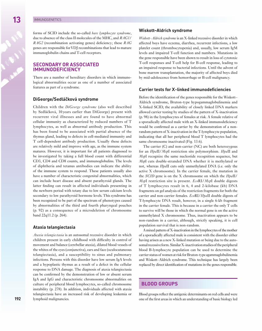
Before the identifi cation of the genes responsible for the Wiskott–Aldrich syndrome, Bruton-type hypogammaglobulinemia and X-linked SCID, the availability of closely linked DNA markers allowed carrier testing by studies of the pattern of X-inactivation (p. 98) in the lymphocytes of females at risk. A female relative of a sporadically affected male with an X-linked immunodefi ciency would be confi rmed as a carrier by the demonstration of a non-random pattern of X-inactivation in the T-lymphocyte population, indicating that all her peripheral blood T lymphocytes had the same chromosome inactivated (Fig. 13.6).
The carrier (C) and non-carrier (NC) are both heterozygous for an HpaII/MspI restriction site polymorphism. HpaII and MspI recognize the same nucleotide recognition sequence, but MspI cuts double-stranded DNA whether it is methylated or not, whereas HpaII cuts only unmethylated DNA (i.e. only the active X chromosome). In the carrier female, the mutation in the SCID gene is on the X chromosome on which the HpaII/MspI restriction site is present. EcoRI/MspI double digests of T lymphocytes result in 6, 4 and 2-kilobase (kb) DNA fragments on gel analysis of the restriction fragments for both the carrier and non-carrier females. EcoRI/HpaII double digests of T-lymphocyte DNA result, however, in a single 6-kb fragment in the carrier female. This is because in a carrier the only T cells to survive will be those in which the normal gene is on the active unmethylated X chromosome. Thus, inactivation appears to be non-random in a carrier, although, strictly speaking, it is cell population survival that is non-random.
A mixed pattern of X-inactivation in the lymphocytes of the mother of a sporadically affected male is consistent with the disorder either having arisen as a new X-linked mutation or being due to the auto-somal recessive form. Similar X-inactivation studies of the peripheral blood B-lymphocyte population can be used to determine the carrier status of women at risk for Bruton-type agammaglobulinemia and Wiskott–Aldrich syndrome. This technique has largely been replaced by direct identifi cation of mutations in the genes responsible.
BLOOD GROUPS
Blood groups refl ect the antigenic determinants on red cells and were one of the fi rst areas in which an understanding of basic biology led
Ch13-S2917.indd 192Ch13-S2917.indd 192 4/30/07 10:21:09 AM4/30/07 10:21:09 AM
IMMUNOGENETICS
193
13
to significant advances in clinical medicine. Our knowledge of the ABO and Rhesus blood groups has resulted in safe blood transfusion and the prevention of Rhesus hemolytic disease of the newborn.
THE ABO BLOOD GROUPS
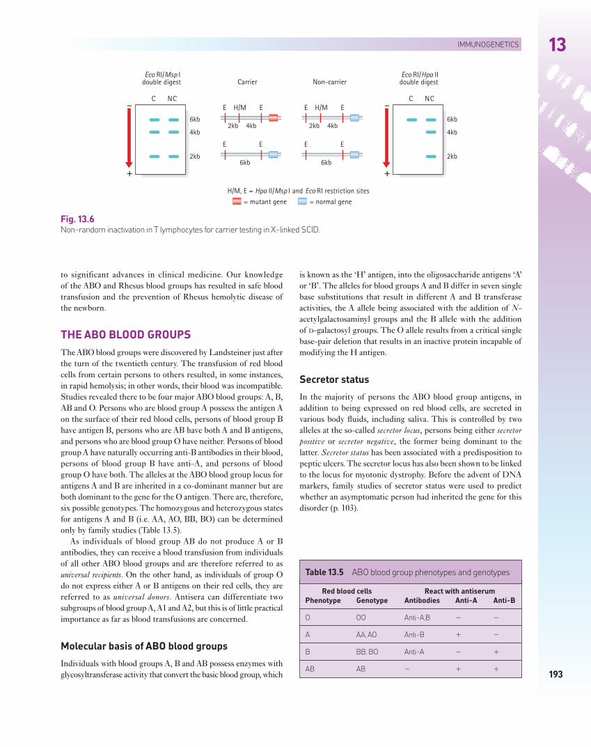
The ABO blood groups were discovered by Landsteiner just after the turn of the twentieth century. The transfusion of red blood cells from certain persons to others resulted, in some instances, in rapid hemolysis; in other words, their blood was incompatible. Studies revealed there to be four major ABO blood groups: A, B, AB and O. Persons who are blood group A possess the antigen A on the surface of their red blood cells, persons of blood group B have antigen B, persons who are AB have both A and B antigens, and persons who are blood group O have neither. Persons of blood group A have naturally occurring anti-B antibodies in their blood, persons of blood group B have anti-A, and persons of blood group O have both. The alleles at the ABO blood group locus for antigens A and B are inherited in a co-dominant manner but are both dominant to the gene for the O antigen. There are, therefore, six possible genotypes. The homozygous and heterozygous states for antigens A and B (i.e. AA, AO, BB, BO) can be determined only by family studies (Table 13.5).
As individuals of blood group AB do not produce A or B antibodies, they can receive a blood transfusion from individuals of all other ABO blood groups and are therefore referred to as universal recipients. On the other hand, as individuals of group O do not express either A or B antigens on their red cells, they are referred to as universal donors. Antisera can differentiate two subgroups of blood group A, A1 and A2, but this is of little practical importance as far as blood transfusions are concerned.
Molecular basis of ABO blood groups
Individuals with blood groups A, B and AB possess enzymes with glycosyltransferase activity that convert the basic blood group, which
is known as the ‘H’ antigen, into the oligosaccharide antigens ‘A’ or ‘B’. The alleles for blood groups A and B differ in seven single base substitutions that result in different A and B transferase activities, the A allele being associated with the addition of N-acetylgalactosaminyl groups and the B allele with the addition of D-galactosyl groups. The O allele results from a critical single base-pair deletion that results in an inactive protein incapable of modifying the H antigen.
Secretor status
In the majority of persons the ABO blood group antigens, in addition to being expressed on red blood cells, are secreted in various body fl uids, including saliva. This is controlled by two alleles at the so-called secretor locus, persons being either secretor positive or secretor negative, the former being dominant to the latter. Secretor status has been associated with a predisposition to peptic ulcers. The secretor locus has also been shown to be linked to the locus for myotonic dystrophy. Before the advent of DNA markers, family studies of secretor status were used to predict whether an asymptomatic person had inherited the gene for this disorder (p. 103).
H/M, E = Hpa II/Msp I and Eco RI restriction sites
2kb 4kb
E H/M E
2kb 4kb
E H/M E
Carrier Non-carrier
6kb
E E
6kb
E E
= mutant gene = normal gene
+
–
Eco RI/Msp Idouble digest
C NC
6kb
4kb
2kb
+
–
Eco RI/Hpa IIdouble digest
C NC
6kb
4kb
2kb
Fig. 13.6Non-random inactivation in T lymphocytes for carrier testing in X-linked SCID.
Table 13.5 ABO blood group phenotypes and genotypes
Red blood cells React with antiserum
Phenotype Genotype Antibodies Anti-A Anti-B
O OO Anti-A,B � �
A AA, AO Anti-B � �
B BB, BO Anti-A � �
AB AB � � �
Ch13-S2917.indd 193Ch13-S2917.indd 193 4/30/07 10:21:09 AM4/30/07 10:21:09 AM

IMMUNOGENETICS13
194
RHESUS BLOOD GROUP
The Rhesus (Rh) blood group system involves three sets of closely linked antigens, Cc, Dd and Ee. D is very strongly antigenic and persons are, for practical purposes, either Rh positive (possessing the D antigen) or Rh negative (lacking the D antigen).
Rhesus hemolytic disease of the newborn
A proportion of women who are Rh negative have an increased chance of having a child who will either die in utero or be born severely anemic because of hemolysis unless transfused in utero. This occurs for the following reason. If Rh-positive blood is given to persons who are Rh negative, the majority will develop anti-Rh antibodies. Such sensitization occurs with exposure to very small quantities of blood and, once a person is sensitized, further exposure results in the production of very high antibody titers.
In the case of an Rh-negative mother carrying an Rh-positive fetus, red cells of fetal origin can enter the mother’s circulation, for example after a miscarriage or at the time of delivery. This can induce the formation of Rh antibodies in the mother. In a subsequent pregnancy these antibodies can cross the placenta and enter the fetal circulation. This leads to hemolysis of the fetal red blood cells if the fetus is Rh negative, which can result either in fetal death, known as erythroblastosis fetalis, or a severe hemolytic anemia of newborn infants that is called hemolytic disease of the newborn. Once a woman has been sensitized there is a signifi cantly greater risk that a child in a subsequent pregnancy, if Rh positive, will be more severely affected.
To avoid sensitizing an Rh-negative woman, Rh-compatible blood must always be used in any blood transfusion. Furthermore, the development of sensitization and therefore Rh incompatibility after delivery can be prevented by giving the mother an injection of Rh antibodies, so-called anti-D, so that any fetal cells that have found their way into the maternal circulation are destroyed before the mother can become sensitized.
It is routine to screen all Rh-negative women during pregnancy for the development of Rh antibodies. Despite these measures, a small proportion of women do become sensitized. If Rh antibodies appear, tests are carried out to see whether the fetus is affected. If so, there is a delicate balance between the choice of early delivery, with the risks of prematurity and exchange transfusion, and treating the fetus in utero with blood transfusions.
Molecular basis of the Rhesus blood group
Recent biochemical evidence has shown there to be two types of Rh red cell membrane polypeptide. One corresponds to the D antigen and the other to the C and E series of antigens. Cloning of the genomic sequences responsible using Rh complementary DNA
(cDNA) from reticulocytes has revealed that there are two genes coding for the Rh system: one for D and d, and a second for both C and c and E and e. The D locus is present in most persons and codes for the major D antigen present in those who are Rh positive. Rh-negative individuals are homozygous for a deletion of the D gene. It is not, perhaps, surprising therefore that an antibody has never been raised to d!
Analysis of cDNA from reticulocytes in Rh-negative persons who were homozygous for dCe, dcE and dce allowed identifi cation of the genomic DNA sequences responsible for the different antigenic variants at the second locus, revealing that they are produced by alternative splicing of the mRNA transcript. The Ee polypeptide is a full-length product of the CcEe gene, very similar in sequence to the D polypeptide. The E and e antigens differ by a point mutation in exon 5. The Cc polypeptides are, in contrast, products of a shorter transcript of the same gene having either exons 4, 5 and 6 or 4, 5 and 8 spliced out. The difference between C and c is due to four amino-acid substitutions in exons 1 and 2. These fi ndings help to explain what was an apparently complex blood group system.
OTHER BLOOD GROUPS
There are approximately a further 12 ‘common’ blood group systems of clinical importance in humans, including Duffy, Lewis, MN and S. These are usually of concern only when cross-matching blood for persons who, because of repeated transfusions, have developed antibodies to one of these other blood group antigens. Until the advent of DNA fi ngerprinting (p. 69), they were used in linkage studies (p. 131) and paternity testing (p. 259).
FURTHER READING
Bell J I, Todd J A, McDevitt H O 1989 The molecular basis of HLA–disease association. Adv Hum Genet 18: 1–41
Good review of the HLA–disease associations.Dreyer W J, Bennet J C 1965 The molecular basis of antibody formation:
a paradox. Proc Natl Acad Sci USA 54: 864–869The proposal of the generation of antibody diversity.Hunkapiller T, Hood L 1989 Diversity of the immunoglobulin gene
superfamily. Adv Immunol 44: 1–63Good review of the structure of the immunoglobulin gene superfamily.Janeway C A, Travers P, Walport M, Capra J D 1999 Immunobiology,
4th edn. Current Biology, LondonGood, well illustrated, textbook of the biology of immunology.Lachmann P J, Peters K, Rosen F S, Walport M J 1993 Clinical aspects of
immunology, 5th edn. Blackwell, OxfordA comprehensive three-volume multiauthor text covering both basic and clinical
immunology.Roitt I 1997 Essential immunology, 9th edn. Blackwell, OxfordExcellent basic immunology textbook.
Ch13-S2917.indd 194Ch13-S2917.indd 194 4/30/07 10:21:10 AM4/30/07 10:21:10 AM

IMMUNOGENETICS
195
13
ELEMENTS
1 The immune response in humans can be divided into two main types, innate and specifi c acquired or adaptive immunity. Both types can be further subdivided into humoral and cell-mediated immunity.
2 Innate humoral immunity involves acute-phase proteins that act to minimize tissue injury by limiting the spread of infective organisms and, through the alternative pathway of complement activation, results in a localized infl ammatory response and the attraction of phagocytes and opsonization of microorganisms. Complement, which consists of a series of inactive blood proteins that are activated sequentially in a cascade, can also be activated through the classic pathway by antibody binding to antigen.
3 Innate cell-mediated immunity involves engulfment of microorganisms by macrophages and their destruction by intracellular granules.
4 Specifi c acquired humoral immunity involves produc-tion of antibodies by mature B cells or plasma cells in response to antigen. Antibodies are Y-shaped molecules and each is composed of two identical heavy (H) chains and two identical light (L) chains. The antibody molecule has two parts that differ in their function: two identical antigen-binding sites (Fab) and a single binding site for complement (Fc). There are fi ve classes of antibody, IgA, IgD, IgE, IgG and IgM, each with a specifi c heavy chain. The light chain of any class of antibody can be made up of either kappa (k) or lambda (l) chains.
5 Each immunoglobulin light or heavy chain has a variable (V) region of approximately 110 amino acids at the amino-terminal end. The carboxy-terminal end consists of a constant (C) region of approximately 110 amino acids in the k and l light chains and three to four times that length in the heavy chain. Most of the amino-acid sequence
variation in both the light and heavy chains occurs within several small hypervariable regions. These are thought to be the sites of antigen binding. The immunoglobulin chains are produced from combinations of separate groups of DNA segments. These consist of one from a variable number of DNA segments coding for the constant (C), variable (V) and joining (J) regions between the V and C regions for the k and l light chains and the various types of heavy chains. The heavy chains also contain a diversity (D) region located between the V and J regions. The total number of possible antibodies that could be produced by various combinations of these DNA segments accounts for the antibody diversity seen in humans.
6 Cell-mediated specifi c acquired immunity primarily involves T cells which, through the T-cell surface antigen receptor, in conjunction with the major histocompatibility complex molecules on the surface of infected cells, engage T helper cells and cytotoxic T cells to combat intracellular infections.
7 The major histocompatibility complex (MHC) or human leukocyte antigen (HLA) system consists of a number of closely linked loci on chromosome 6. The many different alleles that can occur at each locus mean that a very large number of different combinations of these can result. The HLA loci are inherited ‘en bloc’ as a haplotype. The closer the match of HLA antigens between the donor and recipient in organ transplantation, the greater the likelihood of long-term survival of the homograft. Possession of certain HLA antigens is associated with an increased relative risk of developing specifi c diseases.
8 An understanding of the ABO and Rhesus blood groups has resulted in safe blood transfusions and the prevention of Rhesus hemolytic disease of the newborn.
Ch13-S2917.indd 195Ch13-S2917.indd 195 4/30/07 10:21:10 AM4/30/07 10:21:10 AM