chapter seven were basal primates nocturnal? evidence from ...rosslab.uchicago.edu/publications/ross...
TRANSCRIPT
233
CHAPTER SEVEN
Were Basal PrimatesNocturnal? Evidence from
Eye and Orbit ShapeCallum F. Ross, Margaret I. Hall, and
Christopher P. Heesy
INTRODUCTION
The adaptations of basal primates are of interest to paleoprimatologistsbecause they give insight into the context in which primates diverged fromother mammals. In addition, these basal adaptations may have biased the evo-lutionary trajectories taken by the lineages leading to extant primates. Dielactivity pattern is an important component of an animal’s ecology because ithas pervasive influence on many aspects of primate morphology and behavior,including body size, diet, substrate preference, communication, and adapta-tions of the sensory systems (Charles-Dominique, 1975; Heesy and Ross,2001, 2004; Martin, 1979).
Early explanations for primate origins did not specify the activity pattern ofbasal primates (Cartmill, 1970, 1972, 1974; Elliot Smith, 1924; Le GrosClark, 1959; Wood Jones, 1916), with one possible exception. Writing inthe spirit of the “Primatological Synthesis” (sensu Cartmill, 1982), in which
Callum F. Ross ● Organismal Biology & Anatomy, University of Chicago, Chicago, IL 60637Margaret I. Hall ● Department of Anatomical Sciences, Health Sciences Center, Stony BrookUniversity, Stony Brook, NY 11794-8081 Christopher P. Heesy ● Department of Anatomy, NewYork College of Osteopathic Medicine, Old Westbury, NY 11568
primates were defined by a set of pervasive trends, Polyak (1957) argued thattrends toward diurnality, high-visual acuity, and color vision, culminating inthe higher primates, suggested continuity of diurnal “potential” through themammalian stem lineage, up through primates. According to Polyak (1957:968–969), nocturnal strepsirrhines are divergent from the diurnal mainstreamof primate evolution.
By the beginning of the 1970s, however, P. Charles-Dominique andR. D. Martin’s field studies had revealed many ecological and behavioralsimilarities between Microcebus murinus and “Galago demidovii,” includingnocturnality, leading them to suggest that many of these aspects are likelyto be both ancestral strepsirrhine and ancestral primate characteristics(Charles-Dominique and Martin, 1970). Martin (1973) bolstered the argu-ment that the ancestral strepsirrhines were nocturnal by noting that manydiurnal lemurid species possess a tapetum. Tapeta are usually found in noc-turnal animals (Walls, 1942; see Ross, 2004, for a review), suggesting thattheir presence in extant diurnal strepsirrhines is due to “primitive reten-tion.” Extending this “primitive retention” argument to explain the absenceof a tapetum in Aotus and Tarsius, Martin suggested that these animals weredescended from diurnal ancestors, retaining a diurnal adaptation into a noc-turnal environment (Martin, 1973). This argument was reinforced by theobservation that Tarsius also possesses a retinal fovea (Le Gros Clark,1959)—a traditionally diurnal adaptation (Ogden, 1974; Polyak, 1957;Ross, 2004; Walls, 1942). In contrast with Martin’s “primitive retention”explanation for the imperfect correlations between primate morphology andactivity pattern, Charles-Dominique (1975: 86) suggested that the lastcommon ancestor of primates “had an eye slightly differentiated for bothnocturnal and diurnal vision,” capable of evolving into an anthropoid or astrepsirrhine eye.
By the late 1970s, the issue of the activity pattern of basal primates wasindependently addressed by a number of workers. Martin (1979) marshaledevidence, including body size, relative size of the olfactory bulb, and the pres-ence or absence of tapeta, to suggest “that nocturnal life involving at least somepredation on small animals is a primitive feature for the lemurs and lorises, andpossibly for the primates as a whole” (Martin, 1979: 72). Supporting Martin’sargument were functional interpretations of two features assumed to have char-acterized basal primates: high degrees of orbital convergence and relativelylarge orbital apertures.
234 Callum F. Ross et al.
ORBITAL CONVERGENCE
Primates have long been noted to have more convergent orbital aperturesthan most other mammals. Early explanations related convergence to arbore-ality (Elliot Smith, 1924; Le Gros Clark, 1959; Wood Jones, 1916).However, comparisons with other animals suggested to Cartmill (1970,1972) that convergent orbits facilitated visual predation on insects in the finebranches of the shrub layer of tropical rainforests. Cartmill argued that“Stereoptic integration of the two visual fields improves the accuracy of thefinal strike; increase in visual-field overlap facilitates compensation for evasivemovements of the prey” (1972: 113). Cartmill’s hypothesis did not specifywhether these first primates were diurnal, nocturnal, crepuscular, or cathe-meral, and it was left to Jack Pettigrew and John Allman to round out thevisual predation hypothesis, specifying nocturnality as an essential part of theargument (Cartmill, 1992).
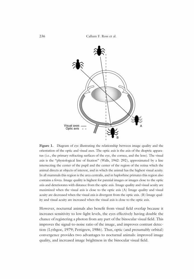
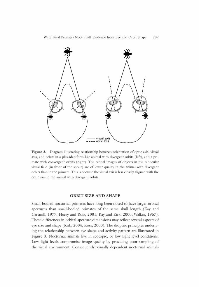
Pettigrew (cited by Allman, 1977: p. 29; Pettigrew, 1978) and Allman(1977) pointed out that the dioptric benefits of orbital convergence accrue tonocturnal rather than diurnal animals. The optical axis is the axis of the diop-tric apparatus of the eye (i.e., lens and cornea), around which image qualityis highest, whereas the visual axis is the “physiological line of fixation” (Walls,1942: 292), approximated by a line passing through the center of the pupiland the retinal fovea or area centralis. Thus, alignment of the optic axis withthe visual axis maximizes image quality in the fovea or area centralis(Figure 1). The Allman-Pettigrew model posits that orbital convergence iscorrelated with convergence of the optic axes on the visual axes (Figure 2),providing improved image quality in nocturnal primates. Another way toensure high-image quality across the retina is to restrict incoming imagesto the paraxial region of the dioptric apparatus. This can be achieved bydecreasing diameter of the pupil, but this option is not available to nocturnalanimals that must maintain large pupil sizes in order to maintain imagebrightness. Consequently, nocturnal animals can only improve image qualityin the area of visual field overlap by optic convergence (Figure 2). This sug-gested to Allman (1977) that if the first primates had high degrees of orbitalconvergence, then they were probably nocturnal.
Convergence of the optic axes on each other increases the size of theregion of visual field overlap (Heesy, 2004; Ross, 2000), something thatCartmill hypothesized was advantageous in the pursuit of evasive prey.
Were Basal Primates Nocturnal? Evidence from Eye and Orbit Shape 235
However, nocturnal animals also benefit from visual field overlap because itincreases sensitivity to low-light levels, the eyes effectively having double thechance of registering a photon from any part of the binocular visual field. Thisimproves the signal-to-noise ratio of the image, and improves contrast detec-tion (Lythgoe, 1979; Pettigrew, 1986). Thus, optic (and presumably orbital)convergence provides two advantages to nocturnal animals: improved imagequality, and increased image brightness in the binocular visual field.
236 Callum F. Ross et al.
Optic axisVisual axes
B
A
Figure 1. Diagram of eye illustrating the relationship between image quality and theorientation of the optic and visual axes. The optic axis is the axis of the dioptric appara-tus (i.e., the primary refracting surfaces of the eye, the cornea, and the lens). The visualaxis is the “physiological line of fixation” (Walls, 1942: 292), approximated by a lineintersecting the center of the pupil and the center of the region of the retina which theanimal directs at objects of interest, and in which the animal has the highest visual acuity.In all mammals this region is the area centralis, and in haplorhine primates this region alsocontains a fovea. Image quality is highest for paraxial images or images close to the opticaxis and deteriorates with distance from the optic axis. Image quality and visual acuity aremaximized when the visual axis is close to the optic axis (A) Image quality and visualacuity are decreased when the visual axis is divergent from the optic axis. (B) Image qual-ity and visual acuity are increased when the visual axis is close to the optic axis.
ORBIT SIZE AND SHAPE
Small-bodied nocturnal primates have long been noted to have larger orbitalapertures than small-bodied primates of the same skull length (Kay andCartmill, 1977; Heesy and Ross, 2001; Kay and Kirk, 2000; Walker, 1967).These differences in orbital aperture dimensions may reflect several aspects ofeye size and shape (Kirk, 2004; Ross, 2000). The dioptric principles underly-ing the relationship between eye shape and activity pattern are illustrated inFigure 3. Nocturnal animals live in scotopic, or low light level conditions.Low light levels compromise image quality by providing poor sampling ofthe visual environment. Consequently, visually dependent nocturnal animals
Were Basal Primates Nocturnal? Evidence from Eye and Orbit Shape 237
visual axisoptic axis
Figure 2. Diagram illustrating relationship between orientation of optic axis, visualaxis, and orbits in a plesiadapiform-like animal with divergent orbits (left), and a pri-mate with convergent orbits (right). The retinal images of objects in the binocularvisual field (in front of the snout) are of lower quality in the animal with divergentorbits than in the primate. This is because the visual axis is less closely aligned with theoptic axis in the animal with divergent orbits.
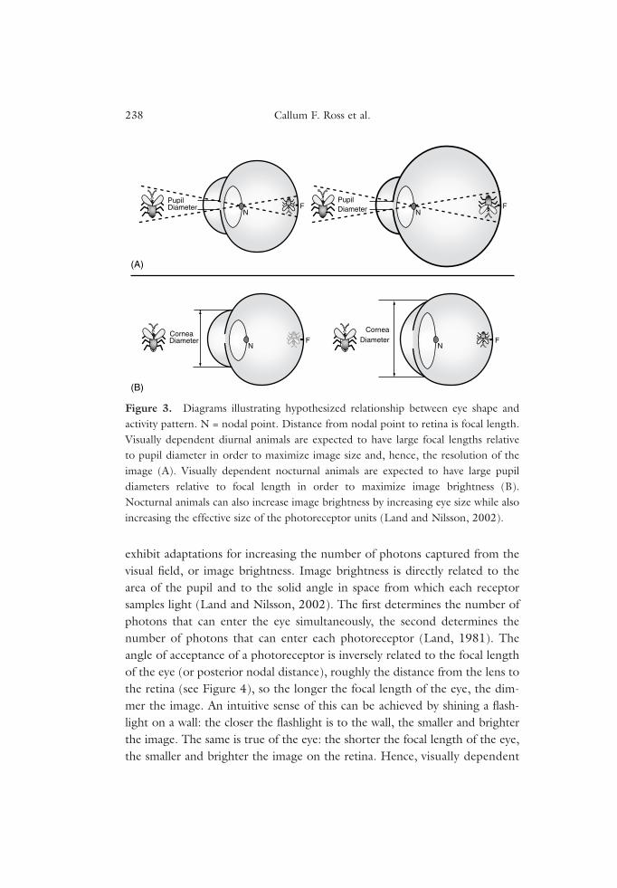
exhibit adaptations for increasing the number of photons captured from thevisual field, or image brightness. Image brightness is directly related to thearea of the pupil and to the solid angle in space from which each receptorsamples light (Land and Nilsson, 2002). The first determines the number ofphotons that can enter the eye simultaneously, the second determines thenumber of photons that can enter each photoreceptor (Land, 1981). Theangle of acceptance of a photoreceptor is inversely related to the focal lengthof the eye (or posterior nodal distance), roughly the distance from the lens tothe retina (see Figure 4), so the longer the focal length of the eye, the dim-mer the image. An intuitive sense of this can be achieved by shining a flash-light on a wall: the closer the flashlight is to the wall, the smaller and brighterthe image. The same is true of the eye: the shorter the focal length of the eye,the smaller and brighter the image on the retina. Hence, visually dependent
238 Callum F. Ross et al.
NF
PupilDiameter
FN
CorneaF
N
N
PupilDiameter F
(A)
(B)
Diameter
CorneaDiameter
Figure 3. Diagrams illustrating hypothesized relationship between eye shape andactivity pattern. N = nodal point. Distance from nodal point to retina is focal length.Visually dependent diurnal animals are expected to have large focal lengths relativeto pupil diameter in order to maximize image size and, hence, the resolution of theimage (A). Visually dependent nocturnal animals are expected to have large pupildiameters relative to focal length in order to maximize image brightness (B).Nocturnal animals can also increase image brightness by increasing eye size while alsoincreasing the effective size of the photoreceptor units (Land and Nilsson, 2002).
nocturnal animals are predicted to have large corneas relative to their focallength regardless of body or eye size (Figure 3B).
In addition to these effects on eye shape, nocturnality is also predicted tobe associated with increased eye size. The pupil obviously cannot be largerthan the eye, so eye size limits the amount that the pupil can be expanded. Ifpupil diameter is increased by making the eye bigger, focal length will alsoincrease, reducing image brightness by decreasing the angle of acceptance ofthe photoreceptors. However, this latter effect can be compensated for byincreasing the effective size of the photoreceptors. In vertebrates this is doneby pooling many receptors into one functional unit by connecting many ofthem up to a single ganglion cell—a phenomenon widespread among verte-brates, including primates (Kay and Kirk, 2000; Rohen and Castenholtz,1967). Thus, photoreceptor pooling allows image brightness to be increasedpurely by increasing eye size, so it is expected that nocturnal animals will havelarger eyes than similarly sized diurnal animals.
It has also been argued that photoreceptor pooling has the added bene-fit of widening the range of “image brightness,” or luminance, to which an
Were Basal Primates Nocturnal? Evidence from Eye and Orbit Shape 239
C P
A
F
N
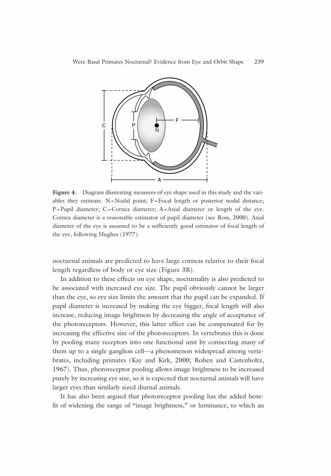
Figure 4. Diagram illustrating measures of eye shape used in this study and the vari-ables they estimate. N=Nodal point; F=Focal length or posterior nodal distance;P=Pupil diameter; C=Cornea diameter; A=Axial diameter or length of the eye.Cornea diameter is a reasonable estimator of pupil diameter (see Ross, 2000). Axialdiameter of the eye is assumed to be a sufficiently good estimator of focal length ofthe eye, following Hughes (1977).
animal is sensitive (Martin, 1999). This hypothesis assumes that the size ofthe photoreceptor (rod) pools of nocturnal animals is flexible at the retinallevel, allowing changes in the size of the pool in response to different lumi-nance levels. This would be valuable in nocturnal environments that arecharacterized by a much wider range of luminance levels than diurnal envi-ronments (Lythgoe, 1979; Martin, 1990, 1999). The hypothesis argues thatincreases in eye size would augment the number of photoreceptor pools, cre-ating the possibility of greater flexibility to the greater range of light levels innocturnal environments (Martin, 1990, 1999). The validity of this hypothe-sis remains in doubt until mechanisms for adjusting receptive field size in sco-topic conditions are demonstrated. Primates lack rod–rod coupling thatmight be one mechanism to accomplish this (Djamgoz et al., 1999), butthere do appear to be up to three pathways for information to pass from rodsto the inner retina, and these pathways may operate under different ambientlight conditions (Bloomfield and Dacheux, 2001).
Diurnal animals, in contrast, are not constrained by the need to shortenfocal length or enlarge the pupil, because image brightness is not a problemin the photopic or light rich environment. Consequently, visually dependentdiurnal animals are able to have long focal lengths, thereby decreasing theacceptance angle of each photoreceptor in the retina and increasing visualresolution (Figure 3A). Another way of saying this is that increased focallength spreads the image over a larger number of photoreceptors, increasingvisual resolution. Moreover, because diurnal animals do not need to enhanceimage brightness, they are predicted to have small pupils relative to focallength.
RECONSTRUCTIONS OF ORBIT SIZE AND SHAPE IN BASAL PRIMATES
Functional interpretations of orbital convergence and enlargement only sug-gest that basal primates were nocturnal if these features were present in basalprimates. The last common ancestor of primates is not known from fossil evi-dence, and nor are their immediate outgroups. Consequently, the assumptionthat basal primates had orbital apertures that were convergent and enlargedrests on interpretation of the available evidence from fossils and extant taxa.
The objectives of this study are: (a) to document the relationship betweeneye size and shape, and activity patterns in extant primates; (b) to document
240 Callum F. Ross et al.
the relationships between eye size and shape, and activity patterns in extantamniotes and use these data to interpret primate eye shape; and (c) to useorbit shape data to reconstruct activity pattern in fossil primates.
MATERIALS AND METHODS
Eye Shape Measures
On the basis of the dioptric principles outlined in the Introduction, visuallydependent nocturnal animals are predicted to have large pupils relative tofocal length, and visually dependent diurnal animals are predicted to havesmall pupil diameters for their focal lengths. These dimensions cannot bemeasured accurately in preserved eyes and are known for only a small num-ber of vertebrates (e.g., Arrese, 2002; Hughes, 1977; Martin, 1999). Here,we use the axial diameter of the eye as a surrogate for focal length, and corneadiameter as a surrogate for pupil diameter (Figure 4). Hughes (1977, Figure9B) has shown that, across a range of vertebrates of differing activity patterns,focal length is approximately 0.6 axial diameter of the eye. Assuming that thisrelationship is constant across vertebrates, we use axial diameter of the eye asa surrogate for focal length. Cornea diameter is a reasonable surrogate forpupil diameter as there is no obvious reason to have a cornea that is signifi-cantly larger than the pupil.
To investigate scaling relationships of cornea diameter and axial length,head and body length is used as a measure of body size for comparisons acrossdifferent groups of amniotes.
Orbit Shape Measures
The relationship between relative size of the orbital aperture and activity pat-tern has long been of interest to paleoprimatologists because it provides amethod for reconstructing activity pattern in moderately well-preserved fos-sils (Beard et al., 1991; Heesy and Ross, 2001; Kay and Cartmill, 1977; Kayand Kirk, 2000; Martin, 1990; Rasmussen and Simons, 1992; Walker, 1967).Given the relationship between eye shape and activity pattern predictedabove, we also predict that there will be a relationship between orbit shapeand activity pattern. Specifically, we predict that the size of the orbital aper-ture relative to the axial depth of the orbit should be correlated with activitypattern. The underlying assumptions, that pupil or cornea area is correlated
Were Basal Primates Nocturnal? Evidence from Eye and Orbit Shape 241
with orbital aperture area, and that the axial length of the eye is correlatedwith the axial depth of the orbit, have not yet been evaluated using measuresof orbit and eye shape taken from the same individuals.
Orbital aperture size is estimated by the diameter of the orbital aperture,measured from orbitale inferius to the orbitale superius (Cartmill, 1970).Orbit depth is calculated as the distance from the midpoint of orbitalesuperius–orbitale inferius chord to the superiormost point along the rim ofthe optic canal. These measures were extracted using customized macros inMicrosoft Excel, from 3D coordinates of points collected using a Microscribe3DX digitizer (Immersion Corp., San Jose, CA).
Orbit diameter data for Cantius abditus are from Heesy and Ross (2001).The optic canal of Cantius abditus (USNM 494881: Rose et al., 1999) is notpreserved. A minimum estimate of the axial depth of the orbit in C. abdituswas obtained by combining the length of the orbit floor in C. abditus (USNM494881) with that of another specimen of this taxon (USNM 93938).Comparisons of C. abditus (USNM 494881) orbit and preserved braincasewith similar-sized extant strepsirrhines (e.g., Otolemur crassicaudatus) suggestthat the orbit depth was not substantially longer than this estimate.
The majority of the eye shape data were derived from Ritland (1982). Datafrom nonadult individuals were excluded. The primate eye shape data werederived in part from Ritland (1982), as well as from unpublished observationsmade by C. F. Ross. Additional data on bats and birds were collected byM. I. Hall (Hall, 2005). The orbit shape data were collected from differentspecimens than the eye shape data.
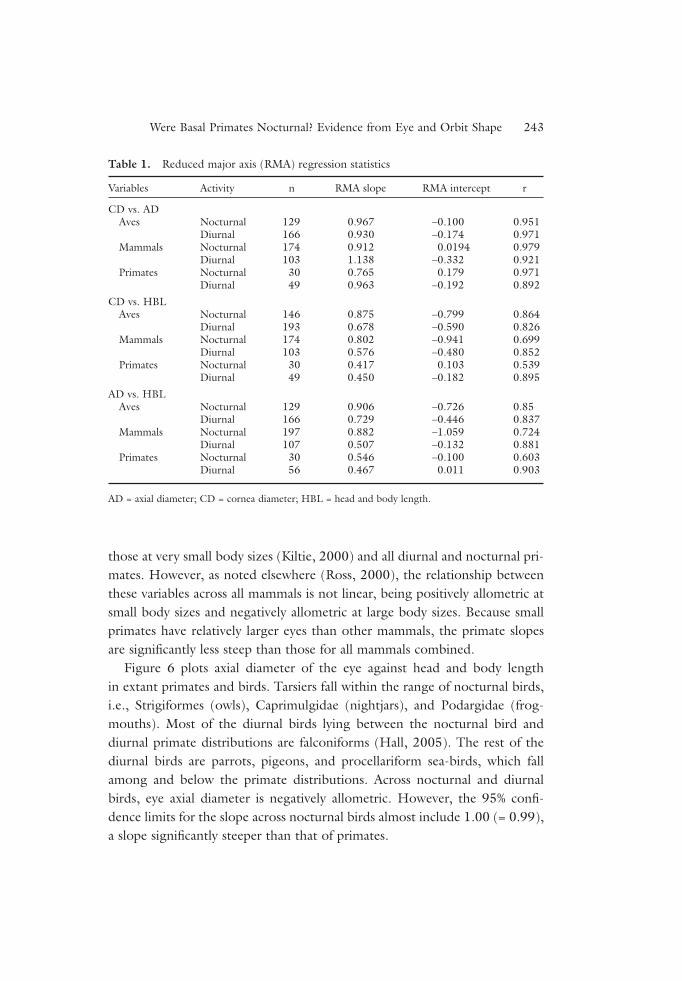
Reduced major axis (RMA) regression equations for all Aves, Primates,and Mammalia were calculated from least squares equations generated bySPSS 12.0 for Windows. The RMA slopes, intercepts, and correlation coef-ficients are given in Table 1.
RESULTS
Eye Size and Shape
Figure 5 plots axial eye diameter against head and body length in extant pri-mates and other mammals. Nocturnal primate eyes have larger axial diametersthan most similarly sized nonprimate mammals. Tarsiers have longer eyes rel-ative to body size than any other mammals. Axial diameter of the eye scaleswith negative allometry across all diurnal and nocturnal mammals, except
242 Callum F. Ross et al.
those at very small body sizes (Kiltie, 2000) and all diurnal and nocturnal pri-mates. However, as noted elsewhere (Ross, 2000), the relationship betweenthese variables across all mammals is not linear, being positively allometric atsmall body sizes and negatively allometric at large body sizes. Because smallprimates have relatively larger eyes than other mammals, the primate slopesare significantly less steep than those for all mammals combined.
Figure 6 plots axial diameter of the eye against head and body lengthin extant primates and birds. Tarsiers fall within the range of nocturnal birds,i.e., Strigiformes (owls), Caprimulgidae (nightjars), and Podargidae (frog-mouths). Most of the diurnal birds lying between the nocturnal bird anddiurnal primate distributions are falconiforms (Hall, 2005). The rest of thediurnal birds are parrots, pigeons, and procellariform sea-birds, which fallamong and below the primate distributions. Across nocturnal and diurnalbirds, eye axial diameter is negatively allometric. However, the 95% confi-dence limits for the slope across nocturnal birds almost include 1.00 (= 0.99),a slope significantly steeper than that of primates.
Were Basal Primates Nocturnal? Evidence from Eye and Orbit Shape 243
Table 1. Reduced major axis (RMA) regression statistics
Variables Activity n RMA slope RMA intercept r
CD vs. ADAves Nocturnal 129 0.967 –0.100 0.951
Diurnal 166 0.930 –0.174 0.971Mammals Nocturnal 174 0.912 0.0194 0.979
Diurnal 103 1.138 –0.332 0.921Primates Nocturnal 30 0.765 0.179 0.971
Diurnal 49 0.963 –0.192 0.892
CD vs. HBLAves Nocturnal 146 0.875 –0.799 0.864
Diurnal 193 0.678 –0.590 0.826Mammals Nocturnal 174 0.802 –0.941 0.699
Diurnal 103 0.576 –0.480 0.852Primates Nocturnal 30 0.417 0.103 0.539
Diurnal 49 0.450 –0.182 0.895
AD vs. HBLAves Nocturnal 129 0.906 –0.726 0.85
Diurnal 166 0.729 –0.446 0.837Mammals Nocturnal 197 0.882 –1.059 0.724
Diurnal 107 0.507 –0.132 0.881Primates Nocturnal 30 0.546 –0.100 0.603
Diurnal 56 0.467 0.011 0.903
AD = axial diameter; CD = cornea diameter; HBL = head and body length.

244 Callum F. Ross et al.
Log head and body length (mm)4.54.03.53.02.52.01.51.0
Log
axia
l dia
met
er (
mm
)
2.0
1.5
1.0
0.0
Nocturnal primate
Nocturnal mammal
Fossorial mammal
Diurnal primate
Diurnal mammal
Crepuscular mammal
Cathemeral primate
Unknown
Nocturnalprimates
Diurnalprimates
0.5
− 0.5
Figure 5. Bivariate plot of axial diameter (log10) against head and body length(log10) across all mammals. Minimum spanning polygons for nocturnal primates anddiurnal primates are added. Nocturnal primates have longer axial lengths for theirbody size than similarly sized nonprimate mammals.
Log head and body length (mm)
3.43.23.02.82.62.42.22.01.81.6
Log
axia
l dia
met
er (
mm
)
1.6
1.4
1.2
1.0 Nocturnal primate
Nocturnal aves
Diurnal primate
Diurnal aves
Cathemeral primate
Nocturnalprimates
Diurnalprimates
0.8
0.6
Figure 6. Bivariate plot of axial diameter (log10) against head and body length(log10) across birds and primates. Minimum spanning polygons for nocturnal primatesand diurnal primates are added. The nocturnal primates with the longest axial lengthsfor their body size are tarsiers, which plot with the nocturnal birds represented here;i.e., Strigiformes (owls) Caprimulgidae (nightjars), and Podargidae (frogmouths).
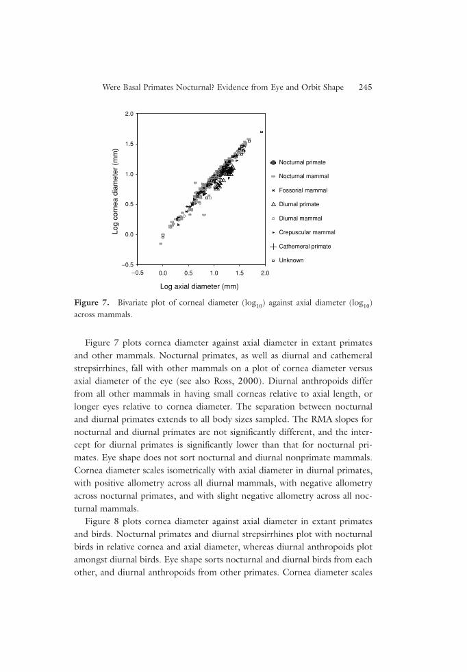
Figure 7 plots cornea diameter against axial diameter in extant primatesand other mammals. Nocturnal primates, as well as diurnal and cathemeralstrepsirrhines, fall with other mammals on a plot of cornea diameter versusaxial diameter of the eye (see also Ross, 2000). Diurnal anthropoids differfrom all other mammals in having small corneas relative to axial length, orlonger eyes relative to cornea diameter. The separation between nocturnaland diurnal primates extends to all body sizes sampled. The RMA slopes fornocturnal and diurnal primates are not significantly different, and the inter-cept for diurnal primates is significantly lower than that for nocturnal pri-mates. Eye shape does not sort nocturnal and diurnal nonprimate mammals.Cornea diameter scales isometrically with axial diameter in diurnal primates,with positive allometry across all diurnal mammals, with negative allometryacross nocturnal primates, and with slight negative allometry across all noc-turnal mammals.
Figure 8 plots cornea diameter against axial diameter in extant primatesand birds. Nocturnal primates and diurnal strepsirrhines plot with nocturnalbirds in relative cornea and axial diameter, whereas diurnal anthropoids plotamongst diurnal birds. Eye shape sorts nocturnal and diurnal birds from eachother, and diurnal anthropoids from other primates. Cornea diameter scales
Were Basal Primates Nocturnal? Evidence from Eye and Orbit Shape 245
Log axial diameter (mm)
Log
corn
ea d
iam
eter
(m
m)
2.0
1.5
Nocturnal primate
Nocturnal mammal
Fossorial mammal
Diurnal primate
Diurnal mammal
Crepuscular mammal
Cathemeral primate
Unknown
1.0
0.5
0.0
−0.52.01.51.00.50.0− 0.5
Figure 7. Bivariate plot of corneal diameter (log10) against axial diameter (log10)across mammals.
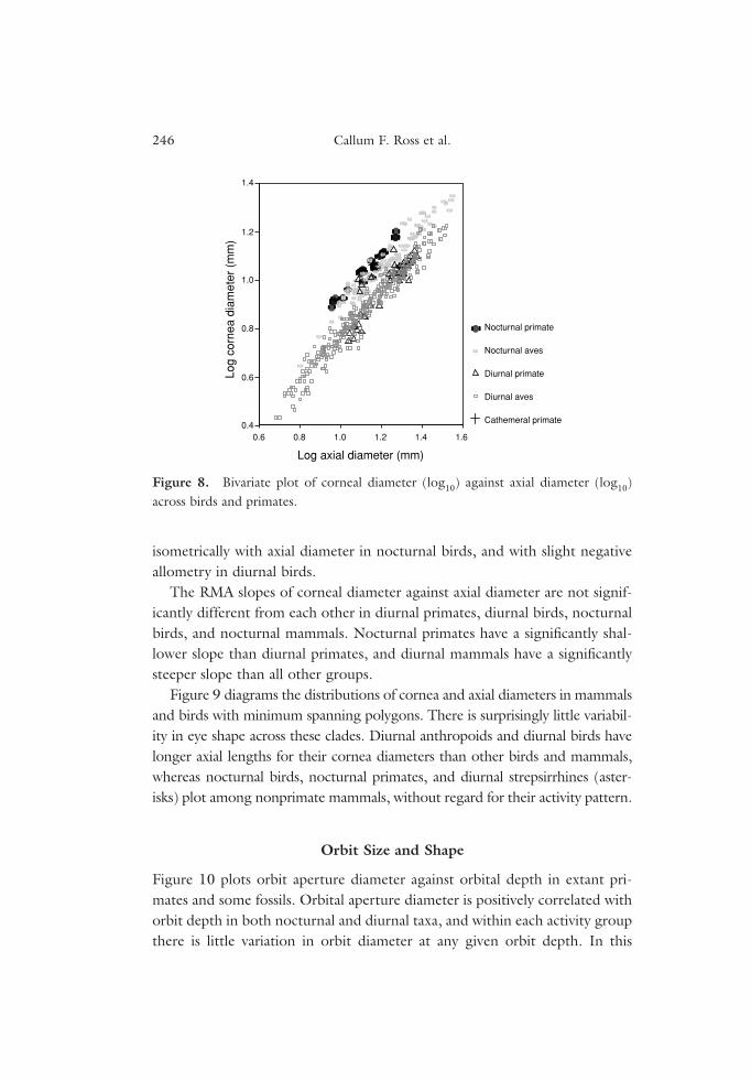
isometrically with axial diameter in nocturnal birds, and with slight negativeallometry in diurnal birds.
The RMA slopes of corneal diameter against axial diameter are not signif-icantly different from each other in diurnal primates, diurnal birds, nocturnalbirds, and nocturnal mammals. Nocturnal primates have a significantly shal-lower slope than diurnal primates, and diurnal mammals have a significantlysteeper slope than all other groups.
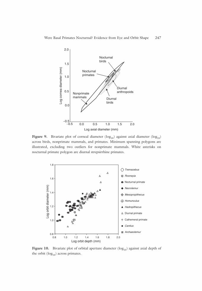
Figure 9 diagrams the distributions of cornea and axial diameters in mammalsand birds with minimum spanning polygons. There is surprisingly little variabil-ity in eye shape across these clades. Diurnal anthropoids and diurnal birds havelonger axial lengths for their cornea diameters than other birds and mammals,whereas nocturnal birds, nocturnal primates, and diurnal strepsirrhines (aster-isks) plot among nonprimate mammals, without regard for their activity pattern.
Orbit Size and Shape
Figure 10 plots orbit aperture diameter against orbital depth in extant pri-mates and some fossils. Orbital aperture diameter is positively correlated withorbit depth in both nocturnal and diurnal taxa, and within each activity groupthere is little variation in orbit diameter at any given orbit depth. In this
246 Callum F. Ross et al.
Log axial diameter (mm)
Log
corn
ea d
iam
eter
(m
m)
Nocturnal primate
Nocturnal aves
Diurnal primate
Diurnal aves
Cathemeral primate
0.6
0.40.6 0.8 1.0 1.2 1.4 1.6
0.8
1.0
1.2
1.4
Figure 8. Bivariate plot of corneal diameter (log10) against axial diameter (log10)across birds and primates.
Were Basal Primates Nocturnal? Evidence from Eye and Orbit Shape 247
Figure 10. Bivariate plot of orbital aperture diameter (log10) against axial depth ofthe orbit (log10) across primates.
Figure 9. Bivariate plot of corneal diameter (log10) against axial diameter (log10)across birds, nonprimate mammals, and primates. Minimum spanning polygons areillustrated, excluding two outliers for nonprimate mammals. White asterisks onnocturnal primate polygon are diurnal strepsirrhine primates.
***
*
Log
corn
ea d
iam
eter
(m
m)
Log axial diameter (mm)
2.0
1.5
1.0
0.5
0.0
−0.50.5 1.0 1.5 2.0
Nonprimate mammals Diurnal
birds
Diurnal anthropoids
Nocturnal primates
Nocturnal birds
−0.5 0.0
Log orbit depth (mm)2.01.81.61.41.21.0
Log
orbi
t dia
met
er (
mm
)
1.8
1.6
1.4
1.2
1.0
Tremacebus
Rooneyia
Nocturnal primate
Necrolemur
Mesopropithecus
Homunculus
Hadropithecus
Diurnal primate
Cathemeral primate
Cantius
Archaeolemur0.8
0.8
respect this plot resembles the plot of cornea diameter against axial length ofthe eye in birds and primates (Figure 7). However, the slopes of the two dis-tributions differ, so that at small body sizes nocturnal primates have largerorbital apertures than diurnal primates with the same axial depths, but abovean axial depth=1.3 (log10) the nocturnal and diurnal distributions begin tooverlap.
The activity patterns of several fossil taxa can be reconstructed by plottingthem on this distribution. As noted by others, Necrolemur was almost cer-tainly nocturnal. Tremacebus and Homunculus also plot as nocturnal, andMesopropithecus plots as diurnal. Rooneyia plots closest to the diurnal pri-mates, and was probably diurnal, although it has a slightly larger orbit diam-eter than extant diurnal primates. At orbit depths greater than that ofMesopropithecus there are no nocturnal extant primates, so it is not possible toestimate the activity patterns of larger fossil forms, such as Hadropithecus.Cantius falls above the distribution of extant nocturnal primates, suggestingthat it was nocturnal, confirming the results of Heesy and Ross (2001).However, because of the uncertainty of orbit depth in this taxon, thisconclusion must be regarded as preliminary.
DISCUSSION
The Eyes of Basal Primates
There is debate as to the precise ecological significance of the relatively highlevels of orbital convergence seen in primates. Cartmill attributes it toselection for nocturnal visual predation (Cartmill, 1992), whereas othersassociate it with manual manipulation or visual detection of small objects,including fruits, insects, and small branches in a nocturnal rainforestenvironment (Crompton, 1995; Rasmussen, 1990; Sussman, 1991, 1995).Common to all these models is the relationship between orbital convergenceand nocturnality. Although relatively high degrees of orbital conver-gence have not been extrapolated down to basal primates using rigorouscharacter optimization methods, all primates, living and fossil, with only oneexception (Megaladapis) have more highly convergent orbits (Heesy, 2003;Ni et al., 2004; Ravosa and Savokova, 2004) than seen in nonprimate mam-mals, including plesiadapiforms. It, therefore, seems probable that basal pri-mates also had relatively high degrees of orbital convergence, and hencewere nocturnal.
248 Callum F. Ross et al.
This conclusion is congruent with character optimization studies of theevolution of activity pattern and chromacy in primates, and their relatives(Heesy and Ross, 2001, 2004), but runs counter to recent claims that basalprimates were diurnal (Li, 2000; Ni et al., 2004; Tan and Li, 1999). We havediscussed our objections to Tan and Li’s arguments elsewhere (Heesy andRoss, 2001, 2004).
The eye shape data presented here suggest that the eyes of these basal pri-mates were probably not distinguished from those of their ancestors on thebasis of shape, as anthropoids are the only mammals with a distinctive eyeshape (Figure 7). The reason for the lack of correlation between eye shapeand diel activity pattern in nonprimate mammals is not obvious. One possi-bility is that the nocturnality generally assumed for the mammalian stem lin-eage resulted in a nocturnal-shaped eye (i.e., with a large cornea relative toaxial length), and that nocturnality and its characteristic eye shape persisted inthe lineage leading from basal mammals to basal primates. Of course, thisdoes not explain why all nonanthropoid diurnal mammals possess a “noctur-nal eye shape,” including many visually dependent diurnal mammals. Anotherpossibility is that the measures of eye shape used here are poor indicators ofimage brightness; in particular, axial diameter of the eye may not accuratelyreflect focal length in mammals. Future work should evaluate this possibility.
In contrast with these conclusions regarding eye shape, it can be hypothe-sized that basal primates, if they were nocturnal, were distinguished fromtheir ancestors by larger eye size (Figure 5): extant nocturnal primate eyeshave larger axial diameters than similarly sized nonprimate mammals. Asnoted earlier, when accompanied by photoreceptor pooling, increase in eyesize increases image brightness (Land and Nilsson, 2002). Increase in axiallength of the eye in basal primates will also increase visual acuity in a noctur-nal environment, the same way as it increases acuity for diurnal animals (i.e.,by enlarging the image and spreading it over a greater number of photore-ceptors). Of course, this would make the image dimmer if the cornea andpupil did not also increase in size to maintain image brightness, but imagebrightness is maintained in primates (Figure 7) regardless of differences in eyesize (Figure 5).
Increased visual acuity is also expected in the context of the increasedorbital convergence that also characterized basal primates. One effect ofincreased orbital convergence is to improve image quality along the visual axis(by aligning optic and visual axes), so it seems reasonable to expect that the
Were Basal Primates Nocturnal? Evidence from Eye and Orbit Shape 249
250 Callum F. Ross et al.
eye would be altered to take advantage of the improved image quality.Increasing axial length and spreading the image over a greater number ofphotoreceptors is one way to do this.
If the basal primate eye was characterized by features functioning toincrease visual acuity in a nocturnal environment, this acuity could have beenput to a number of uses, including visual predation on insects (Cartmill,1992), detection of small fruits, and locomotion in the terminal branches(Crompton, 1995; Sussman, 1995). Several workers have criticized the “noc-turnal visual predation” model of primate origins by pointing out that manynocturnal primates use their auditory sense to detect prey, suggesting that thisweakens the link between orbital convergence and visual predation(Crompton, 1995; Rasmussen, 1991; Sussman, 1991, 1995;). Clearly, how-ever, basal primates could well have been using both senses to find their prey.R.S. Heffner and H.E. Heffner provide evidence that in extant mammals,increased acuity in sound localization is positively correlated with bothincreased width of the binocular visual field (1985) and a narrowing of thefield of highest visual acuity, estimated by the width of the area centralis (indegrees) (1992). The sound localization threshold is a measure of acuity, suchthat the lower the threshold, the smaller the difference in the angular positionof a sound source that can be detected. Hence, animals with large binocularvisual fields and narrow fields of high-acuity vision tend to have the highestauditory acuity. Heffner and Heffner argue that auditory and visual acuityare correlated because hearing is used to guide the eyes toward the targetmore precisely. Indeed, they go so far as to suggest that “it is the functionof sound localizing, i.e., directing the attention of other senses towardthe sound-producing object . . . which underlies the variation in mammaliansound-localizing acuity” (R. S. Heffner and H. E. Heffner, 1992: 711). Thissuggests that if basal primates exhibited adaptations for prey localization,these adaptations probably were found in both the hearing and visual systems.
Heffner and Heffner’s data do not include many primates, but support fora link between visual and auditory acuity, and degree of insectivory amongprimates is found in Tetreault et al.’s (2004) study of retinal ganglion celldensities in Cheirogaleus and Microcebus. Microcebus has a higher retinal gan-glion cell density than Cheirogaleus, is more insectivorous, and has largermore mobile pinnae, an important determinant of sound localizing ability(Brown, 1994; Coleman and Ross, 2004; Heffner and Heffner, 1992). Their
Were Basal Primates Nocturnal? Evidence from Eye and Orbit Shape 251
data also suggest that Microcebus has a narrower field of high-acuity vision thanCheirogaleus. Clearly more research is needed into the sound localizingand visual acuity of strepsirrhines, as well as the interactions between thetwo systems.
The Eyes of Haplorhines
The increase in orbital convergence in anthropoid primates over and abovethat of most prosimians (Ross, 1995), combined with the decreased pupildiameter associated with diurnality, probably further improved image qualityalong the visual axis of anthropoids. In this context it would have been worth-while to both further increase image size by increasing axial length of the eye(producing the unusually long eyes of extant anthropoids), and add a retinalfovea to the visual axis. It is noteworthy in this regard that diurnal anthro-poids fall with diurnal birds on the plot of cornea diameter and axial diame-ter (Figure 9), and most diurnal birds have retinal foveae as well (Ross, 2004).
The tarsier eye exhibits adaptations for increased acuity in a nocturnal envi-ronment over and above those predicted for basal primates. The orbits of tar-siers are highly convergent for their size, suggesting that tarsiers are maximizingconvergence as much as possible to improve image quality on the retina. Theeyes of tarsiers are longer in axial length than any other mammals, plotting withstrigiform and caprimulgiform birds. This may reflect increases in overall eyesize, to increase either image brightness or the range of light levels over whichtheir eyes are sensitive. It may also be an attempt to increase visual acuity.Tarsiers possess a retinal fovea characterized by a high density of photoreceptorsand ganglion cells (Hendrickson et al., 2000), and the exclusion of blood ves-sels from the center of the fovea, or foveola (Hendrickson et al., 2000; Polyak,1957; Ross, 2004). Tarsiers lack a tapetum (Hendrickson et al., 2000; Martin,1973), also probably an adaptation for increased acuity (Cartmill, 1980; Ross,2004) and possess a postorbital septum to insulate their fine-grained retinaagainst movements in the temporal fossa during mastication (Cartmill, 1980,Heesy et al., this volume; Ross, 1996). In most of these features, tarsiers resem-ble owls, animals with similar relative axial diameters of the eye (Figure 6), sup-porting Niemitz’ (1985) suggestion of ecological convergence between the two.
Tarsiers and anthropoids share several features of the visual system thatare divergent from the basal primate condition. They both possess retinal

foveae and lack tapeta, even when nocturnal, and their eyes exhibit large axialdiameters. Their orbits are highly convergent for their size and are character-ized by a postorbital septum. One explanation is that these shared features areadaptations to diurnality that have been retained by the tarsier lineage whenit adopted nocturnal habits (Cartmill, 1980; Ross, 2000). These features ofthe tarsier eye may also be adaptations for high acuity in a nocturnal environ-ment. Parsimony suggests that the last common ancestor of extant hap-lorhines was nocturnal (Heesy and Ross, 2001, 2004; Ross, 2004), butdefinitive resolution of this question must await discovery of fossils closer tothe ancestral haplorhine node.
CONCLUSIONS
The origin of primates was accompanied by increases in eye size and orbitalconvergence. These changes almost certainly occurred in a nocturnal lineageand likely functioned to improve image brightness and visual acuity in a noc-turnal environment. The exact use to which this increased acuity was put can-not be determined from eye shape and size alone. Comparative studies ofnonprimate mammals suggest that increased visual acuity was associated withincreased auditory acuity as well (Heffner and Heffner, 1992).
The changes in the visual system at the origin of primates were similar inkind to but less in degree than those that took place along the anthropoidstem lineage; i.e., anthropoids exhibit a further increase in orbital conver-gence, axial diameter of the eye, and visual acuity. Selections for thesechanges in the anthropoid visual system are most likely to have occurred inthe context of the changes in visual system anatomy put in place at the originin primates.
ACKNOWLEDGMENTS
We thank Matt Ravosa and Marian Dagosto for inviting us to participate inthe Primate Origins Conference, and in this volume. This research was sup-ported by a grant from NSF Physical Anthropology to C.F. Ross (SBR9706676) and a grant from the L.S.B. Leakey Foundation to C.P. Heesy.Mark Coleman alerted us to important papers documenting the relationshipbetween auditory and visual acuity, and read and provided comments on themanuscript.
252 Callum F. Ross et al.
REFERENCES
Allman, J., 1977, Evolution of the visual system in the early primates, Prog. Psychobiol.Physiol. Psychol. 7: 1–53.
Arrese, C., 2002, Pupillary mobility in four species of marsupials with differinglifestyles, J. Zool. (Lond). 256: 191–197.
Beard, K. C., Krishtalka, L., and Stucky, R. K., 1991, First skulls of the Early Eoceneprimate Shoshonius cooperi and the anthropoid-tarsier dichotomy, Nature 349:64–67.
Bloomfield, S. A., and Dacheux, R. F., 2001, Rod vision: Pathways and processing inthe mammalian retina, Prog. Ret. Eye Res. 20: 351–384.
Brown, C., 1994, Sound localization, in: Comparative Hearing in Mammals, R. R.Fay and A. N. Popper, eds., Springer-Verlag, New York, pp. 57–96.
Cartmill, M., 1970, The Orbits of Arboreal Mammals: A Reassessment of the ArborealTheory of Primate Evolution, Unpublished Ph.D. dissertation, University ofChicago, Chicago, IL.
Cartmill, M., 1972, Arboreal adaptations and the origin of the Order Primates, in: TheFunctional and Evolutionary Biology of Primates, R. Tuttle, ed.,. Aldine, Chicago,pp. 97–122.
Cartmill, M., 1974, Rethinking primate origins, Science 184: 436–443.Cartmill, M., 1980, Morphology, function and evolution of the anthropoid postorbital
septum, in: Evolutionary Biology of the New World Monkeys and Continental Drift, R.L. Ciochon and A. B. Chiarelli, eds., Plenum Press, New York, pp. 243–274.
Cartmill, M., 1982, Basic primatology and prosimian evolution, in: Fifty Years ofPhysical Anthropology in North America, F. Spencer, ed.,. Academic Press, NewYork, pp. 147–186.
Cartmill, M., 1992, New views on primate origins, Evol. Anthropol. 3: 105–111.Charles-Dominique, P., 1975, Nocturnality and diurnality: An ecological interpreta-
tion of these two modes of life by an analysis of the higher vertebrate fauna in trop-ical forest ecosystems, in: Phylogeny of the Primates: A Multidisciplinary Approach,W. P. Luckett and F. S. Szalay,eds., Plenum Press, New York, pp. 69–88.
Charles-Dominique, P., and Martin, R. D., 1970, Evolution of lorises and lemurs,Nature 227: 257–260.
Coleman, M.N. & Ross, C. F. 2004 Primate auditory diversity and its influence onhearing perfomance. Anat. Rec 281A, 1123–1137.
Crompton, R. H., 1995, “Visual predation,” habitat structure, and the ancestral pri-mate niche, in: Creatures of the Dark: The Nocturnal Prosimians, L. Alterman, G. A.Doyle, and M. K. Izard,eds., Plenum Press, New York, pp. 11–30.
Djamgoz, M. B. A., Vallerga, S., and Wagner, H.-J., 1999, Functional organization of theouter retina in aquatic and terrestrial vertebrates: Comparative aspects and possiblesignificance to the ecology of vision, in: Adaptive Mechanisms in the Ecology of
Were Basal Primates Nocturnal? Evidence from Eye and Orbit Shape 253
Vision, S. N. Archer, M. B. A. Djamgoz, E. R. Loew, J. C. Partridge, and S. Vallerga,eds., Kluwer Academic Publishers, Dordrecht, pp. 329–382.
Elliot Smith, G. E., 1924, The Evolution of Man, Oxford University Press, Londonand New York.
Frishman, L. J., and Robson, J. G., 1999, Inner retinal signal processing: Adaptationto environmental light, in: Adaptive Mechanisms in the Ecology of Vision, S. N.Archer, M. B. A. Djamgoz, E. R. Loew, J. C. Partridge, and S. Vallerga, eds.,Kluwer Academic Publishers, Dordrecht, pp. 383–412.
Hall, M. I. 2005 The Roles of Function and Phylogeny in the Morphology of the DiapsidVisual system. In Anatomical sciences, vol. Ph.D. Stony Brook: Stony Brook University.
Heesy, C. P. 2003 The Evolution of Orbit Orientation in Mammals and the Functionof the Primate Postorbital Bar. In Interdepartmental Doctoral Program inAnthropological Sciences, vol. Ph.D. Stony Brook: Stony Brook University.
Heesy, C. P., 2004, On the relationship between orbit orientation and binocular visualfield overlap in mammals, Anat. Rec. 281A: 1104–1110.
Heesy, C. P., and Ross, C. F., 2001, Evolution of activity patterns and chromatic visionin primates: Morphometrics, genetics and cladistics, J. Hum. Evol. 40: 111–149.
Heesy, C. P., and Ross, C. F., 2004, Mosaic evolution of activity pattern, diet, andcolor vision in haplorhine primates, in: Anthropoid Origins: New Visions, C. F.Ross and R. F. Kay, eds., Kluwer Academic/Plenum Publishers, New York,pp. 665–698.
Heffner, R. S., and Heffner, H. E., 1985, Auditory localization and visual fields inmammals, Neurosci. Abstracts 11: 547.
Heffner, R. S., and Heffner, H. E., 1992, Evolution of sound localization in mam-mals. in: The Evolutionary Biology of Hearing, D. B. Webster, R. R. Fay, and A. N.Popper, eds., Springer-Verlag, New York, pp. 691–715.
Hendrickson, A. E., Djajadi, H. R., Nakamura, L., Possin, D. E., and Sajuthi, D.,2000, Nocturnal tarsier retina has both short and long/medium-wavelength conesin an unusual topography, J. Comp. Neurol. 424: 718–730.
Hughes, A., 1977, the topography of vision in mammals of contrasting life style:Comparative optics and retinal organization, in: The Visual System in Vertebrates,F. Crescitelli, ed., Springer-Verlag, Berlin, pp. 613–756.
Kay, R. F., and Cartmill, M., 1977, Cranial morphology and adaptations ofPalaechthon nacimienti and other Paromomyidae (Plesiadapoidea,? Primates), witha description of a new genus and species, J. Hum.Evol. 6: 19–35.
Kay, R. F., and Kirk, E. C., 2000, Osteological evidence for the evolution of activitypattern and visual acuity, Am. J. Phys. Anthropol. 113: 235–262.
Kirk, E. C. 2004 Comparative morphology of the eye in primates. Anatomical RecordA 281A, 1095–1103.
254 Callum F. Ross et al.
Land, M. F., 1981, Optics and vision in invertebrates. In: H. Autrum, ed.Comparative physiology and evolution of vision in invertebrates. B: Invertebrate visualcenters and behavior I. Handbook of sensory physiology VII/6B, Springer Verlag, NewYork, pp. 471–592.
Land, M. F., and Nilsson, D. E., 2002, Animal Eyes, Oxford University Press, Oxford.Le Gros Clark, W. E., 1959, The Antecedents of Man, Harper, New York.Li, W. H., 2000, Genetic systems of color vision in primates, Am. J. Phys. Anthropol.
30 (Suppl.): 318.Lythgoe, J. N., 1979, The Ecology of Vision, Clarendon Press, Oxford.Martin, R. D., 1990, Primate Origins and Evolution: A Phylogenetic Reconstruction,
Princeton University Press, Princeton, New Jersey.Martin, G. R., 1990, Birds by Night, T & A.D. Poyser, London.Martin, G. R., 1999, Optical structure and visual fields in birds: Their relationship
with foraging behaviour and ecology, in: Adaptive Mechanisms in the Ecologyof Vision, S. N. Archer, M. B. A. Djamgoz, E. R. Loew, J. C. Partridge, andS. Vallerga, eds., Kluwer Academic Publishers, Dordrecht, pp. 485–508.
Martin, R. D., 1973, Comparative anatomy and primate systematics, Symp. Zool. Soc.Lond. 33: 301–337.
Martin, R. D., 1979, Phylogenetic aspects of prosimian behavior, in: The Study ofProsimian Behavior, ( ed. G. A. Doyle & R. D. Martin) pp. 45–78. New York:Academic Press.
Ni, X., Wang, Y., Hu, Y., and Li, C., 2004, A euprimate skull from the early Eoceneof China, Nature 427: 65–68.
Niemitz, C., 1985, Can a primate be an owl? Convergences in the same ecologicalniche, in: Functional Morphology in Vertebrates. Proceedings of the 1st InternationalSymposium on Vertebrate Morphology, Giessen, 1983, H.-R. Duncker and G. Fleischer,eds., Gustav Fischer Verlag, Stuttgart, pp. 667–670.
Ogden, T. E., 1974, The morphology of retinal neurones of the owl monkey Aotes,J. Comp. Neurol. 153: 399–428.
Pettigrew, J. D., 1978, Comparison of the retinotopic organization of the visual wulstin nocturnal and diurnal raptors, with a note on the evolution of frontal vision, in:Frontiers of Visual Science, S. J. Cool and E. L. Smith, eds., Springer-Verlag, NewYork, pp. 328–335.
Pettigrew, J. D., 1986, Evolution of binocular vision, in: Evolution of the Eye andVisual System, J. R. Cronly-Dillon and R. L. Gregory, eds., Macmillan Press, NewYork, pp. 271–283.
Polyak, S., 1957, The Vertebrate Visual System, University of Chicago Press, Chicago.Rasmussen, D. T., 1990, Primate origins: Lessons from a neotropical marsupial, Am.
J. Primatol. 22: 263–277.
Were Basal Primates Nocturnal? Evidence from Eye and Orbit Shape 255
Rasmussen, D. T., and Simons, E. L., 1992, Paleobiology of the oligopithecines, theearliest known anthropoid primates, Int. J. Primatol. 13(5): 477–508.
Ravosa, M. J., and Savakova, D. G., 2004, Euprimate origins: The eyes have it,J. Hum. Evol. 46: 355–362.
Ritland, S., 1982, The Allometry of the Vertebrate Eye, Unpublished Ph.D. dissertation,Department of Biology, University of Chicago, Chicago.
Rohen, J. W., and Castenholz, A., 1967, Über die Zentralisation der Retina beiPrimaten, Folia Primatol. 5: 92–147.
Rose, K. D., MacPhee, R. D. E., and Alexander, J. P., 1999, Skull of early EoceneCantius abditus (Primates: Adapiformes) and its phylogenetic implications, with areevaluation of “Hesperolemur” actius, Am. J. Phys. Anthropol. 109: 523–539.
Ross, C. F., 1995, Allometric and functional influences on primate orbit orientationand the origins of the Anthropoidea, J. Hum. Evol. 29: 201–227.
Ross, C. F., 2000, Into the light: The origin of Anthropoidea, Annu. Rev. Anthropol.29: 147–194.
Ross, C. F., 2004, The tarsier fovea: Functionless vestige or nocturnal adaptation? in:Anthropoid Origins: New Visions, C. F. Ross and R. F. Kay, eds., Kluwer Academic/Plenum Publishers, New York, pp. 437–577.
Sussman, R. W., 1991, Primate origins and the evolution of angiosperms, Am.J. Primatol. 23: 209–223.
Sussman, R. W., 1995, How primates invented the rainforest and vice versa, in:Creatures of the Dark: The Nocturnal Prosimians, L. Alterman, G. A. Doyle, andM. K. Izard, eds., Plenum Press, New York, pp. 1–10.
Tan, Y., and Li, W.-H., 1999, Trichromatic vision in prosimians, Nature 402: 36.Tetreault, N., Hakeem, H., and Allman, J., 2004, The distribution and size of retinal
ganglion cells in Cheirogaleus medius and Tarsius syrichta: Implications for the evo-lution of sensory systems in Primates, in: Anthropoid Origins: New Visions, C. F.Ross and R. F. Kay, eds., Kluwer Academic/Plenum Publishers, New York.
Walker, A. C., 1967, Patterns of extinction among the subfossil Madagascanlemuroids, in: Pleistocene Extinctions. The Search for a Cause, P. S. Martin and H. E.Wright, eds., Yale University Press, New Haven, pp. 425–432.
Walls, G. L., 1942, The Vertebrate Eye and Its Adaptive Radiation, Hafner, New York.Wood Jones. F., 1916, Arboreal Man, Arnold, London.
256 Callum F. Ross et al.