characteristics electroencephalogram....
TRANSCRIPT
CHARACTERISTICSOF THENORMALELECTROENCEPHALOGRAM.I. A STUDYOF THE OCCIPITAL CORTICAL POTENTIALS
IN 500 NORMALADULTS1
By MARYA. B. BRAZIER AND JACOB E. FINESINGER
(From the Department of Neuropsychiatry, Harvard Medical School, and the Psychiatric Departmentand Electroencephalographic Laboratory, Massachusetts General Hospital, Boston)
(Received for publication August 2, 1943)
This study represents an attempt to codifythe main characteristics of the electroencephalo-gram in the normal adult. This attempt was in-spired by the recent interest in the use of theelectroencephalogram as a possible method ofgrading normals in the selection of air pilots.
The chief characteristics of the normal electro-encephalogram may be studied by consideringthe following components:
(1) Dominant frequency . . . which is heredefined as the frequency in cycles per second ofthe majority of the waves present. The degreeof fluctuation in the dominant frequency of anindividual in repeated recordings has also beenstudied.
(2) Percentage time alpha . . . the percentageof the record occupied by waves of 8.0 to 13.0cycles per second, whether occurring singly or inchains.
(3) Percentage time intermediate frequencies. . .the percentage of the record occupied bywaves in the intermediate band (13.5 to 17.5 persecond).
(4) Percentage time beta . . . the percentageof the record occupied by cortical potentials offrequencies above 17.5 per second, and of volt-ages so low as to make them individuallyuncountable.
(5) Percentage time slow activity . . . i.e., wavesslower than 8.0 cycles per second.
(6) Voltage . . . In this study, the voltagecharacteristic studied was the maximum voltageof the potentials from the bipolar occipital leads.
The characteristics listed above will be foundto vary in the same person, according to the partof the head examined. Throughout the presentstudy, all analyses were made from bipolarrecordings from the occiput.
1 This study was aided by a grant from the HarringtonFund.
METHODOF ANALYZING THE RECORDS
After many attempts at easier and more rapid methodsof analysis, the method finally chosen for this research,because it gave more information than any other, was thefrequency distribution of the waves, compiled by countingthe percentage time covered by waves of each differentnumber of cycles per second.
In order to compile a frequency distribution curve, a2-minute record, taken when the subject was lying quietlyand breathing normally, is first inspected for the presenceof artifacts. Any portion showing artifacts due to eye-blinks, muscle movements, etc., is omitted from the samplefor analysis. The remainder is measured for total lengthof time, and this figure becomes the total on which allpercentages are calculated.
A transparent grating (designed by Davis), marked offin intervals equivalent to each of the frequencies, is thenlaid on the record, and the frequency of any chains ofwaves is thus easily determined. The time covered bywaves of each frequency is then totalled, the results beingexpressed as percentages of the whole period measured.
This process can be shortened by measuring only chainsin which at least 3 waves of the same frequency occurtogether; in the majority of normal records, this arbitraryrule gives an adequately representative picture of therecord, although its only specific merit is as a time saver.
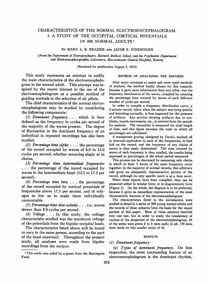
When these figures have been compiled, they can bepresented either in tabular form, or in diagrammatic form(Figure 1). On the whole, the diagram is to be preferred,because it gives an immediate representation of the mostcharacteristic features of the electroencephalogram.
The characteristics listed in the introduction werestudied in detail in a series of 500 young normal adults andthe records of these subjects form the basis for the majorportion of this paper. Most of these subjects receivedonly one test, but in order to study the consistency ofvarious of the properties of the electroencephalogram, 45of the series were given 4 to 5 tests each; in all, 176 testswere made on this smaller series of 45.
RESULTS
(1) Dominant frequency(a) Types of dominant frequency. On first
inspection, the most outstanding feature of anelectroencephalogram is the dominant rhythm,
303

MARYA. B. BRAZIER AND JACOB E. FINESINGER
z
a
03: 20%- A
54-
wa9i01'15%3.1.1FRQEC IN CYLSPRSCN
0 ,Go OF O
AGE O I
RANGE
095%.I1 1 4516.
FREQUENCYIN CYCLES PER SECOND
FIG. 1. GRAPHCOMPILEDFROMTHELECTROENCEPH-ALOGRAMOF ONENORMALADULT>IHOWING"THE,PERCENT-AGESOF EACHFREQUENCYPRESENT, OMITTING THE BETARANGE
The dominant frequency is defined as the one presentin the greatest amount, i.e., at the mode of the curve.(In this case, it is 9.5 cycles per second.)
Also, in the present work, a third classifica-tion is used, since it has been found in this seriesof 500 normal adults that there is evidence forregarding the dominant frequencies in the inter-mediate band (13.5 to 17.5 cycles per second)between the alpha and the beta ranges as aseparate entity (Figure 2).
A detailed statistical analysis was made of theactivity slower than beta (i.e., slower than 18.0cycles) in the records of 500 normal adults.The mean for all the dominant frequencies inthis range was 10.5 cycles per second, with astandard deviation of 0.9. Thus, any dominantfrequency slower than 8.0 or faster than 13.0cycles is outside 3 times standard deviation fornormals, and is therefore, by definition, excludedfrom the alpha range. Further reason for re-garding records with a dominant frequency in theintermediate range as a separate group is foundin a study of the distribution curve of the domi-nant frequencies of 500 normal adults (Figure 3).
In this graph, there appears to be a normaldistribution curve dominating the picture butwith some outlying stragglers in the faster fre-quencies. The main curve consists of 474
i.e., the frequency in cycles per second which ispresent in greater amounts than any other fre-quency. This frequency is usually apparent onrough inspection and most electroencephalog-raphers have adopted the classification into twogroups, as first suggested by Berger, namely, thealpha and the beta types. These have usuallybeen defined as the frequencies between 8.0 and13.0 cycles per second for the alpha group, andfrequencies faster than 18.0 cycles for the betagroup. This classification is not satisfactorysince, in fact, all electroencephalograms consistof a mixture of these rhythms in some degree,and no record consists wholly of either alpha orbeta waves. Hence, if this classification is tobe used, some criterion must be defined as to thepercentage of beta activity which must bepresent in a record before that record should beclassified as a beta type. For the purposes ofthe present study, records are classed as betatype only if there is present less than 20 per centof other activity, i.e., of waves slower than 18cycles per second.
ALPHA RHYTHM 8.0 TO 13.0 PER SECOND
INTERMEDIATE RHYTHM 135 TO 17.S PER SECOND
I
KrA RHYTHM 19.0 TO 35.0 PER SECOND
_- _ or--I
FIG. 2. TYPES OF NoRMALELECTROENCEPHALOGRAMSThe upright line to the right of each tracing is the cali-
bration for 100 mV.
--%j
304

ELECTROENCEPHALOGRAMSIN THE NORMALADULT
subjects whose dominant frequencies give anormal distribution around a mode of 10.0cycles per second, with 26 subjects outside thenormal distribution curve. Hence, it is feltthat those frequencies in the range 13.5 to 17.5should not be included in the alpha group.Frequencies falling within this range are referredto as intermediate rhythms.
In this group of 500 normal adults, examinedin this laboratory, the following distribution ofthe three types (alpha, intermediate, and beta)was found:Number of subjects: 500 per centNumber of alpha type (8 to 13.0 cycles 474 94.8
per second):Number of intermediate (13.5 to 17.5): 18 3.6Number of beta type (18.0 and over): 8 1.6
(b) Consistency of the dominant frequency. Thatthe dominant frequency remains constant withinnarrow limits for the same individual over longperiods of time can be demonstrated by repeatedobservations on the same person. A discussionof some factors which may, in certain circum-stances, alter the dominant frequency will bereported in a subsequent paper.
Electroencephalograms repeated on the samesubjects over a period of a few years revealedonly small fluctuations in the dominant fre-quency from one test to the next:Subject 1, aged 35, female Subject 2, aged 24, female
uly 1940 9.5 Oct. 1940 9.5Dec. 16, 1940 9.5 June 1941 10.0Dec. 26, 1940 9.5 April 1942 10.0Sept. 1942 9.5 Sept. 1942 10.0
Subject 3, aged 36, femaleDec. 1940 20.0June 1941 20.0July 1941 18.0Aug. 1941 20.0
This degree of fluctuation is of the same orderas that reported by other workers (Loomis,Harvey, and Hobart (1), Jasper and Cruikshank(2), and Jasper and Andrews (3)).
That there is also only a small fluctuation inthe dominant frequency of an individual whenexamined several times during the same day,has been established on a larger group.
One hundred and seventy-six observationswere made on 45 normal subjects, all of whomwere examined at non-fasting blood sugar levels(above 70 mgmper 100 cc.), i.e., 4 to 5 tests weremade on the same individual at intervals duringthe same day.
25%-
z20%- 500 SubJects
Ua.I
'5%-
CIo 10%-° * I \a JZI
ALPHA RANGE INTERMEDIATEFREQUENCYIN CYCLES PER SECOND
FIG. 3. THE DISTRIBUTION CURVEOF THE DOMINANTFREQUENCIESFOUNDIN THE OCCIPITAL RHYTHMsOF 500NORMALADULTs
Ten of the 45 subjects showed no variation in theirdominant frequency. In 1 subject only did the dominantfrequency vary from his own mean value by more than7 per cent. The mean variation for the whole series of45 subjects was under 1 per cent (176 observations).
(c) Relation between consistency of the dominantfrequency and age. If the consistency of a person'sdominant frequency be studied, it is found thatage is a factor in the degree of variability foundin the electroencephalogram at non-fasting bloodsugar levels (above 70 mgm).
This degree of variability is determined byfinding the coefficient of variation for the domi-
nant frequency of each individual. (The coeffi-
cient of variation = 100 Xstandard deviationmean
A group of 45 young adults between the agesof 17 and 38 were thus examined for consistencyof. dominant frequency at normal blood sugarlevels. By rough observation, it appeared asthough the dominant frequency in records ofthose subjects over the age of 20 was less stablethan of those under that age, and for this reasonthe division into two groups was made at thisage level. In the series examined, there were
305

MARYA. B. BRAZIER AND JACOB E. FINESINGER
13 subjects under the age of 20, and 32 were aged encephalogram of adults up to the age of 47.20 or over. Calculations of the coefficient of We have no data on normal individuals overvariation gave the following result: this age.
Under 20 20 and over (e) Dominant frequency and its relation to otherMean of coefficient of variation 1.2 3.2 physiological factors. The dominant frequenciesStandard deviation of coefficient 1.3 2.4 in this group were examined for any possibleof variation
correlation with sex, weight, height, or height-The difference between these two means was weight ratio, but no relation was found with
tested for reliability by determining the standard any of these factors.error of the difference:
D= 3.52,
where D equals the difference between the twomeans, and oiD, the standard error of thatdifference.
The result is indicative of a significant differ-ence between these two groups, i.e., the chancesof this being a true difference are over 1000 to 1.
It would appear therefore that the dominantcortical frequency becomes less stable withincreasing age, and this is demonstrable even ina series which contains no one over the age of 38.
(d) Dominant frequency and the age factor.The total group of 500 young adults, betweenthe ages of 17 and 47, was studied for correla-tion between their age and the actual frequencyof their dominant rhythm at non-fasting bloodsugar levels (in contrast to the consistency of thisdominant frequency which has just been ex-amined).
There have been several studies of this kind inrelation to age in children (Berger (4), Loomis(5), Lindsley (6), Smith (7), and Weinbach (8));but the age factor in adults has not receivedmuch attention. Bernhard and Skoglund (9, 10)demonstrated a difference of over half a cycle inthe mean dominant frequency between two agegroups of 15 to 18 and 19 to 30, respectively.
In the present series of 500 adults between theages of 17 and 47, with a mean age of 24, thefollowing results were obtained:
Under 24 year8270 alphas-mean dominant frequency 10.5
7 intermediates4 betas
24 years old and over204 alphas-mean dominant frequency 10.4
17 intermediates
(2) Percentage time alphaThis is defined as the percentage time occupied
by waves of 8.0 to 13.0 cycles per second, occur-ring either singly or in chains; i.e., it is the grossalpha count.
In 500 subjects who were examined at non-fasting blood sugar levels, the percentage timealpha varied in the group from 9 to 93 per cent,with a mean of 61 per cent; There was somealpha activity present in all records, even thosewhich were predominantly beta in type.
The distribution among this group of theamount of alpha activity present at non-fastingblood sugar levels is given in the following dis-tribution diagram (Figure 4).
50%-
a,- 40,%C0 -C
U
_
10%-
£0
Is 20%0-.
AIEz2
10%-.
under 25% 25%-49% 50%-74%' 75%-over
PER CENT TIME ALPHA ACTIVITY
4 betas FIG. 4. GRAPHILLUSTRATING 500 NoRMALSUBJECTS(ExPRESSED AS PERCENTAGES)GROUPEDAccoRDING TO
Thus, it would appear that age has no in- THE AMOUNTOF ALP ACTIVITY PRESENT IN THEIRfluence on the dominant frequency of the electro- REcoRDS
306

ELECTROENCEPHALOGRAMSIN THE NORMALADULT
Of the 24 subjects with less than 25 per centalpha activity, 16 had a predominantly inter-mediate rhythm, and 8 had dominant frequenciesin the beta range.
(a) Consistency of percentage time alpha. Thevariability in percentage time alpha for anindividual was not so marked in this series ashas been described by Rubin (11) in bipolarrecordings, the mean standard deviation forrepeated tests on an individual being 7.1. Itshould be pointed out that in this work thepercentage time alpha quoted includes all alphaactivity present, whether occurring in singlewaves or in chains, whereas Rubin's observationsare based on a criterion of 3 waves of alpha fre-quency occurring together; and in Rubin's ex-periments, the blood sugar was not controlled.
The relation between variability of percentagetime alpha and high or low alpha percentage, asdescribed by Rubin (12), did not hold in thisseries.
(b) Relation between percentage time alpha anddominant frequency. There was found to be aninverse relationship between percentage timealpha and the dominant frequency of that alpha;in other words, individuals whose percentagetime alpha values were high showed dominantfrequencies in the slower alpha range.
In the total series of 500 subjects, the recordswere examined for the relation between thedominant frequency of any alpha present and thepercentage time occupied by this alpha activity.The following results were obtained:
Number MeanAlpha frequency of atgnage Standardsbetime deviationsujcs alpha
Slower than 10.5 cycles 236 68.5 14.110.5 cycles and faster 264 54.7 20.1
The difference between these 2 means was
tested for reliability by determining the standarderror of the difference which was found to be8.90. Such a high standard error of the differ-ence is beyond the possibility of chance.
It is therefore concluded that an inverse rela-tionship exists between the dominant frequencyof the alpha present and the amount of totalalpha present.
(c) Relation between percentage time alpha and
other physiological factors. No correlation wasfound in this series between the percentage timealpha and age, sex, height, weight, or the height-weight ratio.
(3) Percentage time intermediate rhythmFrequencies of 13.5 to 17.5 cycles per second,
i.e., those which lie between the alpha and betaranges, are not commonly found in more thannegligible quantities in the records of normaladults, and are only rarely found as the dominantfrequency of the record. In this series of 500subjects, there were 18 with a dominant fre-quency in this range, or 3.6 per cent.
Rhythms in this range, however, normally oc-cur in bursts during the lighter stages of sleep(13), and it would not be surprising to find thatthey have a different physiological origin fromthe alpha waves. They do not appear to bemerely accelerated alpha waves, since observa-tions on sleep show that they appear abruptlyand do not emerge by gradual transition fromthe higher alpha frequencies. These facts sug-gest that one is here dealing with a dichotomy.
As has already been noted, the electroencephal-ogram in normal adults contains very littleactivity in this intermediate frequency band.What little there is might be expected to appearin those records with the faster alpha frequenciesas an extreme variation of their predominantly12.5 to 13.0 cycle rhythms, but an examinationof this series failed to establish any such correla-tion. There were no more waves of the inter-mediate frequencies in those records with domi-nant frequencies in the faster alpha range thanin those with predominantly 9.0 and 9.5 cycleactivity.
Unlike the so-called alpha and beta rhythmswhich are present to some extent in all records,the intermediate band of 13.5 to 17.5 cycleactivity is sometimes totally absent, a factwhich contributes to the impression, previouslymentioned, that one is here dealing with adichotomy.
(4) Percentage time betaBeta activity (i.e., 18.0 cycles per second and
over) was found, to a greater or less extent, inevery record in this series at non-fasting bloodsugar levels. As has already been remarked, all
307

MARYA. B. BRAZIER AND JACOB E. FINESINGER
a
c 40%-U0
30%-a-
S
°' 20%-
I0S.
.012 10%-z.
24.5%
47%
500 Subjects
4%
A
under 10% 10%-24% 25%-49% 50%-overPER CENT TIME BETA ACTIVITY
FIG. 5. GRAPHSHOWING500 NORMALSUBJECTS (Ex-PRESSEDAS PERCENTAGES)GROUPEDACCORDINGTO THEAMOUNTOF BETA ACTIVITY PRESENTIN THEIR RECORDS
electroencephalograms are a mixture of rhythms,and, in this series, the 8 cases which have beenclassified as beta type, all contained some alpha;their percentages of beta activity varied from51 to 80 per cent. The distribution of betaactivity in this series is given in Figure 5.
Figures 4 and 5 taken together do not, ofcourse, give the total picture for the group,since the time occupied by intermediate frequen-cies and by slow activity is not represented.The amount of beta activity in an individual'srecord was found to be remarkably constant inrepeated runs, and is characteristic for the indi-vidual. It did not vary with the degree ofrelaxation achieved by the subject.
Although in this series there was a tendencyto more beta activity in the records of the oldersubjects, no statistical relation between it andage could be established. The amount of betaactivity present did not correlate with sex,height, or weight.
(5) Activity slower than 8.0 cycles per secondWaves of a frequency slower than 8.0 cycles
per second are here referred to as slow activity.
Traces of 7.0 to 7.5 per second frequencieswere found in the records of 125 out of the seriesof 500 subjects, but only in 4 individuals wasthere more than 5 per cent of such slow activityin the 2-minute recording; of these 4 individuals,1 had 19 per cent slow activity, 2 had 9 per cent,and 1 had 6 per cent.
Six-cycle waves were found in the occipitalleads in 41 (8 per cent) of the 500 records at non-fasting blood sugar levels, but waves slower than6 cycles per second were found in only 4 subjects,or less than 1 per cent.
Were an investigation of the potentials fromthe temporal regions to be made in a way similarto the present detailed study of the occipitalpotentials, it seems likely that there would be ahigher incidence of 6- and 7-cycle waves in normalrecords, this being our experience and that ofother electroencephalographers (14).
No wave in the range commonly called deltaactivity (i.e., 4 cycles or slower) was found.
(6) VoltageThis was measured by a pair of calipers,
adjusted to the calibration made for voltage atthe beginning of each record.
The maximum voltage was measured in everycase and classified as to whether it was under25 mV, over 25 mVbut under 50 mV, over 50mVbut under 100 mV, or over 100 mV. In thelatter case, a further breakdown in classificationwas made between those who had less than 20waves which reached 100 mVin amplitude andthose which had more than this number in a 2-minute run.
The distribution of voltage in this series of500 normal subjects at non-fasting blood sugarlevels was as follows:
(a) Voltage and dominant frequency. The fasterfrequencies tend to be of low voltage; no recordwith a dominant frequency faster than 11.5 hadany potentials which reached as much as 100 mV.
In the alpha range, the records with the sloweralpha frequencies were of higher voltage than
308

ELECTROENCEPHALOGRAMSIN THE NORMALADULT
the faster ones, thus following the usual characterof oscillations in which the amplitude is inverselyproportional to the frequency. In the follow-ing table, 474 normal subjects, whose dominantfrequencies were in the 8.0 to 13.0 cycle band,are listed according to their voltage.
Meandominantfrequency
Maximum voltage under 50 mV (105 subjects) 11.0Maximum voltage over 50 mV (274 subjects) 10.4
but under 100 mVMaximum voltage over 100 mV (95 subjects) 10.0
(b) Voltage and percentage time alpha. Themaximum voltage of the potentials in a recordvary directly with the percentage time alphaactivity present; i.e., those records which have alarge amount of 8.0 to 13.0 cycle waves reach ahigher maximum potential.
In the following table, voltage is related tothe mean percentage time alpha found in thesame series of 474 normal subjects whose domi-nant frequencies were in the 8.0 to 13.0 cycleband.
Maximum voltageUnder 25 mVOver 25 mV, under 50 mVOver 50 mV, under 100 mVOver 100 mV
Mean percentage time alpha40.3 (10 subjects)48.4 (95 subjects)65.4 (274 subjects)73.6 (95 subjects)
DISCUSSION
Since the first development of electroen-cephalography, interest has been centered mainlyon its application to clinical problems, and it isonly recently that there has been a shift ofinterest to the study of the normal adult.For many reasons, it would have been preferablehad the reverse taken place, for the developmentof this test to have proceeded from a basic studyof the normal to a comparison of clinical recordswith a norm already well established.
The desirability is patent for the establish-ment of a yardstick for the normal populationagainst which may be measured the variablesfound in pathological records. It is equallydesirable in attempting to assess the electro-encephalograms of normal subjects (as, forexample, is being done in air-pilot selection) tohave a quantitative basis from which one may
calculate the chances of any observed phenom-enon being a normal finding.
In this paper, a beginning has been made in anattempt to find what range of variation can befound for some of the characteristics of theelectroencephalogram of normal adults. At thepresent stage, this study has been limited to ananalysis of the cortical potentials from the occi-pital lobes, and it cannot be too strongly em-phasized that a different set of data would un-doubtedly be obtained from the frontal lobes,and different again from the temporal andparietal regions.
This study is also limited to analysis of electro-encephalograms during normal breathing. Areport of an investigation during hyperventila-tion will follow this, with special reference to therole of blood sugar and depth of hyperventilation.
In the past, the bulk of the work on bothnormal and clinical electroencephalograms hasbeen done by the method of appraisal. Theexperienced electroencephalographer has lookedat the record, compared it in his mind with hisimpression of those records which have pre-viously come into his laboratory, and assessedit from this mental comparison. Where grossdifferences are present, such as are found inpatients with epilepsy or with neoplasms, thismethod has in the main sufficed, but when finershades of difference are being searched for, amore finely differentiated set of standards isnecessary.
Such standards can only be set up on a basisof actual measurement, a method which is timeabsorbing, but essential in any research projectdesigned to establish normal control standards.Were such a set of standards established on alarge enough group of individuals, it would thenbe possible to estimate the chances of normalitywhen any fine differences occur in the record,as for example, 7 cycle waves occurring singlyin the occipital leads, or trains of 14 cycle waves.The percentage of normal records in which suchwaves occur would be known and the importanceof the finding could thus be assessed.
A development in the measuring of electro-encephalograms has been made by Gibbs andGrass (15) in the form of a spectrum analyzer.This apparatus gives a compilation of the amountof energy present at each frequency. At thepresent stage of our experience, we have foundmore meaning in the frequency of waves than
309

MARYA. B. BRAZIER AND JACOB E. FINESINGER
in their voltage inf normal records, and we there-fore look for a method where the number ofwaves present at any frequency is not obscuredby the voltage. An instrument for this purposehas recently been designed by Walter (16), butis not yet on the market.
It is obvious that the current differentiationof electrical potentials into alpha, beta, anddelta rhythms is arbitrary and, in some respects,unfortunate. The classification of records intoalpha and beta rhythms tends to obscure thefact that many so-called alpha records containpotentials of faster frequencies and that manybeta records contain percentages of the sloweralpha frequencies. A more accurate and morecomplete assessment could be made by describingthe frequency distribution on the potentials.It would seem much wiser to describe rhythmsin terms of the incidence of actually measuredfrequencies until some physiological or statisticalreason can be found for grouping frequencies intocertain rhythms.
The fact that significant contributions toclinical diagnosisn aepilepsy and the localizationof neoplasms have been made by the crudemethods of gross inspection of records would byno means invalidate the need for studies basedupon careful measurements. However, once thedata on a sufficiently large number of cases arecollected, it might well be possible to developsimpler methods of analysis, based upon theknown verifiable distributions of frequencies.This would seem a more logical approach andcould give more precise information in the studyof problems in which fine differentiations occurin the records.
SUMMARY
The occipital cortical potentials have beenanalyzed under conditions of controlled bloodsugar in 500 subjects. Of these, the majorityreceived but one test, but in 45, repeated observa-tions were obtained.
The following characteristics were found inthese electroencephalograms.
1. The dominant frequency of an individualis comparatively stable, but becomes less sowith increasing age. There is a statisticallysignificant correlation between age and stabilityof dominant frequency (45 subjects).
2. Waves of 8.0 to 13.0 cycles per second("alpha") were present in all records examined.The percentage time occupied by alpha wavesvaried inversely with the frequency of thedominant frequency. This inverse relationshiphas been established statistically in 500 subjects.
3. Waves of 13.5 to 17.5 cycles per second("intermediate") are rarely found in any quan-tity in normal records, and constitute the domi-nant frequency in only 3.6 per cent of all normalsexamined (500).
4. Waves of 18.0 cycles per second and faster("beta") were present in all records examined(500). The percentage time occupied by wavesin this range is nearly constant for an individualin repeated runs (45 subjects).
5. Waves slower than 8.0 cycles per second arefound in occipital potentials of 25 per cent ofnormal subjects. Waves as slow as 6.0 cycleswere found in only 41 out of 500 subjects.
6. No waves of 4.0 cycles per second or slower("delta") were found in the occipital recordingsof any normal subject while breathing normally(500 subjects).
7. Maximum voltages are higher in thoserecords which contain the most alpha activity,and in those records whose dominant frequenciesfall in the slower alpha frequencies (500 subjects).
8. The physiological factors of sex, height,weight, or the height-weight ratio did notcorrelate with any characteristic in the brainwave record.
All the electroencephalographic tracings for this researchwere recorded in the Brain WaveLaboratory of the Massa-chusetts General Hospital with the cooperation of thedirector, Dr. Robert S. Schwab.
The authors are indebted to Mrs. Frances Coopersteinand Miss Margaret Gray for technical help, and to Mrs.Mary Newell for analysis of the records.
BIBLIOGRAPHY
1. Loomis, A. L., Harvey, E. N., and Hobart, G., Elec-trical potentials of the human brain. J. Exper.Psychol., 1936, 19, 249.
2. Jasper, H. H., and Cruikshank, R. M., Visual stimu-lation and the after-image as affecting the occipitalalpha rhythm. J. Gen. Psychol., 1937, 17, 29.
3. Jasper, H. H., and Andrews, H. L., Electroencephalog-raphy. Arch. Neurol. and Psychiat., 1938, 39, 96.
-4. Berger, H., tOber das Elektrenkephalogramm des Men-schen. Arch. f. Psychiat., 1932, 98, 231.
310

ELECTROENCEPHALOGRAMSIN THE NORMALADULT
5. Loomis, A. L., Harvey, E. N., and Hobart, G. A.,Cerebral states during sleep as studied by humanbrain potentials. J. Exper. Psychol., 1937, 21,127.
6. Lindsley, D. B., Brain potentials in children andadults. Science, 1936, 84, 354.
7. Smith, J. R., Electroencephalogram during infancyand childhood. Proc. Soc. Exper. Biol. and Med.,1937, 36, 384.
8. Weinbach, A. P., Contour maps, center of gravity,moment of inertia and surface area of human body.Human Biol., 1938, 10, 356.
9. Bernhard, C. G., and Skoglund, C. R., Recherches sur
la frequence alpha de l'electro-encephalogrammechez l'enfant. Acta psychiat. et neurol., 1939, 14,223.
10. Bernhard, C. G., and Skoglund, C. R., On alpha fre-
quency of human brain potentials as function ofage. Skandinav. Archiv. f. Physiol., 1939, 82, 178.
11. Rubin, M. A., The distribution of the alpha rhythmover the cerebral cortex of normal man. J. Neuro-physiol., 1938, 1, 313.
12. Rubin, M. A., A variability study of the normal andschizophrenic occipital alpha rhythm. J. Psychol-ogy, 1938, 6, 325.
13. Loomis, A. L., Harvey, E. N., and Hobart, G., Fur-ther observations on potential rhythms of cerebralcortex during sleep. Science, 1935, 82, 198.
14. Kershman, J., Personal communication.15. Grass, A. M., and Gibbs, F. A., Fourier transform of
electroencephalogram. J. Neurophysiol., 1938, 1,521.
16. Walter, W. G., An automatic low frequency analyzer.Electronic Engineering. June 1943.
311