characteristics of natural killer cells in the
TRANSCRIPT
C H A R A C T E R I S T I C S O F N A T U R A L K I L L E R C E L L S I N T H E
M U R I N E I N T E S T I N A L E P I T H E L I U M A N D L A M I N A P R O P R I A
By A. TAGLIABUE,* A. D. BEFUS,:~§ D. A. CLARK,§II AND J. BIENENSTOCK§
From the Istituto di Ricerche Farmacologiche M. Negri, Milan, Italy; and the Host Resistance Programme, Departments of Medicine and Pathology, McMaster University, Hamilton, Ontario, Canada
Morphologic and functional similarities between the lymphoid tissues of the gut and lung have shown that a common mucosal immune system is involved in the protection of mucosal sites (1). Several studies have demonstrated the relevance of antibody, part icularly IgA, in mucosal immunity. However, less at tention has been directed to the role of cell-mediated immunity. The recent development of methods for the isolation of mucosal Iymphoid cells (2-7) has facilitated the analysis of cytotoxic functions of lymphocytes from intestine and lung. Thus, it has been shown that guinea pig and h u m a n lymphocytes from the intestinal mucosa exert mitogen- induced, ant ibody-dependent , and spontaneous cellular cytotoxicity (6-8). Sponta- neous cytotoxic activity of cells from the mouse lung (9) and gut (10) has m a n y characteristics o f natural killer (NK) 1 cytotoxicity.
The main effector cells of N K activity in h u m a n (11), rat (12), and mouse (13) are large granular lymphocytes (LGL) with a high cytoplasmic/nuclear ratio and azuro- philic granules in the cytoplasm. Because large lymphocytes with cytoplasmic granules are abundan t in the mammal i an intestinal mucosa (2), we postulated that gut granulated lymphocytes (gGL) might be the N K effector cells at that site (10). Thus, we initiated a s tudy to clarify the relationship between gGL and gut N K cytotoxicity. In an a t tempt to define the phenotype Of gu t N K cells and their lineage, experiments with sera against various surface markers were performed. Moreover, spleni c and gut N K activity were compared in C 5 7 B L / 6 - b g / b g (beige) mice, a mouse strain with very low N K activity (14), and in WBB6F1-W/W v mice, a hybrid mouse deficient in hemopoiet ic stem cells and mast cell precursors (15). The results obtained in this and previous studies (10) suggest that gut N K cells in the mouse are lymphocytes of the T cell lineage, similar but not identical to splenic N K cells.
M a t e r i a l s a n d M e t h o d s Mice. Inbred CBA/J mice of both sexes were obtained from The Jackson Laboratory,
Bar Harbor, ME. C57BL/6-bg/bg (beige) mice were bred in McMaster University from heter-
* Supported by a fellowship from the Associazion Italiana per la Ricerca sul Cancro. Present address: Istituto Sieroterapico Vaccinogeno, Toscano, Siena, Italy.
~: Rockefeller Foundation Fellow in Tropical and Geographic Medicine. Correspondence should be addressed to Dr. Befus at the Department of Pathology, McMaster University Medical Center, |tamihon, Ontario, Canada L8N 3Z5.
§ Supported by grants from the Medical Research Council of Canada. 11 Scholar of the Medical Research Council of Canada. I Abbreviations used in this paper: A/T, attacker/target ratio; C, complement; gGL, gut granular lympho-
cytes; HBSS, Hanks' balanced salt solution; IEL, intraepithelial lymphocytes; LGL, large granular lymphocytes; LP, lamina propria; LPI,, LP lymphocytes; LU, lytic units; NK, natural killer.
J. Exp. MEt). © The Rockefeller University Press • 0022-1007/82/06/1785/12 $1.00 1 785 Volume 155 June 1982 1785-1796
1786 NATURAl, KILI,ER CYTOTOXICITY IN INTESTINAl, MUCOSA
ozygotes purchased from The Jackson Laboratory. Similarly, WBB6F1-W/W v mice were bred in McMaster University from WB/ReJ-W/+ and C57BL/6J-W"/+ parental stocks purchased from The Jackson Laboratory. In this study, littermates of W/W" mice included WBB6F1-+/+, WBB6F1-W/+, and WBB6F1-W"/+. Groups were distinguished by their coat color. All the mice used in this study were between 6 and 10 wk of age. Food and water were provided ad lib. and, commencing 48 h before killing, the anti-protozoal agent, metronidazole (Poulenc Ltd., Montreal, Canada) was administered in the drinking water (2 g/liter). Experi- ments designed to assess whether this drug altered NK activity included comparisons of splenic and intestinal NK activity in treated and untreated mice, and the results showed that it did not (unpublished).
Preparation of Effector Cells. Single-cell suspensions from spleen were obtained by mincing and repeated aspiration with pasteur pipets and filtration through gauze. Lymphocytes from the small intestine epithelium (IEL) and from the lamina propria (LP) were isolated using a modification of previously described procedures (2-8, 16, 17). The small intestines from five mice were removed and flushed of fecal material with 10 ml of saline. The mesentery and adherent connective tissue and fat were dissected from the intestines, and the Peyer's patches removed. The intestines were opened longitudinally and cut into pieces 1-2-cm long, which were washed in 50 ml of Hanks' balanced salt solution containing 25 mM Hepes, 5% agammaglobulinemic horse serum (HS), pH 7.4, 300 mosmol/kg (HBSS). To remove epithelial cells and IEL, the tissue was incubated with stirring at 37°C for 15 min in HBSS containing 10 -4 M EDTA. After supernatant collection, this procedure was repeated once in EDTA-HBSS and once in HBSS. The supernatants were washed twice in HBSS. Histologic examination of the tissue revealed that at this stage, the intestines were devoid of most of the epithelium, although the villus structures and associated LP were largely preserved. To obtain a single-cell suspension from LP, the remaining tissue was cut into 2-5-mm pieces and incubated with stirring at 37°C for 45 rain in HBSS containing 25 U/ml of collagenase (840-7018; Gibco Laboratories, Grand Island Biological Co., Grand Island, NY) and 20% HS. After supernatant collection, the collagenase treatment was repeated once. Supernatants from enzymatic digestion were pooled and washed twice. All the cells from the epithelium and the LP were resuspended in 50 ml HBSS and rapidly (10-25 ml/min) filtered through 10-ml syringes containing 300 mg loosely packed nylon wool. This filtration procedure removed a large proportion of the dead cells and debris, and yielded 50-75% of the viable lymphocytes applied to the column. Cells were then washed twice and resuspended in 10 ml of RPMI 1640 supplemented with 10% fetal bovine serum, 10 mM Hepes, and 50/.tg/ml gentamycin (Schering Corp. Ltd., Pointe Claire, Canada), pH 7.4, 300 mosmol/kg (growth medium).
At this stage the cell populations usually contained viable epithelial cells, viable lymphocytes, and nonviable epithelial cells (Table I). To obtain enriched lymphocyte populations, a discontinuous density gradient was devised using Percoll (Pharmacia Fine Chemicals, Phar- macia Inc., Uppsala, Sweden), diluted in growth medium (10). Five different concentrations of Percoll were prepared and corrected for osmolarity. The gradient was prepared in 50-ml conical tubes (25339; Corning Glass Works, Corning NY), layering from the bottom: 5 ml 80% (vol/ vol) Percoll, 7.5 ml 70% Percoll, 7.5 ml 55~7~ Percoll, 15 ml 40% Percoll and 10 ml 30% Percoll. 2 × l0 s cells were placed on the top of this gradient, and the tube was centrifuged at room temperature at 600 g for 20 min. Cells from the interface between layers were then collected and washed. Approximately 50% of the viable lymphocytes were recovered. Differential counts of the cell subpopulations were made on smears prepared by cytocentrifugation (Cytospin Centrifuge, Shandon Southern Instruments, Camberley, England) and stained with Diff Quik (Harleco, American Hospital Supply Corp., Gibbstown, N J).
Cytotoxicity Assay. The YAC-1 clone 19 (18) tissue culture line was obtained through the courtesy of Dr. J. Roder, Queen's University, Kingston, Ontario and maintained in vitro in growth medium. Tumor cells suspended ill 1 ml of growth medium were incubated for 60 rain at 37°C with 300 #Ci of Na'~lCrO4 (New England Nuclear, Boston, MA); 104 washed cells were then incubated for 6 h at various attacker/target (A/T) cell ralios in 0.7-cm conical bottom wells 76-022-05; Linbro Chemical Co., Hamden, CT). The percentage of isotope released was calculated from the fornmla: percent release = ([cpm release from cells during incubation -

TAGIJABUE, BEFUS, CI,ARK, AND BIENENSTOCK 1787
cpm spontaneous release]/[total cpm incorporated × 0.8 - cpm spontaneous release]) × 100. Spontaneous release in the 6-h assay was usually between 5 and 12%.
Antisera. Monoclonal anti-Thy-l.2 antibody was purchased from New England Nuclear and used at a final dilution of 1:200 for 107 cells/0.5 ml. Monoclonal anti-Lyt-l.l (lot 7120) and anti-Lyt-2.1 (lot 7301) were purchased from Cedarlane Laboratories (Hornby, Canada). These sera were used at a final dilution of 1:20 and 1:10, respectively, for 107 cells/0.5 ml. Anti- NK-I.2 (CE × NZB anti-CBA) alloantiserum (18) was a kind gift from Dr. R. C. Burton, Harvard Medical School, Boston, MA and was used at a final dilution of 1:10 for 10 v cells/0.5 ml. Anti-asialo GM1 serum produced by Dr. Ko Okumura (19) was kindly donated by Dr. J. Roder and Dr. K. Okumura and used at a final dilution of I:100 for 107 cells/0.5 ml. The antiserum dilutions described above have been previously shown to be effective for removal of the appropriate cell subpopulations (18-20).
Complement (C)-dependent Cytolysis. To kill Thy- 1.2, Lyt- 1.1, Lyt-2.1, and asialo GMvpositive cells, low toxicity rabbit C (lot 4057; Cedarlane Laboratories) was used at a final dilution of 1:10. To kill NK-l.2-positive cells, rabbit C provided by Dr. R. C. Burton was used at a final dilution of 1:6. Lymphocytes from spleen and intestinal epithelium were incubated at 37°C for 60 min with or without (medium control) the above mentioned antisera in phosphate-buffered saline supplemented with 0.3% bovine serum albumin. Ceils were washed once, resuspended in diluted C, and incubated at 37°C for 45 min. Cells were then washed twice and resuspended in growth medium without adjusting the cell concentration, as described by Mattes et al. (21). Viability was assessed by adding fluorescein diacetate and ethidium bromide to the cells, and counting them under UV light (22).
Statistical Analysis. Cytotoxic activity was calculated from 51Cr release assay titration curves by the method of Miller and Dunkley (23) and expressed as lytic units (LU) + SEM; 1 LU represents the amount of activity required to lyse 20% (LU20) of the target cells during the assay period.
Resu l t s
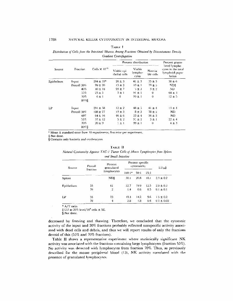
Isolation of Mucosal Lymphoid Cells with NK Activity. We have previously shown that murine IEL possess a high N K cytotoxicity (10). However, the low yield o f purified mucosal lymphocytes (<_1 × 10G/mouse) obtained by the mechanical isolation pro- cedures used in that study prevented a better characterization o f the cells involved. Therefore, we developed a new procedure using E D T A incubations and collagenase digestions for obta ining mouse IEL (5-10 × 106/mouse) and LP lymphocytes (LPL) (~ 15 × 106/mouse) in high purity. Table I shows the distribution of cells from the intestinal mucosa and fractions obtained by discontinuous density gradient centrifu- gation after isolation. In the cell populations, before gradient centrifugation, the ratio of viable epithelial cells to viable lymphocytes was from 1:1 to 1:2 for the epithelium, and from 1:3 to 1:4 for the LP. The viable cells represented ~60% of the total cells. As shown in Table I, after the density gradient centrifugations, the majority o f viable lymphocytes was found in the 55% and 70% fractions. Interestingly, fraction 55% contained a number of large (>10/xm) gGL, almost twice that of the input population. On the contrary, fraction 70% mainly contained small lymphocytes (~5 pm) with no granules. Thus, the Percoll gradient allowed us to obtain clean populat ions of lymphocytes enriched or depleted in gGL.
We then assessed IEL and L P L obtained from the gradients for N K activity against YAC-I tumor cells. Prel iminary studies showed that the total IEL and LPL popula- tions possessed marked cytotoxic potential against YAC-I and that after Percoll fractionation activity was present in the 30% and 55% fractions. However, the input and 30% populations, but not the 55% fraction, expressed high levels of cytotoxicity for N K insensitive targets ( M C A / M N fibrosarcoma [10]), and this activity was not
1788 NATURAL KILLER CYTOTOXICITY IN INTESTINAl. MUCOSA
TABLE I
Distribution of Cells from the Intestinal Mucosa Among Fractions Obtained by Discontinuous Density Gradient Centrifugation
Source Fraction Cells X 10 -6
Percent distribution Percent granu- lated lympho-
Viable Nonvia- cytes in the total Viable epi- lympho- lymphoid popu- thelial cells ble cells
cytes [ation
Epithelium Input 204 + 33* Percoll 30% 99 +- 20
40% 40 ± 16 55% 25 "4- 5 70% 6 +- 1 80,7.§
2 6 + 3 4 1 ± 3 35_+5 3 8 + 6 13+_3 10± 1 79_+5 ND~ 93+_7 5 + 3 3 + 2 ND
3---1 91+_I 0 6 4 + 3 0 99± 1 0 12+_5
LP Input 291 + 58 12 - 2 48 ± 5 41 ± 4 13 _ 4 Percoll 30% 108 _+ 27 13 + 3 8 + 3 78 ± 5 ND
40% 64+ 16 4 6 + 6 2 3 + 4 2 6 + 3 ND 55'7~ 37+12 5+__2 9 1 ± 2 3_+1 2 2 + 4 70% 2 0 ± 9 1 + 1 99+_ 1 0 4 ± 3 8o'~§
* Mean ± standard error from 10 experiments; five mice per experiment. :~ Not done. § Contains only bacteria and erythrocytes.
TABLE II
Natural Cytotoxicity Against YAC-I Tumor Cells of Mouse Lymphocytes from Spleen and Small Intestine
Percent Percent specific Percoll cytotoxicity
Source fraction granulated lymphocytes 100:1" 50:1 25:1
LU20¢
Spleen ND§ 30.1 20.8 10.1 2.3 + 0.2
Epithelium 55 61 32.7 19.9 12.5 2.0 + 0.3 70 2 1.4 0.6 0.5 0.1 + 0.1
LP 55 23 19.4 t4.2 9.6 1.5 + 0.2 70 4 2.8 1.8 0.6 0.1 + 0.03
* A/T ratio. :~ LU at 20% level/106 cells 4- SE. § Not done.
d e c r e a s e d by f reez ing a n d t h a w i n g . T h e r e f o r e , we c o n c l u d e d t h a t t he cy to tox i c
ac t iv i ty o f the i n p u t a n d 30% f rac t ions p r o b a b l y re f l ec ted nonspec i f i c ac t iv i ty associ-
a t e d w i t h d e a d cells a n d debr i s , a n d thus we will r epo r t resul ts o f on ly the f rac t ions
d e v o i d o f th is (55% a n d 70% frac t ions) .
T a b l e II shows a r e p r e s e n t a t i v e e x p e r i m e n t w h e r e s ta t i s t ica l ly s ign i f i can t N K
ac t iv i ty was a s soc i a t ed w i t h t h e f rac t ions c o n t a i n i n g large l y m p h o c y t e s ( f rac t ion 55%).
No ac t iv i ty was d e t e c t e d w i t h l y m p h o c y t e s f rom f rac t ion 70%. T h u s , as p rev ious ly
d e s c r i b e d for t he m o u s e p e r i p h e r a l b l o o d (13), N K ac t iv i ty c o r r e l a t e d w i t h t he
p r e sence o f g r a n u l a t e d l y m p h o c y t e s .
TAGLIABUE, BEFUS, CLARK, AND BIENENSTOCK 1789
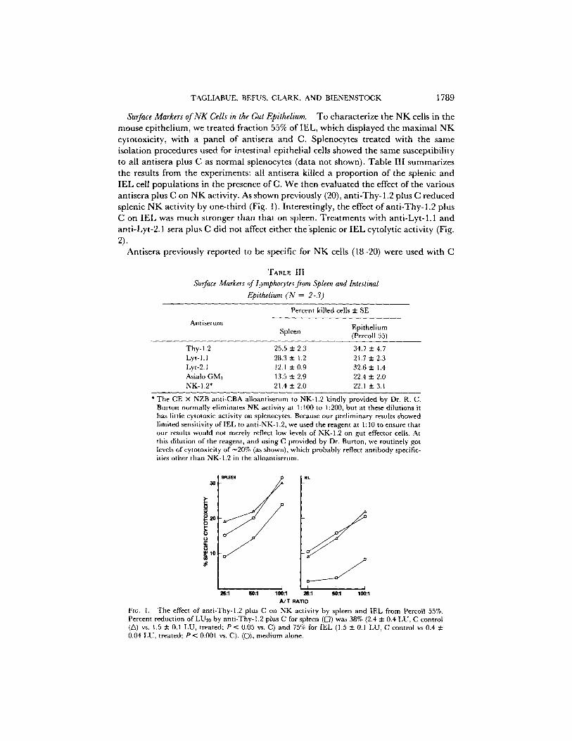
Surface Markers of NK Cells in the Gut Epithelium. To characterize the NK cells in the mouse epithelium, we treated fraction 55% of IEL, which displayed the maximal NK cytotoxicity, with a panel of antisera and C. Splenocytes treated with the same isolation procedures used for intestinal epithelial cells showed the same susceptibility to all antisera plus C as normal splenocytes (data not shown). Table III summarizes the results from the experiments: all antisera killed a proportion of the splenic and IEL cell populations in the presence of C. We then evaluated the effect of the various antisera plus C on NK activity. As shown previously (20), anti-Thy- 1.2 plus C reduced splenic NK activity by one-third (Fig. 1). Interestingly, the effect of anti-Thy-1.2 plus C on IEL was much stronger than that on spleen. Treatments with anti-Lyt-1.1 and anti-Lyt-2.1 sera plus C did not affect either the'splenic or IEL cytolytic activity (Fig. 2).
Antisera previously reported to be specific for NK cells (18-20) were used with C
T A B L E I I I
Surface Markers of Lymphocytes from Spleen and Intestinal Epithelium (N = 2-3)
Antiserum
Percent killed cells + SE
Epithelium Spleen (Percoll 55)
Thy-l,2 25.5 + 2.3 34.7 + 4.7 Lyt-l.1 28.3 ± 1.2 21.7 ± 2.3 Lyt-2. l 12.1 ::k 0.9 32.6 + 1.4 Asialo GM] 13.5 ± 2.9 22.4 ± 2.0 NK-I.2* 21.4 ± 2.0 22.1 ± 3.1
* The CE X NZB anti-CBA alloantiserum to NK-1.2 kindly provided by Dr. R. C. Burton normally eliminates NK activity at 1:100 to 1:200, but at these dilutions it has little cytotoxic activity on splenocytes. Because our preliminary results showed limited sensitivity of IEL to anti-NK- 1.2, we used the reagent at 1 : 10 to ensure that our results would not merely reflect low levels of NK-I.2 on gut effector cells. At this dilution of the reagent, and using C provided by Dr. Burton, we routinely got levels of cytotoxicity o f -20 % (as shown), which probably reflect antibody specific- ities other than NK-I.2 in the alloantiserum.
30
R
o u.
S~.EEN
L k I 25:1 50:1 100:1
J S
I I r 26:1 50:1 100:1
A / T RATIO
Fro. 1. The effect of anti-Thy-l.2 plus C on NK activity by spleen and IEL from Percoll 55%. Percent reduction of LU20 by anti-Thy-1.2 plus C for spleen (I--/) was 38% (2.4 ± 0.4 LU, C control (Z~) vs. 1.5 ± 0.1 LU, treated; P < 0.05 vs. C) and 75% for IEL (1.5 ± 0.1 LU, C control vs 0.4 ± 0.04 LU, treated; P < 0.001 vs. C). (C)), medium alone.
1790 NATURAL KILLER CYTOTOXICITY IN INTESTINAL MUCOSA
40 ~ S P L E E N
, o
m u . ,
I IEL
f I I I I I I
25:1 50:1 100:1 25:1 50:1 100:1 A / T RATIO
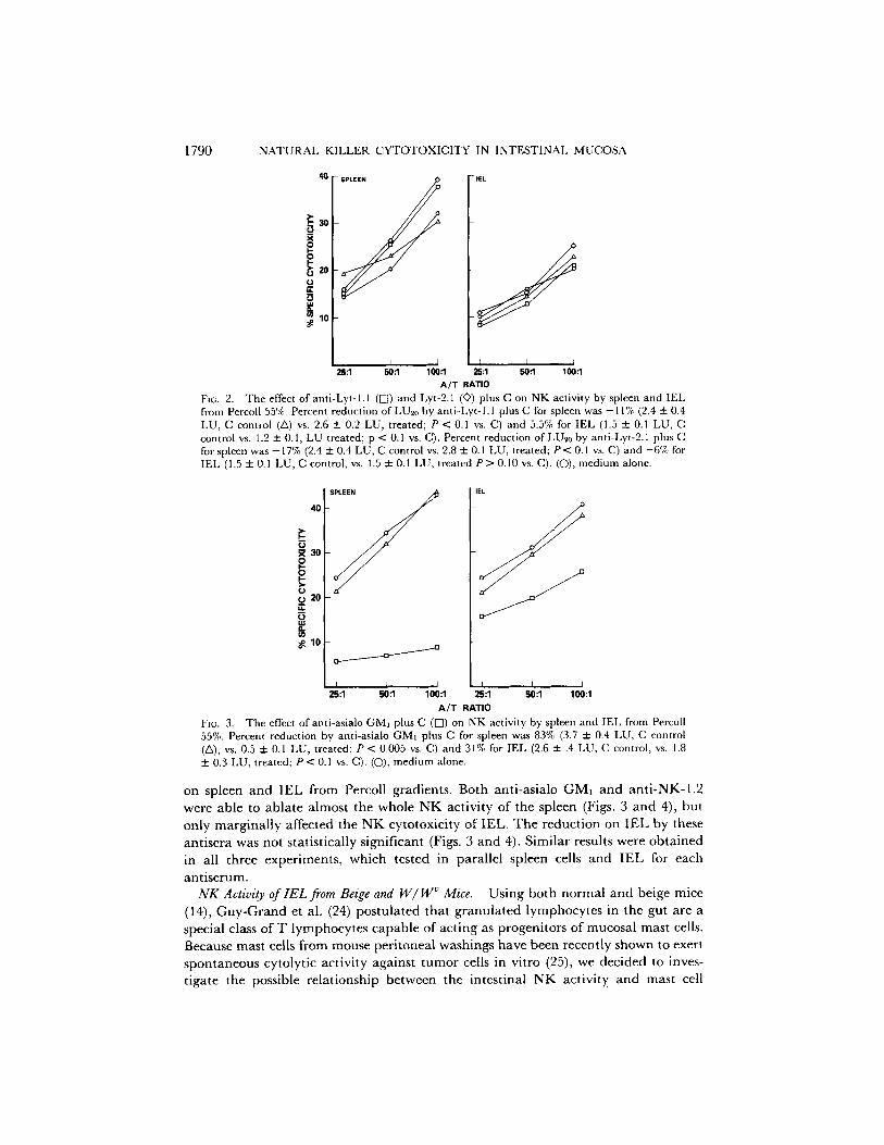
Fie. 2. The effect of anti-Lyt- h 1 (R) and Lyt-2.1 (©) plus C on NK activity by spleen and IEL from Percoll 55%. Percent reduction of LUe0 by anti-Lyt-l.l plus C for spleen was -11% (2.4 + 0.4 LU, C control (ZX) vs. 2.6 + 0.2 LU, treated; P < 0.1 vs. C) and 5.5% for IEL (1.5 + 0.1 LU, C control vs. 1.2 - 0.1, LU treated; p < 0.1 vs. C). Percent reduction of LU20 by anti-Lyt-2.1 plus C for spleen was -17% (2.4 + 0.4 LU, C control vs. 2.8 2:0.1 LU, treated; P < 0.1 vs. C) and -6% for IEL (1.5 + 0.1 LU, C control, vs. 1.5 + 0.1 LU, treated P > 0.10 vs. C). (C)), medium alone.
40
30
E
~ 10
J J I 25:1 50:1 100:1
IEL .o
I ; J 25:1 50:1 100:1
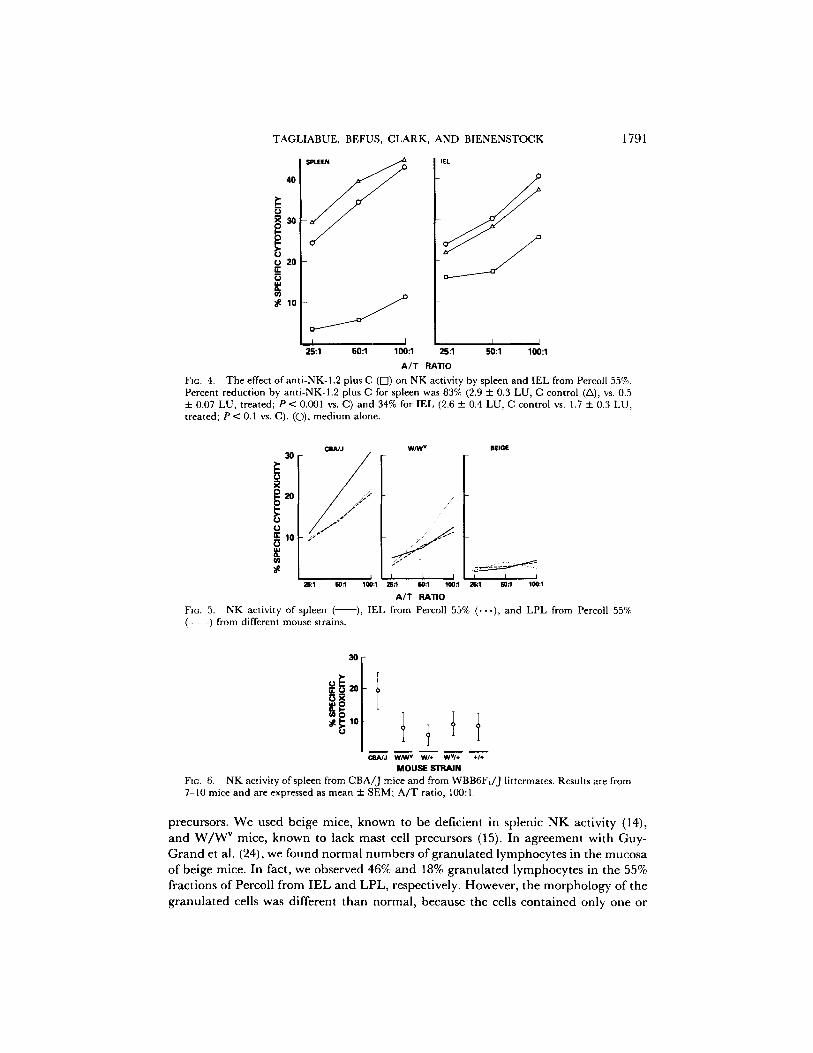
A/T RATIO FiG. 3. The effect ofapti-asialo GMI plus C (IS]) on NK activity by spleen and IEL from Percoll 55%. Percent reduction by anti-asialo GMI plus C for spleen was 83% (3.7 + 0.4 LU, C control (Z~), vs. 0.5 + 0.1 LU, treated; P < 0.005 vs. C) and 31% for IEL (2.6 + .4 LU, C control, vs. 1.8 ::1:0.3 LU, treated; P < 0.1 vs. C). (O), medium alone.
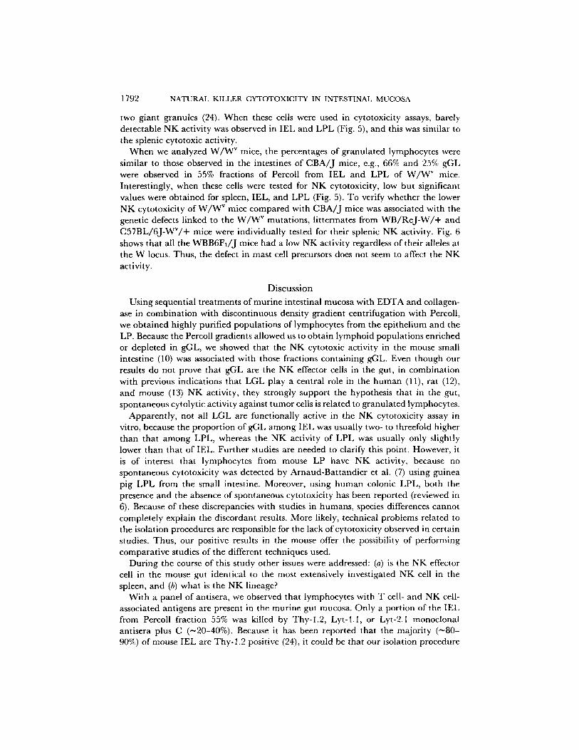
on spleen and IEL from Percoll gradients. Both anti-asialo GM] and anti-NK-1.2 were ab le to a b l a t e a lmos t the who le N K ac t iv i ty o f the sp leen (Figs. 3 a n d 4), bu t
on ly m a r g i n a l l y a f fec ted the N K cy to tox ic i ty o f I E L . T h e r e d u c t i o n on I E L by these
an t i se ra was no t s ta t i s t ica l ly s igni f icant (Figs. 3 a n d 4). S i m i l a r results were o b t a i n e d
in all th ree expe r imen t s , wh ich tes ted in para l l e l sp leen cells a n d I E L for each
an t i s e rum. NK Activity of IEL from Beige and I47/W ~ Mice. U s i n g b o t h n o r m a l a n d be ige mice
(14), G u y - G r a n d et al. (24) p o s t u l a t e d tha t g r a n u l a t e d l y m p h o c y t e s in the gut a re a
specia l class o f T l y m p h o c y t e s c a p a b l e o f ac t i ng as p rogen i to r s o f mucosa l mas t cells.
Because mast cells f rom m o u s e pe r i t onea l wash ings h a v e been recen t ly shown to exer t
s p o n t a n e o u s cy to ly t i c ac t iv i ty aga ins t t u m o r cells in v i t ro (25), we dec ided to inves-
t iga te the possible r e l a t ionsh ip b e t w e e n the in tes t ina l N K ac t iv i ty a n d mas t cell
TAGLIABUE, BEFUS, CLARK, AND BIENENSTOCK
X30
ca 20 14. Q
;¢ lO
I i I L J J 25:1 50:1 100:1 25:1 50:1 100:1
A / T RATIO
FIG. 4. The effect of anti-NK-1.2 plus C ([7) on NK activity by spleen and IEL from Percol[ 55%. Percent reduction by anti-NK-1.2 plus C for spleen was 83% (2.9 ± 0.3 LU, C control (/x), vs. 0.5 ± 0.07 LU, treated; P < 0.001 vs. C) and 34% for IEL (2.6 ± 0.4 LU, C control vs. 1.7 ± 0.3 LU, treated; P < 0.1 vs. C). (O), medium alone.
1791
C a A / J
~20 // . / , :~:
o
I I ZE:l 80:1
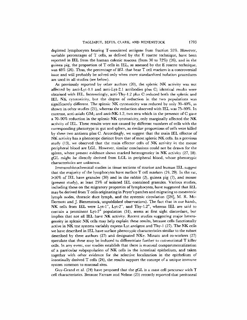
FIG. 5. NK activity of spleen ( (- - -) from different mouse strains.
W/W v BEIGE
/
I I I I I I 100:1 28:1 E0:l 100:1 28:1 80:1 100:1
A / T RATIO
), IEL from Percoll 55% ( . . . ) , and LPL from Percoll 55%
0 t OBAdJ W/W v WI÷ WV/* +l÷
MOUSE STRAIN
Fzc. 6. NK activity of spleen from CBA/J mice and from WBB6FI/J llttermates. Results are from 7-10 mice and are expressed as mean + SEM; A/T ratio, 100:1.
precursors . W e used be ige mice , k n o w n to be def ic ien t in sp lenic N K ac t iv i ty (14),
a n d W / W v mice , k n o w n to lack mas t cell p recursors (15). In a g r e e m e n t w i t h G u y -
G r a n d et al. (24), we found n o r m a l n u m b e r s o f g r a n u l a t e d l y m p h o c y t e s in the m u c o s a
o f be ige mice . In fact , we obse rved 46% a n d 18% g r a n u l a t e d l y m p h o c y t e s in t he 55%
f rac t ions o f Percol l f rom I E L a n d L P L , respect ive ly . H o w e v e r , t he m o r p h o l o g y of t he
g r a n u l a t e d cells was d i f fe ren t t h a n n o r m a l , because the cells c o n t a i n e d on ly one or
1792 NATURAL KILLER CYTOTOXICITY IN INTESTINAL MUCOSA
two giant granules (24). When these cells were used in cytotoxicity assays, barely detectable N K activity was observed in IEL and LPL (Fig. 5), and this was similar to the splenic cytotoxic activity.
When we analyzed W / W v mice, the percentages of granulated lymphocytes were similar to those observed in the intestines of CBA/J mice, e.g., 66% and 25% gGL were observed in 55% fractions of Percoll from IEL and LPL of W / W v mice. Interestingly, when these cells were tested for N K cytotoxicity, low but significant values were obtained for spleen, IEL, and LPL (Fig. 5). To verify whether the lower N K cytotoxicity of W / W v mice compared with CBA/J mice was associated with the genetic defects linked to the W / W " mutations, littermates from W B / R e J - W / + and C57BL/6J-WV/+ mice were individually tested for their splenic NK activity. Fig. 6 shows that all the WBB6F1/J mice had a low N K activity regardless of their alleles at the W locus. Thus, the defect in mast cell precursors does not seem to affect the NK activity.
Discussion
Using sequential treatments of murine intestinal mucosa with EDTA and collagen- ase in combination with discontinuous density gradient centrifugation with Percoll, we obtained highly purified populations of lymphocytes from the epithelium and the LP. Because the Percoll gradients allowed us to obtain lymphoid populations enriched or depleted in gGL, we showed that the NK cytotoxic activity in the mouse small intestine (10) was associated with those fractions containing gGL. Even though our results do not prove that gGL are the N K effector cells in the gut, in combination with previous indications that LGL play a central role in the human (11), rat (12), and mouse (13) N K activity, they strongly support the hypothesis that in the gut, spontaneous cytolytic activity against tumor cells is related to granulated lymphocytes.
Apparently, not all LGL are functionally active in the NK cytotoxicity assay in vitro, because the proportion of gGL among IEL was usually two- to threefold higher than that among LPL, whereas the N K activity of LPL was usually only slightly lower than that of IEL. Further studies are needed to clarify this point. However, it is of interest that lymphocytes from mouse LP have N K activity, because no spontaneous cytotoxicity was detected by Arnaud-Battandier et al. (7) using guinea pig LPL from the small intestine. Moreover, using human colonic LPL, both the presence and the absence of spontaneous cytotoxicity has been reported (reviewed in 6). Because of these discrepancies with studies in humans, species differences cannot completely explain the discordant results. More likely, technical problems related to the isolation procedures are responsible for the lack ofcytotoxicity observed in certain studies. Thus, our positive results in the mouse offer the possibility of performing comparative studies of the different techniques used.
During the course of this study other issues were addressed: (a) is the N K effector cell in the mouse gut identical to the most extensively investigated NK cell in the spleen, and (b) what is the NK lineage?
With a panel of antisera, we observed that lymphocytes with T cell- and NK cell- associated antigens are present in the murine gut mucosa. Only a portion of the IEL from Percoll fraction 55% was killed by Thy-l .2, Lyt-l.1, or Lyt-2.1 monoclonal antisera plus C (~20-40%). Because it has been reported that the majority (~80- 90%) of mouse IEL are Thy-l .2 positive (24), it could be that our isolation procedure
TAGLIABUE, BEFUS, CLARK, AND BIENENSTOCK 1793
depleted lymphocytes bearing T-associated antigens from fraction 55%. However, variable percentages of T cells, as defined by the E rosette technique, have been reported in IEL from the human colonic mucosa (from 30 to 72%) (26), and in the guinea pig, the proportion of T cells in IEL, as assessed by the E rosette technique, was 48% (26). Thus, the percentage of IEL that bear T cell markers is a controversial issue and will probably be solved only when more standardized isolation procedures are used in all studies (see below).
As previously reported by other authors (20), the splenic NK activity was not affected by anti-Lyt-l.1 and anti-Lyt-2.1 antibodies plus C; identical results were obtained with IEL. Interestingly, anti-Thy-l.2 plus C reduced both the splenic and IEL NK cytotoxicity, but the degree of reduction in the two populations was significantly different. The splenic NK cytotoxicity was reduced by only 30-40%, as shown in other studies (21), whereas the reduction observed with IEL was 75-90%. In contrast, anti-asialo GMI and anti-NK-1.2, two sera which in the presence of C gave a 70-90% reduction in the splenic NK cytotoxicity, only marginally affected the NK activity of IEL. These results were not caused by different numbers of cells with the corresponding phenotype in gut and spleen, as similar proportions of cells were killed by these two antisera plus C. Accordingly, we suggest that the main IEL effector of NK activity has a phenotype distinct from that of most splenic NK cells. In a previous study (13), we observed that the main effector cells of NK activity in the mouse peripheral blood are LGL. However, similar conclusions could not be drawn for the spleen, where present evidence shows marked heterogeneity in NK activity (27, 28). gGL might be directly derived from LGL in peripheral blood, whose phenotypic characteristics are unknown.
Immunohistochemical studies in tissue sections of murine and human IEL suggest that the majority of the lymphocytes have surface T cell markers (24, 29). In the rat, >50% of IEL have granules (30) and in the rabbit (2), guinea pig (7), and mouse (present study), at least 25% of isolated IEL contained granules. Various studies, including those on the migratory properties of lymphocytes, have suggested that IEL may be derived from T cells originating in Peyer's patches and migrating to mesenteric lymph nodes, thoracic duet lymph, and the systemic circulation ([24], M. R. Mc- Dermott and J. Bienenstock, unpublished observations). The fact that in our hands, NK cells from IEL were Lyt- l - , Lyt-2-, and Thy-l .2 +, whereas IEL are said to contain a prominent Lyt-2 + population (31), seems at first sight discordant, but implies that not all IEL have NK activity. Recent studies suggesting major hetero- geneity in splenic NK cells may help explain these results, because cells functionally active in NK test systems variably express Lyt antigens and Thy-1 (27). The NK cells we have described in IEL have surface phenotypic characteristics similar to the subset described by these authors (27) and designated NKT. Minato and co-workers (27) speculate that these may be induced to differentiate further to conventional T killer cells. In any event, our studies establish that there is mucosal compartmentalization of a particular subpopulation of NK cells in the intestinal epithelium, and taken together with other evidence for the selective localization in the epithelium of intestinally derived T cells (24), the results support the concept of a unique immune system common to mucosal sites.
Guy-Grand et al. (24) have proposed that the gGL is a mast cell precursor with T cell characteristics. Because Farram and Nelson (25) recently reported that peritoneal

1794 NATURAL KILLER CYTOTOXICITY IN INTESTINAL MUCOSA
mast cells have natural cytotoxic activity in vitro for certain tumor targets, we investigated the possible relationship between gut NK cells and mast cells. We used two genetically deficient mouse models: the beige, deficient in NK activity (14), and the W / W v, lacking mast cell precursors (15). Extremely low NK activity was found in IEL and LPL from beige mice. W / W v mice had normal numbers of gGL and low but detectable NK activity, suggesting that no lineage relationship exists between NK cells and mast cell precursors. It is important to stress that genetic analysis of the littermates of the WBB6Fx mice showed no particular NK defect associated with the W / W v alleles. The reason(s) why these results differ from those of Seaman and Talal (32), who showed a depressed NK activity in W / W v relative to littermates, is unclear; perhaps environmental differences affecting one or more subpopulation of NK cells are responsible.
In conclusion, this study has confirmed and extended previous observations (6-8, 10) indicating that granulated lymphocytes from the intestinal mucosa, phenotypi- cally different from the splenic NK cell, are capable of exerting spontaneous cytotox- icity against tumor cells. Lymphocytes with cytoplasmic granules have been described in the epithelium of other anatomical sites such as the mouse lung (10), the rabbit oviductal fimbriae and endocervices (33), and the male reproductive tract of rats and monkeys (34). Thus, the LGL could be a central cell of NK immunosurveillance in several anatomic sites. However, the gGL could have other functions, such as a role in resistance to intestinal infection. Preliminary experiments in mice infected with Nippostrongylus brasiliensis or Giardia muris show that an increase in splenic NK activity occurs during intestinal infection. Experiments are in progress to assess whether gut mucosal lymphocytes are cytotoxic for intestinal parasites.
S u m m a r y
Highly purified populations of lymphocytes were obtained from the murine intes- tinal mucosa using EDTA-coUagenase isolation procedures in combination with discontinuous density centrifugation. Intraepithelial lymphocytes (IEL) were sepa- rated from lamina propria lymphocytes (LPL) and, within these two populations, fractions enriched or depleted in gut granular lymphocytes (gGL) were obtained. Using these cells in cytotoxic assays, it was shown that both IEL and LPL possess natural killer (NK) activity, and this was associated with gGL. The major effector cells of gut NK activity appeared to be Thy-l .2 ÷, Lyt - l . l - , and Lyt-2.1-. The susceptibility of gut NK cells to anti-Thy- 1.2 plus complement (C) was significantly higher than that of splenic NK cells. In contrast, anti-asialo GM1 and anti-NK-1.2 plus C only slightly aftected the gut NK activity. Thus, the phenotype of the gut NK cells appears to be different from the splenic one and provides further evidence for NK heterogeneity and establishes the compartmentalization of one NK subpopula- tion. Beige mice, deficient in splenic NK activity, also had very low gut NK activity. W / W v mice, which lack mast cell precursors, had normal numbers of gGL and diminished, but still present, gut and splenic NK activity. This deficiency did not segregate with the genes responsible for the basic hemopoietic stem cell defect, and these results argue against a close ontogenetic relationship between IEL, gGL, and intestinal mucosal mast cells. The relevance of these observations to the cell lineage of the effector cell of gut NK activity is discussed.
Received for publication 22 December 1981 and in revised form 18 March 1982.

TAGLIABUE, BEFUS, CLARK, AND BIENENSTOCK 1795
References 1. Bienenstock, J., and A. D. Befus. 1980. Mucosal immunology. Immunology. 41:249. 2. Rudzik, O., and J. Bienenstock. 1974. Isolation and characteristics of gut mucosal lympho-
cytes. Lab. Invest. 30:260. 3. Bull, D. M., and M. A. Bookman. 1977. Isolation and functional characterization of human
intestinal mucosal lymphoid cells.J. Clin. Invest. 59:966. 4. Cebra, J. J., P. J. Gearhart, R. Kamat, S. M. Robertson, and J. Tseng. 1977. The secretory
immunoglobulin A response in the gut. Biochem. Soc. Tram. 5:1565. 5. Goodacre, R., R. Davidson, D. Singal, and J. Bienenstock. 1979. Morphologic and
functional characteristics of human intestinal lymphoid cells isolated by a mechanical technique. Gastroenterology. 76:300.
6. Chiba, M., W. Bartnik, S. G. ReMine, W. R. Thayer, and R. G. Shorter. 1981. Human colonic intraepithelial and lamina proprial lymphocytes: cytotoxicity in vitro and the potential effects of the isolation method on their functional properties. Gut. 22:177.
7. Arnaud-Battandier, F., B. M. Bundy, M. O'Neill, J. Bienenstock, and D. L. Nelson. 1978. Cytotoxic activities of gut mucosal lymphoid cells in guinea pigs. J. Immunol. 121:1059.
8. MacDermott, R. P., G. O. Franklin, K. M. Kenkins, I. J. Kodner, G. S. Nash, and I. J. Weinrieb. 1980. Human intestinal mononuclear cells. I. Investigation of antibody-depend- ent, lectin-induced, and spontaneous cell-mediated cytotoxic capabilities. Gastroenterology. 78:47.
9. Puccetti, P., A. Santoni, C. Riccardi, and R. B. Herberman. 1980. Cytotoxic effector cells with the characteristics of natural killer cells in the lungs of mice. Int. J. Cancer 25:153.
10. Tagliabue, A., W. Luini, D. Soldateschi, and D. Boraschi. 1981. Natural killer activity of gut mucosal lymphoid cells in mice. Eur. J. Immunol. 11:919.
11. Timonen, T., J. R. Ortaldo, and R. B. Herberman. 1981. Characteristics of human large granular lymphoeytes and relationship to natural killer and K cells.J. Exp. Med. 153:569.
12. Reynolds, C. W., T. Timonen, and R. B. Herberman. 1981. Natural killer (NK) cell activity in the rat. I. Isolation and characterization of the effector cell. J. Imrnunol. 127:282.
13. Luini, W., D. Boraschi, S. Alberti, A. Aleotti, and A. Tagliabue. 1981. Morphological characterization of a cell population responsible for natural killer activity. Immunology. 43:663.
14. Roder, J., and A. Durne. 1977. The beige mutation in the mouse selectively impairs natural killer cell function. Nature (Lond.). 278:451.
15. Kitamura, Y., S. Go, and K. Hatanaka. 1978. Decrease of mast cells in W / W v mice and their increase by bone marrow transplantation. Blood. 52:447.
16. Befus, A. D., F. L. Pearce, J. Gauldie, P. Horsewood, and J. Bienenstock. 1981. Mucosal mast cells. I. Isolation and functional characteristics of rat intestinal mast cells.J. ImmunoL In press.
17. Davies, M. D. J., and D. M. V. Parrott. 1981. Preparation and purification of lymphocytes from the epithelium and lamina propria of murine small intestine. Gut. 22:481.
18. Burton, R. C. 1980. Alloantisera selectively reactive with NK cells: characterization and use in defining NK cell classes. In Natural Cell-mediated Immunity Against Tumors. R. B. Herberman, editor. Academic Press, Inc., New York. 19.
19. Kasai, M., M. Iuamor, Y. Nagai, K. Okumura, and T. Tada. 1980. A glycolipid on the surface of mouse natural killer cells. Eur. J. Immunol. 10:175.
20. Pollack, S. B., M. R. Tam, R. C. Nowinski, and S. L. Emmons. 1979. Presence of T cell- associated surface antigens on murine NK cell.J. Immunol. 123:1818.
21. Mattes, M. J., S. O. Sharrow, R. B. Herberman, and H. T. Holden. 1979. Identification and separation of Thy-1 positive mouse spleen cells active in natural cytotoxic and antibody-dependent cell mediated cytotoxicity.J. Immunol. 123:2851.
1796 NATURAL KILLER CYTOTOXICITY IN INTESTINAL MUCOSA
22. Takasugi, M. 1971. An improved fluorochromatic cytotoxic test. Transplantation (Balti- more). 12:148.
23. Miller, R. G., and M. Dunkley. 1974. Quantitative analysis of the 51chromium release cytotoxicity assay for cytotoxic lymphocytes. Cell Immunol. 14:284.
24. Guy-Grand, D., C. Griscelli, and P. Vassalli. 1978. The mouse gut T lymphocyte, a novel type ofT cell. Nature, origin, and traffic in mice in normal and graft-versus-host conditions. J. Exp. Med. 148:1661.
25. Farram, E., and D. S. Nelson. 1980. Mouse mast cells as anti-tumor effector cells. Cell, Immunol. 55:294.
26. Arnaud-Battandier, F. 1982. Immunologic characteristics of isolated gut mucosal lymphoid cells. In Recent Advances in Mucosal Immunity. W. Strober and K. W. Sell, editors. Raven Press, New York. In press.
27. Minato, N., L. Reid, and B. R. Bloom. 1981. On the heterogeneity of murine natural killer cells.J. Exp. Med. 154:750.
28. Lust, J. A., V. Kumar, R. C. Burton, S. P. Bartlett, and M. Bennett. 1981. Heterogeneity of natural killer cells in the mouse..]. Exp. Med. 154:306.
29. Janossy, G., N. Tidman, W. S. Selby, J. A. Thomas, S. Granger, P. C. Kung, and G. Goldstein. 1980. Human T lymphocytes of inducer and suppressor type occupy different microenvironments. Nature ( Lond. ). 288:81.
30. Collan, Y. 1972. Characteristics of nonepithelial cells in the epithelium of normal rat ileum. Scand. J. GastroenteroL 7(Suppl.): 1.
31. Davies, M. D. J.,J. M. Ure, D. M. V. Parrott, and H. S. Micklem. 1981. Lyt subset analysis of lymphocyte populations isolated from mouse intestinal epithelium and lamina propria. Adv. Exp. Med. Biol. In press.
32. Seaman, W. E., and N. Talal. 1981. Natural killing by spleen cells from W/ W v and S1/S1 d anemic mice. Exp. Hematol. 9:961.
33. Odor, D. L. 1974. The question of "basal" cells in oviductal and endocervical epithelium. FertiL Steril. 25:1047.
34. Dym, M., and L. J. Romrell. 1975. Intraepithelial lymphocytes in the male reproductive tract of rats and rhesus monkeys. J. Reprod. Fert. 42:1.