characterization of b cells in healthy pregnant women from ... pregnancy.pdf · post-partum: a...
TRANSCRIPT
RESEARCH ARTICLE Open Access
Characterization of B cells in healthypregnant women from late pregnancy topost-partum: a prospective observationalstudyJorge Lima1,2*, Catarina Martins2, Maria J. Leandro3, Glória Nunes2, Maria-José Sousa4,5, Jorge C. Branco6
and Luís-Miguel Borrego7,2
Abstract
Background: B cells play a role in pregnancy due to their humoral and regulatory activities. To our knowledge,different maturational stages (from transitional to memory) of circulating B cell subsets have not yet beencharacterized (cell quantification and phenotype identification) in healthy pregnant women. Thus, the objective ofour study was to characterize these subsets (as well as regulatory B cells) from late pregnancy to post-partum andto compare them with the circulating B cells of non-pregnant women.
Methods: In all of the enrolled women, flow cytometry was used to characterize the circulating B cell subsetsaccording to the expression of IgD and CD38 (Bm1-Bm5 classification system). Regulatory B cells were characterizedbased on the expression of surface antigens (CD24, CD27, and CD38) and the production of IL-10 after lipopolysaccharidestimulation.
Results: Compared to the absolute counts of B cells in the non-pregnant women (n= 35), those in the pregnant women(n = 43) were significantly lower (p < 0.05) during the 3rd trimester of pregnancy and on delivery day (immediately afterdelivery). The percentages of these cells on delivery day and at post-partum were significantly lower than those in thenon-pregnant women.In general, the absolute counts and percentages of the majority of the B cell subsets were significantly lower in the 3rdtrimester of pregnancy and on delivery day than in the non-pregnant women. However, these counts and percentagesdid not differ significantly between the post-partum and the non-pregnant women.The most notable exceptions to the above were the percentages of naïve B cells (which were significantly higher in the3rd trimester and on delivery day than in the non-pregnant women) and of CD24hiCD38hi regulatory B cells (which weresignificantly higher in the post-partum than in the non-pregnant women).
Conclusion: According to our study, the peripheral B cell compartment undergoes quantitative changes during normallate pregnancy and post-partum. Such findings may allow us to better understand immunomodulation during humanpregnancy and provide evidence that could aid in the development of new strategies to diagnose and treatpregnancy-associated disturbances. Our findings could also be useful for studies of the mechanisms of maternalresponses to vaccination and infection.
Keywords: B cell subsets, Flow cytometry, Human pregnancy, Obstetrics
* Correspondence: [email protected] of Obstetrics and Gynecology, CUF Descobertas Hospital,Lisbon, Portugal2CEDOC, Chronic Diseases Research Center, Immunology, NOVA MedicalSchool, Faculty of Medical Sciences, Lisbon, PortugalFull list of author information is available at the end of the article
© 2016 The Author(s). Open Access This article is distributed under the terms of the Creative Commons Attribution 4.0International License (http://creativecommons.org/licenses/by/4.0/), which permits unrestricted use, distribution, andreproduction in any medium, provided you give appropriate credit to the original author(s) and the source, provide a link tothe Creative Commons license, and indicate if changes were made. The Creative Commons Public Domain Dedication waiver(http://creativecommons.org/publicdomain/zero/1.0/) applies to the data made available in this article, unless otherwise stated.
Lima et al. BMC Pregnancy and Childbirth (2016) 16:139 DOI 10.1186/s12884-016-0927-7
BackgroundThe immune system of pregnant women tolerates agenetically foreign fetus. This physiological adaptation isthought to be promoted by an array of anti-inflammatoryand pro-inflammatory cytokines that are produced by Tand B cells [1]. This state of immunological tolerance mayinfluence the course of pre-existing pathologies (e.g., auto-immune diseases) [2] and may also increase fetal-maternalsusceptibility to infection [3]. Furthermore, a decrease offunction of B cells with loss of responsiveness to mitogensand infectious agents during the course of normal humanpregnancy has also been reported [4].B cells have a role in pregnancy because of their humoral
activity (i.e., the production of protective antibodies againstpaternal antigens during pregnancy and the production ofauto-antibodies that may lead to pregnancy complications)[5]. Normally, B cells leave the bone marrow and enter thecirculation as immature transitional B cells, which later ma-ture into naïve B cells. Finally, when naïve B cells encountertheir cognate antigens in secondary lymphoid organs, thesecells become activated and mature into memory B cells andplasma cells [6, 7]. B cell subsets of different maturationalstages, from transitional to memory B cells, have been iden-tified in peripheral blood using the mature B (Bm)1-Bm5classification system. This classification system has provento be effective in the identification of disturbances in theproportions of peripheral blood B cell subsets in patientswith autoimmune diseases (e.g., Lupus or Sjögren’s syn-drome) [8–10] and in those undergoing therapy (e.g., withbiological agents), by assessing the depletion and repopula-tion of B cells [11].Furthermore, it has been suggested that, in addition to
their humoral activity, specific B cells can also have aregulatory function although this is still controversial.According to recent studies, regulatory B cells (Bregs)can inhibit pro-inflammatory responses by secreting theanti-inflammatory cytokine IL-10 [12, 13]. Breg countsincrease in the first trimester of pregnancy, suppressingunwanted immune responses of maternal effector T cells,protecting against pregnancy loss [14]. While the pheno-type and function of regulatory T cells has been extensivelystudied [15], further studies are needed to investigate themechanisms behind the activation and expansion of Bregsand other B cell subsets in pregnancy.These regulatory functions have been attributed to dif-
ferent B cell subsets, and despite some controversy, greatprogress has been made in the characterization of Bregs.The inability to identify a Breg-specific transcription fac-tor, together with the phenotypic heterogeneity of Bregs,supports the idea that Bregs are not lineage specific andthat they may expand in response to inflammation whenimmunosuppression is necessary [16]. It remains unclearwhether the regulatory B cell function is a specific roleof a particular subset or whether it is a reflection of their
maturation stage. Although the expression of IL-10 hasbeen a valuable tool in defining populations of Bregs,CD24hiCD27+ and CD24hiCD38hi are the most frequentlycharacterized phenotypes in humans [9, 12, 13].Several studies [17–27] both prospective and cross-
sectional have reported on circulating B cells duringnormal human pregnancy with majority describing lowertotal numbers and/or frequency when compared to levelspost-partum or in healthy non-pregnant. Most studies[17, 19–24, 27] have only investigated total B cells asdefined by the expression of CD20 or CD19 with olderstudies [18, 25, 26] using either expression of Ia (HLA-DR)or surface immunoglobulin. A few studies [17, 19, 24, 27]have reported on the frequency of B cell subsets expressingCD5 with majority describing lower frequency or lowertotal numbers of this subset during pregnancy, at deliveryor early in the postpartum period. One study [19] reportedlower CD21 and CD23 frequencies at delivery. However,peripheral B cells have not been characterized in humanpregnancy while considering the different maturationalstages, from transitional to memory B cells (using CD38and immunoglobulin IgD as differentiation markers). Con-sequently, the objective of our study was to characterizethese specific peripheral blood B cell subsets (transitional,naïve, unswitched memory, post-germinal, and restingmemory B cells as well as plasmablasts) and Bregs(CD24hiCD27+, CD24hiCD38hi and IL-10 regulatory Bcells) from late pregnancy to post-partum and comparethem with those in non-pregnant women.
MethodsStudy populationThis prospective observational study followed healthypregnant women over time to characterize (i.e., cell quanti-fication and phenotype identification) their peripheral bloodB cell subsets from late pregnancy to post-partum. Thischaracterization of B cells in the pregnant women was alsocompared with the characterization of single samples ofperipheral blood B cells from a control group of healthynon-pregnant women to investigate changes associatedwith pregnancy.Sequential non-laboring healthy women with singleton
pregnancies who were attending an outpatient clinic(routine obstetrical care) during the 3rd trimester wererecruited for participation. None of the pregnancies hadcomplications prior to recruitment. Furthermore, all ofthe fetuses exhibited appropriate growth (as measuredby uterine fundal height and by ultrasound performedafter 28 weeks of gestation).Sequential non-pregnant women who were attending an
outpatient clinic were also recruited (healthy controls).These were asymptomatic women who were attendingtheir annual routine well-woman exams.
Lima et al. BMC Pregnancy and Childbirth (2016) 16:139 Page 2 of 13
For all of the women, the exclusion criteria were a his-tory of diabetes, hypertension, or autoimmune diseaseand smoking during the 6 months prior to peripheralblood sample collection. Additional exclusion criteria forthe pregnant women included prenatal use of any medi-cation (other than vitamins and iron supplements) andongoing complications in the pregnancy. Non-pregnantwomen taking oral contraceptives were also excluded, asthese drugs affect circulating B cells [28].All of the women were recruited at the Hospital CUF
Descobertas in Lisbon (Portugal) between July 2013 andMarch 2014. The Ethics Committee of this hospital ap-proved the study protocol. All of the recruited womenprovided written informed consent before the start ofthe study.
Study visit proceduresThree visits were planned for the pregnant women: visit1 was planned for the 3rd trimester of pregnancy (3rdtrimester); visit 2, for the day of delivery; and visit 3, forpost-partum (at least 6 weeks after delivery). A singlevisit was planned for the non-pregnant controls.To characterize B cell subsets from late pregnancy to
post-partum, peripheral blood samples were collected fromall of the pregnant women at each planned visit: the “3rdtrimester” sample was collected at visit 1, the “on deliveryday” sample was collected at visit 2 (immediately after de-livery, within 15 min after placental expulsion and oxytocinadministration), and the “post-partum” sample was col-lected at visit 3. A peripheral blood sample was collectedfrom the non-pregnant women at the planned visit, whichtook place during the follicular phase of their menstrualcycle because hormone status during the luteal phase issimilar to that during pregnancy [29].The baseline data collected for all women at the time
of enrollment included demographics (age and ethnicity),anthropometrics [body mass index (BMI)], obstetric his-tory, and systolic and diastolic blood pressures. The datacollected for the pregnant women on the day of delivery in-cluded gestational age, type of analgesia and/or anesthesia,and mode of delivery. The data collected for the newbornsincluded gender, weight, and 1-min and 5-min Apgarscores.
Flow cytometry analysis and laboratory measurementsPeripheral blood samples were collected into EDTA-coatedand heparinized tubes. These samples were analyzed byfour-color flow cytometry (BD FACSCalibur, BD Biosci-ences, San Jose, CA, USA) to characterize B cell subsetsand their maturation profiles. MultisetTM and CellQuest3.3TM (BD Biosciences) software were used for both acqui-sition and analysis.To obtain absolute counts of B cells (CD19+), a single-
platform strategy was used. EDTA samples were assayed
using a lyse-no-wash technique, with a BD IMK Kit withBD Trucount™ Tubes (BD Biosciences). The assay wasperformed according to the manufacturer’s instructions.In brief, 50 μL of blood were incubated for 15 min inthe dark, at room temperature, with the monoclonalantibodies provided in the kit, in Trucount™ tubes con-taining a calibrated number of microbeads for countingpurposes. Red blood cells were then lysed with the lysingsolution (also provided with the BD IMK Kit), for15 min and finally samples were acquired. The cells weregated on CD45/SSC, and a minimum of 2500 lympho-cyte events were acquired. Multiset software providedpercentage and absolute counts of B cells using thenumber of microbeads in each Trucount™ tube, alongwith the number of microbead and lymphocyte eventsacquired in each tube.To study the surface B cell markers, a modified lyse-wash
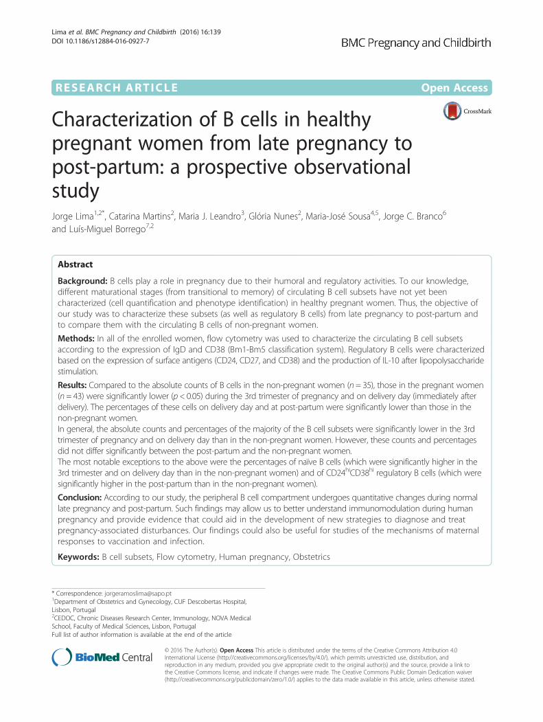
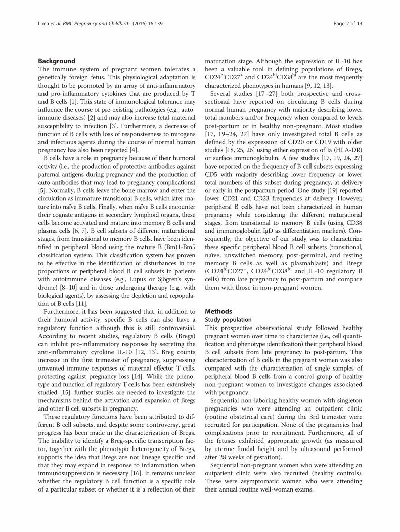
protocol was used. EDTA samples were washed twice inphosphate-buffered saline (PBS) to lower background stain-ing. The washed cells were then stained with a panel ofmonoclonal antibodies (mAbs) that were conjugated withdifferent fluorochromes: anti-CD19 PerCPCy5.5 (cloneHIB19, Biolegend), anti-CD24 PE (clone ML5, Biolegend),anti-CD27 FITC (clone O323, Biolegend), CD38 APC(clone HIT2, Biolegend), and anti-IgD PE (clone IA6-2, BDPharmingen). Red blood cells were incubated for 15 min atroom temperature in the dark. The red cells were thenlysed with BD FACS lysing solution (BD Biosciences)according to the manufacturer’s instructions. After awash step with PBS, events were acquired. For thecharacterization of IL-10-producing Bregs, heparin sam-ples were incubated for 5 h at 37 °C in a 5 % CO2 atmos-phere with phorbol 12-myristate 13-acetate (PMA)(50 ng/mL, Sigma Aldrich), calcium ionophore (1 μg/mL,Sigma Aldrich), and lipopolysaccharide (LPS) (10 μg/mL,Sigma Aldrich) in the presence of Brefeldin A (1.0 μg/ml,BD Pharmingen) [13, 30]. After the stimulation, the redblood cells were lysed via the addition of BD FACS lysingsolution and were stained for surface markers with anti-CD3 FITC (clone SK7, BD Biosciences), anti-CD19PerCPCy5.5 (clone HIB19, Biolegend), and anti-CD8 APC(clone SK1, Biolegend) mAbs. The Cytofix-Cytopermkit (BD Pharmingen) was used for cell fixation andpermeabilization according to the manufacturer’s instruc-tions. To assess the cytoplasmic expression of IL-10 in theB cells, a final intracellular staining step with an anti-IL-10PE mAb (clone JES3-19F1, Biolegend) was performed be-fore cell acquisition. A minimum of 2000 B cells (CD19+)were acquired in all tubes (gate in CD19/SSC). The analysisstrategies are presented in Figs. 1 and 2. The flow cytometryresults are presented as a percentage of total B cellsand as absolute cell counts (cells/μL).The Bm1-5 classification system used to identify the de-
velopment of mature B cells was based on the expression
Lima et al. BMC Pregnancy and Childbirth (2016) 16:139 Page 3 of 13
of IgD/CD38 phenotypic markers. The cells were charac-terized as follows: transitional B cells (Bm2’: IgD+CD38hi),naïve B cells (Bm2: IgD+CD38+), unswitched memory Bcells (Bm1: IgD+CD38−), and switched memory B cells(Bm5: IgD−CD38+/−) and were subsequently divided intopost-germinal memory B cells (early Bm5: IgD−CD38+),resting memory (late Bm5: IgD−CD38−) B cells, andplasmablasts (Bm3 + Bm4: IgD−CD38hi) [8–10, 14].Bregs were evaluated in three different populations:CD24hiCD27+, CD24hiCD38hi and IL-10-producing B cells.
Our laboratory measurement included both absolutecounts and percentages of total B cells and the differentB cell subsets, as we feel that the two types of data arecomplementary. Percentages were measured as these allowinterpreting the relative fluctuations in distinct B cell sub-sets from pregnancy to post-partum. Absolute counts werealso measured and reported, although we are aware thatpregnancy is characterized by variable degrees of hemodilu-tion, and that changes in these counts may not reflect truevariations in the total numbers of circulating cells.
Fig. 1 Identification of B cell subsets according to Bm1-5 classification system. a and b Gating strategy for CD19+ B cells using an initial CD19/SSC plotand refinement of the gate using a plot of FSC vs SSC. c Bm1-5 classification from double staining for IgD and CD38 (unswitched memoryBm1: IgD+CD38−; naïve Bm2: IgD+CD38+; transitional Bm2′: IgD+CD38hi; plasmablasts Bm3 + Bm4: IgD−CD38hi; post-germinal memory/earlyeBm5: IgD−CD38+; and resting memory/late Bm5: IgD−CD38−)
Fig. 2 Identification of regulatory B cell subsets. a and b Gating strategy for CD24hiCD27+ (a) and CD24hiCD38hi (b) Bregs; c and d IL-10-producingCD19+ B cells (CD19+ B cells were analyzed for the expression of IL-10 after a 5-h incubation period without stimulation (c) and with stimulation (d)with phorbol 12-myristate 13-acetate, calcium ionophore and lipopolysaccharide)
Lima et al. BMC Pregnancy and Childbirth (2016) 16:139 Page 4 of 13
Statistical analysisIf the baseline data were normally distributed, they werepresented as means (±standard deviations); otherwise,these data were presented as medians and ranges. Cat-egorical variables were described as absolute and relativefrequencies and were expressed as percentages.Cell counts and percentages were presented as me-
dians and ranges. If normally distributed, 2 independentgroups were compared using Student’s t-tests; otherwise,Mann–Whitney U tests were used. If normally distrib-uted, pairs of samples were compared using pairedStudent’s t-tests; otherwise, Wilcoxon signed-rank testswere used. For normally distributed data, comparisonsbetween more than 2 groups were performed usingANOVA I; otherwise, Kruskal-Wallis tests were used.Statistical significance was defined by a P-value <0.05.The P-values for the comparisons of B cells betweenthe non-pregnant women and pregnant women at differ-ent visits, as well as for the comparisons of the B cells ofthe pregnant women between visits, were adjusted formultiplicity using the Benjamini and Yekutieli method[31]. All of the data were analyzed using R software, ver-sion 3.12 for Windows.
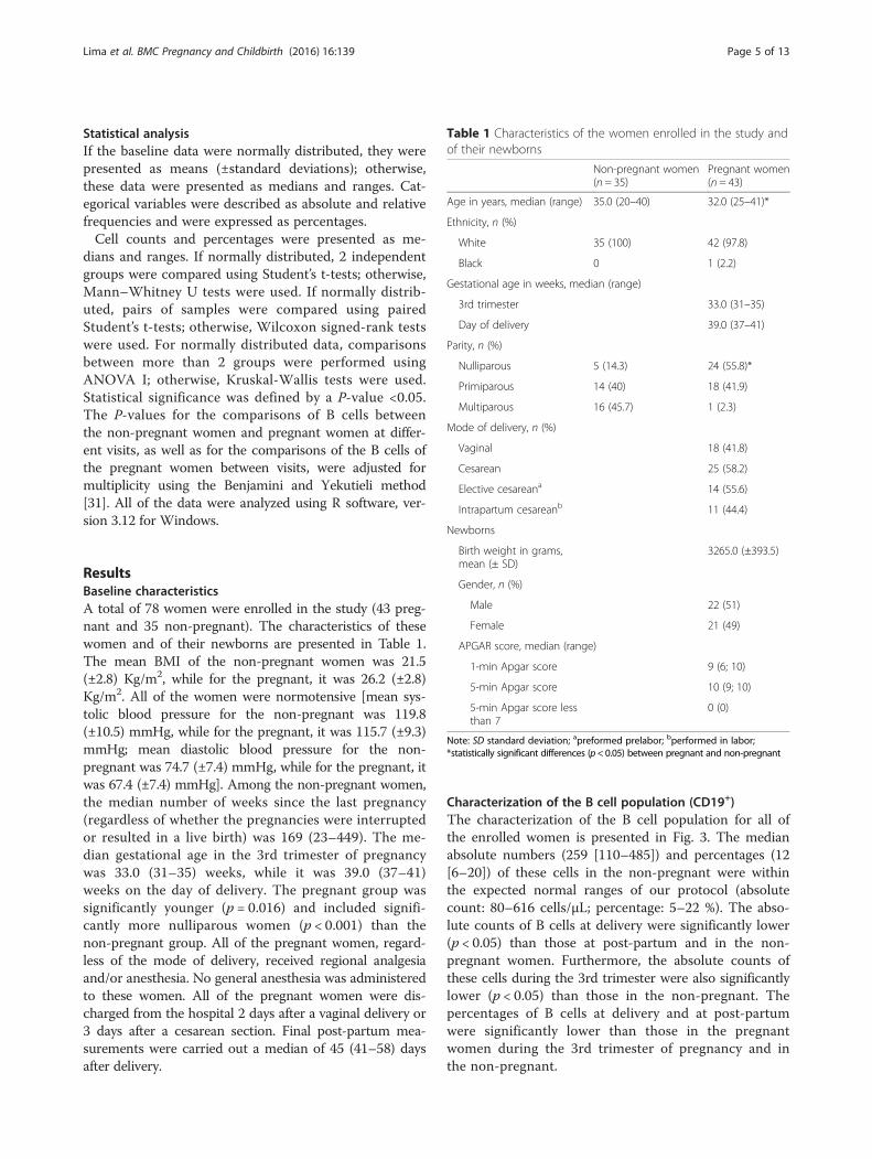
ResultsBaseline characteristicsA total of 78 women were enrolled in the study (43 preg-nant and 35 non-pregnant). The characteristics of thesewomen and of their newborns are presented in Table 1.The mean BMI of the non-pregnant women was 21.5(±2.8) Kg/m2, while for the pregnant, it was 26.2 (±2.8)Kg/m2. All of the women were normotensive [mean sys-tolic blood pressure for the non-pregnant was 119.8(±10.5) mmHg, while for the pregnant, it was 115.7 (±9.3)mmHg; mean diastolic blood pressure for the non-pregnant was 74.7 (±7.4) mmHg, while for the pregnant, itwas 67.4 (±7.4) mmHg]. Among the non-pregnant women,the median number of weeks since the last pregnancy(regardless of whether the pregnancies were interruptedor resulted in a live birth) was 169 (23–449). The me-dian gestational age in the 3rd trimester of pregnancywas 33.0 (31–35) weeks, while it was 39.0 (37–41)weeks on the day of delivery. The pregnant group wassignificantly younger (p = 0.016) and included signifi-cantly more nulliparous women (p < 0.001) than thenon-pregnant group. All of the pregnant women, regard-less of the mode of delivery, received regional analgesiaand/or anesthesia. No general anesthesia was administeredto these women. All of the pregnant women were dis-charged from the hospital 2 days after a vaginal delivery or3 days after a cesarean section. Final post-partum mea-surements were carried out a median of 45 (41–58) daysafter delivery.
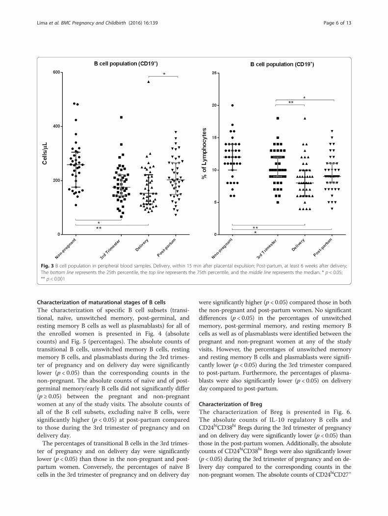
Characterization of the B cell population (CD19+)The characterization of the B cell population for all ofthe enrolled women is presented in Fig. 3. The medianabsolute numbers (259 [110–485]) and percentages (12[6–20]) of these cells in the non-pregnant were withinthe expected normal ranges of our protocol (absolutecount: 80–616 cells/μL; percentage: 5–22 %). The abso-lute counts of B cells at delivery were significantly lower(p < 0.05) than those at post-partum and in the non-pregnant women. Furthermore, the absolute counts ofthese cells during the 3rd trimester were also significantlylower (p < 0.05) than those in the non-pregnant. Thepercentages of B cells at delivery and at post-partumwere significantly lower than those in the pregnantwomen during the 3rd trimester of pregnancy and inthe non-pregnant.
Table 1 Characteristics of the women enrolled in the study andof their newborns
Non-pregnant women(n = 35)
Pregnant women(n = 43)
Age in years, median (range) 35.0 (20–40) 32.0 (25–41)*
Ethnicity, n (%)
White 35 (100) 42 (97.8)
Black 0 1 (2.2)
Gestational age in weeks, median (range)
3rd trimester 33.0 (31–35)
Day of delivery 39.0 (37–41)
Parity, n (%)
Nulliparous 5 (14.3) 24 (55.8)*
Primiparous 14 (40) 18 (41.9)
Multiparous 16 (45.7) 1 (2.3)
Mode of delivery, n (%)
Vaginal 18 (41.8)
Cesarean 25 (58.2)
Elective cesareana 14 (55.6)
Intrapartum cesareanb 11 (44.4)
Newborns
Birth weight in grams,mean (± SD)
3265.0 (±393.5)
Gender, n (%)
Male 22 (51)
Female 21 (49)
APGAR score, median (range)
1-min Apgar score 9 (6; 10)
5-min Apgar score 10 (9; 10)
5-min Apgar score lessthan 7
0 (0)
Note: SD standard deviation; apreformed prelabor; bperformed in labor;*statistically significant differences (p< 0.05) between pregnant and non-pregnant
Lima et al. BMC Pregnancy and Childbirth (2016) 16:139 Page 5 of 13
Characterization of maturational stages of B cellsThe characterization of specific B cell subsets (transi-tional, naïve, unswitched memory, post-germinal, andresting memory B cells as well as plasmablasts) for all ofthe enrolled women is presented in Fig. 4 (absolutecounts) and Fig. 5 (percentages). The absolute counts oftransitional B cells, unswitched memory B cells, restingmemory B cells, and plasmablasts during the 3rd trimes-ter of pregnancy and on delivery day were significantlylower (p < 0.05) than the corresponding counts in thenon-pregnant. The absolute counts of naïve and of post-germinal memory/early B cells did not significantly differ(p ≥ 0.05) between the pregnant and non-pregnantwomen at any of the study visits. The absolute counts ofall of the B cell subsets, excluding naïve B cells, weresignificantly higher (p < 0.05) at post-partum comparedto those during the 3rd trimester of pregnancy and ondelivery day.The percentages of transitional B cells in the 3rd trimes-
ter of pregnancy and on delivery day were significantlylower (p < 0.05) than those in the non-pregnant and post-partum women. Conversely, the percentages of naïve Bcells in the 3rd trimester of pregnancy and on delivery day
were significantly higher (p < 0.05) compared those in boththe non-pregnant and post-partum women. No significantdifferences (p < 0.05) in the percentages of unswitchedmemory, post-germinal memory, and resting memory Bcells as well as of plasmablasts were identified between thepregnant and non-pregnant women at any of the studyvisits. However, the percentages of unswitched memoryand resting memory B cells and plasmablasts were signifi-cantly lower (p < 0.05) during the 3rd trimester comparedto post-partum. Furthermore, the percentages of plasma-blasts were also significantly lower (p < 0.05) on deliveryday compared to post-partum.
Characterization of BregThe characterization of Breg is presented in Fig. 6.The absolute counts of IL-10 regulatory B cells andCD24hiCD38hi Bregs during the 3rd trimester of pregnancyand on delivery day were significantly lower (p < 0.05) thanthose in the post-partum women. Additionally, the absolutecounts of CD24hiCD38hi Bregs were also significantly lower(p < 0.05) during the 3rd trimester of pregnancy and on de-livery day compared to the corresponding counts in thenon-pregnant women. The absolute counts of CD24hiCD27+
Fig. 3 B cell population in peripheral blood samples. Delivery, within 15 min after placental expulsion; Post-partum, at least 6 weeks after delivery;The bottom line represents the 25th percentile, the top line represents the 75th percentile, and the middle line represents the median. * p < 0.05;** p < 0.001
Lima et al. BMC Pregnancy and Childbirth (2016) 16:139 Page 6 of 13
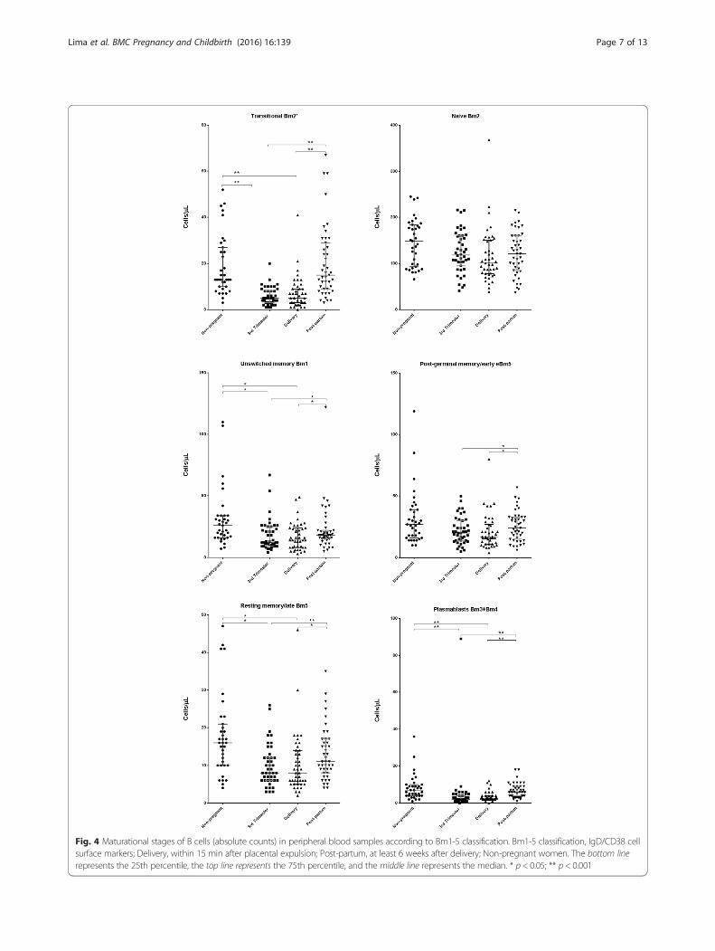
Fig. 4 Maturational stages of B cells (absolute counts) in peripheral blood samples according to Bm1-5 classification. Bm1-5 classification, IgD/CD38 cellsurface markers; Delivery, within 15 min after placental expulsion; Post-partum, at least 6 weeks after delivery; Non-pregnant women. The bottom linerepresents the 25th percentile, the top line represents the 75th percentile, and the middle line represents the median. * p < 0.05; ** p < 0.001
Lima et al. BMC Pregnancy and Childbirth (2016) 16:139 Page 7 of 13
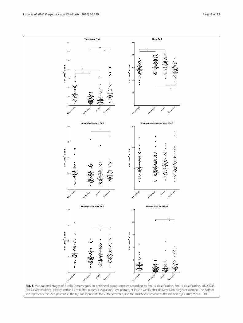
Fig. 5 Maturational stages of B cells (percentages) in peripheral blood samples according to Bm1-5 classification. Bm1-5 classification, IgD/CD38cell surface markers; Delivery, within 15 min after placental expulsion; Post-partum, at least 6 weeks after delivery; Non-pregnant women. The bottomline represents the 25th percentile, the top line represents the 75th percentile, and the middle line represents the median. * p < 0.05; ** p < 0.001
Lima et al. BMC Pregnancy and Childbirth (2016) 16:139 Page 8 of 13
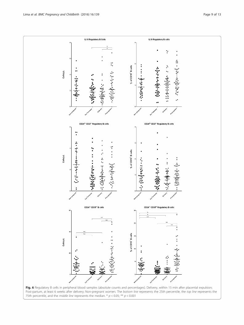
Fig. 6 Regulatory B cells in peripheral blood samples (absolute counts and percentages). Delivery, within 15 min after placental expulsion;Post-partum, at least 6 weeks after delivery; Non-pregnant women. The bottom line represents the 25th percentile, the top line represents the75th percentile, and the middle line represents the median. * p < 0.05; ** p < 0.001
Lima et al. BMC Pregnancy and Childbirth (2016) 16:139 Page 9 of 13
Bregs did not significantly differ between the pregnantand non-pregnant women or between study visits.The percentages of CD24hiCD38hi Bregs during the
3rd trimester of pregnancy and on delivery day were signifi-cantly lower (p < 0.05) than those in the non-pregnant andpost-partum women. Furthermore, the percentages of thesecells at post-partum were significantly higher (p < 0.05) thanthose in the non-pregnant women. The percentages of IL-10 regulatory B cells and of CD24hiCD27+ Bregs did notsignificantly differ between the pregnant and non-pregnantwomen or between study visits.
DiscussionAccording to our study, late-stage pregnancy (betweenthe 3rd trimester and delivery) is associated with periph-eral blood B cell lymphopenia. Indeed, the absolutecounts and percentages of most B cell subsets in the 3rdtrimester of pregnancy and on delivery day were signifi-cantly lower compared to the corresponding counts andpercentages in the non-pregnant. However, these differ-ences did not significantly differ between the post-partumand non-pregnant women, suggesting that at this latertime point, the absolute counts and percentages of most Bcell subsets revert (or at least partially revert) to normalvalues. The most notable exceptions to this observationwere identified for naïve B cells (whose percentages weresignificantly higher during the 3rd trimester and on deliv-ery day than in the non-pregnant) and for CD24hiCD38hi
Bregs (whose percentages were significantly higher in thepost-partum compared to the non-pregnant women).To our knowledge, this is the first study to characterize
the circulating B cell compartment in pregnancy whiletaking into account the maturational stages of the differentB cell subsets. Furthermore, the study was conducted pro-spectively, from the 3rd trimester of pregnancy to post-partum, in a sample of 43 pregnant women.The pregnancy-associated B lymphopenia that we iden-
tified in our study has already been described in animalmodels [32, 33] and in humans [17–27]. According toMedina et al. [32], B lymphopoiesis in bone marrow isselectively reduced during normal pregnancy becauseof hormonal influences. Furthermore, Muzzio et al. [33]demonstrated that B lymphopoiesis is reduced in latepregnancy, when estradiol levels are high. Cellular migra-tion is another mechanism that can contribute to B celllymphopenia. Studies of animal models have shown thatmonocytes and other immune cells migrate to the uterusduring the later stages of pregnancy due to changes in theexpression of chemokines [34, 35]. Furthermore, smallpopulations of B cells have been identified in the decidua,suggesting leucocyte recruitment into the maternal-fetalinterface [36]. The biological meaning of this suppressionof B lymphopoiesis in normal pregnancy is uncertain butis probably related to the physiological immune tolerance.
In this study, we found that the absolute counts of themajority of the B cell subsets were significantly lower inthe 3rd trimester of pregnancy than in the non-pregnantwomen, suggesting pregnancy-associated B lymphopenia(though most pregnant women still presented valueswithin normal ranges). The increased blood volemia ob-served during pregnancy could in part explain these obser-vations. In fact, pregnant presented also decreased absolutelymphocyte counts, along with decreased absolute countsof T and NK cells, compared to non-pregnant (data notshown). Nonetheless, B cell percentages in pregnant alsoseem to decrease during pregnancy, which does not hap-pens with either T cells or NK cells.Compared to the percentages of peripheral blood naïve
B cells in the non-pregnant women, we found highervalues during the 3rd trimester and on delivery day, butno differences were observed in absolute counts. Thisrelative increase in naïve B cells may be a consequenceof decreased differentiation of B cells into memory cellsand/or plasmablasts. In fact, Muzzio et al. [33] reportedthe expansion of naïve B cells in pregnant mice. The highlevels of progesterone present in late pregnancy may poten-tially explain this, as high progesterone levels inhibit B cellactivation in mice [37]. Our results may also be explainedby the mobilization of more differentiated B subsets fromperipheral blood to other body tissues.Normal pregnancy has been compared to a state of
quiescent systemic inflammation, while parturition hasbeen likened to an immunological reaction that resultsin the recruitment of immune cells not only to the ma-ternal-fetal interface but also to the systemic circulation[38]. The results of our study support this idea, as weidentified higher counts and percentages of CD24hiCD38hi
Bregs post-partum relative to during the 3rd trimesterand on delivery day. This observation may represent aregulatory mechanism for the suppression of immunecell activation events and may also explain the increasedsusceptibility to infections that occurs during the post-partum period and the altered clinical outcomes of someautoimmune diseases.Interestingly, we found that while the majority of B
cell subsets increased to levels closer to those of thenon-pregnant (or to normal values) from the third tri-mester of pregnancy to post-partum, the percentages ofCD24hiCD38hi Bregs were significantly higher in thepost-partum compared to the non-pregnant women. Be-cause B lymphopoiesis is under endocrine regulationduring pregnancy [32, 33], our results may be explainedby the decline of hormonal levels that typically occurspost-partum, which may have lead both to lymphopoi-esis recovery and to B cell activation. As hypothesizedby Medina et al. [32], this result may be of clinical utilityto increase lymphocyte formation in transplanted pa-tients and in those with immunodeficiencies.
Lima et al. BMC Pregnancy and Childbirth (2016) 16:139 Page 10 of 13
Unlike for the CD24hiCD38hi Bregs, no significant dif-ferences between the non-pregnant and the pregnant orfrom the 3rd trimester of pregnancy to post-partumwere identified for IL-10 Bregs (cell percentages) or forCD24hiCD27+ Bregs (cell percentages and counts). Thisheterogeneity of Breg subsets has been reported in otherstudies with humans [39]. Furthermore, CD24hiCD27+
Bregs, an activated memory subset, are more maturethan transitional CD24hiCD38hi Bregs; thus, it is morelikely for them to develop into antibody-producing cellsthat no longer possess a regulatory function [40].The differences between pregnant and non-pregnant
women identified for age, and parity are not likely tobias our results. In fact, among all of the women whowere included in our study, counts and percentages of Bcell subsets were not significantly associated with age, asdemonstrated by the non-significant Spearman correl-ation coefficients between these variables (see Additionalfile 1). Furthermore, in the vast majority of cases, therewere no statistically significant differences in counts andpercentages of B cell subsets among women, despiteparity (see Additional file 2). Finally, in the vast majority ofcases, we have also not found significant associations be-tween counts (and percentages) of B cell subsets and gesta-tional age at the 3rd trimester of pregnancy (see Additionalfile 3), gestational age at delivery (see Additional file 4),length of time post-partum until the collection of the finalblood samples (see Additional file 5), and time since lastpregnancy in the non-pregnant (see Additional file 6).Previous studies have identified differences in the
counts and percentages of B cells (total and subsets) be-tween neonates and individuals of up to 50 years of age[41]. However, to the best of our knowledge, there areno data regarding B cell variation in women over shortperiods of time, such as that of our study.Although the pregnant women received analgesia and/
or anesthesia, which may cause temporary changes inmaternal blood pressure, this is unlikely to cause import-ant changes in B cell counts because regional administra-tion is generally associated with low plasma levels of thesedrugs. Ideally, samples collected before pregnancy wouldhave been compared with samples collected during preg-nancy in the same individuals; however, this would havebeen very difficult for us from a practical point of view.The fact that several of the changes that were observedduring the 3rd trimester of pregnancy seem to be reversedduring the post-partum period suggests that comparisonswith non-pregnant women were adequate.In future research, it is important to investigate whether
B cell subset characterization could help to identify riskmarkers for the development of obstetric complications inpregnant women with or without autoimmune diseases. Inthis context, it would also be important to clarify therole of B-cell activating factor (BAFF), an essential survival
factor for transitional B cells, and of CD23, a B-lymphocytedifferentiation marker.
ConclusionAccording to our study, the characteristics of peripheralB cell compartment differ significantly between pregnantand non-pregnant women and vary over time from latepregnancy to post-partum. Such findings may allow us torecognize normal fluctuations in B cell subsets to betterunderstand immune regulation during human pregnancyand to identify new strategies for the diagnosis and treat-ment of pregnancy-associated disturbances as well asthe mechanisms of maternal responses to vaccination andinfection.
Additional files
Additional file 1: Correlation coefficients between counts (andpercentages) of B cell subsets and age, both for pregnant and non-pregnant women. aHealthy non-pregnant women; bThird trimester ofpregnancy; cWithin 15 min after placental expulsion; dAt least 6 weeksafter delivery; eSpearman correlation coefficient. (XLSX 11 kb)
Additional file 2: Comparison of counts (and percentages) of B cellsubsets between nulliparous, primiparous, and multiparous women, bothfor pregnant and non-pregnant women. Values presented as median(Interquartile range); Three classes were compared using one-way ANOVA, orKuskal-Wallis tests; Two classes were compared with t-Student or Wilcoxontests; There was only one multiparous women among the pregnant,and this woman was excluded from this analysis. aNon-pregnantwomen; bThird trimester of pregnancy; cWithin 15 min after placentalexpulsion; dAt least 6 weeks after delivery; eKuskal-Wallis test; f Wilcoxontest; * p < 0.05. (XLSX 46 kb)
Additional file 3: Correlation coefficients between counts (andpercentages) of B cell subsets and gestational age at the 3rd trimester ofpregnancy. Corr. Coef., Spearman correlation coefficient. (XLSX 9 kb)
Additional file 4: Correlation coefficients between counts (andpercentages) of B cell subsets and gestational age at delivery. Corr. Coef.,Spearman correlation coefficient. (XLSX 9 kb)
Additional file 5: Correlation coefficients between counts (andpercentages) of B cell subsets and length of time post-partum until thecollection of the final blood samples. Corr. Coef., Spearman correlationcoefficient. (XLSX 9 kb)
Additional file 6: Correlation coefficients between counts (andpercentages) of B cell subsets and time since last pregnancy, in thenon-pregnant women. Corr. Coef., Spearman correlation coefficient.(XLSX 9 kb)
AbbreviationsBm, mature B cells; Bregs, regulatory B cells
AcknowledgementsThe authors would like to thank José de Mello Saúde for partially fundingthe research.
FundingThis study was partially funded by José de Mello Saúde.
Authors’ contributionsJL had the original research idea. All of the authors designed the study andcreated the study protocol. JL recruited the patients and collected the data.CM and GN analyzed the blood samples using flow cytometry. All of theauthors contributed to the data analysis and interpretation. JL drafted the
Lima et al. BMC Pregnancy and Childbirth (2016) 16:139 Page 11 of 13
manuscript, and all of the authors revised it and intellectually contributed. Allof the authors approved the final version of the manuscript.
Competing interestsThe authors declare that they have no competing interests.
Ethics approval and consent to participateAll procedures were performed in accordance with the Declaration ofHelsinki and were approved by the ethics committee of the Hospital CUFDescobertas in Lisbon (Portugal).Informed consent to participate in the study was obtained from all of therecruited women before the start of the study.
Author details1Department of Obstetrics and Gynecology, CUF Descobertas Hospital,Lisbon, Portugal. 2CEDOC, Chronic Diseases Research Center, Immunology,NOVA Medical School, Faculty of Medical Sciences, Lisbon, Portugal. 3Centerfor Rheumatology Research, Department of Medicine, University CollegeLondon, London, UK. 4Centro de Medicina Laboratorial Germano Sousa,Lisbon, Portugal. 5Department of Clinical Pathology, Hospital Prof. FernandoFonseca, E.P.E., Amadora, Portugal. 6Obstetrics and Gynecology, Lisbon,Portugal. 7Department of Immunoallergy, CUF Descobertas Hospital, Lisbon,Portugal.
Received: 30 July 2015 Accepted: 31 May 2016
References1. Nahmias AJ, Schollin J, Abramowsky C. Evolutionary-developmental
perspectives on immune system interactions among the pregnant woman,placenta, and fetus, and responses to sexually transmitted infectious agents.Ann N Y Acad Sci. 2011;1230:25–47.
2. Perricone C, de Carolis C, Perricone R. Pregnancy and autoimmunity: acommon problem. Best Pract Res Clin Rheumatol. 2012;26(1):47–60.
3. Yasumizu T. Influenza complicating pregnancy. Nihon Rinsho. 2006;64(10):1930–3.4. Birkeland SA, Kristoffersen K. Lymphocyte transformation with mitogens and
antigens during normal human pregnancy: a longitudinal study. Scand JImmunol. 1980;11(3):321–5.
5. Muzzio D, Zenclussen AC, Jensen F. The role of B cells in pregnancy: thegood and the bad. Am J Reprod Immunol. 2013;69(4):408–12.
6. Bemark M, Holmqvist J, Abrahamsson J, Mellgren K. Translational mini-reviewseries on B cell subsets in disease. Reconstitution after haematopoietic stemcell transplantation - revelation of B cell developmental pathways and lineagephenotypes. Clin Exp Immunol. 2012;167(1):15–25.
7. Marie-Cardine A, Divay F, Dutot I, Green A, Perdrix A, Boyer O, et al.Transitional B cells in humans: characterization and insight from Blymphocyte reconstitution after hematopoietic stem cell transplantation.Clin Immunol. 2008;127(1):14–25.
8. Bohnhorst JO, Bjorgan MB, Thoen JE, Natvig JB, Thompson KM. Bm1-Bm5classification of peripheral blood B cells reveals circulating germinal centerfounder cells in healthy individuals and disturbance in the B cellsubpopulations in patients with primary Sjogren’s syndrome. J Immunol.2001;167(7):3610–8.
9. Sanz I. Rationale for B cell targeting in SLE. Semin Immunopathol.2014;36(3):365–75.
10. Sims GP, Ettinger R, Shirota Y, Yarboro CH, Illei GG, Lipsky PE. Identificationand characterization of circulating human transitional B cells. Blood.2005;105(11):4390–8.
11. Guzman Moreno R. B-cell depletion in autoimmune diseases. Advances inautoimmunity. Autoimmun Rev. 2009;8(7):585–90.
12. Blair PA, Norena LY, Flores-Borja F, Rawlings DJ, Isenberg DA, Ehrenstein MR,et al. CD19(+)CD24(hi)CD38(hi) B cells exhibit regulatory capacity in healthyindividuals but are functionally impaired in systemic Lupus Erythematosuspatients. Immunity. 2010;32(1):129–40.
13. Iwata Y, Matsushita T, Horikawa M, Dilillo DJ, Yanaba K, Venturi GM, et al.Characterization of a rare IL-10-competent B-cell subset in humans thatparallels mouse regulatory B10 cells. Blood. 2011;117(2):530–41.
14. Rolle L, Memarzadeh Tehran M, Morell-Garcia A, Raeva Y, Schumacher A,Hartig R, et al. Cutting edge: IL-10-producing regulatory B cells in earlyhuman pregnancy. Am J Reprod Immunol. 2013;70(6):448–53.
15. Ruocco MG, Chaouat G, Florez L, Bensussan A, Klatzmann D. RegulatoryT-cells in pregnancy: historical perspective, state of the art, and burningquestions. Front Immunol. 2014;5:389.
16. Rosser EC, Mauri C. Regulatory B cells: origin, phenotype, and function.Immunity. 2015;42(4):607–12.
17. Bhat NM, Mithal A, Bieber MM, Herzenberg LA, Teng NN. Human CD5+ Blymphocytes (B-1 cells) decrease in peripheral blood during pregnancy.J Reprod Immunol. 1995;28(1):53–60.
18. Christiansen JS, Andersen AR, Osther K, Peitersen B, Bach-Mortensen N,Lebech PE. The relationship between pregnancy, HCS and B lymphocytes.Acta Pathol Microbiol Immunol Scand [C]. 1976;84C(4):313–8.
19. Delgado I, Neubert R, Dudenhausen JW. Changes in white blood cells duringparturition in mothers and newborn. Gynecol Obstet Invest. 1994;38(4):227–35.
20. Iwatani Y, Amino N, Tachi J, Kimura M, Ura I, Mori M, et al. Changes oflymphocyte subsets in normal pregnant and postpartum women: postpartumincrease in NK/K (Leu 7) cells. Am J Reprod Immunol Microbiol. 1988;18(2):52–5.
21. Kraus TA, Engel SM, Sperling RS, Kellerman L, Lo Y, Wallenstein S, et al.Characterizing the pregnancy immune phenotype: results of the viralimmunity and pregnancy (VIP) study. J Clin Immunol. 2012;32(2):300–11.
22. Kuhnert M, Strohmeier R, Stegmuller M, Halberstadt E. Changes inlymphocyte subsets during normal pregnancy. Eur J Obstet Gynecol ReprodBiol. 1998;76(2):147–51.
23. Mahmoud F, Abul H, Omu A, Al-Rayes S, Haines D, Whaley K. Pregnancy-associated changes in peripheral blood lymphocyte subpopulations innormal Kuwaiti women. Gynecol Obstet Investig. 2001;52(4):232–6.
24. Matthiesen L, Berg G, Ernerudh J, Hakansson L. Lymphocyte subsets andmitogen stimulation of blood lymphocytes in normal pregnancy. Am JReprod Immunol. 1996;35(2):70–9.
25. Moore MP, Carter NP, Redman CW. Lymphocyte subsets defined by monoclonalantibodies in human pregnancy. Am J Reprod Immunol. 1983;3(4):161–4.
26. Valdimarsson H, Mulholland C, Fridriksdottir V, Coleman DV. A longitudinalstudy of leucocyte blood counts and lymphocyte responses in pregnancy: amarked early increase of monocyte-lymphocyte ratio. Clin Exp Immunol.1983;53(2):437–43.
27. Watanabe M, Iwatani Y, Kaneda T, Hidaka Y, Mitsuda N, Morimoto Y, et al.Changes in T, B, and NK lymphocyte subsets during and after normalpregnancy. Am J Reprod Immunol. 1997;37(5):368–77.
28. Auerbach L, Hafner T, Huber JC, Panzer S. Influence of low-dose oralcontraception on peripheral blood lymphocyte subsets at particular phasesof the hormonal cycle. Fertil Steril. 2002;78(1):83–9.
29. Shinoda R, Watanabe M, Nakamura Y, Maruoka H, Kimura Y, Iwatani Y.Physiological changes of Fas expression in peripheral lymphocyte subsetsduring the menstrual cycle. J Reprod Immunol. 2003;60(2):159–68.
30. Yanaba K, Bouaziz JD, Haas KM, Poe JC, Fujimoto M, Tedder TF. A regulatoryB cell subset with a unique CD1dhiCD5+ phenotype controls T cell-dependent inflammatory responses. Immunity. 2008;28(5):639–50.
31. Benjamini Y, Yekutieli D. The control of the false discovery rate in multipletesting under dependency. Ann Statist. 2001;29(4):1165–88.
32. Medina KL, Smithson G, Kincade PW. Suppression of B lymphopoiesisduring normal pregnancy. J Exp Med. 1993;178(5):1507–15.
33. Muzzio DO, Soldati R, Ehrhardt J, Utpatel K, Evert M, Zenclussen AC, et al.B cell development undergoes profound modifications and adaptationsduring pregnancy in mice. Biol Reprod. 2014
34. Gomez-Lopez N, Tanaka S, Zaeem Z, Metz GA, Olson DM. Maternalcirculating leukocytes display early chemotactic responsiveness during lategestation. BMC Pregnancy Childbirth. 2013;13 Suppl 1:S8.
35. Tessier DR, Raha S, Holloway AC, Yockell-Lelievre J, Tayade C, Gruslin A.Characterization of immune cells and cytokine localization in the rat utero-placental unit mid- to late gestation. J Reprod Immunol. 2015;110:89–101.
36. Hussein MR, Abd-Elwahed AR, Abodeif ES, Abdulwahed SR. Decidualimmune cell infiltrate in hydatidiform mole. Cancer Invest. 2009;27(1):60–6.
37. Zhang L, Chang K-K, Li M-Q, Li D-J, Yao X-Y. Mouse endometrial stromal cellsand progesterone inhibit the activation and regulate the differentiation andantibody secretion of mouse B cells. Int J Clin Exp Pathol. 2014;7(1):123–33.
38. Christiaens I, Zaragoza DB, Guilbert L, Robertson SA, Mitchell BF, Olson DM.Inflammatory processes in preterm and term parturition. J Reprod Immunol.2008;79(1):50–7.
39. van der Vlugt LE, Mlejnek E, Ozir-Fazalalikhan A, Janssen Bonas M, DijksmanTR, Labuda LA, et al. CD24(hi)CD27(+) B cells from patients with allergicasthma have impaired regulatory activity in response to lipopolysaccharide.Clin Exp Allergy. 2014;44(4):517–28.
Lima et al. BMC Pregnancy and Childbirth (2016) 16:139 Page 12 of 13

40. Quan C, ZhangBao J, Lu J, Zhao C, Cai T, Wang B, et al. The immunebalance between memory and regulatory B cells in NMO and the changes ofthe balance after methylprednisolone or rituximab therapy. J Neuroimmunol.2015;282:45–53.
41. Morbach H, Eichhorn EM, Liese JG, Girschick HJ. Reference values for B cellsubpopulations from infancy to adulthood. Clin Exp Immunol. 2010;162(2):271–9.
• We accept pre-submission inquiries
• Our selector tool helps you to find the most relevant journal
• We provide round the clock customer support
• Convenient online submission
• Thorough peer review
• Inclusion in PubMed and all major indexing services
• Maximum visibility for your research
Submit your manuscript atwww.biomedcentral.com/submit
Submit your next manuscript to BioMed Central and we will help you at every step:
Lima et al. BMC Pregnancy and Childbirth (2016) 16:139 Page 13 of 13