chemical imaging on liver steatosis
TRANSCRIPT
Chemical Imaging on Liver Steatosis Using Synchrotron
Infrared and ToF-SIMS Microspectroscopies
1 13/11/2012
Stéphane Nguyen

Plan
• Introduction
• Materials and Methods
• Results
• Discussion
2 13/11/2012
Introduction
• Steatosis: alcoholism, drug intake, small-bowel by-pass surgery or metabolic syndrome
• Non alcoholic fatty liver disease (obesity, insulin resistance, diabetes, drugs and the metabolic syndrome): most common cause of chronic liver disease in Western countries
• precursor for steatohepatitis: a condition that may progress to cirrhosis and in some cases to the development of primary liver cancer
• In countries with obesity: 25% of the general population and very common in overweight persons over the age of 30*
3 13/11/2012 *http://www.virtualmedicalcentre.com/diseases/fatty-liver-steatosis-steatohepatitis/648
Introduction
13/11/2012 4
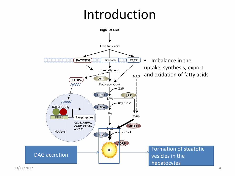
DAG accretion
• Imbalance in the uptake, synthesis, export and oxidation of fatty acids
Formation of steatotic vesicles in the hepatocytes
Problems
13/11/2012 5
Primary metabolic abnormalities leading to lipid accretion
Local lipid composition
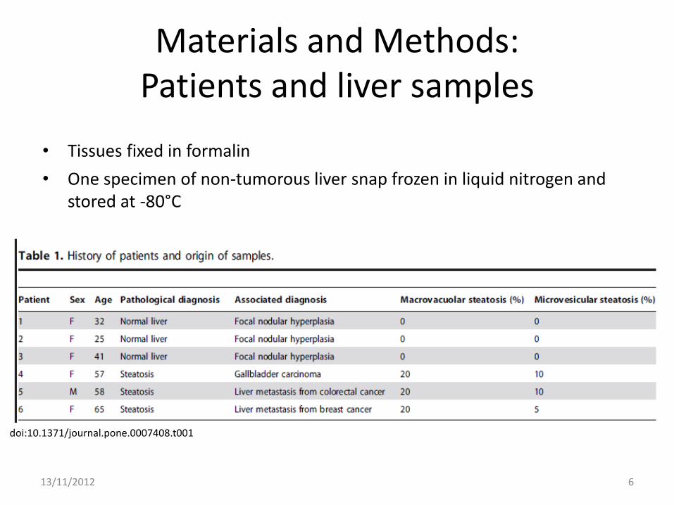
Materials and Methods: Patients and liver samples
6 13/11/2012
doi:10.1371/journal.pone.0007408.t001
• Tissues fixed in formalin
• One specimen of non-tumorous liver snap frozen in liquid nitrogen and stored at -80°C

Materials and Methods: Tissue section
7 13/11/2012
doi:10.1371/journal.pone.0007408.g001
PT: portal tract, BD: biliary duct, PV: portal vein, HA: hepatic artery, CLV: centrilobular vein, SV: steatotic vacuole.
• Normal hepatic lobule • fatty liver area : exhibiting macrovacuolar and microvesicular steatosis
X100
X400
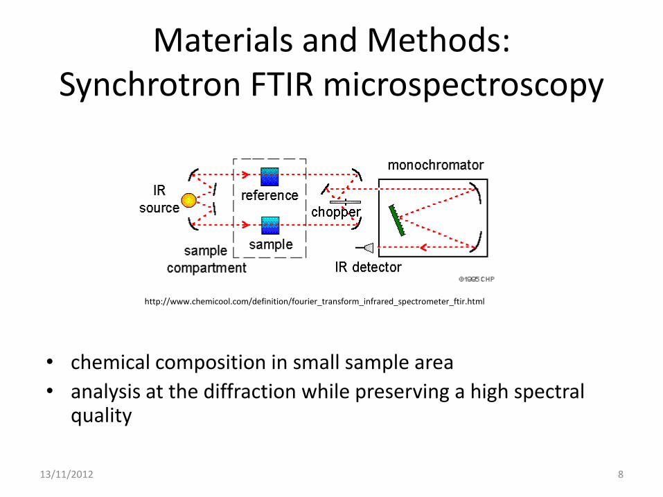
Materials and Methods: Synchrotron FTIR microspectroscopy
• chemical composition in small sample area
• analysis at the diffraction while preserving a high spectral quality
8 13/11/2012
http://www.chemicool.com/definition/fourier_transform_infrared_spectrometer_ftir.html
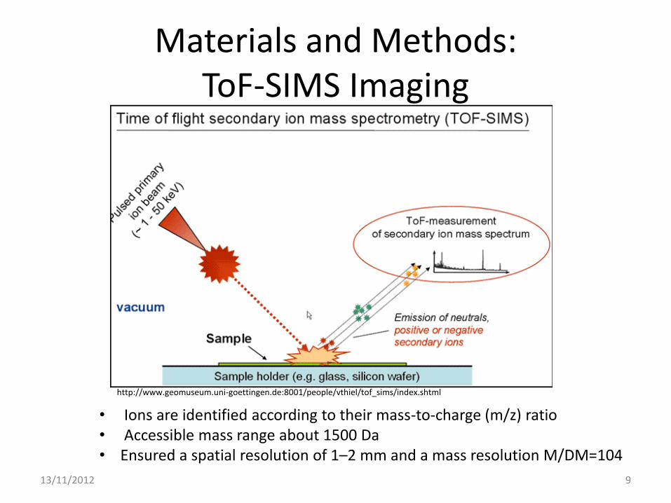
Materials and Methods: ToF-SIMS Imaging
9 13/11/2012
• Ions are identified according to their mass-to-charge (m/z) ratio • Accessible mass range about 1500 Da • Ensured a spatial resolution of 1–2 mm and a mass resolution M/DM=104
http://www.geomuseum.uni-goettingen.de:8001/people/vthiel/tof_sims/index.shtml
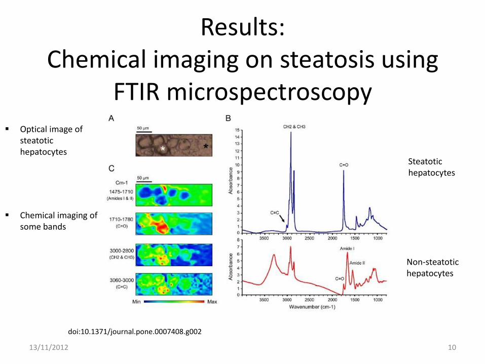
Results: Chemical imaging on steatosis using
FTIR microspectroscopy
10 13/11/2012
doi:10.1371/journal.pone.0007408.g002
Optical image of steatotic hepatocytes
Steatotic hepatocytes
Chemical imaging of some bands
Non-steatotic hepatocytes
11 13/11/2012
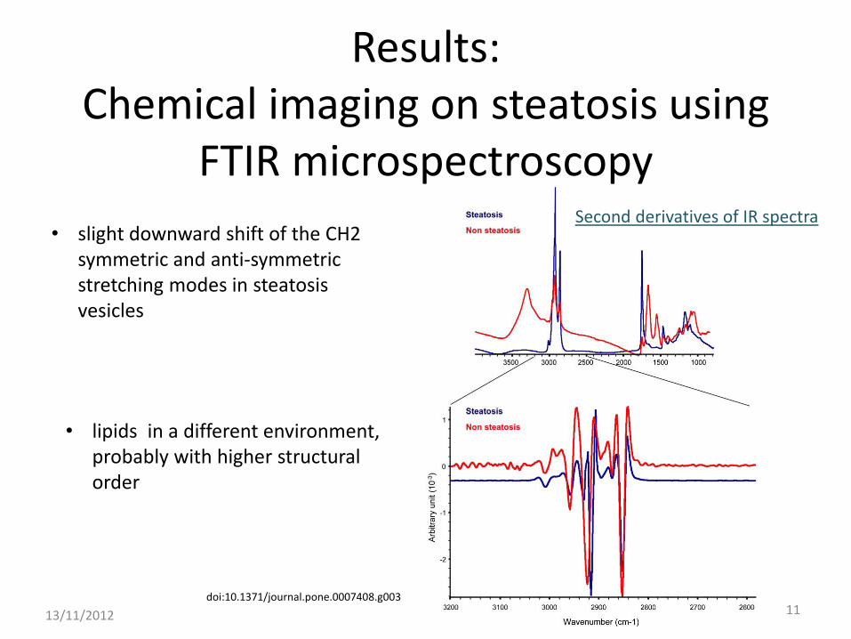
doi:10.1371/journal.pone.0007408.g003
Second derivatives of IR spectra • slight downward shift of the CH2
symmetric and anti-symmetric stretching modes in steatosis vesicles
• lipids in a different environment, probably with higher structural order
Results: Chemical imaging on steatosis using
FTIR microspectroscopy
12 13/11/2012
doi:10.1371/journal.pone.0007408.g006
doi:10.1371/journal.pone.0007408.g007
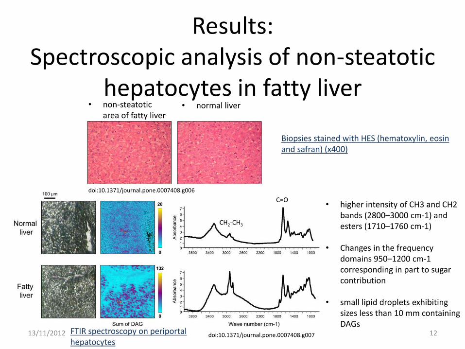
• normal liver • non-steatotic area of fatty liver
Biopsies stained with HES (hematoxylin, eosin and safran) (x400)
FTIR spectroscopy on periportal hepatocytes
• higher intensity of CH3 and CH2 bands (2800–3000 cm-1) and esters (1710–1760 cm-1)
• Changes in the frequency domains 950–1200 cm-1 corresponding in part to sugar contribution
• small lipid droplets exhibiting sizes less than 10 mm containing DAGs
C=O
CH2-CH3
Results: Spectroscopic analysis of non-steatotic
hepatocytes in fatty liver
Results: Chemical imaging on steatosis using ToF-SIMS
mass spectrometry
13 13/11/2012
doi:10.1371/journal.pone.0007408.g004
doi:10.1371/journal.pone.0007408.g005
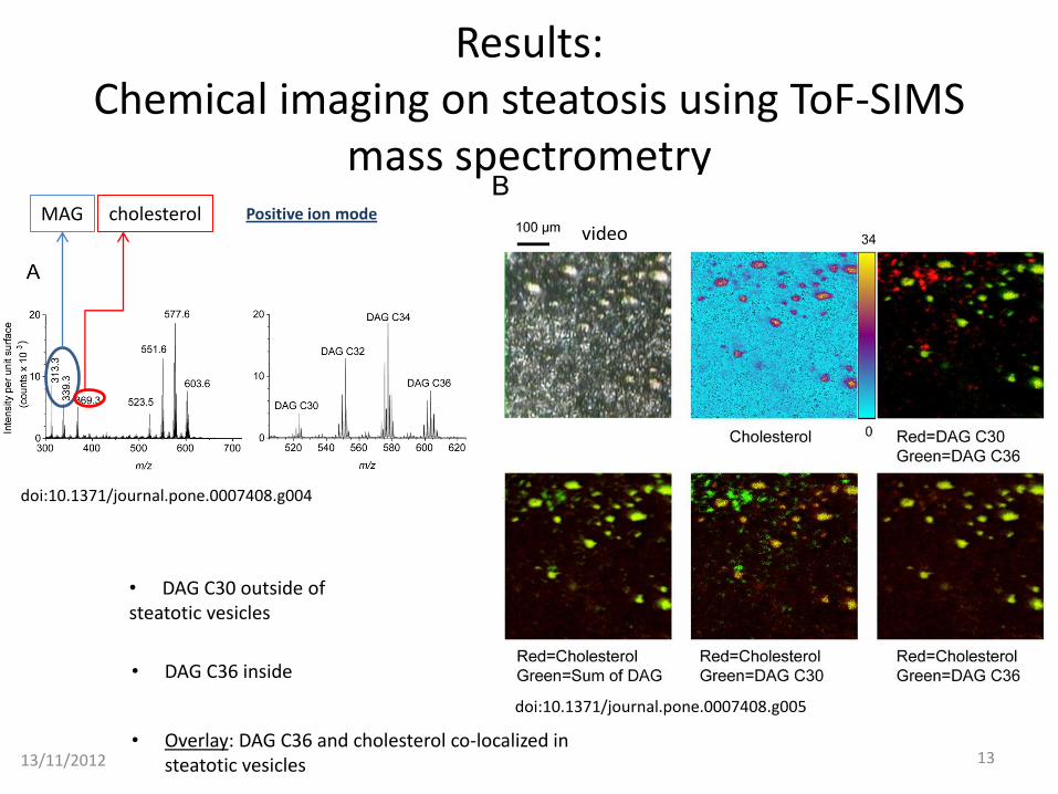
Positive ion mode video
• DAG C36 inside
• Overlay: DAG C36 and cholesterol co-localized in steatotic vesicles
MAG cholesterol
• DAG C30 outside of steatotic vesicles
14 13/11/2012
doi:10.1371/journal.pone.0007408.g005
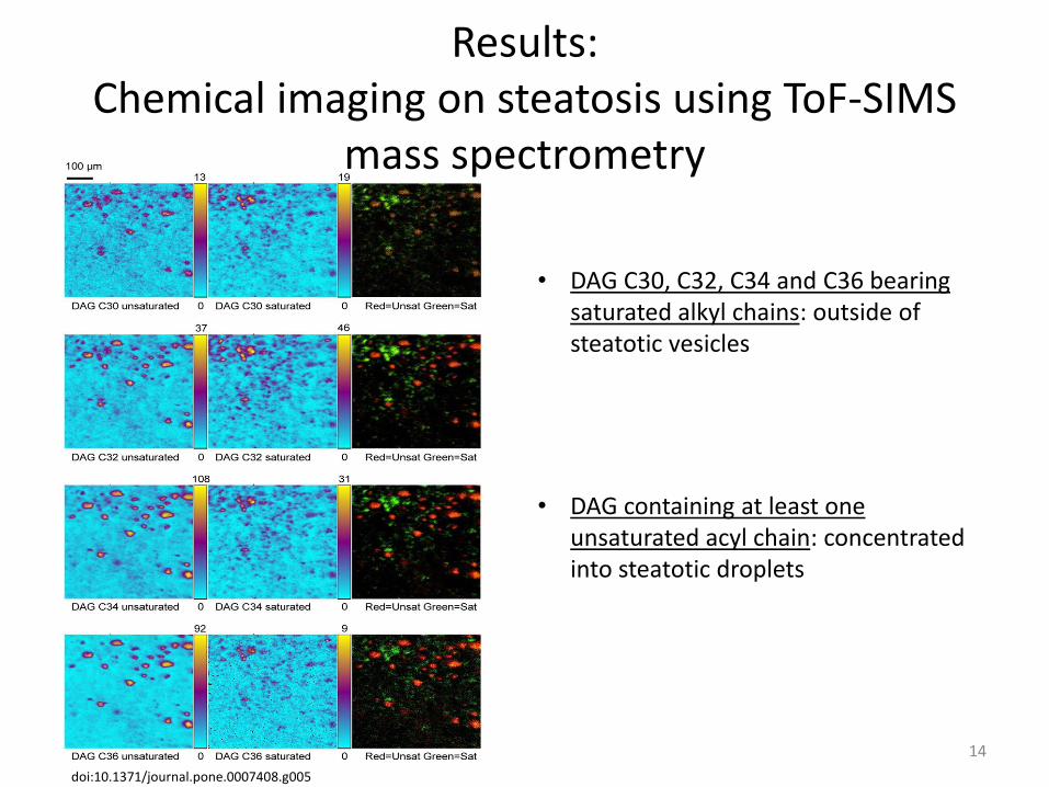
• DAG C30, C32, C34 and C36 bearing saturated alkyl chains: outside of steatotic vesicles
• DAG containing at least one unsaturated acyl chain: concentrated into steatotic droplets
Results: Chemical imaging on steatosis using ToF-SIMS
mass spectrometry
Discussion
• synchrotron source to study the complete frequency range from 900 to 4000 cm-1 (lipids)
• ToF-SIMS for investigating the local composition and distribution of the molecular species of lipids
• investigation at cellular and subcellular levels
• Concentration of unsaturated lipids inside steatotic vesicles may constitute a potential highly reactive site for peroxidation
15 13/11/2012
Perspectives
• infrared spectroscopy might be used as a diagnosis mean especially in the setting of liver transplantation
• spatial resolution and sensitivity of synchrotron FTIR microspectroscopy and mass spectrometry may open new avenue for characterizing early events in pathologies or for identifying markers for diagnosis and prognosis
• FTIR microspectroscopy using conventional infrared source might be set up in hospitals for clinical use
16 13/11/2012