chikv zikv - iris paho home
TRANSCRIPT

a
TOOL FOR THE DIAGNOSIS AND CARE OF PATIENTS WITH SUSPECTED
ARBOVIRAL DISEASES
DENV
CHIKV ZIKV
40 c

b

i
Pan American Health OrganizationPan American Sanitary Bureau
Regional Office of the World Health OrganizationWashington, D.C., 2017
TOOL FOR THE DIAGNOSIS AND CARE OF PATIENTS WITH SUSPECTED
ARBOVIRAL DISEASES
DENV
CHIKV ZIKV
40 c

ii
Original version in SpanishInstrumento para el diagnóstico y la atención a pacientes con sospecha de arborisosis
ISBN 978-92-75-31936-9
PAHO HQ Library Cataloguing-in-Publication Data
Pan American Health Organization
Tool for the diagnosis and care of patients with suspected arboviral diseases. Washington, D.C. : PAHO; 2017.
1. Arbovirus Infections. 2. Patient Care. 3. Zika Virus. 4. Chikungunya Virus. 5. Dengue.
ISBN 978-92-75-11936-5 (NLM Classification: WC 524)
© Pan American Health Organization 2017
All rights reserved. Publications of the Pan American Health Organization are available on the PAHO website (www.paho.org). Requests for permission to reproduce or translate PAHO Publications should be addressed to the Communications Department through the PAHO website (www.paho.org/permissions).
Publications of the Pan American Health Organization enjoy copyright protection in accordance with the provisions of Protocol 2 of the Universal Copyright Convention. All rights are reserved.
The designations employed and the presentation of the material in this publication do not imply the expression of any opinion whatsoever on the part of the Secretariat of the Pan American Health Organization concerning the status of any country, territory, city or area or of its authorities, or concerning the delimitation of its frontiers or boundaries.
The mention of specific companies or of certain manufacturers’ products does not imply that they are endorsed or recommended by the Pan American Health Organization in preference to others of a similar nature that are not mentioned. Errors and omissions excepted, the names of proprietary products are distinguished by initial capital letters.
All reasonable precautions have been taken by the Pan American Health Organization to verify the information contained in this publication. However, the published material is being distributed without warranty of any kind, either expressed or implied. The responsibility for the interpretation and use of the material lies with the reader. In no event shall the Pan American Health Organization be liable for damages arising from its use.

iii
CONTENTS
Foreword ................................................................................................ vMethodology ........................................................................................ viiAcknowledgments ................................................................................ ixAbbreviations and acronyms ................................................................ xiIntroduction ............................................................................................ 11. Clinical description of dengue, chikungunya,
and Zika virus infections ................................................................. 31.1. Dengue ..................................................................................... 31.2. Chikungunya ............................................................................ 71.3. Zika .........................................................................................11
2. Signs and symptoms of DENV, CHIKV, and ZIKV infection ....................................................................... 15
3. Suspected DENV, CHIKV, or ZIKV infection ............................. 175. Clinical diagnosis of patients with suspected
arboviral disease ............................................................................ 216. Clinical care for patients ............................................................... 23
6.1. Clinical care for suspected dengue virus infection ................ 246.2. Clinical care for suspected chikungunya virus infection ....... 346.3. Clinical care for suspected Zika virus infection .................... 40
7. Recommendations for laboratory diagnosis of DENV, CHIKV, and ZIKV infection ........................................ 437.1. Algorithm for the detection of DENV, CHIKV, or ZIKV ..... 447.2. Sample collection and shipment ............................................ 457.3. Observations and additional recommendations ..................... 48

iv
8. Other arboviral diseases with epidemic potential ......................... 518.1 West Nile Virus ...................................................................... 518.2 Yellow Fever .......................................................................... 558.3 Oropouche Fever .................................................................... 588.4 Mayaro Fever ......................................................................... 618.5 The Equine Encephalitides ................................................... 63
9 References ..................................................................................... 6710 Annexes .......................................................................................... 77
Annex 1. Analgesics: dosages in children and adults and administration to pregnant women ....................... 77
Annex 2: WHO analgesic ladder ................................................. 82Annex 3. Scientific literature search strategy.............................. 83
v
Dengue is the most common mosquito-borne viral disease in the Americas and the most suspected in patients with fever. However, the recent introduction of two new arboviral diseases (chikungunya virus in late 2013 and Zika virus in 2014) has created a new challenge for public health in the Americas. The three arboviral diseases (dengue, chikungunya, and Zika) can produce very similar clinical symptoms, mainly during the acute phase (the first days of the disease), hindering clinical diagnosis by health workers, creating problems for appropriate case management, and sometimes triggering fatal events. Serological diagnosis has presented further difficulties, due to the cross-reaction between IgM/IgG antibodies of the dengue and Zika viruses, complicating laboratory confirmation and compromising epidemiological surveillance.
This new and complex panorama makes it essential to provide health workers with methods for clinical diagnosis of patients suspected of dengue, chikungunya, or Zika infection, particularly at the primary care level. For this reason, in January 2016 the Pan American Health Organization/World Health Organization (PAHO/WHO) organized a meeting with a group of clinical experts from the Americas responsible for the diagnosis and management of these diseases. After the meeting, a document was prepared on the basis of experiences in the Americas, supported by the best available scientific evidence, in order to serve as tool to support health workers in the clinical diagnosis of dengue, chikungunya, and Zika cases. The end result is this tool for the diagnosis and care of patients suspected of arboviral diseases (dengue, chikungunya, and Zika), which offers valuable information on the
FOREWORD

vi
clinical manifestations of the three diseases and facilitates differential diagnosis among them. This tool also contains recommendations for the proper management of each of the three diseases, as well as the necessary elements for laboratory confirmation of diagnosis, which will be a valuable contribution to epidemiological surveillance.
As part of current efforts to achieve an appropriate and comprehensive approach to infections caused by dengue, chikungunya, and Zika viruses, PAHO/WHO is pleased to offer the countries and territories of the Americas this first edition of the Tool for the Diagnosis and Care of Patients with Suspected Arboviral Diseases. This tool is intended to contribute to public health in the Americas by providing health workers with the clinical information necessary for the detection and timely management of the three diseases, with the main objective of saving patients’ lives.
Dr. Marcos A. EspinalDirector Communicable Diseases and Health Analysis

vii
This Tool for the Diagnosis and Care of Patients with Suspected Arboviral Diseases was prepared by a team of specialists, including clinical physicians in the prepared by a team of specialists, including clinical physicians in the Americas specialized in the care of patients with chikungunya and Zika virus infection; members of the International Technical Group of Experts on Dengue (GT-Dengue); and technical staff from the Pan American Health Organization/World Health Organization (PAHO/WHO).
In a meeting held at PAHO/WHO Headquarters in Washington, D.C., from 23 to 25 January 2016, participants reviewed the descriptions and clinical information on dengue, chikungunya, and Zika, as well as the documentation, clinical guidelines, and scientific tests available as of that date for each of the three diseases. They also looked at epidemiological, clinical, and laboratory aspects of these pathologies as they relate to clinical, epidemiological, and laboratory surveillance. The participants’ contributions and conclusions are reflected in the present document. For dengue, the material was mainly drawn from the second edition of Dengue guidelines for patient care in the Region of the Americas (11). The definition of a dengue case, the description of its clinical evolution, and guidelines on treating the disease have been adapted for specific risk groups. In addition, aspects related to differential diagnosis have been refined. For chikungunya, the main references were the PAHO/CDC document Preparedness and Response for Chikungunya Virus Introduction in the Americas (12) and an article entitled “French Guidelines for the Management of Chikungunya (Acute and Persistent
METHODOLOGY

viii
Presentations)” (13). Again, the clinical case definition and the information on clinical manifestations and treatment have been updated to take into account the phases of the disease.
Finally, the definition of a Zika case was initially based on the PAHO/WHO proposal for epidemiological surveillance published online as an epidemiological alert on 16 October 2015 (14) and subsequently modified to take into account evidence cited in scientific publications (15-19) and studies as yet unpublished of positive Zika cases conducted in Brazil and Colombia, as well as the experience of physicians who have been caring for patients with the infection. The section containing additional information on chikungunya and Zika was based on the experience of physicians in the Americas who see patients diagnosed with these arboviral diseases on a daily basis.
Including a chapter on other arboviral diseases with epidemic potential in the Americas was also considered appropriate. To prepare that chapter, a structure was followed similar to that of the preceding chapters (clinical symptoms and clinical, differential, and laboratory diagnosis). The chapter includes a brief abstract on each disease, as well as an additional bibliography to provide further information. The chapter’s contents are based on publications and peer reviews of the respective diseases.
Once the content of this proposed instrument for clinical diagnosis and patient care was agreed upon and adapted, PAHO/WHO technical staff proceeded to consolidate the information and edit the document. It was then distributed to the participants prior to the above-mentioned meeting for their review and final contributions.
This document is not based solely on scientific evidence. It is also an informative tool that reports and describes facts and evidence, and documents current practices and interventions. In the future it will be necessary to prepare evidence-informed treatment guidelines, in particular for the management of chronic arthritis and arthralgia associated with chikungunya and for the clinical response to Zika virus infection.

ix
PAHO/WHO wishes to thank the following professionals for their collaboration in the preparation and review of this document: Dr. Anabelle Alfaro (GT-Dengue, Costa Rican Social Security Fund); Dr. Kleber Luz (Federal University of Rio Grande do Norte, Brazil); Dr. Eric Martínez (GT-Dengue, Pedro Kouri Institute of Tropical Medicine); Dr. Sandra Ortegón (La Samaritana University Hospital, Colombia); Dr. Ernesto Pleités (GT-Dengue, National Institute of Health, Ministry of Health, El Salvador); Dr. J. Erin Staples (U.S. Centers for Disease Control and Prevention [CDC], Fort Collins, Colorado), and Sylvain Aldighieri, Liliana Benitez Own, Haroldo Bezerra, Luis Gerardo Castellanos, Leticia Franco, Gamaliel Gutiérrez, Mariana Leone, Jairo Méndez Rico, Roy Mendoza, Pillar Ramón-Pardo, José Luis San Martín, and Ignacio Postigo, all of PAHO/WHO.
Dr. Gamaliel Gutiérrez and Dr. Pilar Ramón-Pardo of PAHO/WHO were responsible for the editing and final review of the document with the assistance of Mr. Kem Ramírez of PAHO/WHO.
ACKNOWLEDGMENTS

x

xi
ALT alanine aminotransferase AST aspartate aminotransferase CDC Centers for Disease Control and Prevention CHIKV chikungunya virus CNS central nervous system COPD chronic obstructive pulmonary disease DEET N,N-diethyl-m-toluamide DENCO Dengue and Control Study (multicountry study) DENV dengue virus DNWS Dengue without warning signs DWWS Dengue with warning signs EEE Eastern Equine Encephalitis ELISA enzyme-linked immunosorbent assay GBS Guillain-Barré syndrome GT-Dengue International Technical Group of Experts on Dengue IgG Immunoglobulin G IgM Immunoglobulin M MF Mayaro fever mmHg millimeters of mercury NSAID Nonsteroidal anti-inflammatory drugs OF Oropouche fever PAHO Pan American Health Organization
ABBREVIATIONS AND ACRONYMS

xii
PO By mouth, orally (“per os”) PRNT Plaque reduction neutralization test RT-PCR reverse transcription-polymerase chain reaction SD Severe dengue VEE Venezuelan equine encephalitis WEE Western equine encephalomyelitis WHO World Health Organization WNF West Nile fever WNV West Nile virus YF Yellow fever ZIKV Zika virus

1
Diseases caused by arboviruses usually occur epidemically and are similar in their clinical expression. They constitute a syndrome that can be either febrile (e.g. dengue and chikungunya) or exanthematic (Zika). Other frequent symptoms are headache and body pain, including myalgia and manifestations in the joints. The latter may be arthralgia only (as in the case of dengue), arthritis (chikungunya), or both (Zika). There may also be edema in the limbs (chikungunya and Zika) and non-purulent conjunctivitis (Zika). It is important to keep in mind that any arboviral infection can be either asymptomatic or oligosymptomatic (60 to 80%) (1,2). Moreover, it can occur along with other infections, which makes differential diagnosis even more challenging (3-6).
In the case of dengue virus (DENV) infection, identifying the warning signs, which almost always occur during or after a decrease in fever, is helpful in making a clinical diagnosis and managing patient care. While the majority of cases of arboviral disease are self-limiting, sometimes they can manifest severe forms, such as shock, hemorrhage, or severe organ damage (in the case of dengue) or neurological complications (Zika), which can lead to death. Chikungunya virus (CHIKV) infection can also be clinically severe, particularly at the extreme ages of life. Chikungunya patients can develop post-acute or chronic arthropathy lasting 21 to 90 days in acute cases, and 3 months to ≥2 years in chronic cases. These manifestations can be incapacitating. Furthermore, these three arboviral diseases can cause autoimmune disease affecting the central nervous system (CNS)—for example, Guillain-Barré syndrome (GBS) or encephalopathy—and visual damage due to optic neuritis.
INTRODUCTION
2
It is Zika virus (ZIKV) that most often causes these neurological effects it appears to be the only arbovirus than can cause congenital malformations such as microcephaly. In any case, more scientific tests are needed to establish the causal relationship between the virus and this malformation (7-10).
This document is a practical tool designed to help health workers improve clinical diagnosis and provide timely care for patients infected with the dengue, chikungunya, or Zika virus. It is intended mainly for health workers in primary care facilities where laboratory diagnosis of arboviruses is not always available. However, this guide may also be very useful in hospitals that provide second- and third-level care, as it describes the clinical manifestations of each of the three most important arboviral diseases currently found in the Region, the elements for differential diagnosis, and their clinical behavior.
Finally, this guide includes useful clinical information on other arboviruses with epidemic potential in the Americas, such as yellow fever, Mayaro fever, and equine encephalitis, among others. It is hoped that the main beneficiaries will be people who suffer from these diseases and who will receive better care from health workers.

3
Clinical description of dengue, chikungunya, and Zika virus infections
DEN
VCH
IKV
ZIKV
1.1 Dengue
Natural evolution of the disease and warning signs
Dengue is a single disease with varying clinical presentations and, often with an unpredictable evolution and outcome (11, 20). It is characterized by acute fever accompanied by two or more of the following manifestations: nausea, vomiting, rash, headache, retro-orbital pain, myalgia, arthralgia, petechiae, positive tourniquet test, and leukopenia (11). The clinical picture can evolve and present warning signs such as constant intense abdominal pain, persistent vomiting, fluid accumulation (ascites or pleural or pericardial effusion), mucosal bleeding, lethargy, irritability, postural hypotension, hepatomegaly (palpated at 2 cm below the costal margin), and progressive increase in hematocrit (11, 21). The disease can also progress to more severe forms, with plasma leakage (22), severe hemorrhage, or serious organ failure that can lead to death (11).
When fever suddenly falls, the dengue patient may either improve and recover from the disease or present clinical deterioration and serious warning signs (21). If the patient does not feel better at that point or no improvement is seen, it should be suspected that the disease is still progressing and about to become more severe. A study in Puerto Rico (23) looked at a series of deaths from confirmed cases of dengue and found that they had presented a set of known warning signs, including intense abdominal pain, persistent vomiting, sharp drop in fever, and altered state of consciousness. These signs should have alerted physicians to the severity of the disease. According to DENCO (multicountry dengue control study), intense abdominal pain, mucosal bleeding, and lethargy, which presented 24 hours before the severity of dengue was established, were clinical manifestations of high statistical significance (21).
CLINICAL DESCRIPTION OF DENGUE, CHIKUNGUNYA, AND ZIKA VIRUS INFECTIONS 1
DEN
VCH
IKV
ZIKV
4
Tool for the diagnosis and care of patients with suspected arboviral diseases
DEN
VCH
IKV
ZIKV
The majority of the warning signs, described in the following paragraphs, are the result increased capillary permeability, which means that they mark the beginning of the critical phase.
- Intense and continuous abdominal pain or tenderness. Intense and continuous abdominal pain is an indication that the patient’s condition may progress or is already progressing to the dengue shock stage and its severe complications.
Abdominal pain with the characteristics described above is not due to the sudden appearance of hepatomegaly during the critical phase of dengue or to presumed erosions in the gastric mucosa as seen in Cuba in studies during the first epidemic of dengue hemorrhagic fever in the Region of the Americas in 1981 (24). The new hypothesis is that the intense epigastric pain is actually referred pain from the sudden presence of large amounts of fluid extravasating toward the pararenal and perirenal areas, which irritates the nerve plexuses in the retroperitoneal region (25). Abdominal ultrasound studies conducted in Indonesian children (26) with dengue shock showed that 77% of them had perirenal and pararenal fluid “masses,” which did not appear in the children without shock. This finding points to a clear association between fluid accumulation in the retroperitoneal region and dengue shock. It also indicates the speed at which large volumes of fluid can accumulate in that region. Though intense, the pain is transitory. In isolated cases, abdominal pain can coincide with hepatitis, enteritis, or pancreatitis, conditions that have been seen in some dengue patients and have generated proposals to account for the symptom (27). In such cases, however, the abdominal pain is not associated with plasma leakage, which means that these explanations should not be accepted to account for the warning sign.
Furthermore, it has been shown that thickening of the gallbladder wall is produced by sudden plasma leakage in sufficient volume to produce pain in the right hypochondrium without other signs of inflammation, and that therefore this thickening is a warning sign. Some have interpreted it erroneously as a case of cholecystitis without gallstones (27), but when the gallbladder was removed in these cases, no inflammatory cell infiltrate was found in its wall, only pure fluid in the form of edema (22, 27). Leakage is
DEN
VCH
IKV
ZIKV

5
Clinical description of dengue, chikungunya, and Zika virus infections
DEN
VCH
IKV
ZIKV
also found in the intestinal wall, forming edema and suddenly expanding the intestinal volume with fluid accumulating under the serous layer, as is often seen in autopsies of patients who died from dengue, causing abdominal pain in any location. This pain becomes so intense that it can resemble the symptoms of acute abdomen (cholecystitis, cholelithiasis, appendicitis, ectopic pregnancy, intestinal infarction) (28, 29).
- Persistent vomiting, defined as three or more episodes in 1 hour or four episodes in 6 hours. It prevents adequate oral rehydration and contributes to hypovolemia. Persistent vomiting has been recognized as a clinical sign of severity (11, 30).
- Fluid accumulation, which tends be manifested as pleural effusion, ascites, or pericardial effusion. It is detected by clinical methods, radiology, or ultrasound and is not necessarily associated with respiratory distress or hemodynamic compromise. Should the last-mentioned condition be present, the patient’s case should be classified as severe dengue (11, 31).
- Active mucosal bleeding, mainly in the gums and nose but also transvaginal (metrorrhagia and hypermenorrhea) or in the digestive system (blood-streaked vomit) or kidneys (macroscopic hematuria). Mucosal bleeding with accompanying hemodynamic changes is considered a warning sign of severe dengue (11, 32).
- Change in mental state. Irritability (restlessness) or drowsiness (lethargy) may occur, with Glasgow Coma Scale score of less than 15. It is accepted that both these manifestations are the expression of cerebral hypoxia induced by hypovolemia resulting from plasma leakage (11).
- Hepatomegaly is defined as palpation of the liver at more than 2 cm below the costal margin. It can be the result of an increase in the size of the organ itself (due to a combination of congestion, intrahepatic hemorrhage, and fatty metamorphosis) or a shift in the position of the liver due to pleural effusion and the accumulation of other fluid (ascites) in the intraperitoneal or retroperitoneal cavity (24). This has been a significant risk factor for shock in children with dengue (11, 31).
DEN
VCH
IKV
ZIKV
6
Tool for the diagnosis and care of patients with suspected arboviral diseases
DEN
VCH
IKV
ZIKV
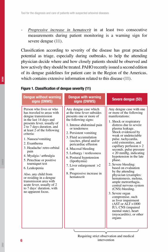
- Progressive increase in hematocrit in at least two consecutive measurements during patient monitoring is a warning sign for severe dengue (11).
Classification according to severity of the disease has great practical potential as triage, especially during outbreaks, to help the attending physician decide where and how closely patients should be observed and how actively they should be treated. PAHO recently issued a second edition of its dengue guidelines for patient care in the Region of the Americas, which contains extensive information related to this disease (11).
Figure 1. Classification of dengue severity (11)
Person who lives or who has traveled to areas with dengue transmission in the last 14 days and presents fever, usually of 2 to 7 days duration, and at least 2 of the following criteria:1. Nausea/vomiting2. Exanthema3. Headache/ retro-orbital
pain4. Myalgia / arthralgia5. Petechiae or positive
tourniquet test6. Leukopenia
Also, any child from or residing in a dengue transmission area with acute fever, usually of 2 to 7 days’ duration, with no apparent focus.
Dengue without warning signs (DNWS)
Any dengue case which at the time fever subsides presents one or more of the following signs:1. Intense abdominal pain
or tenderness2. Persistent vomiting3. Fluid accumulation
(ascites, plural and/or pericardiac effusion
4. Mucosal bleeding5. Lethargy / restlessness6. Postural hypotension
(lipothymia) 7. Liver enlargement >2
cm8. Progressive increase in
hematocrit
Dengue with warning signs (DWWS)
Any dengue case with one or more of the following manifestations:1. Shock or respiratory
distress due to severe plasma leakage. Shock evidenced by weak or undetectable pulse, tachycardia, cold extremities, and capillary perfusion > 2 seconds, pulse pressure < 20 mmHg, indicating hypotension in the late phase.
2. Severe bleeding: based on evaluation by the attending physician (examples: hematemesis, melena, ample metrorrhagia, central nervous system (CNS) bleeding.
3. Severe organ compromise, such as liver impairment (AST or ALT >1000 IU), CNS (impaired mental state), heart (myocarditis), or other organs
Severe dengue (SD)
Requiring strict observation and medical intervention
DEN
VCH
IKV
ZIKV

7
Clinical description of dengue, chikungunya, and Zika virus infections
DEN
VCH
IKV
ZIKV
1.2 Chikungunya
Manifestations in the acute, post-acute, and chronic phases
Below is a summary of the clinical and osteoarticular manifestations that can be observed in cases of acute chikungunya. After a silent incubation period averaging 4 to 7 days (minimum 1 day; maximum 12 days), the acute phase usually lasts from 5 to 10 days (12, 13).
- Joint manifestations: arthritis and inflammatory arthralgias that can become intense and produce functional disability, mainly affecting the limbs (wrists, ankles, and phalanges). The symptoms can last for two to three weeks. Joint manifestations are the cardinal signs and symptoms of the disease.
- Cutaneous manifestations: eruptions appearing between days 2 and 5 after onset of fever, which can affect the face, chest, abdomen, limbs, palms of the hands, and soles of the feet (33). Presenting as erythema, morbilliform maculopapular rash, or bullous rash, after several days they may progress to hyperpigmented stains, nodular erythema, multiform erythema, generalized urticaria, reactivation of lichen planus, activation of preexisting psoriasis, widespread pruritus, peeling of the skin (Ritter syndrome in newborns or Lyell syndrome in older children), or necrosis of the nose or phalanges (34).
- Cardiovascular manifestations: heart failure, arrhythmia, myocarditis or pericarditis, angina pectoris, acute myocardial infarction, and sudden death (35-37).
- Neurological manifestations: encephalitis, stroke, convulsions, meningoencephalitis, Guillain-Barré syndrome (following the acute phase of the infection), neuropathy, myelitis, altered mental state, neurological deficit, myeloneuropathy, and acute flaccid paralysis (34).
- Ocular manifestations: impaired vision linked to neuroretinitis, chorioretinitis, optic neuritis, or uveitis.
- Renal manifestations: prerenal acute renal failure or exacerbation of a preexisting renal disorder.
DEN
VCH
IKV
ZIKV

8
Tool for the diagnosis and care of patients with suspected arboviral diseases
DEN
VCH
IKV
ZIKV
- Pulmonary manifestations: pneumonia and pneumonitis (34, 37).
- Metabolic manifestations: hyperglycemia and syndrome of inappro-priate antidiuretic hormone (ADH) secretion.
- Hepatic manifestations: elevated transaminases and hepatitis (37).
- Hemorrhagic manifestations: though not as common as in the dengue, they can be present in the form of epistaxis, bleeding gums, positive tourniquet test, subconjunctival bleeding, and rash (37).
- Transitory vascular disorders (for example, Raynaud’s syndrome) have been reported in association with mixed cryoglobulinemia.
The post-acute phase (from week 4 to the end of month 3) can be characterized by the presence of multiform and other associated lesions characterized by the persistence of initial inflammatory manifestations that slowly recede, in particular: inflammatory arthralgia, arthritis, synovitis with or without effusion, tenosynovitis, or bursitis. The evolution is often ongoing with inflammatory outbreaks induced by cold temperatures, but there may also be intermittent periods without symptoms. There may often be intense asthenia in post-acute phase, as well as neuropsychological changes, especially when the pain is intense (13).
The chronic phase (starting in the fourth month) can last from a few months to several years. The manifestations are the same as those described in the post-acute phase, often with painful resurgence in the most commonly used joints because of their inflammatory state after the infection. In short, the evolution may lead to a cure without sequelae, either spontaneous or after treatment, or the joint and other symptoms may persist for a long time and even get worse, leading to an inflammatory or degenerative process. Most chronic patients see a worsening of their quality of life during the first years after a chikungunya infection (13).
DEN
VCH
IKV
ZIKV
DEN
VZI
KV

9
Clinical description of dengue, chikungunya, and Zika virus infections
DEN
VCH
IKV
ZIKV
Special considerations: cases in children under 1 year old, persons over 65, and those with chronic disease
The clinical manifestations associated with chikungunya are more frequent in patients with certain underlying diseases–for example: diabetes mellitus, cardiomyopathy, heart disease, cardiac arrhythmia, heart valve disease, stroke, epilepsy, hypertension, chronic obstructive pulmonary disease, and bronchial asthma.
Children under 1 year of age, persons over 65, people with underlying disease, and pregnant women are all at greater risk for complications. They must be monitored by the clinician and, if deemed necessary, hospitalized. Severe organ dysfunction has been seen in a few cases, which can lead to death (37).
Clinical diagnosis in children under 1 year of age is particularly challenging. Still, by matching up the clinical signs and the epidemiological context, it is possible to approximate a diagnosis based on certain manifestations. Affected babies are cranky and do not move around very much; on the contrary, when they are moved they become more irritable and resume their original position. This sign has been described as “the elastic baby.” Patients at this age also present periarticular swelling and, less often, skin lesions like rash, maculopapular or morbilliform erythema, bullous erythema, or bullous dermatitis (38).
As for pregnant women infected with CHIKV, the literature contains reports of miscarriages and in utero fetal death (without malformations) prior to week 22 of gestation. For women giving birth during the viremia period (febrile mother with viremia demonstrated during delivery), the number of cesarean sections is increased due to acute fetal stress with abnormal fetal heart rate, and in some cases amniotic fluid with meconium has been demonstrated (39).
Chikungunya infection is uncommon in infants, but when it is present, a clear clinical picture of CHIKV infection emerges shortly after birth, with signs appearing after 3 to 7 days. In such situations, all children with symptoms are being tested for presence of the virus, for example,
DEN
VCH
IKV
ZIKV
DEN
VZI
KV

10
Tool for the diagnosis and care of patients with suspected arboviral diseases
DEN
VCH
IKV
ZIKV
using reverse transcription-polymerase chain reaction (RT-PCR). Studies during a recent outbreak on Reunion Island showed a vertical transmission rate of 48.5% (40-42), prompting an active search for the virus in the placenta. The most common signs of infection by among neonates are fever (79%), rash (82%), and peripheral edema (58%). Complications may also develop as a result of convulsions, hemorrhagic manifestations, or hemodynamic instability. Echocardiography has revealed abnormalities in 42% of cases, including heart attack, hypertrophy, ventricular dysfunction, pericarditis, and expansion of the coronary artery. Magnetic resonance imaging has revealed the presence of parenchymatous hemorrhage and lesions in the white matter of the brain. There has also been an increased incidence of aspiration of amniotic fluid with meconium.
DEN
VZI
KV

11
Clinical description of dengue, chikungunya, and Zika virus infections
DEN
VCH
IKV
ZIKV
1.3 Zika
The description that follows is intended to be a pedagogical tool for dealing with ZIKV infection.
Like any infectious disease, Zika depends on the epidemiological context, the circulating viral strains or genotypes, and host factors such as age, sex, and the presence of concomitant conditions. The signs and symptoms of ZIKV infection can arise simultaneously or progressively. For example, some patients may present a number of signs and symptoms which together will be sufficient to arrive at a clinical diagnosis, while others my present only limited signs and symptoms, making the diagnosis more difficult. In the absence of a practical, rapid, and reliable laboratory test for the definitive diagnosis of ZIKV infection, familiarity with the clinical description provided below will be essential in guiding health workers to decide on appropriate care and in enabling the epidemiological surveillance team to properly report the cases.
Incubation and onset of clinical manifestations
The average incubation period is estimated to last 2 to 7 days. Onset is sudden, with the appearance of a maculopapular rash, with or without a low-grade fever (< 38.5° C). Fever is often absent. The rash develops in a cephalocaudal pattern (starting at the head and moving to the trunk, arms, and legs). The itching is so severe that it often interferes with the patient’s daily activities and even sleep. It often affects the palms of the hands and soles of the feet, where palmar or plantar hyperemia can develop. Laminar desquamation may be observed in the hands and feet during the convalescent phase. Whereas in dengue the rash and itching typically develop after day 5 or 6 of the disease and mark its end, the ZIKV rash is often accompanied by nonpurulent conjunctival hyperemia. Adenopathy and glandular enlargement are almost never seen, but the retroauricular nodes can be affected (18, 19).
Involvement of joints and other systems
The joints are involved in more than half the cases, typically in the form of polyarthralgia with periarticular, bilateral, and symmetrical edema.
DEN
VCH
IKV

12
Tool for the diagnosis and care of patients with suspected arboviral diseases
DEN
VCH
IKV
ZIKV
Compared with chikungunya, the pain for Zika patients tends to be less intense and incapacitating. Physical examination will reveal mild edema in the area of the joints, but without hyperemia or local warmth. These joint symptoms start to regress about a week after onset, but in a few cases they can last up to 30 days in a relapsing pattern. The joints in the hands and wrist are affected most often, followed by the knees and the ankles. Unlike CHIKV infection, no cases of chronic joint symptoms have been observed (15, 18).
Other manifestations may also be present, such as headache, myalgia, nausea, and vomiting. No hemodynamic changes have been observed in ZIKV infections such as those seen in the severe forms of dengue.
Nervous system involvement
Though infrequent, encephalitis, meningoencephalitis, encephalopathy, or myelitis can occur in the acute phase. In general, the prognosis is good, but in some cases the clinical symptoms can be severe.
Guillain-Barré syndrome (GBS) following ZIKV infection was observed in French Polynesia (43, 44), where a time relationship was established between the two, followed by biological confirmation. Cases of GBS caused by ZIKV were also observed in Brazil in the city of Recife, the virus being detected by RT-PCR in the cerebrospinal fluid of eight patients with neurological manifestations. Other neurological manifestations associated with ZIKV infection are optic neuritis, Miller-Fisher syndrome, acute disseminated encephalomyelitis, transverse myelitis, cerebellitis, and paralysis of the VI cranial pair. The time lapse between the clinical manifestations of ZIKV infection and the development of neurological symptoms ranged between 4 and 19 days. Since neurological symptoms can appear either during the acute phase of the infection or afterwards, it is essential to keep in mind all the signs and symptoms that might be related to ZIKV infection. It is also important to remember that other infectious agents can cause manifestations related to the nervous system, as well other body systems. All relevant details in the patient’s history (for example, diarrheal symptoms, past vaccinations, etc.) should be investigated.
DEN
VCH
IKV
13
Clinical description of dengue, chikungunya, and Zika virus infections
DEN
VCH
IKV
ZIKV
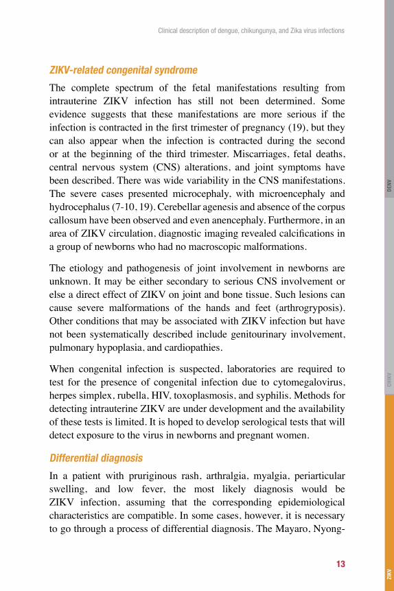
ZIKV-related congenital syndrome
The complete spectrum of the fetal manifestations resulting from intrauterine ZIKV infection has still not been determined. Some evidence suggests that these manifestations are more serious if the infection is contracted in the first trimester of pregnancy (19), but they can also appear when the infection is contracted during the second or at the beginning of the third trimester. Miscarriages, fetal deaths, central nervous system (CNS) alterations, and joint symptoms have been described. There was wide variability in the CNS manifestations. The severe cases presented microcephaly, with microencephaly and hydrocephalus (7-10, 19). Cerebellar agenesis and absence of the corpus callosum have been observed and even anencephaly. Furthermore, in an area of ZIKV circulation, diagnostic imaging revealed calcifications in a group of newborns who had no macroscopic malformations.
The etiology and pathogenesis of joint involvement in newborns are unknown. It may be either secondary to serious CNS involvement or else a direct effect of ZIKV on joint and bone tissue. Such lesions can cause severe malformations of the hands and feet (arthrogryposis). Other conditions that may be associated with ZIKV infection but have not been systematically described include genitourinary involvement, pulmonary hypoplasia, and cardiopathies.
When congenital infection is suspected, laboratories are required to test for the presence of congenital infection due to cytomegalovirus, herpes simplex, rubella, HIV, toxoplasmosis, and syphilis. Methods for detecting intrauterine ZIKV are under development and the availability of these tests is limited. It is hoped to develop serological tests that will detect exposure to the virus in newborns and pregnant women.
Differential diagnosis
In a patient with pruriginous rash, arthralgia, myalgia, periarticular swelling, and low fever, the most likely diagnosis would be ZIKV infection, assuming that the corresponding epidemiological characteristics are compatible. In some cases, however, it is necessary to go through a process of differential diagnosis. The Mayaro, Nyong-
DEN
VCH
IKV
14
Tool for the diagnosis and care of patients with suspected arboviral diseases
DEN
VCH
IKV
ZIKV
Nyong and other arboviruses that cause arthritis do not usually affect people living in urban areas, but it’s still important to be aware of changes in the epidemiological behavior of these agents.
Parvovirosis, characterized by a rash known as “slapped cheek syndrome,” is typically a disease of children. Measles and rubella can be ruled out with high degree of confidence in children and adults whose vaccinations are up to date, although the catarrhal symptoms in measles and the rather painful retroauricular lymph nodes in rubella can help to rule out these classical rash illnesses. Scarlet fever has a very characteristic sandpaper-type rash and the high fever abates and is followed by a rash similar to roseola infantum (exanthema subitum, sixth disease). An acute febrile condition accompanied by rash and monoarticular arthralgia suggests the possibility of early meningococcemia, even in the absence of petechiae.
Dermatitis associated with the consumption of certain drugs should be ruled out by taking a thorough clinical history that includes questions about the use of medicines on the days prior to onset of the current disease and considering the behavior of the disease in the community.
DEN
VCH
IKV

15
Clinical description of dengue, chikungunya, and Zika virus infections
DEN
VCH
IKV
ZIKV
The characteristic signs and symptoms of CHIKV, DENV, and ZIKV infections are summarized below.
Table 1. Signs and symptoms of dengue, chikungunya, and Zika arbovirus infections
SIGNS AND SYMPTOMS DENGUE CHIKUNGUNYA ZIKA
Most frequent reason for consultation Fever, myalgia Joint pain, fever Exanthema or pruritus
Fever ModerateVery frequent
Duration: 5 to 7 daysa
Very highVery frequent
Duration: 3 to 5 days
MildVery infrequent
Duration: 1 to 3 days
Rash Appears between days 5 and 7 Non-characteristic
Appears on day 2 or 3Non-characteristic
Typically from day 1:maculopapular, cephalocaudal
Pruritus Mild to intense Mild to moderate Moderate to intense
Conjunctivitis Infrequent Not very frequentb Very frequent
Neurological manifestations Infrequent Infrequent (can be frequent and serious in neonates)
Possible and serious
Headache Intense and frequent Mild to moderate Mild to moderate
Retro-ocular pain Intense and frequent Infrequent Infrequent
Poliartralgias Absent Very frequent Frecuent
Polyarthritis Absent Frecuent Frecuent
Edema in hands and feet Infrequent Frecuent Infrequent
Evolution to chronic form No Very frequent Not described
Myalgia Very frequent and intense FrequentModerate to intense
Infrequent
Hepatomegaly Warning sign Very infrequent Very infrequent
Frequent vomiting Warning sign Very infrequent Very infrequent
Diarrhea Frequent Very infrequent Very infrequent
Intense abdominal pain Warning sign Not present Not present
Skin bleeding Frequent Very infrequent Very infrequent
Mucosal bleeding Warning sign Very infrequent (when present, it is serious)
Very infrequent
Shock More frequent in the severe formc
Infrequent Unknown
Leukopenia Moderate to intense Mild to moderate Mild to moderate
C-reactive protein Normal Elevated Elevated
High hematocrit level Warning sign Infrequent Infrequent
Platelet count Normal to very low Normal to low Normal to low
Special considerations Risk of death Can evolve to chronic arthropathy Risk of congenital infection and GBS
a With dengue, a drop in fever between days 3 and 5 of the disease can actually be associated with onset of severity.b Conjunctivitis is uncommon in CHIKV infection, but this symptom is more frequent in children.c The onset of shock is sudden and it occurs most often between days 3 and 7 of the disease.
SIGNS AND SYMPTOMS OF DENV, CHIKV, AND ZIKV INFECTION 2

16
Tool for the diagnosis and care of patients with suspected arboviral diseases
DEN
VCH
IKV
ZIKV

17
Clinical description of dengue, chikungunya, and Zika virus infections
DEN
VCH
IKV
ZIKV
The following table summarizes the criteria for suspecting DENV, CHIKV, or ZIKV infection.
Table 2. Elements that justify suspicion of dengue, chikungunya, or Zika virus infections
DENGUE CHIKUNGUNYAa ZIKAa
Case
defi
nitio
n
The person lived or traveled in an area of dengue transmission in the last 14 days, has a sudden high fever typically of 2 to 7 days’ duration, and presents two or more of the following manifestations:
1. Nausea, vomiting
2. Exanthema
3. Myalgia, arthralgia
4. Headache, retro-orbital pain
5. Petechiae or positive tourniquet test
6. Leukopenia
7. Any warning sign
8. Any criterion of severe dengue
Acute phase: The person was in a chikungunya epidemic or endemic area during the two weeks prior to onset of the symptoms and presents a fever of >38.5 °C and (usually incapacitating) arthralgia or arthritis with intense incapacitating pain not explained by other health conditions.
Post-acute phase: The patient’s joint symptoms (arthritis, arthralgias, or articular edema) persist for more than 3 weeks up to the end of the third month. The disease has evolved consistently from the onset of symptoms or there have been no periods of intermittent symptoms.
Chronic phase (more than 3 months): The patient has had CHIKV disease and has presented joint manifestations such as pain, edema, and articular rigidity for more than three months after the acute phase. The patient may also have chronic arthritis due to CHIKV, which should be studied and confirmed. Arthritis due to another inflammatory etiology should be ruled out.
The person has been in a Zika epidemic or endemic area during the two weeks prior to onset of the symptoms and presents an acute (sudden onset) exanthema, which is almost always the reason for the consultation and the first sign of the disease. The exanthema tends to be pruriginous, maculopapular, and cephalocaudal, without any other explanation. Two or more of the following manifestations are present:
1. Fever, usually <38.5° C
2. Nonpurulent conjunctivitis or conjunctival hyperemia
3. Arthralgia
4. Myalgia
5. Periarticular swelling
Neurological manifestations, such as GBS, may occasionally be present.
Com
men
ts
Any child that has visited or resided in an area of dengue transmission and has an acute febrile condition usually lasting 2 to 7 days without any apparent etiology may also be considered a suspected case.
Acute cases: In addition to joint involvement some cases may be accompanied by neurological, cardiovascular, dermatological, ophthalmological, hepatic, renal, respiratory, and/or hematological manifestations, among others, or life-threatening dysfunction of at least one organ or system.
In the case of newborns with a congenital manifestation of the infection, such as microcephaly, the mother should be asked if she was in a Zika epidemic or endemic area during the first 3 months of her pregnancy.
a The first cases of autochthonous transmission will not have a travel background. Therefore, physicians should report suspected cases to their national authorities to arrange for clinical and laboratory confirmation of the symptoms.
SUSPECTED DENV, CHIKV, OR ZIKV INFECTION 3

18
Tool for the diagnosis and care of patients with suspected arboviral diseases
DEN
VCH
IKV
ZIKV

19
Clinical description of dengue, chikungunya, and Zika virus infections
DEN
VCH
IKV
ZIKV
The table below summarizes the differential diagnoses for DENV, CHIKV, and ZIKV infections and other disorders with similar signs and symptoms.
Table 3. Differential diagnosis of arboviral diseases: DENV, CHIKV, and ZIKV
Disorder Differential diagnosis
Influenza-like illness Influenza, measles, infectious mononucleosis, HIV primary infection
Diseases with skin rash Rubella, measles, scarlet fever, meningococcal infection, parvovirosis, toxicoderma, rickettsiosis, ehrlichiosis
Diarrheal diseases Rotavirus, other enteric infections
Diseases with neurological manifestations
Meningoencephalitis, febrile seizures
Hemorrhagic fevers Leptospirosis, Brazilian hemorrhagic fever, Argentine hemorrhagic fever, Bolivian hemorrhagic fever, etc.
Other infections Acute gastroenteritis, malaria, leptospirosis, typhoid fever, typhus, viral hepatitis, severe sepsis, septic shock, hantavirus infection, visceral leishmaniasis, yellow fever
Malignant neoplasms Leukemia, lymphoma, and other neoplasms
Other clinical symptoms Acute abdomen (appendicitis, cholecystitis), diabetic ketoacidosis, lactic acidosis, leukopenia and thrombocytopenia with or without bleeding, platelet disorders (purpura), renal damage, respiratory distress, metabolic acidosis as a cause of Kussmaul respiration, systemic lupus erythematosus, hemolytic anemia
DIFFERENTIAL DIAGNOSIS OF ARBOVIRAL DISEASES 4

20
Tool for the diagnosis and care of patients with suspected arboviral diseases
DEN
VCH
IKV
ZIKV
21
Clinical description of dengue, chikungunya, and Zika virus infections
DEN
VCH
IKV
ZIKV
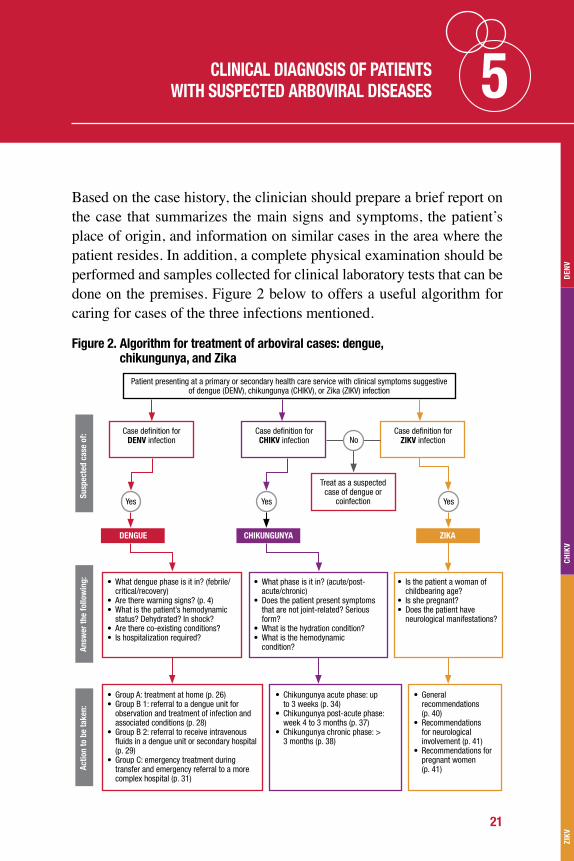
Based on the case history, the clinician should prepare a brief report on the case that summarizes the main signs and symptoms, the patient’s place of origin, and information on similar cases in the area where the patient resides. In addition, a complete physical examination should be performed and samples collected for clinical laboratory tests that can be done on the premises. Figure 2 below to offers a useful algorithm for caring for cases of the three infections mentioned.
Figure 2. Algorithm for treatment of arboviral cases: dengue, chikungunya, and Zika
Yes Yes Yes
Patient presenting at a primary or secondary health care service with clinical symptoms suggestive of dengue (DENV), chikungunya (CHIKV), or Zika (ZIKV) infection
Case definition for DENV infection
Susp
ecte
d ca
se o
f: An
swer
the
follo
win
g:
Actio
n to
be
take
n:
Case definition for CHIKV infection
Treat as a suspected case of dengue or
coinfection
• What dengue phase is it in? (febrile/critical/recovery)
• Are there warning signs? (p. 4)• What is the patient’s hemodynamic
status? Dehydrated? In shock? • Are there co-existing conditions? • Is hospitalization required?
• Group A: treatment at home (p. 26)• Group B 1: referral to a dengue unit for
observation and treatment of infection and associated conditions (p. 28)
• Group B 2: referral to receive intravenous fluids in a dengue unit or secondary hospital (p. 29)
• Group C: emergency treatment during transfer and emergency referral to a more complex hospital (p. 31)
• What phase is it in? (acute/post-acute/chronic)
• Does the patient present symptoms that are not joint-related? Serious form?
• What is the hydration condition? • What is the hemodynamic
condition?
• Chikungunya acute phase: up to 3 weeks (p. 34)
• Chikungunya post-acute phase: week 4 to 3 months (p. 37)
• Chikungunya chronic phase: > 3 months (p. 38)
• Is the patient a woman of childbearing age?
• Is she pregnant? • Does the patient have
neurological manifestations?
• General recommendations (p. 40)
• Recommendations for neurological involvement (p. 41)
• Recommendations for pregnant women (p. 41)
Case definition for ZIKV infectionNo
DENGUE CHIKUNGUNYA ZIKA
CLINICAL DIAGNOSIS OF PATIENTS WITH SUSPECTED ARBOVIRAL DISEASES 5

22
Tool for the diagnosis and care of patients with suspected arboviral diseases
DEN
VCH
IKV
ZIKV

23
Clinical care for patients
DEN
VCH
IKV
ZIKV
There is no specific drug for the treatment of patients infected with DENV, CHIKV, or ZIKV. Therefore, clinical care is symptomatic, with constant monitoring and evaluation of the patient during the symptomatic phase of the disease and, in the case of dengue, for two more days after the fever has subsided (11, 45).
Based on the case history, the clinician should prepare a brief report on the case that summarizes the main signs and symptoms, the patient’s background and place of origin. It should also include any information on similar cases in the area where the patient resides (epidemiological link). In addition, a complete physical examination should be performed and samples collected for clinical laboratory tests that can be done on the premises.
It is important to:- Know the patient’s place of origin and the epidemiological situation
in that area. - Ascertain whether or not the patient presents symptoms of arboviral
disease and, if so, how long they have been developing. - During follow-up consultations, look for the warning signs and
symptoms that precede plasma leakage/shock in cases of severe dengue.
- Make a presumptive diagnosis and decide on treatment, which may be ambulatory or require hospitalization.
At this point, with the available information in hand, the physician may suspect the presence of an arboviral disease and should first consider the descriptions of dengue, chikungunya, or Zika in determining the treatment and extent of monitoring the patient will require.
CLINICAL CARE FOR PATIENTS 6

24
Tool for the diagnosis and care of patients with suspected arboviral diseases
DEN
VCH
IKV
ZIKV
The following pages provide details for the clinical care of each of the arboviral diseases analyzed in the present document.
6.1 Clinical care for suspected dengue virus infection
The following recommendations on clinical care of suspected cases of dengue virus infection are taken from the second edition of Dengue: Guidelines for patient care in the Region of the Americas (2016) and Dengue: Guidelines for diagnosis, treatment, prevention, and control (WHO, 2009) (11, 45). This information does not modify any aspect of the recommendations for treatment found in the 2009 WHO guidelines or the 2016 PAHO guidelines.
If a patient is suspected of having dengue, the following questions should be answered: - What phase of dengue is it in? (febrile/critical/ recovery)- Does the patient have warning signs? (dengue with warning signs)- What is the patient’s hemodynamic status? Is there dehydration?
Shock? (severe dengue)- Does the patient have other concomitant conditions? - Does the patient require hospitalization?
6.1.1 Criteria for hospitalization in dengue units or second-level hospitals
- Presence of warning signs—at least one of the following signs;• Intense and continuous abdominal pain or tenderness• Persistent vomiting• Fluid accumulation (ascites, pleural or pericardiac effusion)• Mucosal bleeding• Lethargy, restlessness• Postural hypotension (lipothymia)• Liver enlargement than 2 cm• Progressive increase in hematocrit
- Signs and symptoms related to the plasma leakage• Weak pulse• Tachycardia• Narrowing pulse pressure
CHIK
VZI
KV
25
Clinical care for patients
DEN
VCH
IKV
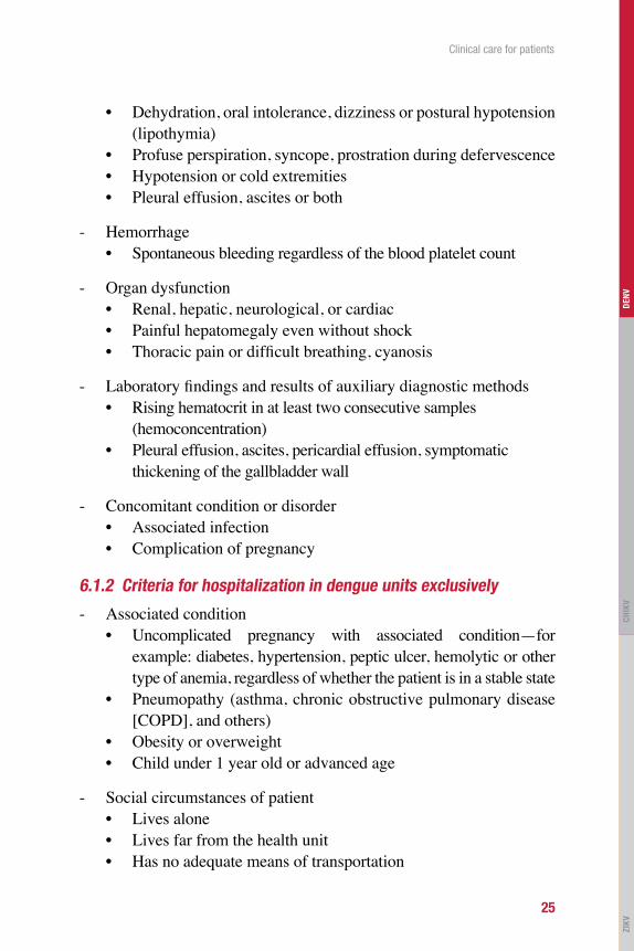
ZIKV
• Dehydration, oral intolerance, dizziness or postural hypotension (lipothymia)
• Profuse perspiration, syncope, prostration during defervescence• Hypotension or cold extremities• Pleural effusion, ascites or both
- Hemorrhage• Spontaneous bleeding regardless of the blood platelet count
- Organ dysfunction• Renal, hepatic, neurological, or cardiac• Painful hepatomegaly even without shock• Thoracic pain or difficult breathing, cyanosis
- Laboratory findings and results of auxiliary diagnostic methods• Rising hematocrit in at least two consecutive samples
(hemoconcentration)• Pleural effusion, ascites, pericardial effusion, symptomatic
thickening of the gallbladder wall
- Concomitant condition or disorder• Associated infection• Complication of pregnancy
6.1.2 Criteria for hospitalization in dengue units exclusively
- Associated condition • Uncomplicated pregnancy with associated condition—for
example: diabetes, hypertension, peptic ulcer, hemolytic or other type of anemia, regardless of whether the patient is in a stable state
• Pneumopathy (asthma, chronic obstructive pulmonary disease [COPD], and others)
• Obesity or overweight• Child under 1 year old or advanced age
- Social circumstances of patient• Lives alone• Lives far from the health unit• Has no adequate means of transportation
CHIK
VZI
KV
26
Tool for the diagnosis and care of patients with suspected arboviral diseases
DEN
VCH
IKV
ZIKV
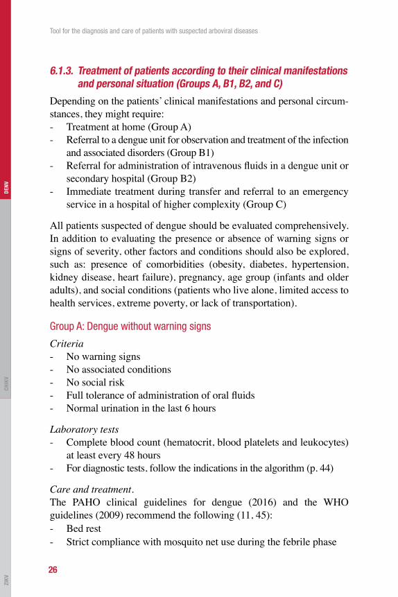
6.1.3. Treatment of patients according to their clinical manifestations and personal situation (Groups A, B1, B2, and C)
Depending on the patients’ clinical manifestations and personal circum-stances, they might require:- Treatment at home (Group A)- Referral to a dengue unit for observation and treatment of the infection
and associated disorders (Group B1)- Referral for administration of intravenous fluids in a dengue unit or
secondary hospital (Group B2) - Immediate treatment during transfer and referral to an emergency
service in a hospital of higher complexity (Group C)
All patients suspected of dengue should be evaluated comprehensively. In addition to evaluating the presence or absence of warning signs or signs of severity, other factors and conditions should also be explored, such as: presence of comorbidities (obesity, diabetes, hypertension, kidney disease, heart failure), pregnancy, age group (infants and older adults), and social conditions (patients who live alone, limited access to health services, extreme poverty, or lack of transportation).
Group A: Dengue without warning signs
Criteria- No warning signs- No associated conditions- No social risk- Full tolerance of administration of oral fluids- Normal urination in the last 6 hours
Laboratory tests- Complete blood count (hematocrit, blood platelets and leukocytes)
at least every 48 hours- For diagnostic tests, follow the indications in the algorithm (p. 44)
Care and treatment. The PAHO clinical guidelines for dengue (2016) and the WHO guidelines (2009) recommend the following (11, 45):- Bed rest- Strict compliance with mosquito net use during the febrile phase
CHIK
VZI
KV

27
Clinical care for patients
DEN
VCH
IKV
ZIKV
- Adequate fluid intake• Adults: at least five 250 ml glasses a day• Children: Ample oral fluids
- Acetaminophen• Adults, 500 mg/dose every 6 hours; maximum daily dose 4 g• Children, 10 mg/kg/dose every 6 hours; maximum daily dose 3 g
- Do not give aspirin or nonsteroidal anti-inflammatory drugs (NSAIDs)- Do not give corticosteroids- Do not give antibiotics- Intramuscular and rectal routes are contraindicated- A patient with stable hematocrit can continue to be cared for at home- The patient should be always followed by an adult trained in
dengue care
Follow-up appointmentsEvaluate the patient immediately to Ascertain whether he or she:- Presents a warning sign or shock- Presents criteria for hospitalization- Has not urinated in 6 hours or more- Feels worse, or his/her caregiver says so
In the absence of the foregoing criteria, the patient should be checked every 48 hours
At each follow-up consultation, evaluate:- Natural history of the disease- Hematocrit, as soon as the report is available (to see if it is rising
progressively)- Blood platelet levels, to see if they are going down or the disease is
still active- Leukopenia- Warning signs, if any
In addition,- Emphasize to the patient (or caregiver) that he/she should go
immediately to the dengue unit or a hospital if one or more of the warning signs is present, and
- Provide a written list of steps to be taken for care at home.
CHIK
VZI
KV

28
Tool for the diagnosis and care of patients with suspected arboviral diseases
DEN
VCH
IKV
ZIKV
Group B1: Dengue without warning signs but with associated disorder or social risk
Criteria- Associated conditions: Pregnancy, age under 1 year old or
over 65, morbid obesity, hypertension, diabetes mellitus, renal damage, hemolytic diseases, chronic liver disease, treatment with anticoagulants, etc.
- Social risk: Patient lives alone or far from where he/she can receive medical care, lack of transportation, extreme poverty.
Laboratory tests- Complete blood count within a period of 3 days of onset of the
disease- For diagnostic tests, follow the indications in the pertinent
algorithm (p. 44)Care and treatmentThe PAHO clinical guidelines for dengue (2016) and the WHO guidelines (2009) recommend the following (11, 45):- Keep the patient orally hydrated. In case of intolerance to the oral
route, initiate intravenous treatment with a crystalloid (lactated Ringer’s or 0.9% saline solution at a maintenance dose of 2 to 4 mL/kg/hr) and restart the oral route as soon as possible.
It is important to keep in mind that for overweight or obese patients, the amount of liquids should be calculated according to ideal bodyweight. Other patient conditions (heart diseases, nephropathies, etc.) should also be taken into account if they make it necessary to adjust the amount of liquids that should be administered.- Provide symptomatic treatment as for Group A.In addition:- Insist on strict compliance with mosquito net use during the
febrile phase- Provide the same information as for Group A.
Specific supervised care for the associated condition
- Monitoring and evaluation- Vital signs: pulse, heart rate, respiration rate, temperature, blood
pressure
CHIK
VZI
KV

29
Clinical care for patients
DEN
VCH
IKV
ZIKV
- Temperature curve- Water balance: intake and output (report times the patient urinates)- Warning signs (mainly on the day the fever abates)- Laboratory, depending on the associated condition (hematocrit,
leukocytes, glucose, electrolytes, etc.)- Hematocrit, blood platelets, and leukocytes every 24 to 48 h- Education about the warning signsFollow-up appointments- See Group A.
Group B2: Dengue with warning signs
CriteriaAny dengue case that presents one or more of the following signs or symptoms around the time the fever abates, preferably at the moment when it starts to fall:- Continuous and intense abdominal pain or tenderness- Persistent vomiting (three or more times an hour or four times in six
hours)- Fluid accumulation (ascites, pleural or pericardiac effusion)- Active mucosal bleeding- Lethargy/restlessness- Postural hypotension (lipothymia)- Liver enlargement >2 cm- Progressive increase in hematocritLaboratory tests- Complete blood count before hydrating the patient- For diagnostic tests, follow the indications in the pertinent
algorithm (p. 44)Care and treatmentThe PAHO clinical guidelines for dengue (2016) and the WHO guidelines (2009) recommend the following (11, 45):
Lack of a complete blood count should not delay the start of hydration. - Administer lactated Ringer’s, Hartmann, or 0.9% saline solution at
10 mL/kg for 1 hour.- Reevaluate. If the warning signs persist and diuresis is <1 mL/kg/h,
repeat the charge once or twice again with an isotonic crystalloid.
CHIK
VZI
KV

30
Tool for the diagnosis and care of patients with suspected arboviral diseases
DEN
VCH
IKV
ZIKV
- Reevaluate. If clinical improvement is observed and diuresis is ≥1 mL/kg/h, reduce the drip to 5-7 mL/kg/h and continue for 2 to 4 hours. If clinical improvement continues, reduce to 3-5 mL/kg/h for 2 to 4 hours. Then continue the drip at 2-4 mL/kg/h for another 2 to 4 hours, depending on the patient’s needs.
- Reevaluate the patient’s clinical status. Repeat hematocrit. If the minimum remains the same or increases only slightly, continue the drip at 2-4 mL/kg/h for 2 to 4 more hours.
- If there is a deterioration in vital signs or a rapid rise in hematocrit: treat the same as Group C and refer the patient to the next higher level of complexity in the health system.
- Reevaluate the patient’s clinical status. Repeat hematocrit and adjust the infusion rate.
- Gradually reduce the infusion rate when the volume of plasma leakage subsides or the critical phase has passed.
It is important to keep in mind that for overweight or obese patients, the amount of liquids should be calculated according to ideal bodyweight. Other patient conditions (heart diseases, nephropathies, etc.) should also be taken into account if they make it necessary to adjust the amount of liquids that should be administered.
Indicators of clinical improvement- Progressive disappearance of warning signs- Progressive remission of the overall symptomatology- Stabilization of vital signs- Normal or increased diuresis- Reduction of hematocrit to lower than the baseline value in a stable
patient- Good tolerance to the oral route- Recovery of appetiteEvaluation schedule- Vital signs and peripheral perfusion: until 4 hours after the critical
phase has passed- Diuresis: until 4 to 6 hours after the critical phase has passed- Hematocrit: before and after resuscitation with fluids, then every 12
to 24 hours
CHIK
VZI
KV

31
Clinical care for patients
DEN
VCH
IKV
ZIKV
- Blood glucose: every 12 or 24 hours- Other tests (depending on the affected organ and associated disease)
Group C: Severe dengue
CriteriaAny dengue case that presents one or more of the following manifestations:- Shock or respiratory distress due to severe plasma leakage. Shock
evidenced by weak or undetectable pulse, tachycardia, cold extremities, and capillary perfusion > 2 seconds, pulse pressure < 20 mmHg, indicating hypotension in the late phase.
- Severe bleeding: based on evaluation by the attending physician (examples: hematemesis, melena, ample metrorrhagia, CNS bleeding).
- Severe organ compromise, such as liver impairment (AST or ALT >1000 IU), CNS (impaired mental state), heart (myocarditis), or other organs.
Laboratory tests and diagnostic imaging- Complete blood count- For diagnostic tests, follow the indications in the pertinent
algorithm (p. 44)- Other tests, depending on the affected organ: for example,
transaminases, arterial gases, electrolytes, blood glucose, urea nitrogen and creatinine, cardiac enzymes, cultures, chest X-ray, thoracic and/or abdominal ultrasound, echocardiogram, and electrocardiogram
Treatment of shockThe PAHO clinical guidelines for dengue (2016) and the WHO guidelines (2009) recommend the following (11, 45):Obtain hematocrit level before hydrating the patient, but if it cannot be obtained, that should not delay the start of hydration.- ABC and monitoring of vital signs every 5 to 30 min - Oxygen therapy- Start hydration with crystalloid intravenous fluid (lactated Ringer’s
or 0.9% saline solution) at 20 mL/kg for 15 to 30 minutes (for pregnant women and adults over 65, boluses are administered at 10 mL/kg for 15 to 30 minutes).
CHIK
VZI
KV
32
Tool for the diagnosis and care of patients with suspected arboviral diseases
DEN
VCH
IKV
ZIKV
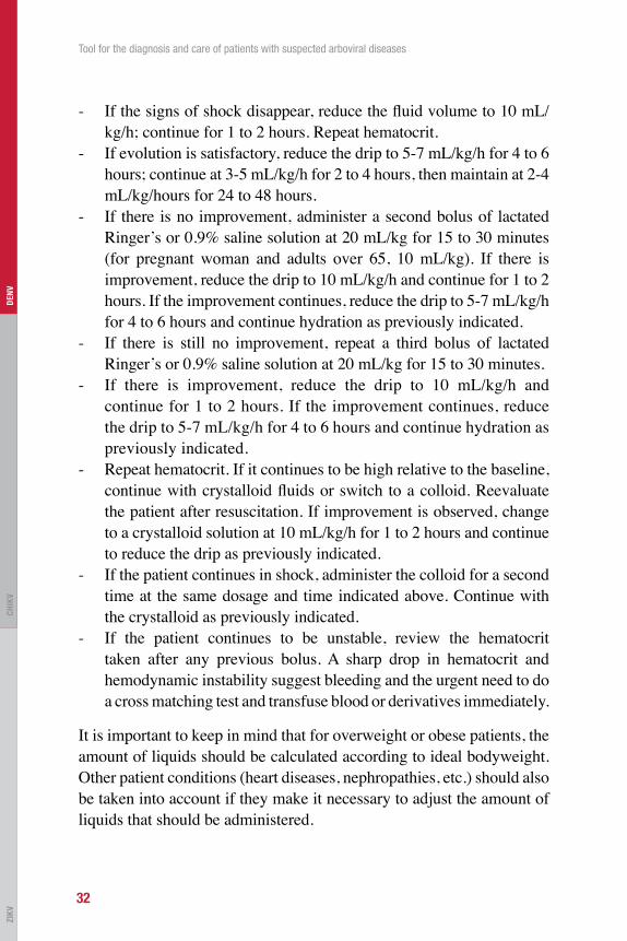
- If the signs of shock disappear, reduce the fluid volume to 10 mL/kg/h; continue for 1 to 2 hours. Repeat hematocrit.
- If evolution is satisfactory, reduce the drip to 5-7 mL/kg/h for 4 to 6 hours; continue at 3-5 mL/kg/h for 2 to 4 hours, then maintain at 2-4 mL/kg/hours for 24 to 48 hours.
- If there is no improvement, administer a second bolus of lactated Ringer’s or 0.9% saline solution at 20 mL/kg for 15 to 30 minutes (for pregnant woman and adults over 65, 10 mL/kg). If there is improvement, reduce the drip to 10 mL/kg/h and continue for 1 to 2 hours. If the improvement continues, reduce the drip to 5-7 mL/kg/h for 4 to 6 hours and continue hydration as previously indicated.
- If there is still no improvement, repeat a third bolus of lactated Ringer’s or 0.9% saline solution at 20 mL/kg for 15 to 30 minutes.
- If there is improvement, reduce the drip to 10 mL/kg/h and continue for 1 to 2 hours. If the improvement continues, reduce the drip to 5-7 mL/kg/h for 4 to 6 hours and continue hydration as previously indicated.
- Repeat hematocrit. If it continues to be high relative to the baseline, continue with crystalloid fluids or switch to a colloid. Reevaluate the patient after resuscitation. If improvement is observed, change to a crystalloid solution at 10 mL/kg/h for 1 to 2 hours and continue to reduce the drip as previously indicated.
- If the patient continues in shock, administer the colloid for a second time at the same dosage and time indicated above. Continue with the crystalloid as previously indicated.
- If the patient continues to be unstable, review the hematocrit taken after any previous bolus. A sharp drop in hematocrit and hemodynamic instability suggest bleeding and the urgent need to do a cross matching test and transfuse blood or derivatives immediately.
It is important to keep in mind that for overweight or obese patients, the amount of liquids should be calculated according to ideal bodyweight. Other patient conditions (heart diseases, nephropathies, etc.) should also be taken into account if they make it necessary to adjust the amount of liquids that should be administered.
CHIK
VZI
KV

33
Clinical care for patients
DEN
VCH
IKV
ZIKV
Treatment of hemorrhage. The PAHO clinical guidelines for dengue (second edition) and the WHO guidelines (2009) recommend the following (11, 45): red blood cells 5-10 mL/kg or fresh blood 10-20 mL/kg- If the patient does not improve, reevaluate the hemodynamic status. - Evaluate the pumping function (to detect cardiomyopathy,
myocarditis); define the use of amines. - Evaluate the concomitant medical conditions (heart disease,
pneumopathy, vasculopathy, nephropathy, diabetes, obesity, pregnancy); stabilize the underlying disorder.
- Look for persistent acidosis and risk for occult hemorrhage and treat them.
Report the case to epidemiology within 24 hours.
6.1.4. Criteria for discharging a dengue patient
All the following conditions should be present:
- Clinical criteria• Absence of fever for 48 hours without the administration of an
antipyretic• Improvement of clinical status (general well-being, good
appetite, normal hemodynamic status, normal or increased diuresis, absence of respiratory distress and no evidence of bleeding)
- Laboratory criteria• Rising blood platelet count• Stable hematocrit without the support of intravenous fluids
CHIK
VCH
IKV
ZIKV

34
Tool for the diagnosis and care of patients with suspected arboviral diseases
DEN
VCH
IKV
ZIKV
6.2 Clinical care for suspected chikungunya virus infection
At the time of publication of this document, there is insufficient published evidence on pain management in patients with chikungunya. A systematic review concluded that there is no evidence to support or reject any evaluated intervention for the treatment of rheumatic diseases in patients with chikungunya (46). It will be necessary to develop evidence-informed chikungunya treatment guidelines, in particular for the management of chronic arthritis and arthralgia.
The following general recommendations for clinical care of suspected chikungunya virus infection were taken from Preparedness and Response for Chikungunya Virus Introduction in the Americas (2011) and French Guidelines for the Management of Chikungunya (Acute and Persistent Presentations) (2014) (12, 13).
If the patient is suspected of having chikungunya, the following questions should be answered:- What phase of chikungunya is it in? (acute/post-acute/chronic)- Does the patient present any non-articular manifestations? Any
clinical sign of severity? Atypical or complicated forms? - Does the patient present any risk factors for severe forms of the
disease? (chronic ailments, extreme ages of life, pregnancy)- What is the patient’s hemodynamic status? Is there dehydration?
6.2.1. Clinical care based on the phase of the disease (12, 13)
6.2.1.1. Acute phase (up to 3 weeks)
- Rest during the febrile phase (patient should stay home from work; in the case of edema, removal of rings and other tight apparel, relative rest of the affected joints)
- Abundant oral fluids to prevent dehydration - If the health unit has the capability, complete blood count (including
platelet count), C-reactive protein, erythrocyte sedimentation rate - No need for radiological imaging of the joints during this phase - Referral of patient with severe forms of the disease to a second- or
third-level hospital
DEN
VZI
KV
35
Clinical care for patients
DEN
VCH
IKV
ZIKV
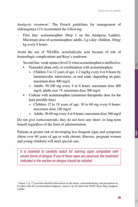
Analgesic treatment.1 The French guidelines for management of chikungunya (13) recommend the following:- First line: acetaminophen (Step 1 on the Analgesic Ladder).
Maximum dose of acetaminophen: adults, 4 g a day; children, 10mg/kg every 6 hours.
Avoid the use of NSAIDs, acetylsalicylic acid because of risk of hemorrhagic complications and Reye’s syndrome.- Second line: weak opiates (level 2) when acetaminophen is ineffective.
• Tramadol alone only or combination with acetaminophen: » Children 3 to 12 years of age: 1-2 mg/kg every 4 or 6 hours by
intramuscular, intravenous, or oral route, depending on pain; maximum dose 400 mg/d.
» Adults: 50-100 mg every 4 or 6 hours; maximum dose 400 mg/d; adults over 75, maximum dose 300 mg/d
• Codeine with acetaminophen (minimum therapeutic dose for the least possible time)
» Children 12 to 18 years of age: 30 to 60 mg every 6 hours; maximum dose 240 mg/d
» Adults: 30-60 mg every 4 or 6 hours; maximum dose 360 mg/dDo not give corticosteroids; they do not have any short- or long-term benefit regardless of the form of administration.
Patients at greater risk of developing less frequent signs and symptoms (those over 60 years of age or with chronic illnesses, pregnant women and young children) will need special care.
It is essential to carefully watch for warning signs compatible with severe forms of dengue. If any of these signs are observed, the treatment indicated in the section on dengue should be initiated.
1 Annex 1 (p. 77) provides detailed information on the doses, contraindications, and precautions to be taken with the recommended analgesics. Annex 2 (p. 82) shows the WHO Three-Step Analgesic Ladder.
DEN
VZI
KV

36
Tool for the diagnosis and care of patients with suspected arboviral diseases
DEN
VCH
IKV
ZIKV
Other indications- Ascertain the hemodynamic status of the patient and immediately
initiate intravenous hydration if he/she presents slow capillary perfusion, slow pulse, or oliguria, which are the first signs of shock or signs of existing shock, hypotension, and altered state of consciousness.
- Determine the severity and take specific measures to address cases of renal failure, neurological signs and symptoms, liver failure, heart disease, thrombocytopenia, skin lesions (bullae), and involvement of other organs that threaten the patient’s life or important functions such as sight.
- Initiate treatment of severe complications in consultation with the pertinent specialists. Severe forms should only be treated in a hospital center, using the conventional measures for medical resuscitation.
Special population groups. The French guidelines for management of chikungunya (13) recommend the following:
Pregnant women- Recommended treatment: acetaminophen- Avoid the use of NSAIDs, acetylsalicylic acid, and topical drugs- If a pregnant woman presents the infection at the end of her
pregnancy, it is essential to consult the obstetrician.
Newborns and children- In a case of suspected vertical transmission, prolonged neonatal
observation (7 days) - In a case of confirmed viremia in the mother, newborn monitoring
for at least 5 days, including:• Body temperature• Quality of the breast-feeding• Signs of pain• State of the skin• Hydration
- Do not administer NSAIDs to infants under 3 months old. - It is not recommended to give codeine to children under 12 years
old (except for those refractory to acetaminophen, after consultation with a specialist).
DEN
VZI
KV
37
Clinical care for patients
DEN
VCH
IKV
ZIKV
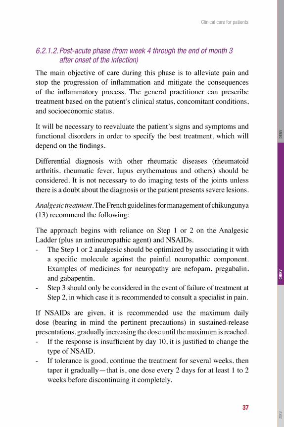
6.2.1.2. Post-acute phase (from week 4 through the end of month 3 after onset of the infection)
The main objective of care during this phase is to alleviate pain and stop the progression of inflammation and mitigate the consequences of the inflammatory process. The general practitioner can prescribe treatment based on the patient’s clinical status, concomitant conditions, and socioeconomic status.
It will be necessary to reevaluate the patient’s signs and symptoms and functional disorders in order to specify the best treatment, which will depend on the findings.
Differential diagnosis with other rheumatic diseases (rheumatoid arthritis, rheumatic fever, lupus erythematous and others) should be considered. It is not necessary to do imaging tests of the joints unless there is a doubt about the diagnosis or the patient presents severe lesions.
Analgesic treatment. The French guidelines for management of chikungunya (13) recommend the following:
The approach begins with reliance on Step 1 or 2 on the Analgesic Ladder (plus an antineuropathic agent) and NSAIDs.- The Step 1 or 2 analgesic should be optimized by associating it with
a specific molecule against the painful neuropathic component. Examples of medicines for neuropathy are nefopam, pregabalin, and gabapentin.
- Step 3 should only be considered in the event of failure of treatment at Step 2, in which case it is recommended to consult a specialist in pain.
If NSAIDs are given, it is recommended use the maximum daily dose (bearing in mind the pertinent precautions) in sustained-release presentations, gradually increasing the dose until the maximum is reached.- If the response is insufficient by day 10, it is justified to change the
type of NSAID. - If tolerance is good, continue the treatment for several weeks, then
taper it gradually—that is, one dose every 2 days for at least 1 to 2 weeks before discontinuing it completely.
DEN
VZI
KV
38
Tool for the diagnosis and care of patients with suspected arboviral diseases
DEN
VCH
IKV
ZIKV
Corticosteroid treatment. The French guidelines for management of chikungunya (13) recommend the following:
This treatment is reserved for highly inflammatory polyarticular forms associated with tenosynovitis, active synovitis, resistance to NSAIDs, or cases in which they are contraindicated.- Prednisone 10 mg/d for 5 days with progressive reduction over a 10-
day period; severe cases, 0.5 mg/kg/d of prednisone for 5 days with progressive reduction over a 10-day period.
- In all cases, corticosteroid treatment should not continue for more than 4 weeks.
Topical anti-inflammatory treatment (including infiltration) (13)
This approach is indicated in cases of tenosynovitis, bursitis, carpal tunnel syndrome, capsulitis, or synovitis that are not sufficiently controlled with oral treatment. Surgical decompression is not recommended.
Disease-modifying medications such as methotrexate are not indicated prior to 8 eight weeks of evolution. This treatment is only recommended in cases of chronic polyarthritis in the presence of signs of inflammatory rheumatism, following approval by a specialist in rheumatology.
6.2.1.3. Chronic phase (starting in month 4)
Given the intensity of the pain and the potential for long-term pain as a result of CHIKV infection, treatment and psychological support should be available. Consideration should also be given to developing protocols and setting up health centers and teams to see patients with chronic pain.
Chronic post-infection inflammatory rheumatism, with or without joint destruction, invariably requires the opinion of a rheumatologist, preferably as part of a multidisciplinary consultation, to ensure maximum possible efficacy and the remission of symptoms. Care should be initiated during the first months of the chronic phase with a view to avoiding a potentially destructive evolution, reducing the functional and psychosocial impact, and improving the patient’s quality of life.
DEN
VZI
KV

39
Clinical care for patients
DEN
VCH
IKV
ZIKV
Analgesic treatment. The French guidelines for management of chikungunya (13) recommend the following:
In most cases, treatment for pain can be administered under the responsibility of the primary care physician using:- Acetaminophen (if responds to the treatment) at the doses given
above- NSAIDs (based on the protocols described above)- Short-term corticosteroids: the first line of treatment for
uncontrolled multiple musculoskeletal disorders (multiple hypertrophic tenosynovitis or distal polyarthralgia with edema). As the corticosteroids are progressively tapered, it is recommended to treat with NSAIDs to avoid clinical rebound.
In the case of a refractory situation or corticosteroid dependency, it is recommended to consult a rheumatologist.
Physical therapy and other rehabilitation techniques: movement and moderate exercise tends to improve the morning stiffness and pain, although it should be kept in mind that intense exercise can make the symptoms worse.
Radiological imaging of the joints can be done in this phase of the disease, depending on the clinical manifestations and diagnostic hypotheses.
DEN
VZI
KV
40
Tool for the diagnosis and care of patients with suspected arboviral diseases
DEN
VCH
IKV
ZIKV
6.3. Clinical care for suspected Zika virus infection
At present, there are no specific clinical guidelines for the management of patients with ZIKV infection. This makes it necessary to develop solidly evidence-based guidelines for proper case management. Since ZIKV symptoms are very similar to those produced by DENV and CHIKV, the following is based on clinical guides for dengue (11, 45) and the opinion of experts. These recommendations may be subject to modification as knowledge of the disease and its etiologic agent advances.
Since ZIKV infection is usually asymptomatic and the clinical course of cases is self-limited, patients often do not need treatment and may not even seek medical care. To date, there are no vaccines or specific treatments for the disease. The following measures focus on the relief of symptoms.
In general, the guidelines for treatment of ZIKV infection are:- Relative rest while the fever lasts - Strict compliance with mosquito net use during the symptomatic
phase - Use of safe repellents (those that contain DEET, IR3535, or
picaridin); - Recommendation that the patient consult a physician immediately
in the event of tingling or numbness in the arms or legs- Adequate fluid intake (11, 45)
• Adults: six 250 ml glasses or more a day• Children: on demand, following the Holliday-Segar formula as
a minimum- Acetaminophen (in the event of pain or fever) (11, 45)
• Adults: 500 mg/dose every 6 hours; maximum daily dose 4 g• Children, 10 mg/kg/dose every 6 hours
- Do not give acetylsalicylic acid to children;
DEN
VCH
IKV

41
Clinical care for patients
DEN
VCH
IKV
ZIKV
- Antihistamines (47)• Chlorpheniramine:
» Children, • 1 month to 2 years old: 1 mg twice daily• 2 to 6 years old: 1 mg every 4 to 6 hours; maximum daily
dose 6 mg• 6 to 12 years old: 2 mg every 4 to 6 hours; maximum
daily dose 12 mg• >12 years old and adults: 4 mg every 4 to 6 hours;
maximum daily dose 24 mg » Adults: 4 mg every 4 to 6 hours; maximum daily dose 24 mg
• Loratadine: » Adults and children >10 years old: 5 to 10 mg every 12 hours
- Artificial tears- In cases of GBS (12):
• Plasmapheresis: 250 mL/kg replacement on alternate days (48, 49)
• Immunoglobulin: 400 mg/iv/kg/d by 5 days (total 2 g) (50-53)- In the event of suspected optic neuritis, refer patient to an
ophthalmologist for evaluation and treatment.
It is essential to carefully watch for warning signs compatible with severe forms of dengue. If any of these signs are observed, the treatment indicated in the section on dengue should be initiated.
Pregnant women with suspected ZIKV infection
The recommendations for pregnant women with suspected ZIKV infec-tion are from the PAHO/WHO documents: Provisional considerations for the care of pregnant women in settings with high Zika virus circu-lation: document for health care professionals (Feb. 17, 2016) (54). In the presence of signs and symptoms, the PAHO/WHO document (53) recommends the following:
- Fever: To reduce the fever, first use physical means; if there is no response, give acetaminophen 500 mg/PO every 6 to 8 hours not to exceed 4 g/day.
DEN
VCH
IKV

42
Tool for the diagnosis and care of patients with suspected arboviral diseases
DEN
VCH
IKV
ZIKV
- Headache: Give acetaminophen in the same doses as for controlling fever. Do not use aspirin or NSAIDS.
- Pruritus: There are not scientific tests that either support or counterindicate the use of topical drugs and clinical experience suggests that they are safe:• Calamine lotion or menthol-based topical creams• Antihistamines, type 1 (H1), in cases of intense pruritus
- Hydration: Recommend steady intake of fluids to prevent dehydration that can be caused by impervious losses (fever, sweating, etc.) or vomiting.
- Control of pregnancy: All pregnant women should be advised get their regular prenatal checkups. Each country’s established national protocols should be followed. Prenatal checkups should be done by a trained health professional (gynecologist-obstetrician). More information on this subject can be found in the provisional considerations for the care of pregnant women (54).
DEN
VCH
IKV

43
Recommendations for laboratory diagnosis of DENV, CHIKV, and ZIKV infection
DEN
VCH
IKV
ZIKV
DEN
VCH
IKV
ZIKV
With arboviral diseases, unlike other viral diseases, laboratory diagnosis does not determine treatment of the patient. Detection of infection markers (detection of the virus or its genome, antigens, or antibodies) supports the confirmation of the clinical diagnosis and is very useful in epidemiological surveillance of the disease, but it does not affect individual treatment.
The diagnosis of arboviral diseases is based on the detection of the infectious agent or an immune response to the infection. In the case of ZIKV and DENV, direct detection of the viral genome or parts thereof using the polymerase chain reaction (PCR) technique is limited by the short duration of viremia and the low virus burden in blood (55), whereas CHIKV has high and prolonged viremia (12). Indirect or serological methods are used once virus has disappeared from the bloodstream and is observed the antibody response, which can last for months (IgM) or even years (IgG) (11, 45). Serology should be performed whenever possible, but it should be kept in mind that cross-antigenicity can complicate the interpretation, especially when multiple flaviviruses are in circulation in the same geographic area, as in the case of dengue, Zika, and yellow fever (11). Specific serological diagnosis requires the use of serum neutralization tests, in which the virus challenges the patient’s antibodies. However, this technique takes time and requires highly trained staff. Virus isolation is not typically done in the current diagnosis of arboviral infections. However if the capability is available, it is important to perform the isolation using a group of samples in order to identify and characterize the different virus serotypes, genotypes, and strains.
In order to follow the diagnostic process and for purposes of clinical care, this document presents the diagnostic algorithm generally accepted for laboratory screening of the arboviruses that are currently most prevalent in the Region of the Americas.
RECOMMENDATIONS FOR LABORATORY DIAGNOSIS OF DENV, CHIKV, AND ZIKV INFECTION 7

44
Tool for the diagnosis and care of patients with suspected arboviral diseases
DEN
VCH
IKV
ZIKV
DEN
VCH
IKV
ZIKV
7.1 Algorithm for the detection of DENV, CHIKV, or ZIKV2
This algorithm can be used by laboratories that have the installed capacity to identify both the dengue and the chikungunya viruses molecularly, antigenically, and serologically and to identify Zika virus in differential diagnosis. A biosafety containment laboratory (BSL) is required for the handling of suspected samples.
Figure 3. Algorithm for laboratory diagnosis of suspected arbovirus cases: acute phase
2 Depending on the epidemiological characteristics of the country and the infection clinics, additional arboviruses may be included in the differential algorithm for Zika virus. These recommendations are subject to change as progress is made in knowledge about the disease and its etiologic agent.
Suspected case of arbovirus infection: acute phase
Serum samplea taken ≤ 5 days after onset of symptoms
RT-PCR DENV
Viral isolationc
Positive
Positive
Positive
Positive
Negativeb Negative
Negative
RT-PCR ZIKV
RT-PCR CHIKV
a A urine sample is also recommended for PCR ZIKV.b Consider dengue NS1 antigen for determining DENV infection.c Isolation is not required in order to confirm infection. It is considered complementary information for identifying serotypes, genotypes, and strains of the arbovirus in question.

45
Recommendations for laboratory diagnosis of DENV, CHIKV, and ZIKV infection
DEN
VCH
IKV
ZIKV
DEN
VCH
IKV
ZIKV
Figure 4. Algorithm for laboratory diagnosis of suspected arbovirus cases: convalescent phase
Suspected case of arbovirus infection: convalescent phase
DENV-Positive Positive DENV Negative DENV
Negative ZIKV
Presumptive DENV
Positive ZIKV
Flavivirus infection
Positive ZIKV
Presumptive ZIKV
7.2 Sample collection and shipment
7.2.1 Virological diagnosis (acute phase of the disease)
Type of sample: serum or urine
In an arbovirus infection, especially those caused by flaviviruses and alphaviruses, viremia can usually be detected 4 to 5 days after the appearance of symptoms (45). Some arboviruses, such as CHIKV, can have more prolonged viremia and can be detected up to more than 7 days after symptom onset (12). The period of viremia for ZIKV has not yet been fully established, but during the current epidemic in the Americas the virus has been detected in serum up to 5 days after symptoms appeared. Since the initial phase of ZIKV infection
IgM (ELISA/IgM-IIF)
Serum sample taken ≥ 6 days after onset of symptoms
46
Tool for the diagnosis and care of patients with suspected arboviral diseases
DEN
VCH
IKV
ZIKV
DEN
VCH
IKV
ZIKV
tends to have few if any symptoms, the disease can go unnoticed, thus reducing the opportunity to collect biological samples. With DENV, on the other hand, an effort should be made to detect NS1 antigen in acute samples, since the antigenemia tends to last somewhat longer than the viremia (11, 45).
In addition to serum samples, urine samples have been gaining increasing attention in recent years. Virus has been detected in urine (viruria) in flavivirus infections such as dengue, yellow fever, West Nile fever, and now Zika (56, 57). Both Zika RNA and viable virus have been found in urine cultures. The usefulness of detecting ZIKV in urine is becoming more important because of the disease’s brief period of viremia and the difficulties associated with of serology. Furthermore, a high viral load has been found in urine for a longer period during the acute phase, even up to 2 weeks after the appearance of symptoms, which makes the urine sample a good alternative (58, 59). However, since further studies are still needed, it is recommended to take the sample at the end of the acute phase.
7.2.2 Serological diagnosis
Type of sample: serum
It is possible to detect arbovirus IgM antibodies using ELISA or immunofluorescence tests up to the fifth day after the appearance of symptoms. Bearing in mind that that a single sample of acute-phase serum is only a presumptive diagnosis, it is recommended to take a second sample one to two weeks later to demonstrate seroconversion (positive or negative) or else increase the antibody titer at least fourfold in a quantitative test (11, 45).
The interpretation of the serological tests is especially relevant in diagnosing arbovirus infections, in particular those in the flavivirus group. In primary infections (first infection with a flavivirus), it has been shown that the antibody in these patients rarely if ever cross-reacts with another antigenically related virus, and if does, the titers are low

47
Recommendations for laboratory diagnosis of DENV, CHIKV, and ZIKV infection
DEN
VCH
IKV
ZIKV
DEN
VCH
IKV
ZIKV
(11, 45). The exception is individuals who have a previous history of flavivirus infection, especially dengue, yellow fever (vaccination or infection), or West Nile virus; serum from such individuals can cross-react in these tests (11, 45).
Although the plaque reduction neutralization technique (PRNT) offers greater specificity in the detection of neutralizing antibodies (IgG), a second dengue infection produces high neutralizing antibody titers against at least two or three of the four dengue serotypes and also against flaviviruses in other antigenic complexes (11, 45). During the early convalescent phase after sequential dengue or other flavivirus infections, the highest neutralizing antibody titer is against the virus of the primo-infection, not the most recent one. This phenomenon occurs due to preexisting antibodies and the “original sin” theory (11, 45).
All of the above makes it difficult to interpret antibody titers in flavivirus infections (11, 45). Special caution is advised in using the PRNT for ZIKV in DENV endemic and epidemic areas. In the case of CHIKV, though it does not cross-react with the falviviruses, attention needs to be given to differential diagnosis with respect to Mayaro virus, an antigenically related alphavirus (12, 60), in areas where it is in circulation.
For further information:- CHIKV diagnosis: Preparedness and Response for Chikungunya
Virus Introduction in the Americas (12).- DENV diagnosis: Dengue: guidelines for patient care in the Region
of the Americas, 2nd ed. (in Spanish only) (11).
7.2.3 Preservation of the sample (61)
- Keep it refrigerated at 2 to 8 °C if it is to be processed or sent to a reference laboratory within 48 hours.
- Keep it frozen at -10 to -20 °C if it is to be processed after 48 hours but not after 7 days.
- Keep it frozen at -70 °C if it is to be processed after a week. At this temperature the sample will be adequately preserved for a long time.
48
Tool for the diagnosis and care of patients with suspected arboviral diseases
DEN
VCH
IKV
ZIKV
DEN
VCH
IKV
ZIKV
7.2.4 Airmailing the sample to the reference laboratory (61)
- If possible pack with dry ice, or at least ensure a cold chain by using cooling gels. Always use triple packaging.
- Dispatch within 48 hours. - The original samples should be packed, marked, labeled (if dry ice
is used), and documented as Category B.- Always include the complete clinical and epidemiological record.
7.3 Observations and additional recommendations (62, 63)
There are various sensor protocols (both primers and probes) for the detection of arboviruses (DENV, CHIKV and ZIKV) using both conventional and real-time RT-PCR. Depending on sensitivity, it is recommended to follow CDC protocols, which will need to be standardized for local diagnosis.
In the case of DENV and CHIKV, the IgM can be identified using a variety of techniques (ELISA or IF, both homemade and commercial). However, no commercial kits have been approved or validated for the serological identification of ZIKV. In any case, the highest sensitivity is achieved with laboratory platforms that use the complete virus as antigen, compared with those that use recombinant proteins (or peptides).
Isolation of the virus is not regarded as a routine diagnosis and is recommended only as a supplementary research test to be used for purposes of public health surveillance (identification of genotypes and strains).

49
Recommendations for laboratory diagnosis of DENV, CHIKV, and ZIKV infection
DEN
VCH
IKV
ZIKV
DEN
VCH
IKV
ZIKV
For ZIKV, national laboratories that are responsible for surveillance but do not have capacity for virological or serological confirmation (RT PCR or viral isolation or sequencing in the first case, or PRNT in the latter) should send their samples to a PAHO/WHO reference laboratory or collaborating center. However, in order to coordinate the process, before prior to shipping any samples it is important to communicate with the country office, with the collaborating center, and with PAHO/WHO Headquarters in Washington D.C.:
PAHO/WHO Collaborating CenterNational Center for Emerging and Zoonotic Infectious DiseasesDivision of Vector-borne DiseasesArboviral Diseases BranchCenters for Disease Control and Prevention (CDC)CDC/DVBD/ADB3156 Rampart RoadFort Collins, CO 80521 USATel. +1 888-232-6348
Pan American Health OrganizationCommunicable Diseases and Health Analysis (CHA)Epidemic Alert and Response and Water-borne Diseases (IR)525 23rd Street NW, Washington D.C. 20037 [email protected]

DEN
VCH
IKV
ZIKV
50
Tool for the diagnosis and care of patients with suspected arboviral diseases
DEN
VCH
IKV
ZIKV

DEN
VCH
IKV
ZIKV
51
Other arboviral diseases with epidemic potential
DEN
VCH
IKV
ZIKV
OTHER ARBOVIRAL DISEASES WITH EPIDEMIC POTENTIAL 8
So far, this document has focused on the three main arboviral diseases: dengue, chikungunya, and Zika. There are also other arboviral diseases with high epidemic potential in the Region of the Americas that should be considered in differential diagnosis. This list includes West Nile fever (WNF), the equine encephalitides, Oropouche fever, Mayaro fever, and yellow fever (78).
From the clinical symptoms of these diseases described in the literature (78-85) it can be seen that there is a fair amount of overlap, which poses a challenge for clinical diagnosis. Laboratory confirmation is necessary in the most severe cases in order to ensure that patients are receiving proper care. Accurate diagnosis is also important for epidemiological reasons. Also, the geographic distribution of these diseases tends to overlap, creating an additional challenge for the correct diagnosis of these arboviral diseases. Thus, care should be taken to consider the epidemiological, laboratory, and clinical factors in arriving at a diagnosis. While the treatment is nonspecific, it is important to institute timely supportive treatment in order to ensure the survival of patients with severe disease (78, 82-84, 86-88).
8.1 West Nile virus
West Nile virus (WNV) is a flavivirus that is found in the Region of the Americas. It causes a zoonosis that spreads naturally through the migration of wild birds, whereas human cases may be regarded as accidental. The virus is typically transmitted by the bite of Culex and Aedes mosquitoes, but transmission via blood transfusion or organ transplantation has also been reported. This is the virus that causes WNF (89).
8.1.1 Clinical description
Once infected by the WNV, about 25% of patients develop WNF and 1% develop neuroinvasive disease (89). The risk factors for getting the disease are not well known, but some studies suggest that women and

DEN
VCH
IKV
ZIKV
52
Tool for the diagnosis and care of patients with suspected arboviral diseases
DEN
VCH
IKV
ZIKV
young people are more susceptible (90), while elderly patients are more likely to develop a neuroinvasive form, especially encephalitis (90, 91).
West Nile fever
The incubation period ranges from 2 and 14 days. It is characteristic for the symptoms, including fever, headache, rash, and myalgia, to appear abruptly, the rash is usually morbilliform, although it can also be maculopapular. It is seen most often on the torso and limbs and its appearance is associated with abatement of the fever (92). Other less common symptoms are joint pain, tremor, ocular pain, vomiting, and diarrhea (93).
Neuroinvasive disease
The disease has three possible presentations: meningitis, encephalitis, or flaccid paralysis (92, 93). The meningeal form is indistinguishable from other viral meningitides, with the same clinical findings (i.e., stiff neck, headache, and fever). The encephalitic form has more serious clinical symptoms, with confusion, convulsions, and altered state of consciousness. The symptoms of the flaccid paralytic form are identical to those of poliomyelitis, since it affects the motor neurons in the anterior gray column of the spinal cord (93).
The majority of patients who present WNF only or meningitis recover completely, but they can suffer weakness, malaise, and fatigue. The patients who develop encephalitis or poliomyelitis may die (10%) or recover with neurological sequelae (92, 93).
8.1.2 Diagnosis
The diagnosis should take into account not only the symptoms mentioned above but also information about the patient’s travels and other activities. It is important to inquire about possible exposure to the vector and, if the clinical picture is suspicious, to record an exhaustive epidemiological history.
Laboratory diagnosis
It has been recommended that the diagnosis be based on the detection of virus-specific immunoglobulin M (IgM) antibodies in cerebrospinal fluid (94).
DEN
VCH
IKV
ZIKV
53
Other arboviral diseases with epidemic potential
DEN
VCH
IKV
ZIKV
Since IgM cannot cross the blood-brain barrier under normal circumstances, its presence is a good indicator of WNV infection. However, it should be noted that false positive cases have been observed in patients vaccinated against yellow fever or Japanese encephalitis (94). Detection of immunoglobulin G (IgG) is not useful in diagnosing acute phases of the disease. Use of both RT-PCR and enzyme-linked immunosorbent assay (ELISA) increases the probability of detecting of the virus and has been recommended in immunodepressed patients who are unable to develop a competent immune response (95). Clinical samples should be handled following the precautions recommended in section 7.2.
Differential diagnosis
Differential diagnosis should consider diseases that have symptoms similar to WNF and/or those that can be associated with neurological symptoms, including the bacterial meningitides, enterovirus infections, the equine encephalitides, and others. They should be ruled out using diagnostic laboratory tests.
Definition of a case
According to the CDC, a probable case of WNV is one that presents either the symptoms listed above or neuroinvasive disease and is also IgM-positive in serum (or cerebrospinal fluid in the case of the neuroinvasive disease) and not positive in laboratory tests for any other disease (96).
A confirmed case, as defined by the CDC, is one that presents the symptoms listed above and yields positive laboratory test results—either, isolation of the virus, detection of the genome using RT-PCR, a 4-fold increase in the levels of virus-specific IgG antibody, a positive neutralization test, or detection of IgM antibody without any reported pleocytosis—and at the same time not positive for any of the other arboviral diseases endemic in the Americas (96).
8.1.3 Clinical management
Our search of the scientific literature on the clinical management of WNF cases (Annex 3) failed to show, as of the time this document

DEN
VCH
IKV
ZIKV
54
Tool for the diagnosis and care of patients with suspected arboviral diseases
DEN
VCH
IKV
ZIKV
was published, any vaccines or specific antiviral treatments. Treatment is palliative and focuses on pain relief, rehydration, and control of vomiting, if present. In cases of neuroinvasive disease, convulsions and any other related complications should be controlled (84, 94, 97).
For further information, see the review of literature on West Nile virus by Petersen et al. (94).

DEN
VCH
IKV
ZIKV
55
Other arboviral diseases with epidemic potential
DEN
VCH
IKV
ZIKV
8.2 Yellow fever
Yellow fever (YF) is an acute hemorrhagic disease produced by a flavivirus that is endemic in tropical and subtropical areas of Africa and South America. In jungle areas, the virus may be transmitted from nonhuman primates to humans via Haemagogus or Sabethes mosquitoes, whereas in urban areas the mosquito vector is Aedes aegypti, which transmits the virus directly between humans.
Nearly 20% of infected patients develop symptoms which, in the most serious forms of the disease, can lead to multiple organ failure and hemorrhage. At present, most of the people affected are forest workers, farmers, or unimmunized travelers. Although a safe and effective exists, yellow fever is regarded as a reemerging disease (98).
8.2.1 Clinical description
According to the World Health Organization document Clinical Management of Patients with Yellow Fever (82), the disease presents three well differentiated phases:1. Infection phase (the first 3 to 6 days): This phase begins with
sudden onset of fever, myalgia (including intense back pain), headache, nausea with possible vomiting, and in some cases bradycardia. The patient appears sickly, with saburral tongue and reddened conjunctivae. Laboratory tests may show leukopenia and, less often, neutropenia.
2. Remission phase (days 6 to 7 post infection): Virus levels in the blood and symptoms of the disease decline. Most patients recover during this phase.
3. Intoxication phase (starting day 7 post infection): Virus blood levels remain undetectable, but the symptoms recur in a more serious form, with hemorrhage, jaundice, and involvement of the vital organs. Laboratory tests reveal high transaminase levels (AST higher than ALT, total bilirubin 10-15 mg/dL, renal failure, and albuminuria. Patients who go into this phase have a grave prognosis: approximately 20 to 50% of them die within 10 to 14 days, although the rest of them recover without significant sequelae (82, 99).

DEN
VCH
IKV
ZIKV
56
Tool for the diagnosis and care of patients with suspected arboviral diseases
DEN
VCH
IKV
ZIKV
It should also be pointed out that vaccination can induce so-called “vaccine-associated disease” (VAD), which should be taken into account in studies of vaccine effectiveness and the correct interpretation of epidemiological data (82). The clinical manifestations of VAD fall into two categories: YF vaccine-associated viscerotropic disease, and YF vaccine-associated neurotrophic disease. These cases are very rare: 0.4 and 0.8 cases per 100, 000 vaccinations, respectively (100, 101).
8.2.2 Diagnosis
WHO (82) recommends that YF should be suspected in any patient who resides in or has recently traveled to an endemic area and presents a sudden onset of fever, bradycardia, and jaundice. The diagnosis of YF during the acute phase can be complicated because it can be mistaken for other diseases. Furthermore, during the intoxication phase jaundice is not always present. These factors should be taken into account in differential diagnosis.
WHO recommends a complete blood count and urinanalysis. Albuminuria is present in 90% of the patients, which helps to distinguish YF from other forms of hepatitis (82).
Laboratory diagnosis
WHO recommends using RT-PCR during the acute phase of the illness to detect the virus. The diagnosis can be confirmed if virus-specific IgM is found in unvaccinated patients, or if IgG levels show a 4-fold or greater increase (82). Clinical samples should be handled following the precautions recommended in section 7.2.
Differential diagnosis
The diseases that need to be considered in the differential diagnosis of YF include:- Hemorrhagic fevers (dengue, Argentine and Venezuelan hemorrhagic
fever); - Viral hepatitis and other toxic or pharmacological causes of liver
failure; - Western Nile fever and the other flaviviruses; - Malaria (it can occur as a coinfection).
DEN
VCH
IKV
ZIKV
57
Other arboviral diseases with epidemic potential
DEN
VCH
IKV
ZIKV
Definition of a case
According to the WHO definition of yellow fever (82), any case of sudden fever in which the patient develops jaundice within 2 two weeks after the fever’s onset should be considered suspicious.
Any illness should be considered a probable case (unless the patient has been vaccinated against the YF on the last 30 days) when YF-specific IgM is found, when liver histopathology is positive for YF, or when there is an epidemiological link between a confirmed case and a YF outbreak (82).
Any probable case is considered confirmed when: the virus can be isolated, there is at least a 4-fold increase in YF virus-specific IgG, the neutralization test is positive, or virus-specific IgM is detected without any reported pleocytosis, and as long as the other endemic arboviral diseases of the Americas have been tested and ruled out (82).
8.2.3 Clinical management
The following recommendations on the clinical management of suspected cases of yellow fever infection have been taken from the WHO document Clinical Management of Patients with Yellow Fever (82). Nothing in the following paragraphs is intended to differ with the treatment recommendations in the aforecited document.
There no specific treatment for yellow fever. The following measures are recommended: nutritional support (with the addition of vitamin K to support coagulation); rehydration (keeping in mind the risk of inducing pulmonary or cerebral edema); prevention of hypoglycemia and hyperglycemia; prevention of gastric distention; management of arterial hypotension; and, if necessary, administration of vasoactive drugs. It is also recommended to administer oxygen; take measures to prevent and correct metabolic acidosis; treat and contain hemorrhages with frozen fresh plasma and, in the event of renal failure, proceed with indication of dialysis. Administration of gastric protectors helps to prevent severe digestive hemorrhage. Salicylates and heparin should never be used in light of the risk of hemorrhage.

DEN
VCH
IKV
ZIKV
58
Tool for the diagnosis and care of patients with suspected arboviral diseases
DEN
VCH
IKV
ZIKV
8.2.4 Vaccination
There is a live attenuated vaccine against yellow fever. It is applied as a single dose and protection is conferred for life. However, WHO recommends revaccination every 10 years for individuals who are exposed in endemic areas (82).
While vaccination is understood to be the basis for epidemiological control of the disease, once an individual who has not been unimmunized against YF becomes infected, it is not recommended to administer the vaccine (82).
For further information, see the WHO publication, Clinical Management of Patients with Yellow Fever (82).

DEN
VCH
IKV
ZIKV
59
Other arboviral diseases with epidemic potential
DEN
VCH
IKV
ZIKV
8.3 Oropouche fever
Oropouche fever (OF) is a disease produced by an Orthobunyavirus. It is a jungle zoonosis transmitted by Aedes and Coquillettidia mosquitoes and Culicoides midges between sloths, monkeys, and birds. In humans, the virus is transmitted within an urban cycle maintained predominantly by Culicoides paraensis, its most important vector), followed by the mosquito Culex quinquefasciatus (102).
Since the disease was first identified in Trinidad and Tobago, it has caused numerous epidemic outbreaks in Brazil, Panama (in 1955), Peru, Suriname, and Trinidad and Tobago (103).
8.3.1 Clinical description
Once the patient is infected, OF has an incubation period of 5 to 7 days, after which he or she develops a high fever with headache, photophobia, myalgia, arthralgia, and sometimes a rash (102). In some patients, symptoms can be more serious and include vomiting and hemorrhage (with petechiae, epistaxis, and gingival bleeding). The infection tends to resolve within two to three weeks, but relapses can occur during the first week after the disappearance of symptoms. On rare occasions, the virus can cause meningitis or encephalitis (104). In these cases, the patients present symptoms and neurological signs such as vertigo, lethargy, nystagmus, and a stiff neck (102). The virus is detected in cerebrospinal fluid (104).
8.3.2 Diagnosis
The diagnosis should take into account not only the symptoms mentioned above but also information about the patient’s recent travels and other activities. It is important to find out if he or she has been exposed to the vector and to record a thorough epidemiological history.
Laboratory diagnosis
It is recommended that the diagnosis be based on indirect detection of the virus by serological methods (virus-specific IgM and IgG levels) (103). These tests may be supplemented with antigen detection using ELISA and genome detection through RT-PCR (103). Clinical samples should be handled following the precautions recommended in section 7.2.

DEN
VCH
IKV
ZIKV
60
Tool for the diagnosis and care of patients with suspected arboviral diseases
DEN
VCH
IKV
ZIKV
Differential diagnosis
Clinical diagnosis of the disease is complicated because of the nonspecific nature of the symptoms. Laboratory diagnosis is of key importance for epidemiological surveillance and to rule out other diseases such as malaria or dengue. Patients with fever accompanied by a rash should be carefully examined to differentiate their clinical symptoms from those caused by rubella, medicinal allergies, or an enterovirus.
When the illness is associated with meningitis or encephalitis, the cerebrospinal fluid should be analyzed for study and confirmation of the etiologic agent (103, 105).
8.3.3 Clinical management
Our search of the scientific literature on the clinical management of OF cases (Annex 3) failed to show, as of the time this document was published, any vaccines or specific antiviral treatments. The treatment is palliative and focuses on pain relief, rehydration, and control of vomiting, if present. In cases of neuroinvasive disease, the patient should be admitted to a unit where he or she can be constantly monitored (104, 105).

DEN
VCH
IKV
ZIKV
61
Other arboviral diseases with epidemic potential
DEN
VCH
IKV
ZIKV
8.4 Mayaro fever
Mayaro fever (MF) is caused by Mayaro virus, a member of the genus Alphavirus, which includes chikungunya virus as well. The alphaviruses produce fever, rash, and myalgia, often accompanied by a debilitating polyarthralgia that can last for weeks or even months.
The virus is mainly transmitted by mosquitoes of the genus Haemagogus, but Culex and Coquillettidia mosquitoes can also act as vectors. To date, two genotypes have been identified: the L, identified only in Belterra, Brazil, and the D, which is widely distributed throughout the Amazon area (106).
8.4.1 Clinical description
The most notable clinical symptom of MF is arthralgia. Affected individuals present painful arthropathy for as long as a year after the acute phase of the disease. The acute phase symptoms are similar to those of dengue: fever of 3 to 5 days’ duration, headache, retro-ocular pain, myalgia, and prostration.
Arthralgia is present in almost all the patients. It tends to be symmetrical and mainly affects the wrists, hands, ankles, and feet. It is rarely destructive.
At the end of the acute febrile period, the arthralgia is often accompanied by rash and, in some cases, petechial lesions. Less common symptoms that have been reported include dizziness, retro-orbital pain, nausea, vomiting, and anorexia. Patients usually recover without sequelae or need for hospitalization (106).
8.4.2 Diagnosis
The diagnosis should take into account not only the symptoms mentioned above but also information about the patient’s recent travels and other activities. It is important to find out if he or she has been exposed to the vector and to record a thorough epidemiological history.
Laboratory diagnosis
It is recommended that the diagnosis be based on indirect detection of the virus by serological methods (virus-specific IgM and IgG levels) (107).
DEN
VCH
IKV
ZIKV
62
Tool for the diagnosis and care of patients with suspected arboviral diseases
DEN
VCH
IKV
ZIKV
However, it is important to note that serological methods can show cross-reactivity with CHIKV (107). These tests can be supplemented with antigen detection using ELISA and genome detection using RT-PCR (104). Clinical samples should be handled following the precautions recommended in section 7.2.
Differential diagnosis
The signs and symptoms of MF can be mistaken for those of other arboviral diseases, including those caused by CHIKV, the YF virus, or DENV. However, the presence of severe polyarthralgia may be of assistance in narrowing down the differential diagnosis.
8.4.3 Clinical management
Our search of the scientific literature on the clinical management of MF cases (Annex 3) failed to show, as of the time this document was published, any vaccines or specific antiviral treatments. The treatment is palliative and focuses on pain relief with analgesics and NSAIDs (108-110).
For further information, see the article by Napoleão-Pego et al. (111) on this subject.

DEN
VCH
IKV
ZIKV
63
Other arboviral diseases with epidemic potential
DEN
VCH
IKV
ZIKV
8.5 The equine encephalitides
The equine encephalitides are febrile diseases caused by viruses of the Alphavirus genus. They directly affect several different equine species (horses, mules, donkeys, etc.) and indirectly affect humans. There are three types: Eastern equine encephalitis (EEE), Western equine encephalitis (WEE), and Venezuelan equine encephalitis (VEE). Each has more than one etiologic agent; the viruses are interrelated, and sometimes they have enzootic and epizootic variants. They all share vectors and symptoms in common, but the illnesses differ in terms of their severity and geographic distribution (78, 83).
In the Region of the Americas, the equine encephalitides occur as avian-transmitted zoonoses. EEE and WEE are transmitted incidentally to humans and equines via Culiseta and Culex mosquitoes, which act as bridge vectors between the sylvatic and the urban cycles. VEE has endemic virus variants that are perpetuated in rodent and mosquito cycles. Other epizootic (epidemic) variants are amplified through equine species and transmitted via Aedes, Anopheles, Culex, and other mosquito genera. To a lesser extent, VEE is also transmitted by acarids and sometimes by direct contact and aerosols (78, 83, 112).
8.5.1 Clinical symptoms
Eastern equine encephalitis
The incubation period may be 1 to 7 days. The symptoms develop suddenly, with a high fever and chills, myalgia, arthralgia, and abdominal pain. To a lesser extent, vomiting and diarrhea can occur as well. Children may develop generalized, facial, or peri-orbital edema (78, 79, 85, 87).
The recovery period is typically 1 to 2 weeks. However, the disease can present in two phases with temporary remission of the symptoms in between. Sometimes there are neurological symptoms that suggest encephalitis. These can appear within the first 4 days after infection and include headache, irritability, focal neurological deficits, stiff neck, confusion, somnolence, disorientation, convulsions, and paralysis. Ninety percent of the patients who develop these symptoms die. Those who survive suffer severe incapacitating sequelae (brain damage) (78, 85, 87, 112).

DEN
VCH
IKV
ZIKV
64
Tool for the diagnosis and care of patients with suspected arboviral diseases
DEN
VCH
IKV
ZIKV
Western equine encephalitis
The incubation period ranges between 4 and 10 days. The symptoms are similar to those of the EEE, but milder. At first they are nonspecific and similar to other febrile diseases (fever, chills, headache, vomiting, myalgia) and sometimes respiratory symptoms develop as well. After this initial phase, neurological symptoms such as restlessness, irritability, tremor, and signs of focal meningeal inflammation may appear. Children, especially those under 1 year of age, can develop more serious symptomatology, similar to that of EEE (78, 80, 88).
Most immunocompetent patients recover completely. Those who develop neurological symptoms recover with moderate sequelae such as fatigue, headaches, tremor, or irritability, which can last as long 2 years post infection (79).
Some of the viruses responsible for this disease can pass through the placenta and cause a congenital infection in the fetus (79).
Venezuelan equine encephalitis
The incubation period varies from 2 to 10 days. The initial symptoms are nonspecific. In 94% of the cases VEE manifests as a febrile disease which is resolved within 4 to 5 days. It is often accompanied by intense frontal headaches with photophobia (80, 86).
After the initial phase, moderate or severe neurological symptoms may develop in children and in adults over 50 years old. In immunocompetent patients, the incidence of neurological symptoms is less than 1%. VEE tends to be resolved within 4 to 6 days. Some of viruses responsible for this disease can cross the placental barrier, causing injury to the placenta and affecting the fetus by causing encephalitis and malformation of the central nervous system or even triggering abortion (81).
8.5.2 Diagnosis
Symptomatology compatible with encephalitis should be suspected in people who live in or have traveled to areas in which these viruses circulate. Consideration should be given to the time of year and the abundance of mosquitoes in the areas visited, as well as the existence of cases already confirmed or with similar clinical symptoms.
DEN
VCH
IKV
ZIKV
65
Other arboviral diseases with epidemic potential
DEN
VCH
IKV
ZIKV
Differential diagnosis
The neurological signs may be more obvious in EEE and WEE than in VEE. These diseases can present similar symptomatology, which means that they can be easily confused. For this reason, it is essential to thoroughly interview the patient, review his or her epidemiological history, and perform a complete medical examination in order to reach a correct diagnosis.
The differential diagnosis should also consider Japanese encephalitis, WNF, rabies, and other infectious and noninfectious agents that can produce a similar symptomatology.
Laboratory diagnosis
Serological diagnosis is possible for all three diseases. According to recommendations in the literature, detection of virus-specific IgM in cerebrospinal fluid, coupled with at least a 4-fold increase in IgG levels in paired samples, is sufficient to confirm the diagnosis of the respective diseases (83, 86-88, 113).
During the first phases of infection when viremia is high, an attempt should be made to detect the presence of VEE virus in blood or cerebrospinal fluid using RT-PCR or ELISA. While the EEE and WEE viruses are not easily detected in blood, they will show up in the cerebrospinal fluid of patients with neurological syndromes (83, 86-88, 113). Clinical samples should be handled following the precautions recommended in section 7.2.
Definition of a case
A probable case is one that presents the symptoms mentioned above and also tests positive for virus-specific IgM in serum (or cerebrospinal fluid in the case of neuroinvasive disease) (83).
A case is considered confirmed when it presents the symptoms mentioned above and gives positive results in any of the following laboratory tests: isolation of the virus, genome detection through RT-PCR, a 4-fold or greater increase in virus-specific IgG antigen, the positive neutralization test, or detection of IgM without any reported pleocytosis, and as long as the other endemic arboviral diseases in the Americas area ruled out (83).

DEN
VCH
IKV
ZIKV
66
Tool for the diagnosis and care of patients with suspected arboviral diseases
DEN
VCH
IKV
ZIKV
8.5.3 Clinical management
Our search of the scientific literature on the clinical management of equine encephalitis (Annex 3) failed to show, as of the time this document was published, any specific antiviral treatment. The treatment is palliative, focusing on primary care measures, including absolute rest, adequate hydration, and measures to control symptoms. In the most serious cases, hospitalization with treatment is specialized rooms or intensive care may be required (83, 85-88).
For further information, see the State University of Iowa bulletin, Eastern, Western, and Venezuelan Equine Encephalomyelitis (83).

67
References
1. Reyes M, Mercado JC, Standish K, Matute JC, Ortega O, Moraga B, et al. Index cluster study of dengue virus infection in Nicaragua. The American journal of tropical medicine and hygiene. 2010;83(3):683-9.
2. Montoya M, Gresh L, Mercado JC, Williams KL, Vargas MJ, Gutierrez G, et al. Symptomatic versus inapparent outcome in repeat dengue virus infections is influenced by the time interval between infections and study year. PLoS Negl Trop Dis. 2013;7(8):e2357.
3. Kalawat U, Sharma KK, Reddy SG. Prevalence of dengue and chickungunya fever and their co-infection. Indian Journal of Pathology and Microbiology. 2011;54(4):844.
4. Dupont-Rouzeyrol M, O’Connor O, Calvez E, Daures M, John M, Grangeon J-P, et al. Co-infection with Zika and dengue viruses in 2 patients, New Caledonia, 2014. Emerging infectious diseases. 2015;21(2):381.
5. Perez MA, Gordon A, Sanchez F, Narvaez F, Gutierrez G, Ortega O, et al. Severe coinfections of dengue and pandemic influenza A H1N1 viruses. The Pediatric infectious disease journal. 2010;29(11):1052-5.
6. Gutierrez G, Standish K, Narvaez F, Perez MA, Saborio S, Elizondo D, et al. Unusual dengue virus 3 epidemic in Nicaragua, 2009. PLoS Negl Trop Dis. 2011;5(11):e1394.
7. Calvet G, Aguiar RS, Melo AS, Sampaio SA, de Filippis I, Fabri A, et al. Detection and sequencing of Zika virus from amniotic fluid of fetuses with microcephaly in Brazil: a case study. The Lancet Infectious diseases. 2016.
8. de Paula Freitas B, de Oliveira Dias JR, Prazeres J, Sacramento GA, Ko AI, Maia M, et al. Ocular findings in infants with microcephaly associated with presumed Zika virus congenital infection in Salvador, Brazil. JAMA ophthalmology. 2016.
9. Mlakar J, Korva M, Tul N, Popović M, Poljšak-Prijatelj M, Mraz J, et al. Zika virus associated with microcephaly. New England Journal of Medicine. 2016;374(10):951-8.
10. Rasmussen SA, Jamieson DJ, Honein MA, Petersen LR. Zika virus and birth defects—reviewing the evidence for causality. N Engl J Med. 2016;2016(374):1981-7.
REFERENCES 9
68
Tool for the diagnosis and care of patients with suspected arboviral diseases
11. Pan American Health Organization. Dengue: Guidelines for patient care in the región of the Americas, 2nd ed. Washington D.C.; Pan American Health Organization. 2016.
12. PAHO, CDC. Preparedness and response for Chikungunya virus introduction in the Americas. Washington, D.C.: PAHO. 2011:159.
13. Simon F, Javelle E, Cabie A, Bouquillard E, Troisgros O, Gentile G, et al. French guidelines for the management of chikungunya (acute and persistent presentations). November 2014. Med Mal Infect. 2015;45:243-63.
14. Pan American Health Organization. Epidemiological update: Zika virus infection. [Internet]. Washington D.C.: PAHO 2015 [consulted 6 April 2016]. Available at: http://www.paho.org/hq/index.php?option=com_docman&task=doc_view&Itemid=270&gid=32022&lang=en.
15. Duffy MR, Chen T-H, Hancock WT, Powers AM, Kool JL, Lanciotti RS, et al. Zika virus outbreak on Yap Island, federated states of Micronesia. New England Journal of Medicine. 2009;360(24):2536-43.
16. Zanluca C, Melo VCAd, Mosimann ALP, Santos GIVd, Santos CNDd, Luz K. First report of autochthonous transmission of Zika virus in Brazil. Memórias do Instituto Oswaldo Cruz. 2015;110(4):569-72.
17. Zammarchi L, Stella G, Mantella A, Bartolozzi D, Tappe D, Günther S, et al. Zika virus infections imported to Italy: clinical, immunological and virological findings, and public health implications. Journal of Clinical Virology. 2015;63:32-5.
18. Campos GS, Bandeira AC, Sardi SI. Zika virus outbreak, Bahia, Brazil. Emerging infectious diseases. 2015;21(10):1885.
19. Brasil P, Pereira J, Jose P, Raja Gabaglia C, Damasceno L, Wakimoto M, Ribeiro Nogueira RM, et al. Zika virus infection in pregnant women in Rio de Janeiro—preliminary report. New England Journal of Medicine. 2016.
20. Rigau-Pérez JG, Clark GG, Gubler DJ, Reiter P, Sanders EJ, Vorndam AV. Dengue and dengue haemorrhagic fever. Lancet. 1998;352(9132):971-7.
21. Alexander N, Balmaseda A, Coelho IC, Dimaano E, Hien TT, Hung NT, et al. Multicentre prospective study on dengue classification in four South-east Asian and three Latin American countries. Tropical Medicine & International Health. 2011;16(8):936-48.
69
References
22. Srikiatkhachorn A, Krautrachue A, Ratanaprakarn W, Wongtapradit L, Nithipanya N, Kalayanarooj S, et al. Natural history of plasma leakage in dengue hemorrhagic fever: a serial ultrasonographic study. The Pediatric infectious disease journal. 2007;26(4):283-90.
23. Rigau-Pérez JG, Laufer MK. Dengue-related deaths in Puerto Rico, 1992– 1996: diagnosis and clinical alarm signals. Clinical Infectious Diseases. 2006;42(9):1241-6.
24. Martínez Torres E, Vidal López B, Moreno Rodríguez O, Guzmán Rodríguez E, Malcolm BD, Peramo Gómez ST. Dengue hemorrágico en el niño: estudio clínico-patológico. Dengue hemorrágico en el niño: estudio clínico-patológico: Cuba. Centro Nacional de Información de Ciencias Médicas; 1984.
25. Martínez E, Velázquez J. Dengue. Rio de Janeiro: Fiocruz. 2005.
26. Setiawan MW, Samsi TK, Wulur H, Sugianto D, Pool TN. Dengue haemorrhagic fever: ultrasound as an aid to predict the severity of the disease. Pediatric radiology. 1998;28(1):1-4.
27. Khanna S, Vij J, Kumar A, Singal D, Tandon R. Etiology of abdominal pain in dengue fever. Dengue bulletin. 2005;29:85.
28. Méndez Á, González G. Manifestaciones clínicas inusuales del dengue hemorrágico en niños. Biomédica. 2006;26(1):61-70.
29. Premaratna R, Bailey M, Ratnasena B, De Silva H. Dengue fever mimicking acute appendicitis. Transactions of the Royal Society of Tropical Medicine and Hygiene. 2007;101(7):683-5.
30. Binh PT, Matheus S, Huong VTQ, Deparis X, Marechal V. Early clinical and biological features of severe clinical manifestations of dengue in Vietnamese adults. Journal of clinical virology. 2009;45(4):276-80.
31. Gupta V, Yadav TP, Pandey RM, Singh A, Gupta M, Kanaujiya P, et al. Risk factors of dengue shock syndrome in children. Journal of tropical pediatrics. 2011:fmr020.
32. Ramírez-Zepeda MG, Velasco-Mondragón HE, Ramos C, Peñuelas JE, Maradiaga-Ceceña MA, Murillo-Llanes J, et al. Caracterización clínica y epidemiológica de los casos de dengue: experiencia del Hospital General de Culiacán, Sinaloa, México. Rev Panam Salud Publica. 2009;25(1):16-23.
33. Talarmin F, Staikowsky F, Schoenlaub P, Risbourg A, Nicolas X, Zagnoli A, et al. Manifestations cutaneo-muqueuses de l’infection par le virus Chikungunya chez l’adulte a la Reunion. Médecine tropicale. 2007;67(2): 167-74.

70
Tool for the diagnosis and care of patients with suspected arboviral diseases
34. Economopoulou A, Dominguez M, Helynck B, Sissoko D, Wichmann O, Quenel P, et al.Atypical Chikungunya virus infections: clinical manifestations, mortality and risk factors for severe disease during the 2005–2006 outbreak on Reunion. Epidemiology and infection. 2009;137(04):534-41.
35. Mendoza I, Morr I, Torres J, Gonzalez K, Meza Y, Villalobos I, et al. A New Arrhythmic Threat to America. Chikungunya Myocarditis. Circulation. 2015;132(Suppl 3):A12496-A.
36. Mendoza I, Morr I, Morr C, Morr C, Meza Y, Marques J, et al. Chikungunya myocarditis: an emerging threat to America. Journal of the American College of Cardiology. 2015;65(10_S).
37. Tandale BV, Sathe PS, Arankalle VA, Wadia R, Kulkarni R, Shah SV, et al. Systemic involvements and fatalities during Chikungunya epidemic in India, 2006. Journal of Clinical Virology. 2009;46(2):145-9.
38. Sebastian MR, Lodha R, Kabra S. Chikungunya infection in children. The Indian Journal of Pediatrics. 2009;76(2):185-9.
39. Lenglet Y, Barau G, Robillard P-Y, Randrianaivo H, Michault A, Bouveret A, et al. Infection à Chikungunya chez la femme enceinte et risque de transmission materno-fœtale: Étude dans un contexte d’épidémie en 2005- 2006 à l’île de la Réunion. Journal de gynécologie obstétrique et biologie de la reproduction. 2006;35(6):578-83.
40. Renault P, Solet J-L, Sissoko D, Balleydier E, Larrieu S, Filleul L, et al. A major epidemic of chikungunya virus infection on Reunion Island, France, 2005–2006. The American journal of tropical medicine and hygiene. 2007;77(4):727-31.
41. Borgherini G, Poubeau P, Staikowsky F, Lory M, Le Moullec N, Becquart JP, et al. Outbreak of chikungunya on Reunion Island: early clinical and laboratory features in 157 adult patients. Clinical Infectious Diseases. 2007;44(11):1401-7.
42. Josseran L, Paquet C, Zehgnoun A, Caillere N, Le Tertre A, Solet J-L, et al. Chikungunya disease outbreak, Reunion island. Emerg Infect Dis. 2006;12(12):1994-5.
43. Cao-Lormeau V-M, Blake A, Mons S, Lastère S, Roche C, Vanhomwegen J, et al. Guillain-Barré Syndrome outbreak associated with Zika virus infection in French Polynesia: a case-control study. The Lancet. 2016.
44. Oehler E, Watrin L, Larre P, Leparc-Goffart I, Lastere S, Valour F, et al. Zika virus infection complicated by Guillain-Barre syndrome-case report, French Polynesia, December 2013. Euro Surveill. 2014;19(9):20720.

71
References
45. World Health Organization. Dengue: guidelines for diagnosis, treatment, prevention and control; 2009. WHO, Geneva (Switzerland). 2009.
46. Martí-Carvajal A, Ramon-Pardo P, Javelle E, Simon F, Aldighieri S, Rodriguez-Abreu J, et al. Interventions for treating patients with rheumatic disorders due to chikungunya infection: a systematic review. In press. 2016.
47. Comité de Medicamentos de la Asociación Española de Pediatría. Pediamécum. Loratadina. Edicion 2016. [Internet]. Spain. [Consulted 4 may 2016]. Available at: http://pediamecum.es/loratadina/.
48. Mellits ED, McKhann GM. Guillain-Barré: Plasmapheresis. Controlled Clinical Trials in Neurological Disease: Springer; 1990. p. 225-47.
49. Raphaël JC, Chevret S, Hughes R, Annane D. Plasma exchange for Guillain- Barré syndrome. Cochrane Database Syst Rev. 2002;2(2).
50. Group SG-BST. Randomised trial of plasma exchange, intravenous immunoglobulin, and combined treatments in Guillain-Barré syndrome. The Lancet. 1997;349(9047):225-30.
51. Comité de Medicamentos de la Asociación Española de Pediatría. Pediamécum. Inmunoglobulina humana inespecífica intravenosa. Edicion 2016. [Internet]. Spain. [Consulted 4 May 2016]. Available at: http://pediamecum.es/inmunoglobulina-humana-inespecifica-intravenosa- igiv/.
52. Hughes R, Raphaël JC, SwanAV, van Doorn PA. Intravenous immunoglobulin for Guillain-Barré syndrome. Cochrane Database Syst Rev. 2006;1.
53. Sociedad Española de Farmacia Hospitalaria. Guía clínica para el uso de inmunoglobulinas. Adaptación para España. Segunda Edición 2008 y Actualización 2011. [Internet]. Spain. [Consulted 10 May 2016]. Available at: http://www.sefh.es/bibliotecavirtual/Guia_Igb/Guia_ Imnunoglobulinas.pdf.
54. Pan American Health Organization. Provisional considerations for the care of pregnant women in settings with high Zika virus circulation: document for health care professionals [Internet]. Washington D.C.: PAHO 2016 [consulted 4 May 2016]. Available at: http://www.paho.org/hq/index.php?option=com_docman&task=doc_view&Itemid=270&gid=34295&lang=en
55. Charrel R, Leparc-Goffart I, Pas S, de Lamballerie X, Koopmans M, Reusken C. State of knowledge on Zika virus for an adequate laboratory response. Bull World Health Organ. 2016.
72
Tool for the diagnosis and care of patients with suspected arboviral diseases
56. Barzon L, Pacenti M, Franchin E, Pagni S, Martello T, Cattai M, et al. Excretion of West Nile virus in urine during acute infection. Journal of Infectious Diseases. 2013;208(7):1086-92.
57. Andries A-C, Duong V, Ly S, Cappelle J, Kim KS, Try PL, et al. Value of Routine Dengue Diagnostic Tests in Urine and Saliva Specimens. PLoS Negl Trop Dis. 2015;9(9):e0004100.
58. Rozé B, Najioullah F, Fergé J, Apetse K, Brouste Y, Cesaire R, et al. Zika virus detection in urine from patients with Guillain-Barré syndrome on Martinique, January 2016. Euro Surveill. 2016;21(9).
59. Gourinat A-C, O’Connor O, Calvez E, Goarant C, Dupont-Rouzeyrol M. Detection of Zika virus in urine. Emerg Infect Dis. 2015;21(1):84-6.
60. Prat CM, Flusin O, Panella A, Tenebray B, Lanciotti R, Leparc-Goffart Evaluation of commercially available serologic diagnostic tests for chikungunya virus. Emerg Infect Dis. 2014;20(12):2129-32.
61. Pan American Health Organization. Zika virus (ZIKV) Surveillance in the Americas: Laboratory detection and diagnosis. [Internet]. Washington D.C.: PAHO 2016 [consulted 10 May 2016]. Available at: http://www.paho.org/hq/index.php?option=com_docman&task=doc_view&Itemid=270&gid=30176&lang=en.
62. Centers for Disease Control and Prevention. Zika Virus. Diagnostic Testing. [Internet]. CDC 2016 [consulted 10 May 2016]. Available at: http://www.cdc.gov/Zika/hc-providers/diagnostic.html.
63. Centers for Disease Control and Prevention. Division of Vector-Borne Diseases (DVBD). Instructions for Submitting Diagnostic Specimens to the DVBD Arbovirus Diagnostic Laboratory. [Internet]. CDC 2016 [consulted 10 May 2016]. Available at: https://www.cdc.gov/ncezid/dvbd/.
64. Firman G. Intermedicina. Avances Médicos. Tratamiento del Dolor en Pediatría. [Internet]. 2002 [consulted 10 May 2016]. Available at: http://www.intermedicina.com/Avances/Pediatria/APE34.pdf
65. World Health Organization. Cancer pain relief. Second Edition. [Internet]. Geneva, Switzerland; World Health Organization. 1996. [Consulted 6 April 2016]. Available at: http://apps.who.int/medicinedocs/documents/s22085en/s22085en.pdf.
66. Torrez Gutierrez CA, Villarreal Acebey MS. Analgésicos en Pediatría. Revista de Actualización Clínica Investiga.319.
73
References
67. Velasco-Pérez G. Escalera analgésica en pediatría. Acta Pediátrica de México. 2014;35(3):249-55.
68. Taketomo CK, Hodding J, Kraus D. Pediatric and neonatal dosage handbook. Lexi-Comp, Hudson, OH. 2012.
69. Golden AS, Haut SR, Moshé SL. Nonepileptic uses of antiepileptic drugs in children and adolescents. Pediatric neurology. 2006;34(6):421-32.
70. Kart T, Christrup l, Rasmussen M. Recommended use of morphine in neonates, infants and children based on a literature review: part 1— pharmacokinetics. Pediatric Anesthesia. 1997;7(1):5-11.
71. KART T, CHRISTRUP LL, RASMUSSEN M. Recommended use of morphine in neonates, infants and children based on a literature review: part 2–clinical use. Pediatric Anesthesia. 1997;7(2):93-101.
72. Comité de Medicamentos de la Asociación Española de Pediatría. Pediamécum. Ibuprofeno. Edicion 2016. [Internet]. Spain. [Consulted 4 May 2016]. Available at: http://pediamecum.es/ibuprofeno/.
73. Comité de Medicamentos de la Asociación Española de Pediatría. Pediamécum. Naproxeno. Edicion 2016. [Internet]. Spain. [Consulted 4 May 2016]. Available at: http://pediamecum.es/naproxeno/.
74. Comité de Medicamentos de la Asociación Española de Pediatría. Pediamécum. Morfina. Edicion 2016. [Internet]. Spain. [Consulted 4 May 2016]. Available at: http://pediamecum.es/morfina/
75. Comité de Medicamentos de la Asociación Española de Pediatría. Pediamécum. Prednisona. Edicion 2016. [Internet]. Spain. [Consulted 4 May 2016]. Available at: http://pediamecum.es/prednisona/
76. Comité de Medicamentos de la Asociación Española de Pediatría. Pediamécum. Metotrexato. Edicion 2016. [Internet]. Spain. [Consulted 4 May 2016]. Available at: http://pediamecum.es/metotrexato/.
77. Comité de Medicamentos de la Asociación Española de Pediatría. Pediamécum. Gabapentina. Edicion 2016. [Internet]. Spain. [Consulted 4 May 2016]. Available at: http://pediamecum.es/gabapentina/.
78. Mesa F, Cárdenas J, Villamil L. Las encefalitis equinas en la salud pública. Bogotá: Facultad de Medicina Veterinaria y de Zootecnia, Universidad Nacional de Colombia; 2005.
79. Acha PN, Szyfres B (OPS). Zoonoses and communicable diseases common to man and animals. Volume 2. Chlamydioses, rickettsioses, and viroses. 3rd ed. Washington D.C.: PAHO; 2003:110-5.

74
Tool for the diagnosis and care of patients with suspected arboviral diseases
80. Acha PN, Szyfres B (OPS). Zoonoses and communicable diseases common to man and animals. Volume 2. Chlamydioses, rickettsioses, and viroses. 3rd ed. Washington D.C.: PAHO; 2003:333-45.
81. Acha PN, Szyfres B (OPS). Zoonoses and communicable diseases common to man and animals. Volume 2. Chlamydioses, rickettsioses, and viroses. 3rd ed. Washington DC: PAHO; 2003:365-72.
82. World Health Organization. Clinical Management of Patients with Yellow Fever. Interim Guidance. Geneva; 2016. In press.
83. Iowa State University. College of Veterinary Medicine. Eastern, Western and Venezuelan Equine Encephalomyelitis; 2015. Available at: http://www.cfsph.iastate.edu/DiseaseInfo/disease.php?name=equine-encephalitides&lang=en [Consulted 1 November 2016].
84. Centers for Disease Control and Prevention. West Nile Virus. Symptoms & Treatment. Atlanta (GA), USA; 2015. Available at: http://www.cdc.gov/westnile/symptoms/ [Consulted 1 November 2016].
85. Centers for Disease Control and Prevention. Eastern Equine Encephalitis. Symptoms & Treatment. Atlanta (GA), USA; 2015. Available at: http://www.cdc.gov/easternequineencephalitis/ [Consulted 1 November 2016].
86. Pan American Health Organization. Viral diseases. Encefalitis equina venezolana. [Internet]. Washington D.C.; 2016. [Consulted 1 November 2016].Available at: http://www.paho.org/hq/index.php?option=com_content&view=article&id=8300&Itemid=39851&lang=en
87. Pan American Health Organization. Viral diseases. Encefalitis equina del Este. [Internet]. Washington D.C. [Consulted 1 November 2016].Available at: http://www.paho.org/hq/index.php?option=com_content&view=article&id =8301&Itemid=39850&lang=en
88. Pan American Health Organization. Viral diseases. Encefalitis equina del Oeste. [Internet]. Washington D.C. [Consulted 1 November 2016].Available at: http://www.paho.org/hq/index.php?option=com_content&view=article&id=8302&Itemid=39842&lang=en
89. Petersen L, Carson P, Biggerstaff B, Custer B, Borchardt S, Busch M. Estimated cumulative incidence of West Nile virus infection in US adults, 1999–2010. Epidemiol Infect. 2013;141(03):591-5.
90. Zou S, Foster GA, Dodd RY, Petersen LR, Stramer SL. West Nile fever characteristics among viremic persons identified through blood donor screening. J Infect Dis. 2010;202(9):1354-61.

75
References
91. Lindsey NP, Staples JE, Lehman JA, Fischer M. Surveillance for human west nile virus disease—United States, 1999–2008. MMWR Surveill Summ. 2010;59(2):1-17.
92. Carson PJ, Borchardt SM, Custer B, Prince HE, Dunn-Williams J, Winkelman V, et al. Neuroinvasive disease and west nile virus infection, North Dakota, USA, 1999–2008. Emerg Infect Dis. 2012;18(4):684-6.
93. Samuel MA, Diamond MS. Pathogenesis of West Nile Virus infection: a balance between virulence, innate and adaptive immunity, and viral evasion. J Virol. 2006;80(19):9349-60.
94. Petersen LR, Brault AC, Nasci RS. West Nile virus: review of the literature. JAMA. 2013;310(3):308-15.
95. Tilley PA, Fox JD, Jayaraman GC, Preiksaitis JK. Nucleic acid testing for West Nile virus RNA in plasma enhances rapid diagnosis of acute infection in symptomatic patients. J Infect Dis. 2006;193(10):1361-4.
96. Centers for Disease Control and Prevention. National Notifiable Diseases Surveillance System. Arboviral Diseases, Neuroinvasive and Non- neuroinvasive 2015 Case Definition. Atlanta (GA), USA; 2015. [Consulted 1 November 2016]. Available at: https://wwwn.cdc.gov/nndss/conditions/arboviral-diseases-neuroinvasive-and-non-neuroinvasive/case-definition/2015/.
97. Petersen LR, Marfin AA. West Nile virus: a primer for the clinician. Ann Intern Med. 2002;137(3):173-9.
98. Barrett AD, Higgs S. Yellow fever: a disease that has yet to be conquered. Annu Rev Entomol. 2007;52:209-29.
99. Monath TP. Treatment of yellow fever. Antivir Res. 2008;78(1):116-24.
100. Gershman MD, Staples JE, Bentsi-Enchill AD, Breugelmans JG, Brito GS, Camacho LAB, et al. Viscerotropic disease: case definition and guidelines for collection, analysis, and presentation of immunization safety data. Vaccine. 2012;30(33):5038.
101. Martin M, Tsai TF, Cropp B, Chang G-JJ, Holmes DA, Tseng J, et al. Fever and multisystem organ failure associated with 17D-204 yellow fever vaccination: a report of four cases. Lancet. 2001;358(9276):98-104.
102. LL GL, Perdue M. Oropouche fever outbreak, Manaus, Brazil, 2007–2008. Virus. 2009;53:7-15.
76
Tool for the diagnosis and care of patients with suspected arboviral diseases
103. Proenca-Modena JL, Sesti-Costa R, Pinto AK, Richner JM, Lazear HM, Lucas T, et al. Oropouche virus infection and pathogenesis are restricted by MAVS, IRF-3, IRF-7, and type I interferon signaling pathways in nonmyeloid cells. J Virol. 2015;89(9):4720-37.
104. Pinheiro F, Rocha A, Freitas R, Ohana B, Travassos da Rosa A, Rogério J, et al. Meningitis associated with Oropouche virus infections. Revista do Instituto de Medicina Tropical de Sao Paulo. 1981;24(4):246-51.
105. Pinheiro F, Travassos da Rosa A, Vasconcelos P, Beran G. Oropouche fever. Textbook of pediatric infectious diseases. Philadelphia: WB Saunders Co; 2004:2418-23.
106. Mourão MPG, Bastos MdS, de Figueiredo RP, Gimaque JBL, dos Santos Galusso E, Kramer VM, et al. Mayaro fever in the city of Manaus, Brazil, 2007–2008. Vector-Borne and Zoonotic Diseases. 2012;12(1):42-6.
107. Figueiredo LTM. Emergent arboviruses in Brazil. Rev Soc Bras Med Trop. 2007;40(2):224-9.
108. McGill PE. Viral infections: α-viral arthropathy. Bailliere’s clinical rheumatology. 1995;9(1):145-50.
109. Suhrbier A, Jaffar-Bandjee M-C, Gasque P. Arthritogenic alphaviruses—an overview. Nature Reviews Rheumatology. 2012;8(7):420-9.
110. Halsey ES, Siles C, Guevara C, Vilcarromero S, Jhonston EJ, Ramal C, et al. Mayaro virus infection, Amazon basin region, Peru, 2010–2013. Emerg Infect Dis. 2013;19(11):1839-42.
111. Napoleão-Pego P, Gomes LP, Provance-Jr DW, De-Simone SG. Mayaro Virus Disease. J Hum Virol Retrovirol. 2014;1(3):00018. DOI: 10.15406/ jhvrv.2014.01.00018
112. Hollidge BS, González-Scarano F, Soldan SS. Arboviral encephalitides: transmission, emergence, and pathogenesis. JNIP. 2010;5(3):428-42.
113. Centers for Disease Control and Prevention. Eastern Equine Encephalitis. Arboviral Diagnostic Testing. Atlanta (GA), USA; 2015. [Consulted 1 November 2016].Available at: http://www.cdc.gov/easternequineencephalitis/tech/diagnosis.html.
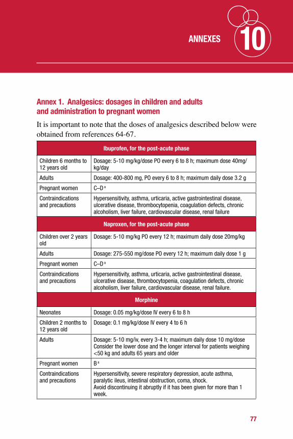
77
References
Annex 1. Analgesics: dosages in children and adults and administration to pregnant women
It is important to note that the doses of analgesics described below were obtained from references 64-67.
Ibuprofen, for the post-acute phase
Children 6 months to 12 years old
Dosage: 5-10 mg/kg/dose PO every 6 to 8 h; maximum dose 40mg/kg/day
Adults Dosage: 400-800 mg, PO every 6 to 8 h; maximum daily dose 3.2 g
Pregnant women C–D a
Contraindications and precautions
Hypersensitivity, asthma, urticaria, active gastrointestinal disease, ulcerative disease, thrombocytopenia, coagulation defects, chronic alcoholism, liver failure, cardiovascular disease, renal failure
Naproxen, for the post-acute phase
Children over 2 years old
Dosage: 5-10 mg/kg PO every 12 h; maximum daily dose 20mg/kg
Adults Dosage: 275-550 mg/dose PO every 12 h; maximum daily dose 1 g
Pregnant women C–D a
Contraindications and precautions
Hypersensitivity, asthma, urticaria, active gastrointestinal disease, ulcerative disease, thrombocytopenia, coagulation defects, chronic alcoholism, liver failure, cardiovascular disease, renal failure.
Morphine
Neonates Dosage: 0.05 mg/kg/dose IV every 6 to 8 h
Children 2 months to 12 years old
Dosage: 0.1 mg/kg/dose IV every 4 to 6 h
Adults Dosage: 5-10 mg/iv, every 3-4 h; maximum daily dose 10 mg/dose Consider the lower dose and the longer interval for patients weighing <50 kg and adults 65 years and older
Pregnant women B a
Contraindications and precautions
Hypersensitivity, severe respiratory depression, acute asthma, paralytic ileus, intestinal obstruction, coma, shock. Avoid discontinuing it abruptly if it has been given for more than 1 week.
ANNEXES 10

78
Tool for the diagnosis and care of patients with suspected arboviral diseases
Prednisone, for joint damage in the chronic phase
Children Dosage: 0.05-2 mg/kg/d PO divided every 12 to 24 h; maximum daily dose 60 mg
Adults Dosage: 5 to 60 mg PO every 24 h; maximum daily dose 60 mg
Pregnant women C a
Contraindications and precautions
Hypersensitivity, mycotic infection, or recent varicella or measles. Avoid discontinuing it abruptly if it has been given for more than 1 week. Do not give steroids to children under 3 years of age. Do not give steroids during the acute or subacute phase (0 to 90 days).
Prednisolone, for joint damage in the chronic phase
Children Dosage: 0.1-2 mg/kg/day PO divided every 6 to 8 h; maximum daily dose 60 mg
Adults Dosage: 5-60 mg PO every 24 h; maximum daily dose 80 mg
Pregnant women C a
Contraindications and precautions
Hypersensitivity, mycotic infection, any active infection, or recent chickenpox or measles. Avoid discontinuing it abruptly if it has been given for more than 1 week. Do not give steroids to children under 3 years of age. Do not give steroids during the acute or subacute phase (0 to 90 days).
Tramadol, for the acute phase
Children Dosage: 1 to 2 mg/kg/dose PO every 6 to 8 h; maximum daily dose 2 mg/kg/dose
Adults Dosage: 100 mg PO every 6 to 8 h; maximum daily dose 400 mg
Pregnant women C a
Contraindications and precautions
Hypersensitivity to the drug, acute renal failure, liver failure, suicidal behavior, acute alcohol poisoning. Do not use in combination with a hypnotic drug, opioid analgesic, or psychotropic drug. Do not give to patients who are taking a monoamine oxidase inhibitor or have taken such a drug in the last 14 days.
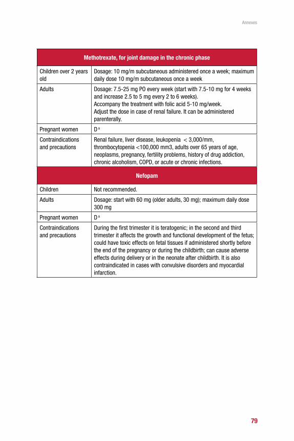
79
References
Methotrexate, for joint damage in the chronic phase
Children over 2 years old
Dosage: 10 mg/m subcutaneous administered once a week; maximum daily dose 10 mg/m subcutaneous once a week
Adults Dosage: 7.5-25 mg PO every week (start with 7.5-10 mg for 4 weeks and increase 2.5 to 5 mg every 2 to 6 weeks). Accompany the treatment with folic acid 5-10 mg/week. Adjust the dose in case of renal failure. It can be administered parenterally.
Pregnant women D a
Contraindications and precautions
Renal failure, liver disease, leukopenia < 3,000/mm, thrombocytopenia <100,000 mm3, adults over 65 years of age, neoplasms, pregnancy, fertility problems, history of drug addiction, chronic alcoholism, COPD, or acute or chronic infections.
Nefopam
Children Not recommended.
Adults Dosage: start with 60 mg (older adults, 30 mg); maximum daily dose 300 mg
Pregnant women D a
Contraindications and precautions
During the first trimester it is teratogenic; in the second and third trimester it affects the growth and functional development of the fetus; could have toxic effects on fetal tissues if administered shortly before the end of the pregnancy or during the childbirth; can cause adverse effects during delivery or in the neonate after childbirth. It is also contraindicated in cases with convulsive disorders and myocardial infarction.
Annexes
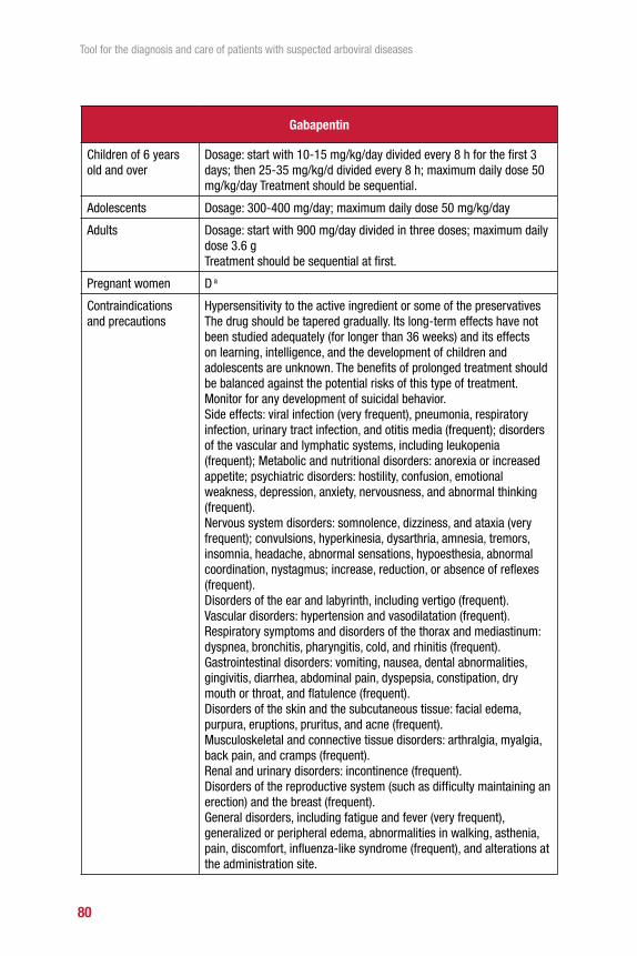
80
Tool for the diagnosis and care of patients with suspected arboviral diseases
Gabapentin
Children of 6 years old and over
Dosage: start with 10-15 mg/kg/day divided every 8 h for the first 3 days; then 25-35 mg/kg/d divided every 8 h; maximum daily dose 50 mg/kg/day Treatment should be sequential.
Adolescents Dosage: 300-400 mg/day; maximum daily dose 50 mg/kg/day
Adults Dosage: start with 900 mg/day divided in three doses; maximum daily dose 3.6 g Treatment should be sequential at first.
Pregnant women D a
Contraindications and precautions
Hypersensitivity to the active ingredient or some of the preservatives The drug should be tapered gradually. Its long-term effects have not been studied adequately (for longer than 36 weeks) and its effects on learning, intelligence, and the development of children and adolescents are unknown. The benefits of prolonged treatment should be balanced against the potential risks of this type of treatment. Monitor for any development of suicidal behavior. Side effects: viral infection (very frequent), pneumonia, respiratory infection, urinary tract infection, and otitis media (frequent); disorders of the vascular and lymphatic systems, including leukopenia (frequent); Metabolic and nutritional disorders: anorexia or increased appetite; psychiatric disorders: hostility, confusion, emotional weakness, depression, anxiety, nervousness, and abnormal thinking (frequent). Nervous system disorders: somnolence, dizziness, and ataxia (very frequent); convulsions, hyperkinesia, dysarthria, amnesia, tremors, insomnia, headache, abnormal sensations, hypoesthesia, abnormal coordination, nystagmus; increase, reduction, or absence of reflexes (frequent). Disorders of the ear and labyrinth, including vertigo (frequent). Vascular disorders: hypertension and vasodilatation (frequent). Respiratory symptoms and disorders of the thorax and mediastinum: dyspnea, bronchitis, pharyngitis, cold, and rhinitis (frequent). Gastrointestinal disorders: vomiting, nausea, dental abnormalities, gingivitis, diarrhea, abdominal pain, dyspepsia, constipation, dry mouth or throat, and flatulence (frequent). Disorders of the skin and the subcutaneous tissue: facial edema, purpura, eruptions, pruritus, and acne (frequent). Musculoskeletal and connective tissue disorders: arthralgia, myalgia, back pain, and cramps (frequent). Renal and urinary disorders: incontinence (frequent). Disorders of the reproductive system (such as difficulty maintaining an erection) and the breast (frequent). General disorders, including fatigue and fever (very frequent), generalized or peripheral edema, abnormalities in walking, asthenia, pain, discomfort, influenza-like syndrome (frequent), and alterations at the administration site.

81
References
Pregabalin
Children No safety or efficacy studies have been conducted.
Adults Dosage: 150 mg/dayThe dosage can be increased up to 300 mg a day after 3 to 7 days and, if necessary, up to a maximum dose of 600 mg/day after an additional 7 days. Maximum daily does: 600 mg.
Pregnancy Information on its effects on pregnancy is insufficient.
Contraindications and precautions
Pregabalin is contraindicated in patients with hypersensitivity to the active ingredient or any of the excipients. Some diabetics may gain weight during treatment with pregabalin; hypoglycemia medication may be needed or need to be adjusted. It has been associated to dizziness and drowsiness. Patients are warned not to drive, operate heavy machinery, or engage in other potentially hazardous activities until it is known whether the drug affects their ability to perform these activities. Animal studies have demonstrated reproductive toxicity. The risk for humans is unknown. Therefore, pregabalin should not be taken during pregnancy unless the benefit for the mother outweighs the potential risk to the fetus. Women of childbearing age should use an effective contraceptive method. It is not known whether pregabalin is excreted in human breast milk, but it is present in rat milk. Therefore, breastfeeding is not recommended as long as this drug is being used.
Annexes
a U.S. Food and Drug Administration (FDA) definitions of categories of pregnancy risk:- A: Controlled studies in women have failed to demonstrate a risk to the fetus in the first trimester
and the possibility of fetal harm appears remote. - B: Animal studies have failed to demonstrate a risk to the fetus and there have been no controlled
studies in humans, or else animal studies do indicate an adverse effect on the fetus but well-controlled studies in pregnant women have failed to demonstrate any fetal risk.
- C: Animal studies have demonstrated that the drug has teratogenic or embryocidal effects but there are no controlled studies in women, or there are no available studies in either animals or women.
- D: There is positive evidence of human fetal risk, but in certain cases (for example, threatening situations or severe diseases in which safer drugs cannot be used or those that can be used are ineffective), the benefits may warrant use of the drug despite the risks.
- X: Studies in animals or humans have demonstrated fetal abnormalities and/or there is evidence of fetal risk based on the experience with human beings and the risks clearly outweigh any potential benefit.

82
Tool for the diagnosis and care of patients with suspected arboviral diseases
Annex 2: WHO analgesic ladder
The analgesic ladder essentially consists of the following three steps:
1) First or lowest step (mild pain): non-opioid ± adjuvants.
2) Second or intermediate step (moderate pain): weak opioid ± non-opioid ± adjuvants.
3) Third or highest step (severe pain): strong opioids ± non-opioids ± adjuvants.
Figure 5. WHO Three-Step Analgesic Ladder
Freedom from cancer pain
Pain persisting or increasing
Pain persisting or increasing
Pain
Opioid for moderate to servere pain± Non-opioid± Adjuvant
Opioid for mild to moderate pain± Non-opioid± Adjuvant
± Non-opioid± Adjuvant
23
1Source: Adapted from Cancer pain relief (65).
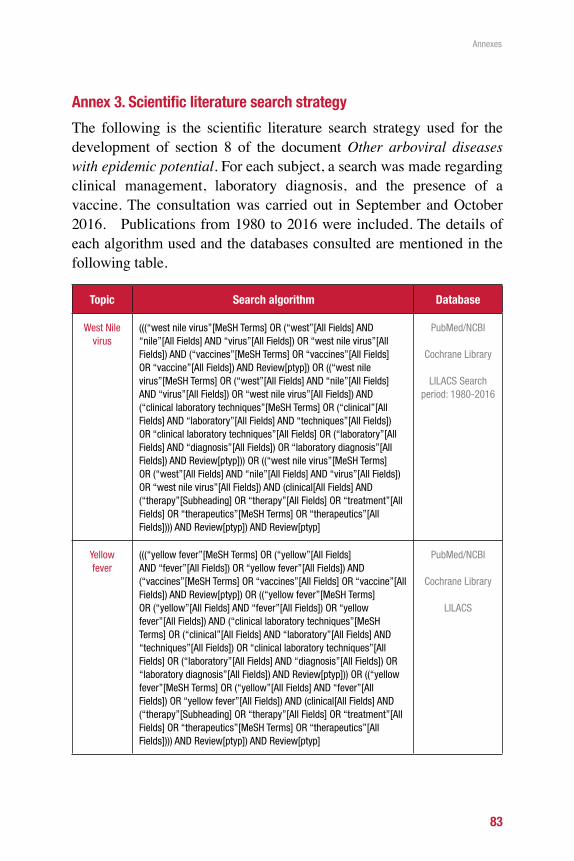
83
References
Annex 3. Scientific literature search strategy
The following is the scientific literature search strategy used for the development of section 8 of the document Other arboviral diseases with epidemic potential. For each subject, a search was made regarding clinical management, laboratory diagnosis, and the presence of a vaccine. The consultation was carried out in September and October 2016. Publications from 1980 to 2016 were included. The details of each algorithm used and the databases consulted are mentioned in the following table.
Annexes
Topic Search algorithm Database
West Nile virus
(((“west nile virus”[MeSH Terms] OR (“west”[All Fields] AND “nile”[All Fields] AND “virus”[All Fields]) OR “west nile virus”[All Fields]) AND (“vaccines”[MeSH Terms] OR “vaccines”[All Fields] OR “vaccine”[All Fields]) AND Review[ptyp]) OR ((“west nile virus”[MeSH Terms] OR (“west”[All Fields] AND “nile”[All Fields] AND “virus”[All Fields]) OR “west nile virus”[All Fields]) AND (“clinical laboratory techniques”[MeSH Terms] OR (“clinical”[All Fields] AND “laboratory”[All Fields] AND “techniques”[All Fields]) OR “clinical laboratory techniques”[All Fields] OR (“laboratory”[All Fields] AND “diagnosis”[All Fields]) OR “laboratory diagnosis”[All Fields]) AND Review[ptyp])) OR ((“west nile virus”[MeSH Terms] OR (“west”[All Fields] AND “nile”[All Fields] AND “virus”[All Fields]) OR “west nile virus”[All Fields]) AND (clinical[All Fields] AND (“therapy”[Subheading] OR “therapy”[All Fields] OR “treatment”[All Fields] OR “therapeutics”[MeSH Terms] OR “therapeutics”[All Fields]))) AND Review[ptyp]) AND Review[ptyp]
PubMed/NCBI
Cochrane Library
LILACS Search period: 1980-2016
Yellow fever
(((“yellow fever”[MeSH Terms] OR (“yellow”[All Fields] AND “fever”[All Fields]) OR “yellow fever”[All Fields]) AND (“vaccines”[MeSH Terms] OR “vaccines”[All Fields] OR “vaccine”[All Fields]) AND Review[ptyp]) OR ((“yellow fever”[MeSH Terms] OR (“yellow”[All Fields] AND “fever”[All Fields]) OR “yellow fever”[All Fields]) AND (“clinical laboratory techniques”[MeSH Terms] OR (“clinical”[All Fields] AND “laboratory”[All Fields] AND “techniques”[All Fields]) OR “clinical laboratory techniques”[All Fields] OR (“laboratory”[All Fields] AND “diagnosis”[All Fields]) OR “laboratory diagnosis”[All Fields]) AND Review[ptyp])) OR ((“yellow fever”[MeSH Terms] OR (“yellow”[All Fields] AND “fever”[All Fields]) OR “yellow fever”[All Fields]) AND (clinical[All Fields] AND (“therapy”[Subheading] OR “therapy”[All Fields] OR “treatment”[All Fields] OR “therapeutics”[MeSH Terms] OR “therapeutics”[All Fields]))) AND Review[ptyp]) AND Review[ptyp]
PubMed/NCBI
Cochrane Library
LILACS

84
Tool for the diagnosis and care of patients with suspected arboviral diseases
Topic Search algorithm Database
Oropouche fever
((“oropouche”[All Fields] AND (“vaccines”[MeSH Terms] OR “vaccines”[All Fields] OR “vaccine”[All Fields])) OR (“oropouche”[All Fields] AND (“clinical laboratory techniques”[MeSH Terms] OR (“clinical”[All Fields] AND “laboratory”[All Fields] AND “techniques”[All Fields]) OR “clinical laboratory techniques”[All Fields] OR (“laboratory”[All Fields] AND “diagnosis”[All Fields]) OR “laboratory diagnosis”[All Fields]))) OR (oropouche[All Fields] AND (clinical[All Fields] AND (“therapy”[Subheading] OR “therapy”[All Fields] OR “treatment”[All Fields] OR “therapeutics”[MeSH Terms] OR “therapeutics”[All Fields])))
PubMed/NCBI
Cochrane Library
LILACS
Mayaro fever
(((“mayaro fever”[MeSH Terms] OR (“mayaro”[All Fields] AND “fever”[All Fields]) OR “mayaro fever”[All Fields]) AND (“vaccines”[MeSH Terms] OR “vaccines”[All Fields] OR “vaccine”[All Fields]) AND Review[ptyp]) OR ((“mayaro fever”[MeSH Terms] OR (“mayaro”[All Fields] AND “fever”[All Fields]) OR “mayaro fever”[All Fields]) AND (“clinical laboratory techniques”[MeSH Terms] OR (“clinical”[All Fields] AND “laboratory”[All Fields] AND “techniques”[All Fields]) OR “clinical laboratory techniques”[All Fields] OR (“laboratory”[All Fields] AND “diagnosis”[All Fields]) OR “laboratory diagnosis”[All Fields]) AND Review[ptyp])) OR ((“mayaro fever”[MeSH Terms] OR (“mayaro”[All Fields] AND “fever”[All Fields]) OR “mayaro fever”[All Fields]) AND (clinical[All Fields] AND (“therapy”[Subheading] OR “therapy”[All Fields] OR “treatment”[All Fields] OR “therapeutics”[MeSH Terms] OR “therapeutics”[All Fields])) AND Review[ptyp]) AND Review[ptyp]
PubMed/NCBI
Cochrane Library
LILACS
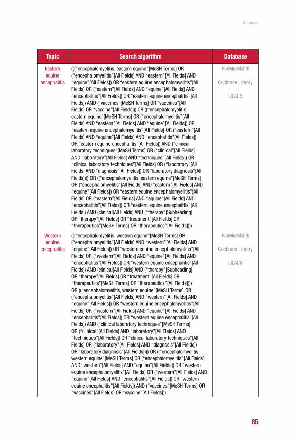
85
References
Topic Search algorithm Database
Eastern equine
encephalitis
(((“encephalomyelitis, eastern equine”[MeSH Terms] OR (“encephalomyelitis”[All Fields] AND “eastern”[All Fields] AND “equine”[All Fields]) OR “eastern equine encephalomyelitis”[All Fields] OR (“eastern”[All Fields] AND “equine”[All Fields] AND “encephalitis”[All Fields]) OR “eastern equine encephalitis”[All Fields]) AND (“vaccines”[MeSH Terms] OR “vaccines”[All Fields] OR “vaccine”[All Fields])) OR ((“encephalomyelitis, eastern equine”[MeSH Terms] OR (“encephalomyelitis”[All Fields] AND “eastern”[All Fields] AND “equine”[All Fields]) OR “eastern equine encephalomyelitis”[All Fields] OR (“eastern”[All Fields] AND “equine”[All Fields] AND “encephalitis”[All Fields]) OR “eastern equine encephalitis”[All Fields]) AND (“clinical laboratory techniques”[MeSH Terms] OR (“clinical”[All Fields] AND “laboratory”[All Fields] AND “techniques”[All Fields]) OR “clinical laboratory techniques”[All Fields] OR (“laboratory”[All Fields] AND “diagnosis”[All Fields]) OR “laboratory diagnosis”[All Fields]))) OR ((“encephalomyelitis, eastern equine”[MeSH Terms] OR (“encephalomyelitis”[All Fields] AND “eastern”[All Fields] AND “equine”[All Fields]) OR “eastern equine encephalomyelitis”[All Fields] OR (“eastern”[All Fields] AND “equine”[All Fields] AND “encephalitis”[All Fields]) OR “eastern equine encephalitis”[All Fields]) AND (clinical[All Fields] AND (“therapy”[Subheading] OR “therapy”[All Fields] OR “treatment”[All Fields] OR “therapeutics”[MeSH Terms] OR “therapeutics”[All Fields])))
PubMed/NCBI
Cochrane Library
LILACS
Western equine
encephalitis
(((“encephalomyelitis, western equine”[MeSH Terms] OR (“encephalomyelitis”[All Fields] AND “western”[All Fields] AND “equine”[All Fields]) OR “western equine encephalomyelitis”[All Fields] OR (“western”[All Fields] AND “equine”[All Fields] AND “encephalitis”[All Fields]) OR “western equine encephalitis”[All Fields]) AND (clinical[All Fields] AND (“therapy”[Subheading] OR “therapy”[All Fields] OR “treatment”[All Fields] OR “therapeutics”[MeSH Terms] OR “therapeutics”[All Fields]))) OR ((“encephalomyelitis, western equine”[MeSH Terms] OR (“encephalomyelitis”[All Fields] AND “western”[All Fields] AND “equine”[All Fields]) OR “western equine encephalomyelitis”[All Fields] OR (“western”[All Fields] AND “equine”[All Fields] AND “encephalitis”[All Fields]) OR “western equine encephalitis”[All Fields]) AND (“clinical laboratory techniques”[MeSH Terms] OR (“clinical”[All Fields] AND “laboratory”[All Fields] AND “techniques”[All Fields]) OR “clinical laboratory techniques”[All Fields] OR (“laboratory”[All Fields] AND “diagnosis”[All Fields]) OR “laboratory diagnosis”[All Fields]))) OR ((“encephalomyelitis, western equine”[MeSH Terms] OR (“encephalomyelitis”[All Fields] AND “western”[All Fields] AND “equine”[All Fields]) OR “western equine encephalomyelitis”[All Fields] OR (“western”[All Fields] AND “equine”[All Fields] AND “encephalitis”[All Fields]) OR “western equine encephalitis”[All Fields]) AND (“vaccines”[MeSH Terms] OR “vaccines”[All Fields] OR “vaccine”[All Fields]))
PubMed/NCBI
Cochrane Library
LILACS
Annexes
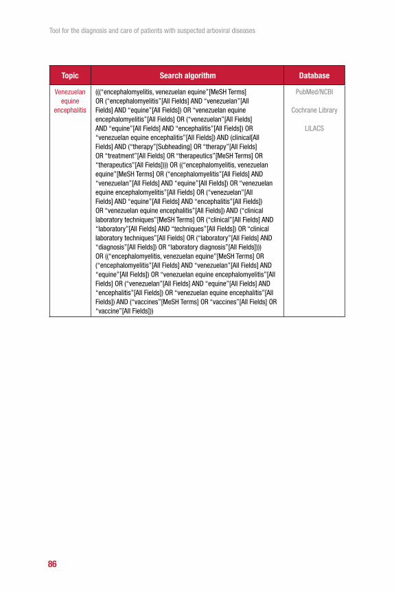
86
Tool for the diagnosis and care of patients with suspected arboviral diseases
Topic Search algorithm Database
Venezuelan equine
encephalitis
(((“encephalomyelitis, venezuelan equine”[MeSH Terms] OR (“encephalomyelitis”[All Fields] AND “venezuelan”[All Fields] AND “equine”[All Fields]) OR “venezuelan equine encephalomyelitis”[All Fields] OR (“venezuelan”[All Fields] AND “equine”[All Fields] AND “encephalitis”[All Fields]) OR “venezuelan equine encephalitis”[All Fields]) AND (clinical[All Fields] AND (“therapy”[Subheading] OR “therapy”[All Fields] OR “treatment”[All Fields] OR “therapeutics”[MeSH Terms] OR “therapeutics”[All Fields]))) OR ((“encephalomyelitis, venezuelan equine”[MeSH Terms] OR (“encephalomyelitis”[All Fields] AND “venezuelan”[All Fields] AND “equine”[All Fields]) OR “venezuelan equine encephalomyelitis”[All Fields] OR (“venezuelan”[All Fields] AND “equine”[All Fields] AND “encephalitis”[All Fields]) OR “venezuelan equine encephalitis”[All Fields]) AND (“clinical laboratory techniques”[MeSH Terms] OR (“clinical”[All Fields] AND “laboratory”[All Fields] AND “techniques”[All Fields]) OR “clinical laboratory techniques”[All Fields] OR (“laboratory”[All Fields] AND “diagnosis”[All Fields]) OR “laboratory diagnosis”[All Fields]))) OR ((“encephalomyelitis, venezuelan equine”[MeSH Terms] OR (“encephalomyelitis”[All Fields] AND “venezuelan”[All Fields] AND “equine”[All Fields]) OR “venezuelan equine encephalomyelitis”[All Fields] OR (“venezuelan”[All Fields] AND “equine”[All Fields] AND “encephalitis”[All Fields]) OR “venezuelan equine encephalitis”[All Fields]) AND (“vaccines”[MeSH Terms] OR “vaccines”[All Fields] OR “vaccine”[All Fields]))
PubMed/NCBI
Cochrane Library
LILACS

87
References
88
Tool for the diagnosis and care of patients with suspected arboviral diseases