chip-it® ffpe chromatin preparation iioverview chromatin immunoprecipitation (chip) is a powerful...
TRANSCRIPT
www.activemotif.com
ChIP-IT® FFPEChromatin Preparation II
(version A1)
Catalog No. 53031
Active Motif North America 1914 Palomar Oaks Way, Suite 150 Carlsbad, California 92008, USA Toll free: 877 222 9543 Telephone: 760 431 1263 Fax: 760 431 1351
Active Motif Europe Avenue Reine Astrid, 92 B-1310 La Hulpe, Belgium UK Free Phone: 0800 169 31 47 France Free Phone: 0800 90 99 79 Germany Free Phone: 0800 181 99 10 Telephone: +32 (0)2 653 0001 Fax: +32 (0)2 653 0050
Active Motif Japan Azuma Bldg, 7th Floor 2-21 Ageba-Cho, Shinjuku-Ku Tokyo, 162-0824, Japan Telephone: +81 3 5225 3638 Fax: +81 3 5261 8733
Active Motif China 787 Kangqiao Road Building 10, Suite 202, Pudong District Shanghai, 201315, China Telephone: (86)-21-20926090 Hotline: 400-018-8123
Copyright 2018 Active Motif, Inc.

www.activemotif.com
Information in this manual is subject to change without notice and does not constitute a commit-ment on the part of Active Motif, Inc. It is supplied on an “as is” basis without any warranty of any kind, either explicit or implied. Information may be changed or updated in this manual at any time.
This documentation may not be copied, transferred, reproduced, disclosed, or duplicated, in whole or in part, without the prior written consent of Active Motif, Inc. This documentation is proprietary information and protected by the copyright laws of the United States and interna-tional treaties.
The manufacturer of this documentation is Active Motif, Inc.
© 2018 Active Motif, Inc., 1914 Palomar Oaks Way, Suite 150; Carlsbad, CA 92008. All rights reserved.
All trademarks, trade names, service marks or logos referenced herein belong to their respective companies.
www.activemotif.com
TABLE OF CONTENTS Page
Overview . . . . . . . . . . . . . . . . . . . . . . . . . . . . . . . . . . . . . . . . . . . . . . . . . . . . . . . . . . . . . . . . . . . . . . . . . . . . 1
Flow Chart of Process . . . . . . . . . . . . . . . . . . . . . . . . . . . . . . . . . . . . . . . . . . . . . . . . . . . . . . . . . . . . . . . .2
Introduction . . . . . . . . . . . . . . . . . . . . . . . . . . . . . . . . . . . . . . . . . . . . . . . . . . . . . . . . . . . . . . . . . . . . . . . . .3
References. . . . . . . . . . . . . . . . . . . . . . . . . . . . . . . . . . . . . . . . . . . . . . . . . . . . . . . . . . . . . . . . . . . . . . . . . . .3
Kit Performance and Benefits . . . . . . . . . . . . . . . . . . . . . . . . . . . . . . . . . . . . . . . . . . . . . . . . . . . . . . . . .4 ChIP-IT FFPE Chromatin Preparation II. . . . . . . . . . . . . . . . . . . . . . . . . . . . . . . . . . . . . . . . . . . . . .5
Kit Components and Storage . . . . . . . . . . . . . . . . . . . . . . . . . . . . . . . . . . . . . . . . . . . . . . . . . . . . . . . . .6 Additional Materials Required. . . . . . . . . . . . . . . . . . . . . . . . . . . . . . . . . . . . . . . . . . . . . . . . . . . . .6 Protocol Overview and Time Table . . . . . . . . . . . . . . . . . . . . . . . . . . . . . . . . . . . . . . . . . . . . . . . .7
Protocols – Experimental Set Up Buffer Preparation . . . . . . . . . . . . . . . . . . . . . . . . . . . . . . . . . . . . . . . . . . . . . . . . . . . . . . . . . . . . . . .8 Recommendations. . . . . . . . . . . . . . . . . . . . . . . . . . . . . . . . . . . . . . . . . . . . . . . . . . . . . . . . . . . . . . .9
Protocols – Preparation of Sheared Chromatin Section A. Removal of Paraffin & Rehydration of FFPE Tissue Slides. . . . . . . . . . . . . . . . . 10 Section B. Removal of Paraffin & Rehydration of FFPE Tissue Blocks . . . . . . . . . . . . . . . . 11 Section C. Tissue Homogenization and Chromatin Isolation . . . . . . . . . . . . . . . . . . . . . . . 12 Section D. Input Preparation. . . . . . . . . . . . . . . . . . . . . . . . . . . . . . . . . . . . . . . . . . . . . . . . . . . . 13 Section E. Analysis of Input DNA and Chromatin Quality . . . . . . . . . . . . . . . . . . . . . . . . . . 14 Section F. Additional Sonication of Insoluble Pellet . . . . . . . . . . . . . . . . . . . . . . . . . . . . . . 16
Appendix Section G. qPCR Primer Design and Data Analysis . . . . . . . . . . . . . . . . . . . . . . . . . . . . . . . . . 17 Section H. Troubleshooting Guide . . . . . . . . . . . . . . . . . . . . . . . . . . . . . . . . . . . . . . . . . . . . . . 18 Section I. Related Products . . . . . . . . . . . . . . . . . . . . . . . . . . . . . . . . . . . . . . . . . . . . . . . . . . . . 19
Technical Services . . . . . . . . . . . . . . . . . . . . . . . . . . . . . . . . . . . . . . . . . . . . . . . . . . . . . . . . . . . . . . . . . . 20

1www.activemotif.com
Overview
Chromatin Immunoprecipitation (ChIP) is a powerful tool for studying protein/DNA interactions, including transcription factors, co-regulatory proteins, modified histones, chromatin-modifying enzymes and polymerases because it enables identification of the localization of proteins bound to specific DNA loci. When used in combination with whole-genome analysis such as ChIP-Seq, insights are possible into gene regulation, gene expression, mechanisms of chromatin modification and pathway analysis.
Formalin-fixed paraffin-embedded (FFPE) tissue blocks and histology slides are a valuable resource for retrospective research on clinical samples. Clinical information, treatments and outcomes are often available for these sample types and large collections of FFPE material is commercially avail-able. The ability to study FFPE samples provides researchers with an opportunity to link FFPE data to disease, diagnosis and biomarker discovery. Traditionally, FFPE samples have not been useful in chromatin immunoprecipitation because of the limited size of the samples, and the fact that the formalin fixation process often causes degradation and loss of antigenicity.
Active Motif’s ChIP-IT® FFPE Chromatin Preparation II and ChIP-IT® FFPE II Kits are our second generation FFPE ChIP kits in which the cumbersome de-paraffinization and dehydration procedure is streamlined to allow the preparation of high quality ChIP-enriched DNA using less reagents and hands on time. This streamlined chromatin preparation protocol is optimal when working with limited amounts FFPE tissue. The ChIP-IT FFPE II Kit has been optimized using specially formulated reagents and protocol guidelines to increase sensitivity and enable analysis by both qPCR and Next Generation sequencing from extremely limited starting material, while producing minimal background signal, thereby enabling specific detection of the target protein of interest.
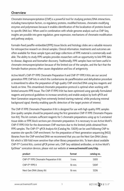
The ChIP-IT FFPE Chromatin Preparation II Kit is designed for use with high quality FFPE samples (low quality samples should be prepared using the first generation ChIP-IT FFPE Chromatin Prepara-tion Kit). This kit contains sufficient reagents for 5 chromatin preparations using up to 5 unstained tissue slides or FFPE block sections per chromatin preparation. It is necessary to use Active Motif’s ChIP-IT FFPE II Kit for the downstream ChIP reactions due to the limited yields obtained from FFPE samples. The ChIP-IT® qPCR Analysis Kit (Catalog No. 53029) can be used following ChIP to examine site-specific ChIP enrichment. For the preparation of Next generation sequencing (NGS) libraries from the ChIP-enriched DNA we recommend that you use the Next Gen DNA Library Kit which is 500-fold more sensitive than other library preparation kits. To learn about available ChIP-IT® Control Kits, control qPCR primer sets, ChIP-Seq validated antibodies, or Active Motif’s EpiShear™ sonication devices, please visit our website at www.activemotif.com/chip.
product format catalog no.
ChIP-IT® FFPE Chromatin Preparation II Kit 5 rxns 53031
ChIP-IT® FFPE II 16 rxns 53047
Next Gen DNA Library Kit 16 rxns 53216
2www.activemotif.com
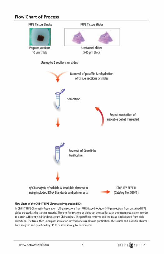
Flow Chart of Process
Flow Chart of the ChIP-IT FFPE Chromatin Preparation II Kit.
In ChIP-IT FFPE Chromatin Preparation II, 10 µm sections from FFPE tissue blocks, or 5-10 µm sections from unstained FFPE slides are used as the starting material. Three to five sections or slides can be used for each chromatin preparation in order to obtain sufficient yield for downstream ChIP analysis. The paraffin is removed and the tissue is rehydrated from each slide/tube. The tissue then undergoes sonication, reversal of crosslinks and purification. The soluble and insoluble chroma-tin is analyzed and quantified by qPCR, or alternatively, by fluorometer.
3www.activemotif.com
Introduction
Formalin-fixed paraffin embedded (FFPE) samples represent an opportunity for researchers to study clinical outcomes of disease and/or treatment conditions in the search to better understand the disease, or as a mechanism to identify biomarkers for screening purposes. FFPE samples serve as the “gold standard” for pathology sample preservation and large collections of these tissues are available.
There are many challenges associated with working with FFPE samples. The samples are often limited in size and require the use of multiple histological slides or tissue sections to extract suf-ficient quantities of material for downstream analysis. Another challenge is the lack of consistency in the methodologies used for formalin fixation. Some treatments tend to be harsh, causing degradation of the sample, loss of antigenicity, or they create “overfixed” chromatin which is difficult to efficiently shear. Although FFPE samples have been used for high-throughput DNA and RNA analysis1,2, the challenges explained above have prevented FFPE material from being used in chromatin immunoprecipitation (ChIP).
ChIP itself can be technically demanding. ChIP requires high-quality antibodies to recognize the fixed, target-bound proteins of interest, and an efficient means to precipitate the antibody/chro-matin complex (usually protein A or G beads). In addition, specialized buffers, inhibitor cocktails and blocking reagents are required to minimize non-specific enrichment and reduce protein degradation.
Researchers have started to address the need for a methodology to study the influences of epi-genetics on normal and tumor samples beyond the traditional immunohistochemistry (IHC) analy-sis. Pathology tissue chromatin immunoprecipitation (PAT-ChIP) was the first method to extract and analyze FFPE chromatin for use in high-throughput analysis, such as ChIP-Seq3,4. Subsequently, many laboratories have attempted to prepare high quality chromatin from FFPE samples5, however many are still reporting issues in generating quality chromatin from ChIP-seq analysis.
Active Motif has utilized our expertise with ChIP to develop a suite of kits for performing ChIP on FFPE samples for use in Next-generation sequencing. The ChIP-IT FFPE Chromatin Preparation II Kit contains specially formulated reagents and protocol guidelines to extract high quality chromatin from limited amounts of histological slides or tissue sections. Coupled with our newly developed and most sensitive ChIP kit, ChIP-IT FFPE II, you have everything you need to generate high quality ChIP-Seq data from extremely limited starting material while producing minimal background signal, thereby enabling specific detection of the target protein of interest. All Active Motif FFPE ChIP kits contain controls to help validate results at each step of the process.
References
1. Weng, L. et al. (2010) J Pathol., 222: 41-51.2. Gu, H.., et al. (2010) Nat. Methods, 7: 133-136.3. Fanelli, M. et al. (2010) PNAS, 107(50): 21535-21540.4. Fanelli, M. et al. (2011) Nat. Protocols, 6(12): 1905-1919.5. Cejas, P. et al. (2016) Nat. Med., 22(6): 685-91.
4www.activemotif.com
Kit Performance and Benefits
ChIP-IT FFPE Chromatin Preparation II Advantages:• Easily obtain chromatin using limited amounts of histological slides or tissue blocks as the
starting material
• Optimized reagents help preserve the quality of the chromatin during extraction
• Highly robust procedure has been validated using FFPE chromatin from both normal and tumor samples with proven performance in both qPCR and ChIP-Seq analysis
• Includes positive control DNA and PCR primers to quantify the chromatin and confirm the shearing efficiency prior to use in ChIP
Detection limit: The limits of the assay will depend on the size and tissue type and may require optimization. The ChIP-IT FFPE Chromatin Preparation II kit was specifically developed for extract-ing high quality chromatin for use in ChIP from limited, but high quality, FFPE tissue slides or blocks. The protocol was optimized to require only five 5 µm sections per chromatin preparation . Our protocol offers guidelines and troubleshooting tips for processing samples to obtain the required 200 ng minimum of chromatin per ChIP reaction. Chromatin should be validated prior to use in the ChIP-IT FFPE II Kit (Catalog No. 53047).
Product Performance: The ChIP-IT FFPE Chromatin Preparation Kit assumes that FFPE tissues are already available as standard sections. Due to the variability that exists in the formalin fixation process and the storage conditions of the sample, not all FFPE material may yield high quality chromatin. The ChIP-IT FFPE Chromatin Preparation II Kit provides recommendations and guide-lines to assist in the extraction process, but Active Motif cannot guarantee successful chromatin preparations for variables that fall outside of our assay kit.
If preparing samples for FFPE preservation, it is important to minimize the time between the resection of the tissue and the fixation in formalin to generate high quality FFPE tissues. Samples should be fixed immediately and stored in a cool, dry location. With human samples, due to the need for surgical removal, there is often variability in the time from resection to fixation. For animal models, in vivo perfusion can be used to generate high quality FFPE tissues.
5www.activemotif.com
ChIP-IT® FFPE Chromatin Preparation II
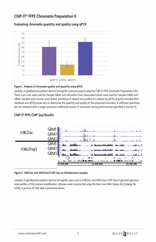
Evaluating chromatin quantity and quality using qPCR
Figure 1. Analysis of chromatin quality and quantity using qPCR.
Samples of glioblastoma patient-derived xenografts were processed using the ChIP-IT FFPE Chromatin Preparation II Kit. Three 5 µm curls were used for Sample GBM1, and cell pellets from dissociated tumor were used for Samples GBM2 and GBM3. Samples were reverse cross-linked, proteinase K treated and purified for analysis by qPCR using the included DNA standards and qPCR primer sets to determine the quantity and quality of the extracted chromatin. If sufficient quantities are not obtained after a single sonication, additional rounds of sonication can be performed (as specified in Section F).
ChIP-IT FFPE ChIP-Seq Results
Figure 2: H3K27ac and H3K27me3 ChIP-Seq on Glioblastoma samples.
Samples of glioblastoma patient-derived xenografts were used in H3K27ac and H3K27me3 ChIP-Seq to generate genome-wide profiles of this histone modification. Libraries were constructed using the Next Gen DNA Library Kit (Catalog No. 53216). A portion of that data is presented above.
6www.activemotif.com
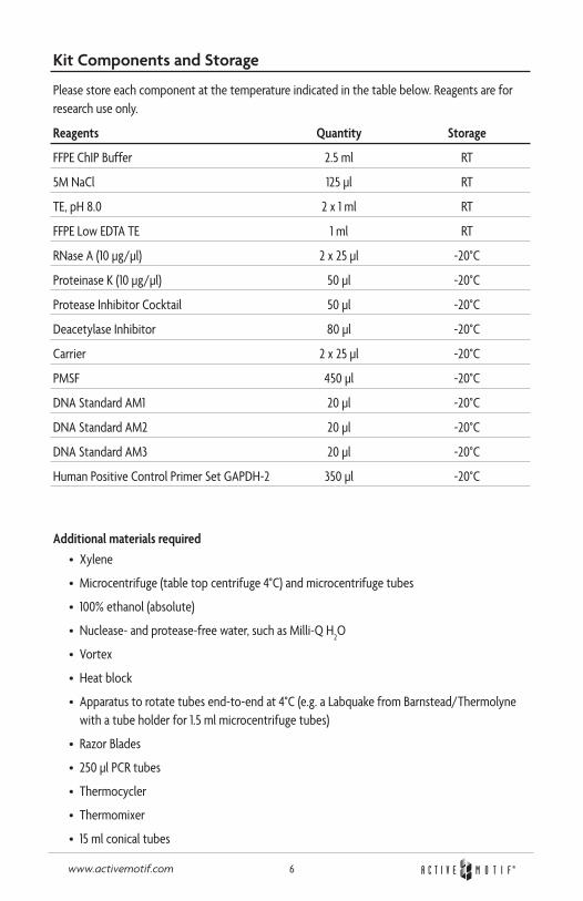
Kit Components and Storage
Please store each component at the temperature indicated in the table below. Reagents are for research use only.
Reagents Quantity Storage
FFPE ChIP Buffer 2.5 ml RT
5M NaCl 125 µl RT
TE, pH 8.0 2 x 1 ml RT
FFPE Low EDTA TE 1 ml RT
RNase A (10 µg/µl) 2 x 25 µl -20°C
Proteinase K (10 µg/µl) 50 µl -20°C
Protease Inhibitor Cocktail 50 µl -20°C
Deacetylase Inhibitor 80 µl -20°C
Carrier 2 x 25 µl -20°C
PMSF 450 µl -20°C
DNA Standard AM1 20 µl -20°C
DNA Standard AM2 20 µl -20°C
DNA Standard AM3 20 µl -20°C
Human Positive Control Primer Set GAPDH-2 350 µl -20°C
Additional materials required• Xylene
• Microcentrifuge (table top centrifuge 4°C) and microcentrifuge tubes
• 100% ethanol (absolute)
• Nuclease- and protease-free water, such as Milli-Q H2O
• Vortex
• Heat block
• Apparatus to rotate tubes end-to-end at 4°C (e.g. a Labquake from Barnstead/Thermolyne with a tube holder for 1.5 ml microcentrifuge tubes)
• Razor Blades
• 250 µl PCR tubes
• Thermocycler
• Thermomixer
• 15 ml conical tubes
7www.activemotif.com
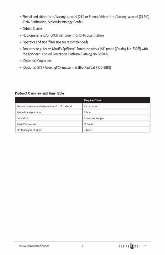
• Phenol and chloroform/isoamyl alcohol (24:1) or Phenol/chloroform/isoamyl alcohol (25:24:1) (DNA Purification, Molecular Biology Grade)
• Orbital Shaker
• Fluorometer and/or qPCR instrument for DNA quantitation
• Pipettors and tips (filter tips are recommended)
• Sonicator (e.g. Active Motif’s EpiShear™ Sonicator with a 1/8” probe (Catalog No. 53051) with the EpiShear™ Cooled Sonication Platform (Catalog No. 53080))
• (Optional) Coplin jars
• (Optional) SYBR Green qPCR master mix (Bio-Rad Cat # 170-8882)
Protocol Overview and Time Table
Required Time
Deparaffinization and rehydration of FFPE material 1.5 - 2 hours
Tissue homogenization 1 hour
Sonication 1 hour per sample
Input Preparation 15 hours
qPCR Analysis of Input 2 hours
8www.activemotif.com
Protocols – Experimental Set Up
PLEASE READ THE ENTIRE PROTOCOL BEFORE STARTING!
Buffer Preparation
FFPE ChIP BufferIs provided ready to use.
Deacetylase InhibitorThaw on ice.
Protease Inhibitor Cocktail (PIC)Thaw the PIC at room temperature until fully dissolved, which takes about 30 minutes. Vortex gently and spin down briefly before use, then add to the buffers immediately before use.
DNA Standards AM1, AM2 and AM3Due to the low quantities of chromatin recovered from FFPE samples, it is necessary to quantify the chromatin using qPCR analysis against known quantities of DNA, such as those provided in DNA Standards AM1, AM2 and AM3. In the qPCR instrument, set the values for DNA Standard AM1 to 2.5 ng/µl, DNA Standard AM2 to 0.25 ng/µl and DNA Standard AM3 to 0.025 ng/µl. Use 2 µl of each DNA standard in the qPCR reactions.
Alternatively, a fluorometric method, such as Qubit™ Fluorometric Quantitation, can be used to quantify the chromatin input. Nanodrop readings are not sensitive enough to detect the low quantities of DNA and, therefore, use of the Nanodrop for DNA quantification is not recom-mended.
Human Positive Control Primer Set GAPDH-2The Human Positive Control Primer Set GAPDH-2 is provided for use in the qPCR quantification of the chromatin input. This primer set can be used to amplify DNA Standards AM1, AM2, AM3 and chromatin input samples. The primer is provided at 2.5 µM. Use 1 µl per qPCR reaction. Alterna-tively, a positive control primer set specific for a human gene-of-interest can be used to amplify the DNA Standards and chromatin samples for quantification.
If not using human material, you will need to design appropriate qPCR primer sets to match your species. We recommend designing primers to perform at an annealing temperature of 58°C so that all qPCR reactions can be performed under identical conditions. An amplicon length of 75-150 bp is recommended. To see a list of validated species-specific qPCR primers designed according to these recommendations, visit www.activemotif.com/chipprimers.
9www.activemotif.com
Recommendations
Chromatin Shearing TipsChIP experiments require chromatin that has been sheared to a size of 200-1200 bp. Due to the limited sample size and yield, there is usually insufficient chromatin available for analysis of shearing efficiency by agarose gel electrophoresis. Instead, we suggest analyzing the quantity and quality of the chromatin preparation through qPCR. The included DNA Standards and qPCR Primer set enables quantification of the chromatin input. Both the soluble chromatin fraction and the insoluble pellet can be analyzed to determine the efficiency of the chromatin preparation. If a majority of the chromatin is still found in the insoluble fraction, the pellet should undergo additional sonication and extraction preparation. The soluble chromatin that is obtained from the additional processing can be combined with the original soluble fraction and quantified.
In general, shearing efficiency is improved through the use of a small shearing volume and a V-bot-tom tube rather than a round-bottom tube. Also, note that shearing is inefficient if the chromatin sample becomes emulsified with air bubbles. To determine the appropriate shearing level for your sample, set up a “practice” tube containing only ChIP Buffer. Slowly increase the sonication ampli-tude until foaming starts to occur. Reduce the amplitude setting down slightly and mark this as the highest possible intensity to use without foaming. If a chromatin preparation becomes emulsified inadvertently, discontinue shearing and centrifuge the sample at maximum speed for 4 minutes at 4ºC in a microcentrifuge to remove trapped air. Finally, to prevent overheating and denaturation of chromatin, samples should be kept on ice as much as possible during shearing, and shearing should be performed discontinuously (i.e. sonicate for 30 seconds, then place on ice/water for 30 seconds, sonicate again for 30 seconds, etc.). If possible, shear while on ice or use Active Motif’s EpiShear Cooled Sonication Platform (Catalog No. 53080) to help regulate sample temperature.
Chromatin QuantityA minimum of 200 ng chromatin is needed per ChIP reaction in Active Motif’s ChIP-IT FFPE II Kit. The volume of the ChIP reaction should not exceed 200 µl. If more chromatin is available, it is recommended to use larger quantities per ChIP in order to improve the efficiency of the ChIP reaction. If insufficient chromatin is obtained, we suggest evaluating the insoluble chromatin pellet or processing additional tissue sections or slides to obtain the required chromatin quantity.
Safety PrecautionsXylene is a skin irritant. Appropriate safety precautions (i.e. safety glasses, gloves and lab coat) should be used. It is recommended to work with the xylene in a biosafety hood to avoid inhala-tion. Please discard the xylene into a glass container for organic solvents and dispose of in accor-dance with local and federal regulations. Do not discard down the sink. Also, chromatin sonication should be performed in a biosafety hood if the chromatin is extracted from biohazardous or infectious materials.
10www.activemotif.com
Protocols – Preparation of Sheared Chromatin
Section A: Removal of Paraffin and Rehydration of FFPE Tissue Sections Mounted on Slides
This protocol describes processing FFPE tissue sections mounted on slides. Depending on the size and tissue type, it may be necessary to process multiple slides together within a single chromatin preparation in order to obtain enough chromatin for DNA quantification and downstream ChIP analysis using the ChIP-IT FFPE II Kit (Catalog No. 53047). This protocol is designed for a maximum of 5 slides to be combined per chromatin preparation. The kit contains enough material for 5 chromatin preparations and the analysis of 25 Input samples.
1. Prepare appropriate number of slides (5-10 µm thickness) required to obtain sufficient soluble chromatin for DNA quantification and ChIP analysis. A minimum of 200 ng chromatin is required for each ChIP reaction in the ChIP-IT FFPE II Kit. We recommend to pool 3-5 slides per chromatin preparation.
2. Start the deparaffinization process in a fume hood by applying xylene to each slide to be processed so that the entire section is immersed in xylene. 1 ml xylene is sufficient for approximately three slides. Loosely cover the slides (e.g. use the lid of a pipette tip box). Incubate for 10 minutes at room temperature.
3. Carefully discard the solution using the appropriate glass disposal container. Do not discard down the sink. Dab the edge of each slide with a paper towel. Repeat Step 2 two more times for a total of 3 treatments.
4. An alternative to the deparaffinization procedure above is to fill a Coplin Jar with xylene (approximately 30-35 ml) and incubate the slides in the jar for 30 minutes.
5. Transfer the deparaffinized tissue into a Coplin Jar containing absolute (100%) ethanol and incubate 10 minutes at room temperature. Alternatively, if not using Coplin Jars use a plastic microscope slide box for the rehydration procedure.
6. To start rehydration of the tissue, transfer the samples into a Coplin Jar containing 70% (vol/vol) ethanol solution and incubate 10 minutes at room temperature.
7. Continue the rehydration process of Step 6 by progressively increasing the percentage of water to obtain 50% and 20% ethanol (vol/vol) solutions, respectively, for each wash step. The final rehydration incubation should be in Milli-Q water only.
8. Between the rehydration steps above prepare the appropriate number of 1.5 ml microcentri-fuge tubes with 500 µl FFPE ChIP Buffer
9. Carefully transfer the rehydrated tissue sections to a 1.5 ml microcentrifuge tube with 500 µl FFPE ChIP Buffer containing 5 µl PIC and 5 µl PMSF by scraping the tissue off the slides using a razor blade. If using multiple slides, you will want to combine the sample material into one tube at this time.
10. Proceed immediately to Section C: Tissue Homogenization and Chromatin Isolation.
11www.activemotif.com
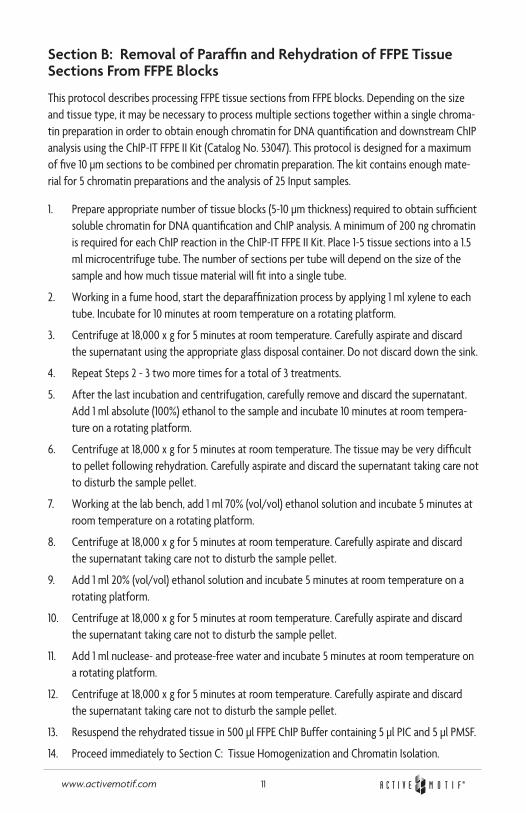
Section B: Removal of Paraffin and Rehydration of FFPE Tissue Sections From FFPE Blocks
This protocol describes processing FFPE tissue sections from FFPE blocks. Depending on the size and tissue type, it may be necessary to process multiple sections together within a single chroma-tin preparation in order to obtain enough chromatin for DNA quantification and downstream ChIP analysis using the ChIP-IT FFPE II Kit (Catalog No. 53047). This protocol is designed for a maximum of five 10 µm sections to be combined per chromatin preparation. The kit contains enough mate-rial for 5 chromatin preparations and the analysis of 25 Input samples.
1. Prepare appropriate number of tissue blocks (5-10 µm thickness) required to obtain sufficient soluble chromatin for DNA quantification and ChIP analysis. A minimum of 200 ng chromatin is required for each ChIP reaction in the ChIP-IT FFPE II Kit. Place 1-5 tissue sections into a 1.5 ml microcentrifuge tube. The number of sections per tube will depend on the size of the sample and how much tissue material will fit into a single tube.
2. Working in a fume hood, start the deparaffinization process by applying 1 ml xylene to each tube. Incubate for 10 minutes at room temperature on a rotating platform.
3. Centrifuge at 18,000 x g for 5 minutes at room temperature. Carefully aspirate and discard the supernatant using the appropriate glass disposal container. Do not discard down the sink.
4. Repeat Steps 2 - 3 two more times for a total of 3 treatments.
5. After the last incubation and centrifugation, carefully remove and discard the supernatant. Add 1 ml absolute (100%) ethanol to the sample and incubate 10 minutes at room tempera-ture on a rotating platform.
6. Centrifuge at 18,000 x g for 5 minutes at room temperature. The tissue may be very difficult to pellet following rehydration. Carefully aspirate and discard the supernatant taking care not to disturb the sample pellet.
7. Working at the lab bench, add 1 ml 70% (vol/vol) ethanol solution and incubate 5 minutes at room temperature on a rotating platform.
8. Centrifuge at 18,000 x g for 5 minutes at room temperature. Carefully aspirate and discard the supernatant taking care not to disturb the sample pellet.
9. Add 1 ml 20% (vol/vol) ethanol solution and incubate 5 minutes at room temperature on a rotating platform.
10. Centrifuge at 18,000 x g for 5 minutes at room temperature. Carefully aspirate and discard the supernatant taking care not to disturb the sample pellet.
11. Add 1 ml nuclease- and protease-free water and incubate 5 minutes at room temperature on a rotating platform.
12. Centrifuge at 18,000 x g for 5 minutes at room temperature. Carefully aspirate and discard the supernatant taking care not to disturb the sample pellet.
13. Resuspend the rehydrated tissue in 500 µl FFPE ChIP Buffer containing 5 µl PIC and 5 µl PMSF.
14. Proceed immediately to Section C: Tissue Homogenization and Chromatin Isolation.
12www.activemotif.com
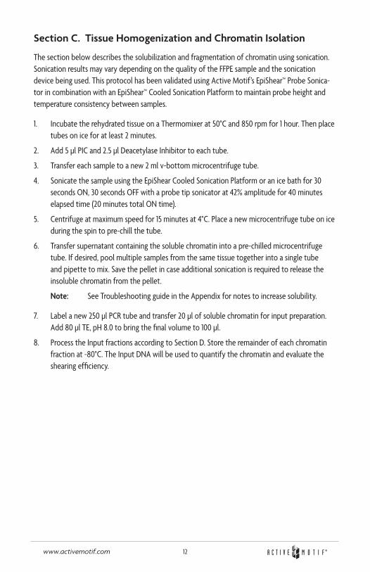
Section C. Tissue Homogenization and Chromatin Isolation
The section below describes the solubilization and fragmentation of chromatin using sonication. Sonication results may vary depending on the quality of the FFPE sample and the sonication device being used. This protocol has been validated using Active Motif’s EpiShear™ Probe Sonica-tor in combination with an EpiShear™ Cooled Sonication Platform to maintain probe height and temperature consistency between samples.
1. Incubate the rehydrated tissue on a Thermomixer at 50°C and 850 rpm for 1 hour. Then place tubes on ice for at least 2 minutes.
2. Add 5 µl PIC and 2.5 µl Deacetylase Inhibitor to each tube.
3. Transfer each sample to a new 2 ml v-bottom microcentrifuge tube.
4. Sonicate the sample using the EpiShear Cooled Sonication Platform or an ice bath for 30 seconds ON, 30 seconds OFF with a probe tip sonicator at 42% amplitude for 40 minutes elapsed time (20 minutes total ON time).
5. Centrifuge at maximum speed for 15 minutes at 4°C. Place a new microcentrifuge tube on ice during the spin to pre-chill the tube.
6. Transfer supernatant containing the soluble chromatin into a pre-chilled microcentrifuge tube. If desired, pool multiple samples from the same tissue together into a single tube and pipette to mix. Save the pellet in case additional sonication is required to release the insoluble chromatin from the pellet.
Note: See Troubleshooting guide in the Appendix for notes to increase solubility.
7. Label a new 250 µl PCR tube and transfer 20 µl of soluble chromatin for input preparation. Add 80 µl TE, pH 8.0 to bring the final volume to 100 µl.
8. Process the Input fractions according to Section D. Store the remainder of each chromatin fraction at -80°C. The Input DNA will be used to quantify the chromatin and evaluate the shearing efficiency.

13www.activemotif.com
Section D. Input Preparation
1. To each Input preparation from Step 7 above, add 2 µl RNase A. Cap the PCR tubes and vortex to mix.
2. Incubate in a thermocycler at 37°C for 30 minutes.
3. Add 2 µl Proteinase K and 5 µl 5 M NaCl. Cap the PCR tubes and vortex to mix.
4. Incubate in a thermocycler at 65°C overnight.
5. Following reversal of cross-links, transfer DNA to a new 1.5 ml microcentrifuge tube. Add 125 µl phenol and 64 µl chloroform/isoamyl alcohol (24:1). Shake vigorously for 15 seconds to mix (do not vortex). Incubate at room temperature for 5 minutes.
6. Centrifuge in a microcentrifuge at maximum speed for 2 minutes. Transfer the aqueous layer (top layer) to a new 1.5 ml microcentrifuge tube. A hazy white layer at interface may still be present.
7. Add 125 µl chloroform/isoamyl alcohol (24:1) to the aqueous layer . Shake vigorously for 15 seconds to mix (do not mix). Incubate at room temperature for 5 minutes.
8. During the incubation, set up new 1.5 ml microcentrifuge tubes for each ChIP reaction and add 2 µl Carrier to each tube.
9. Centrifuge the DNA purifications in a microcentrifuge at maximum speed for 2 minutes. Transfer the aqueous layer (top layer) to the microcentrifuge tube containing Carrier.
10. Add 300 µl 100% ethanol to the DNA solution and briefly vortex to mix.
11. Place samples at -80°C for 30 minutes to precipitate the DNA. Alternatively, samples may be left at -20°C for 2 hours to overnight.
12. Centrifuge in a microcentrifuge at maximum speed for 15 minutes at 4°C. Mark the tube where you expect the pellet to form as it may not be visible.
13. Carefully remove the supernatant taking care not to disturb the location of the DNA pellet.
14. Add 500 µl 70% ethanol to each tube and invert to mix.
15. Centrifuge in a microcentrifuge at maximum speed for 5 minutes at 4°C. Mark the tube where you expect the pellet to form as it may not be visible.
16. Remove 400 µl supernatant and centrifuge a second time at maximum speed for 2 minutes at 4°C. Carefully remove as much residual ethanol as possible with a P200 pipet taking care not to disturb the pellet.
17. Air-dry the pellet for 10-15 minutes (until residual ethanol has evaporated). Resuspend the pellet in 40 µl FFPE Low-EDTA TE Buffer.
18. Due to the low DNA concentration of the Input material from FFPE chromatin extractions, we do not recommend analysis of the shearing efficiency by agarose gel electrophoresis. Instead, we recommend evaluating the quality and quantity of the chromatin by use of a fluorometer, such as Qubit. The Nanodrop is not recommended for quantification as the DNA quantities are usually below the threshold that the Nanodrop can accurately quantify.
14www.activemotif.com
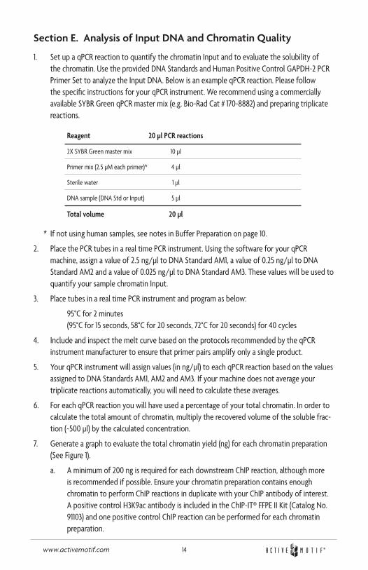
Section E. Analysis of Input DNA and Chromatin Quality
1. Set up a qPCR reaction to quantify the chromatin Input and to evaluate the solubility of the chromatin. Use the provided DNA Standards and Human Positive Control GAPDH-2 PCR Primer Set to analyze the Input DNA. Below is an example qPCR reaction. Please follow the specific instructions for your qPCR instrument. We recommend using a commercially available SYBR Green qPCR master mix (e.g. Bio-Rad Cat # 170-8882) and preparing triplicate reactions.
Reagent 20 µl PCR reactions
2X SYBR Green master mix 10 µl
Primer mix (2.5 µM each primer)* 4 µl
Sterile water 1 µl
DNA sample (DNA Std or Input) 5 µl
Total volume 20 µl
* If not using human samples, see notes in Buffer Preparation on page 10.
2. Place the PCR tubes in a real time PCR instrument. Using the software for your qPCR machine, assign a value of 2.5 ng/µl to DNA Standard AM1, a value of 0.25 ng/µl to DNA Standard AM2 and a value of 0.025 ng/µl to DNA Standard AM3. These values will be used to quantify your sample chromatin Input.
3. Place tubes in a real time PCR instrument and program as below:
95°C for 2 minutes (95°C for 15 seconds, 58°C for 20 seconds, 72°C for 20 seconds) for 40 cycles
4. Include and inspect the melt curve based on the protocols recommended by the qPCR instrument manufacturer to ensure that primer pairs amplify only a single product.
5. Your qPCR instrument will assign values (in ng/µl) to each qPCR reaction based on the values assigned to DNA Standards AM1, AM2 and AM3. If your machine does not average your triplicate reactions automatically, you will need to calculate these averages.
6. For each qPCR reaction you will have used a percentage of your total chromatin. In order to calculate the total amount of chromatin, multiply the recovered volume of the soluble frac-tion (~500 µl) by the calculated concentration.
7. Generate a graph to evaluate the total chromatin yield (ng) for each chromatin preparation (See Figure 1).
a. A minimum of 200 ng is required for each downstream ChIP reaction, although more is recommended if possible. Ensure your chromatin preparation contains enough chromatin to perform ChIP reactions in duplicate with your ChIP antibody of interest. A positive control H3K9ac antibody is included in the ChIP-IT® FFPE II Kit (Catalog No. 91103) and one positive control ChIP reaction can be performed for each chromatin preparation.
15www.activemotif.com
b. If enough chromatin is available in the soluble fraction to perform the desired ChIP reactions, the aliquots stored at -80°C from Section C, Step 8 can be used to perform the ChIP reactions using the ChIP-IT® FFPE II Kit (Catalog No. 53047).
c. If there is not enough chromatin available in the soluble fraction, additional sonication of the insoluble pellet from Section C, Step 6 is required. Follow the instructions in Sec-tion F: Additional Sonication of Insoluble Pellet.
16www.activemotif.com
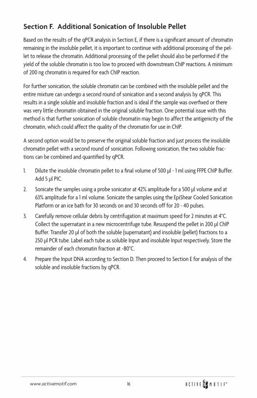
Section F. Additional Sonication of Insoluble Pellet
Based on the results of the qPCR analysis in Section E, if there is a significant amount of chromatin remaining in the insoluble pellet, it is important to continue with additional processing of the pel-let to release the chromatin. Additional processing of the pellet should also be performed if the yield of the soluble chromatin is too low to proceed with downstream ChIP reactions. A minimum of 200 ng chromatin is required for each ChIP reaction.
For further sonication, the soluble chromatin can be combined with the insoluble pellet and the entire mixture can undergo a second round of sonication and a second analysis by qPCR. This results in a single soluble and insoluble fraction and is ideal if the sample was overfixed or there was very little chromatin obtained in the original soluble fraction. One potential issue with this method is that further sonication of soluble chromatin may begin to affect the antigenicity of the chromatin, which could affect the quality of the chromatin for use in ChIP.
A second option would be to preserve the original soluble fraction and just process the insoluble chromatin pellet with a second round of sonication. Following sonication, the two soluble frac-tions can be combined and quantified by qPCR.
1. Dilute the insoluble chromatin pellet to a final volume of 500 µl - 1 ml using FFPE ChIP Buffer. Add 5 µl PIC.
2. Sonicate the samples using a probe sonicator at 42% amplitude for a 500 µl volume and at 63% amplitude for a 1 ml volume. Sonicate the samples using the EpiShear Cooled Sonication Platform or an ice bath for 30 seconds on and 30 seconds off for 20 - 40 pulses.
3. Carefully remove cellular debris by centrifugation at maximum speed for 2 minutes at 4°C. Collect the supernatant in a new microcentrifuge tube. Resuspend the pellet in 200 µl ChIP Buffer. Transfer 20 µl of both the soluble (supernatant) and insoluble (pellet) fractions to a 250 µl PCR tube. Label each tube as soluble Input and insoluble Input respectively. Store the remainder of each chromatin fraction at -80°C.
4. Prepare the Input DNA according to Section D. Then proceed to Section E for analysis of the soluble and insoluble fractions by qPCR.

17www.activemotif.com
Appendix
Section G: qPCR Primer Design
A. Design of the primers
• Design and analyze your potential primer pairs using an in silico PCR program (i.e. Primer3 at http://frodo.wi.mit.edu/ or the UCSC Genome Browser at http://genome.cse.ucsc.edu/cgi-bin/hgPcr).
• Primers that dimerize should be avoided, as they will be bound by SYBR Green, which will compromise accurate quantitation. You can test your primers for self-complementarity and secondary structure at http://frodo.wi.mit.edu/cgi-bin/primer3/primer3_www.cgi.
• Ideally, the amplicons should be 75-150 bp in length.
• For use with the ChIP-IT qPCR Analysis Kit, primers should be designed to anneal optimally at 58°C with a recommended length of 18-22 bp.
• Active Motif offers ChIP Control qPCR primer sets validated to work in our ChIP-IT qPCR Analysis Kit. To see a list of the available species-specific primers, please visit www.activemotif.com/chipprimers.
18www.activemotif.com
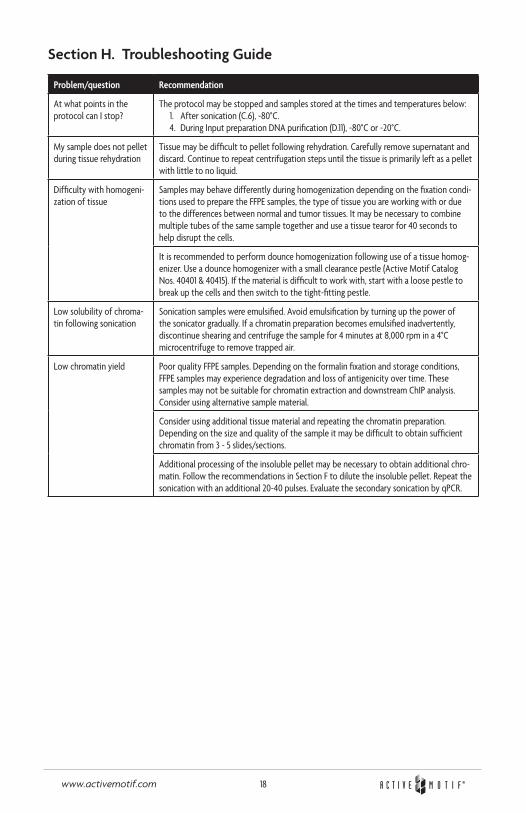
Section H. Troubleshooting Guide
Problem/question Recommendation
At what points in the protocol can I stop?
The protocol may be stopped and samples stored at the times and temperatures below:1. After sonication (C.6), -80°C.4. During Input preparation DNA purification (D.11), -80°C or -20°C.
My sample does not pellet during tissue rehydration
Tissue may be difficult to pellet following rehydration. Carefully remove supernatant and discard. Continue to repeat centrifugation steps until the tissue is primarily left as a pellet with little to no liquid.
Difficulty with homogeni-zation of tissue
Samples may behave differently during homogenization depending on the fixation condi-tions used to prepare the FFPE samples, the type of tissue you are working with or due to the differences between normal and tumor tissues. It may be necessary to combine multiple tubes of the same sample together and use a tissue tearor for 40 seconds to help disrupt the cells.
It is recommended to perform dounce homogenization following use of a tissue homog-enizer. Use a dounce homogenizer with a small clearance pestle (Active Motif Catalog Nos. 40401 & 40415). If the material is difficult to work with, start with a loose pestle to break up the cells and then switch to the tight-fitting pestle.
Low solubility of chroma-tin following sonication
Sonication samples were emulsified. Avoid emulsification by turning up the power of the sonicator gradually. If a chromatin preparation becomes emulsified inadvertently, discontinue shearing and centrifuge the sample for 4 minutes at 8,000 rpm in a 4°C microcentrifuge to remove trapped air.
Low chromatin yield Poor quality FFPE samples. Depending on the formalin fixation and storage conditions, FFPE samples may experience degradation and loss of antigenicity over time. These samples may not be suitable for chromatin extraction and downstream ChIP analysis. Consider using alternative sample material.
Consider using additional tissue material and repeating the chromatin preparation. Depending on the size and quality of the sample it may be difficult to obtain sufficient chromatin from 3 - 5 slides/sections.
Additional processing of the insoluble pellet may be necessary to obtain additional chro-matin. Follow the recommendations in Section F to dilute the insoluble pellet. Repeat the sonication with an additional 20-40 pulses. Evaluate the secondary sonication by qPCR.
19www.activemotif.com
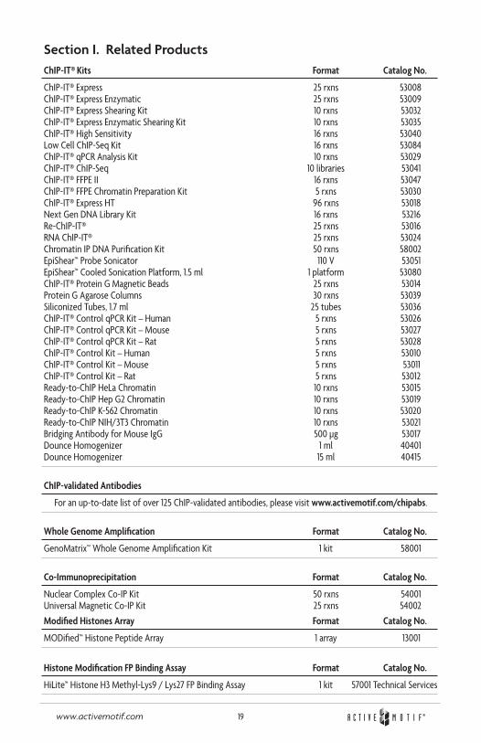
Section I. Related ProductsChIP-IT® Kits Format Catalog No.
ChIP-IT® Express 25 rxns 53008 ChIP-IT® Express Enzymatic 25 rxns 53009 ChIP-IT® Express Shearing Kit 10 rxns 53032 ChIP-IT® Express Enzymatic Shearing Kit 10 rxns 53035 ChIP-IT® High Sensitivity 16 rxns 53040 Low Cell ChIP-Seq Kit 16 rxns 53084 ChIP-IT® qPCR Analysis Kit 10 rxns 53029 ChIP-IT® ChIP-Seq 10 libraries 53041 ChIP-IT® FFPE II 16 rxns 53047 ChIP-IT® FFPE Chromatin Preparation Kit 5 rxns 53030 ChIP-IT® Express HT 96 rxns 53018 Next Gen DNA Library Kit 16 rxns 53216 Re-ChIP-IT® 25 rxns 53016 RNA ChIP-IT® 25 rxns 53024 Chromatin IP DNA Purification Kit 50 rxns 58002 EpiShear™ Probe Sonicator 110 V 53051 EpiShear™ Cooled Sonication Platform, 1.5 ml 1 platform 53080 ChIP-IT® Protein G Magnetic Beads 25 rxns 53014 Protein G Agarose Columns 30 rxns 53039 Siliconized Tubes, 1.7 ml 25 tubes 53036 ChIP-IT® Control qPCR Kit – Human 5 rxns 53026 ChIP-IT® Control qPCR Kit – Mouse 5 rxns 53027 ChIP-IT® Control qPCR Kit – Rat 5 rxns 53028 ChIP-IT® Control Kit – Human 5 rxns 53010 ChIP-IT® Control Kit – Mouse 5 rxns 53011 ChIP-IT® Control Kit – Rat 5 rxns 53012 Ready-to-ChIP HeLa Chromatin 10 rxns 53015 Ready-to-ChIP Hep G2 Chromatin 10 rxns 53019 Ready-to-ChIP K-562 Chromatin 10 rxns 53020 Ready-to-ChIP NIH/3T3 Chromatin 10 rxns 53021 Bridging Antibody for Mouse IgG 500 µg 53017 Dounce Homogenizer 1 ml 40401 Dounce Homogenizer 15 ml 40415
ChIP-validated Antibodies
For an up-to-date list of over 125 ChIP-validated antibodies, please visit www.activemotif.com/chipabs.
Whole Genome Amplification Format Catalog No.
GenoMatrix™ Whole Genome Amplification Kit 1 kit 58001
Co-Immunoprecipitation Format Catalog No.
Nuclear Complex Co-IP Kit 50 rxns 54001 Universal Magnetic Co-IP Kit 25 rxns 54002
Modified Histones Array Format Catalog No.
MODified™ Histone Peptide Array 1 array 13001
Histone Modification FP Binding Assay Format Catalog No.
HiLite™ Histone H3 Methyl-Lys9 / Lys27 FP Binding Assay 1 kit 57001 Technical Services

20www.activemotif.com
Technical Services
If you need assistance at any time, please call Active Motif Technical Service at one of the numbers listed below.
Active Motif North America 1914 Palomar Oaks Way, Suite 150 Carlsbad, CA 92008 USA Toll Free: 877 222 9543 Telephone: 760 431 1263 Fax: 760 431 1351 E-mail: [email protected]
Active Motif Europe Avenue Reine Astrid, 92 B-1310 La Hulpe, Belgium UK Free Phone: 0800 169 31 47 France Free Phone: 0800 90 99 79 Germany Free Phone: 0800 181 99 10 Telephone: +32 (0)2 653 0001 Fax: +32 (0)2 653 0050 E-mail: [email protected]
Active Motif Japan Azuma Bldg, 7th Floor 2-21 Ageba-Cho, Shinjuku-Ku Tokyo, 162-0824, Japan Telephone: +81 3 5225 3638 Fax: +81 3 5261 8733 E-mail: [email protected]
Visit Active Motif on the worldwide web at http://www.activemotif.com
At this site:
• Read about who we are, where we are, and what we do
• Review data supporting our products and the latest updates
• Enter your name into our mailing list to receive our catalog, MotifVations newsletter and notification of our upcoming products
• Share your ideas and results with us
• View our job opportunities
Don’t forget to bookmark our site for easy reference!