chromosome abnormalities involving heterochromatic regions in monocytic leukemia
TRANSCRIPT
Chromosome Abnormalities Involving Heterochromatic Regions in Monocytic Leukemia
Giovanna Rege-Cambrin, Simonetta Kerim, Patrizia Scaravaglio, Jean Louis Michaux, Herman Van Den Berghe, and Giuseppe Saglio
ABSTRACT: We report two cases of monocytic leukemia associated with cytogenetic changes involving the juxtacentromeric heterachromatin of different chromosomes. In a patient with chronic myelomonocytic leukemia (CMMoL) we describe a translocation t(1;9)(q12;q13) in which the duplicated derivative chromosome 9q + showed a huge centromeric C-band, derived by fusion of the heterochromatic regions of chromosomes 1 and 9. The constitutional karyotype showed two heterochromatin polymorphisms, lqh+ and inv(9qh). In the second case, an acute monoblastic leukemia was associated with an abnormally elongated juxtacentromeric heterochromatic region of chromosome 4 that was not constitutianally present.
INTRODUCTION
Although heterochromatin constitutes about 20% of the human genome [1], its role and molecular organization [2] have not yet been completely clarified. C-band regions are the major location of highly repetitive satellite DNA [3, 4]. Heterochromatin may be involved in favoring chromosomal exchanges either in meiotic cells, thus aiding speciation [5], or in somatic cells during mitosis. A role for heterochromatin variants in cancer predisposition has been suggested by several groups of investigators [6--14],
as patients with different types of cancer tend to show a higher incidence of hetero- chromatic polymorphisms with respect to normal controls.
We report two cases of monocytic leukemia in which juxtacentromeric heterochro- matin is obviously involved. In a patient with chronic myelomonocytic leukemia (CMMoL), we detected a translocation t(1;9) involving the heterochromatic regions of both chromosomes I and 9. The constitutional karyotype showed two heterochromatic variants, lqh + and inv(9qh). In the second case, an acute monoblastic leukemia was associated with an abnormally elongated juxtacentromeric heterochromatic region of chromosome 4, not present in the constitutional karyotype. The clinical and cytoge- netic features of these two patients are described.
From the Dipartimento di Scienze Biomediche e Oncologia Umana, Universita di Torino, Italy (G. R. C., S. K., P. S., G. S.); Center for Human Genetics, University of Leuven, Belgium (H. V. D. B.), and Service d'Hematologie, Universit~ Catholique de Louvain, Belgium (J. L. M). Simonetta Kerim is a fellow of A.I.R.C. (Associazione Italiana per la Ricerca sul Cancro. Patrizia Scaravaglio is a fellow of Comitaso Gigi Ghirotti.
Address reprint requests to: Dr. Giovanna Rege-Cambrin, Dipartimento di Scienze Bio- mediche e Oncologia Umana, Via Geneva 3, 10126 Torino, Italy.
Received July 26, 1989; accepted September 28, 1989.
9 9
© 1990 Elsevier Science Publishing Co., Inc. Cancer Genet Cytogenet 46:99-106 (1990) 655 Avenue of the Americas, New York, NY 10010 0165--4608/90/$03.50

100 G. Rege-Cambrin et al.
CASE REPORTS
Patient 1
T. R., a 51-year-old man, was referred to our clinic in Feburary 1987 with a 4-year history of progressive pancytopenia. On admission, the patient had liver (4 cm) and spleen (3 cm) enlargement. His hemoglobin (Hb) level was 11.0 g/dl, his platelet count was 43 × 109/L, and his white blood cell (WBC) count was 0.6 x 10⁹/L. Bone marrow (BM) aspirate smears revealed hypoplasia of the erythroid and megakaryocytic lin- eages. The myeloid series presented all maturation stages but in addition showed dysplastic features. The hematologic picture was consistent with a myelodysplastic syndrome. During follow-up, further decrease in Hb and platelets was observed, and the patient received red blood cell (RBC) transfusions.
In August 1987, his WBC count was 2.5 × 10⁹/L (neutrophils 64%, lymphocytes 5%, monocytes 31%). Bone marrow aspirate examination showed monoblasts and promonocytes (10% and 14% of nucleated cells, respectively). The blasts stained positively with Sudan black and peroxidase reaction; a-naphthyl-chloroacetate ester- ase was slightly positive and periodic acid-Schiff (PAS) reaction showed a diffuse pattern. He was diagnosed as having CMMoL, and purely supportive treatment was established. The patient was still alive in June 1989, when his hematologic condition was stable (last control: Hb level 8.3 g/dL; WBC count 1.8 x 10⁹/L; platelet count 18 x 109/L). He requires 4 U packed RBCs per month and complains of recurrent infections.
Patient 2
W. A., a 45-year-old man, was admitted to the hospital in June 1987 because of a 6- week history of increasing fatigue, weight loss, and petechiae. Hematologic profile was Hb level 7.3 g/dL, WBC count 14 x 109/L (neutrophils 47%, eosinophils 3%, monocytes 11%, lymphocytes 29%, monoblasts 10%), platelet count 101 x 109/L. On BM smears 80% blast cell infiltration was detected. Marked dyserythropoiesis and dysgranulopoiesis were also observed. On cytochemical stains, the blasts showed strong positivity for a-naphthyl esterase reaction, inhibited by NaF. Serum lysozyme level was greater than 100 mg/L.
A diagnosis of acute monoblastic leukemia (M5) following a previous myelodys- plastic syndrome was made, and cytostatic therapy with mitoxantrone and cytarabine was started. After one course of chemotherapy, the patient's hematologic condition improved dramatically, but BM smears showed persistence of marked dysmyelopoi- etic features. The disease was controlled with maintenance chemotherapy and trans- fusional support until April 1988, when a first relapse occurred. At this time, BM aspirate showed a complete replacement by a blast cell population with high nucleo- cytoplasmic ratio, finely dispersed chromatin, two to three nucleoli and no cyto- plasmic granules. Cytochemistry and enzymatic data were the same as those at diagno- sis. Cytostatic regimen with cytarabine and amsacrine was started with partial hematologic response, but in October 1988 the patient had a fatal relapse.
MATERIALS AND METHODS
Chromosome studies were performed on BM cells after 24-hour culture in RPMI 1640 medium supplemented by 20% fetal calf serum. To study the constitutional karyotype, peripheral blood (PB) cells from patient 1 were cultured for 72 hours with phytohe-

Chromosome Abnormalities in Monocytic Leukemia 101
magglutinin (Burroughs Wellcome). Harvesting was performed according to standard procedures, and metaphases were G-C banded with Wright's stain and R-banded with acridine-orange. At least 10 mitoses were fully karyotyped from each culture, and 15 additional metaphases were analyzed under a microscope. Chromosome abnormali- ties were classified according to the International System for Chromosome Nomen- clature [15]. The presence of heterochromatic variants was estimated visually when at least 25% difference in C-band size was observed between the two homologues [6]. We also assessed the C-variants on the basis of Patil and Lubs classification [16]. When we compared the heterochromatin length with the short arm of chromosome 16, the C-bands observed in the two chromosomes I from the constitutional karyotype of patient 1 were indeed in different levels (3 and 5, respectively).
RESULTS
Chromosome findings in the two patients at different stages of the disease are summa- rized in Table 1.
Patient
Patient
1
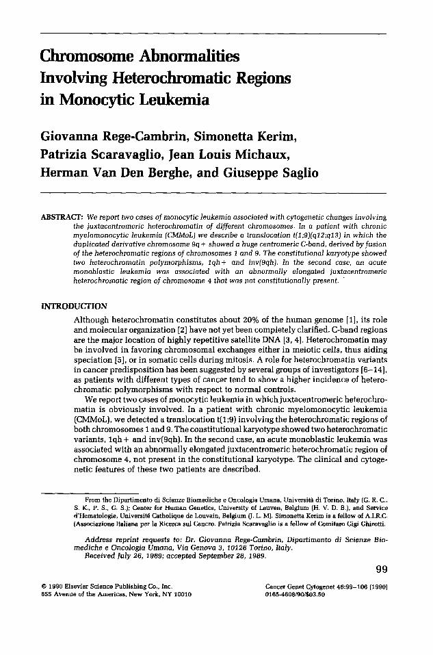
All BM cells examined showed a translocation between the long arms of chromosomes 1 and 9 (Fig. 1). The 9q+ marker chromosome carried an abnormally elongated heterochromatin and was duplicated. Moreover, the normal chromosome 9 showed a pericentric inversion. Study of the patient's constitutional karyotype clarified the breakpoints involved in this chromosomal abnormality. The large C-band contained in the 9q + derivative was not present constitutionally. In addition to the inv(9qh), a constitutional C-band polymorphism for chromosome I was observed, lqh+ (Fig. 2). The long arm of the chromosome lqh+ was broken inside the heterochromatin, whereas the breakpoint in chromosome 9 appeared to be just below the C-centromeric band of the noniuverted 9qh chromosome. The rearrangement was interpreted as a t(1;9)(q12;q13) translocation. The Y chromosome was missing in 100% of the cells. The cytogenetic pattern was unchanged in a subsequent examination.
2
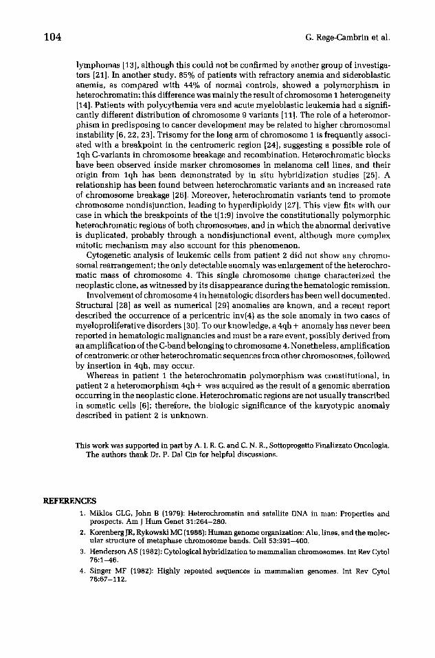
A 4qh ÷ chromosome (Fig. 3) was observed in 50% of BM cells at diagnosis of acute leukemia, and in relapse this clone completely replaced the presumably normal constitutional karyotype. Because during remission the heterochromatin of both chro-
Table 1 Cytogenetic data from bone marrow cultures in different phases of the disease
Patient D a t e Diagnosis Karyotype No. of cells
1 8 /87 CMMoL 46,X, - Y,t(1;9)(q12;q13), 10 + der(9)t(1;9)(q12;q13) idem
12/87 CMMoL idem 10 2 6 /87 A N L L - M 5 46,XY,4qh +/46,XY 5/5
7/87 Remission 45,X, - Y/46,XY 3/7 4 /88 Relapse 46,XY,4qh + idem 10 8 /88 Relapse idem 12
Abbreviations: CMMoL, chronic myelomonocytic leukemia; ANLL, acute nonlymphoblastic leukemia.

102
t~ 2 3
A
8
le-(=- 4 5
1 t °
'l
Y I " . . . . . . . . .
Figure I G-banded bone marrow metaphase from patient 1; l q - and 9q+ (duplicated) derivative chromosomes (arrows). Pericentric inversion of the normal chromosome 9 is also evident.
F i g u r e 2 Patient 1: Partial G-C banded karyotype from a peripheral blood-stimulated culture. A difference in the C-heterochromatin lengths between the two chromosome 1 homologues is shown, as is the centromeric inversion of one chromosome 9. The karyotype was 46,XY.
Q ,
t inv (gqh)
1 9

Chromosome Abnormalities in Monocytic Leukemia 103
\
4 5
8
X ×
13 14
D
~ 9 20
6 t 8 9 ~0
c
11 12
J j . . . . . .
15 16 t7 18
21 22
Figure 3 G-C banded bone marrow metaphase from patient 2, showing the marker chromo- some 4qh+ (arrow).
mosomes 4 appeared normal and equal in length and volume, acquisition of hetero- chromatic material on 4q was the only chromosome abnormality observed in the patient's leukemic cells.
DISCUSSION
In two cases of hematologic disorders with monocytic features, we observed changes involving the heterochromatic regions of chromosomes 9 and 4, respectively, in the neoplastic cells.
In the first case, a reciprocal translocation t(1;9)(q12;q13), with breakpoints mapped in the heterochromatic regions of chromosomes I and 9, was present in all metaphases analyzed. Moreover, duplication of the 9q+ marker resulted in a trisomy lq and 9p. Few cases of t(lq;9q) with the breakpoints located within or immediately adjacent to the centromeric regions of both chromosomes 1 and 9 are reported in the literature; they are generally observed in myeloproliferative disorders [17-20]. Unfortunately, constitutional karyotype data are not available in these cases.
The constitutional karyotype of this patient showed two heterochromatic variants, namely l q h + and inversion of the heterochromatic block of chromosome 9. The translocation found in leukemic cells did not involve the inverted 9qh chromosome 9, however.
Although individuals with heterochromatin variants may not show any phenotypic effect, several reports have suggested that a relationship may exist between hetero- chromatic region variability and cancer proneness. Patients with colon adenoma or carcinoma show a significantly higher frequency of heterochromatic variants, mainly involving chromosomes I and 9 [7, 8]. Similar data have been reported for lymphocytic
104 G. Rege-Cambrin et al.
lymphomas [13], although this could not be confirmed by another group of investiga- tors [21]. In another study, 85% of patients with refractory anemia and sideroblastic anemia, as compared with 44% of normal controls, showed a polymorphism in heterochromatin; this difference was mainly the result of chromosome I heterogeneity [14]. Patients with polycythemia vera and acute myeloblastic leukemia had a signifi- cantly different distribution of chromosome 9 variants [11]. The role of a heteromor- phism in predisposing to cancer development may be related to higher chromosomal instability [6, 22, 23]. Trisomy for the long arm of chromosome I is frequently associ- ated with a breakpoint in the centromeric region [24], suggesting a possible role of lqh C-variants in chromosome breakage and recombination. Heterochromatic blocks have been observed inside marker chromosomes in melanoma cell lines, and their origin from lqh has been demonstrated by in situ hybridization studies [25]. A relationship has been found between heterochromatic variants and an increased rate of chromosome breakage [26]. Moreover, heterochromatin variants tend to promote chromosome nondisjunction, leading to hyperdiploidy [27]. This view fits with our case in which the breakpoints of the t(1;9) involve the constitutionally polymorphic heterochromatic regions of both chromosomes, and in which the abnormal derivative is duplicated, probably through a nondisjunctional event, although more complex mitotic mechanism may also account for this phenomenon.
Cytogenetic analysis of leukemic cells from patient 2 did not show any chromo- somal rearrangement; the only detectable anomaly was enlargement of the heterochro- matic mass of chromosome 4. This single chromosome change characterized the neoplastic clone, as witnessed by its disappearance during the hematologic remission.
Involvement of chromosome 4 in hematologic disorders has been well documented. Structural [28] as well as numerical [29] anomalies are known, and a recent report described the occurrence of a pericentric inv(4) as the sole anomaly in two cases of myeloproliferative disorders [30]. To our knowledge, a 4qh + anomaly has never been reported in hematologic malignancies and must be a rare event, possibly derived from an amplification of the C-band belonging to chromosome 4. Nonetheless, amplification of centromeric or other heterochromatic sequences from other chromosomes, followed by insertion in 4qh, may occur.
Whereas in patient 1 the heterochromatin polymorphism was constitutional, in patient 2 a heteromorphism 4qh + was acquired as the result of a genomic aberration occurring in the neoplastic clone. Heterochromatic regions are not usually transcribed in somatic cells [6]; therefore, the biologic significance of the karyotypic anomaly described in patient 2 is unknown.
This work was supported in part by A. I. R. C. and C. N. R., Sottoprogetto Finalizzato Oncologia. The authors thank Dr. P. Dal Cin for helpful discussions.
REFERENCES
1. Miklos GLG, John B (1979): Heterochromatin and satellite DNA in man: Properties and prospects. Am J Hum Genet 31:264-280.
2. Korenberg JR, Rykowski MC (1988): Human genome organization: Alu, lines, and the molec- ular structure of metaphase chromosome bands. Cell 53:391-400.
3. Henderson AS (1982): Cytological hybridization to mammalian chromosomes. Int Rev Cytol 76:1-46.
4. Singer MF (1982): Highly repeated sequences in mammalian genomes. Int Rev Cytol 76:67-112.

C h r o m o s o m e Abnorma l i t i e s in M o n o c y t i c Leukemia 105
5. Yunis JJ, Yasmineh WG (1971): Heterochromatin, satellite DNA, and cell function. Science 174:1200-1209.
6. Atkin NB, Brito-Babapulle V (1981): Heterochromatin polymorphism and human cancer. Cancer Genet Cytogenet 3:261-272.
7. Labal de Vinuesa M, Mudry de Pargament M, Slavutsky I, Meiss R, Chopita N, Larripa I (1988): Heterochromatic variants and their association with neoplasias: IV. Colon adenomas and carcinomas. Cancer Genet Cytogenet 31:171-174.
8. Shabtai F, Antebi E, Klar D, Kimchi D, Hart J, Halbrecht H (1985): Cytogenetic study of patients with carcinoma of the colon and rectum: Particular C-band variant as possible markers for cancer proneness. Cancer Genet Cytogenet 14:235-245.
9. Labal de Vinuesa M, Larripa I, Mudry de Pargament M, Brieux de Salum S (1984): Hetero- chromatic variants and their association with neoplasias: I. Chronic and acute leukemias. Cancer Genet Cytogenet 13:297-302.
10. Ranni NSM, Labal de Vinuesa M, Mudry de Pargament M, Slavutsky I, Larripa 1 (1987): Heterochromatic variants and their association with neoplasias: Multiple myeloma. Cancer Genet Cytogenet 28:101-105.
11. Berger R, Bernheim A, Le Coniat M, Vecchione D (1979): C-banding studies in various blood disorders. Cancer Genet Cytogenet 1:95-102.
12. Berger R, Bernheim S, Kristoffersson U, Mitelman F, Olsson H (1985): C-band heteromor- phism in breast cancer patients. Cancer Genet Cytogenet 18:37-42.
13. Labal de Vinuesa M, Slavutsky I, Mudry de Pargament M, Larripa I (1988): Heterochromatic variants and their association with neoplasias: V. Non-Hodgkin lymphomas. Cancer Genet Cytogenet 31:175-178.
14. Labal de Vinuesa M, Larripa I, Mudry de Pargament M, Brieux de Salum S (1985): Hetero- chromatic variants and their association with neoplasias: II. Preleukemic states. Cancer Genet Cytogenet 13:297-302.
15. ISCN (1985): An International System for Human Cytogenetic Nomenclature, Harnden DG and Klinger HP (eds.); published in collaboration with Cytogenet Cell Genet (Karger, Basel, 1985); also in Birth Defects: Original Article Series, Vol. 21, No. 1 (March of Dimes Birth Defects Foundation, New York, 1985).
16. Patil SR, Lubs HA (1977): Classification of qh regions in human chromosomes 1, 9, and 16 by C-banding. Hum Genet 38:35-38.
17. Kirkland DJ, Welch SG, Povey S, Najfeld V, Price DJ, Lawler SD (1980): Glutamic pyruvate transaminase phenotypes in polycythemia rubra vera. Br J Hematol 44:407-413.
18. Nowell P, Finan J (1978): Chromosome studies in preleukemic states. IV. Myeloproliferative versus cytopenic disorders. Cancer 42:2254-2261.
19. Westin J, Wahlstrom J, Swolin B (1976): Chromosome studies in untreated polycythemia vera. Scand J Haematol 17:183-196.
20. Swolin B, Weinfeld A, Westin J (1986): Trisomy lq in polycythemia vera and its relation to disease transition. Am J Hematol 22:155-167.
21. Kristoffersson U, Berger R, Bernheim S, Akerman M, Olsson H, Mitelman F (1985): C-Band polymorphism in non-Hodgkin lymphamas. Hereditas 103:85-87.
22. Doneda L, Fuhrman Conti AM, Gualandri V, Larizza L (1987): Mosaicism in the C-banded region of chromosome 1 in cancer families. Cancer Genet Cytogenet 27:261-268.
23. Doneda L, Di Renzo MF, Comoglio PM, Larizza L (1985): Role of heterochromatin variation in the instability of a marker chromosome during tumor progression. Cancer Genet Cytogenet 15:283-291.
24. Brito-Bapapulle V, Atkin NB (1981): Breakpoints in chromosome 1 abnormalities of 218 human neoplasms. Cancer Genet Cytogenet 4:215-225.
25. Doneda L, GinelliE, Agresti A, Larizza L (1989): In situ hybridization analysis of interstitial C-heterochromatin in marker chromosomes of two human melanomas. Cancer Res 49:433-438.
26. Shabtai F, Halbrecht I (1979): Risk of malignancy and chromosomal polymorphism: A possible mechanism of association. Clin Genet 15:73-77.
27. Ford JH, Lester P (1978): Chromosomal variants and non-disjunction. Cytogenet Cell Genet 15:66-80.

106 G. Rege-Cambrin et al.
28. Parkin JL, Arthur DC, Abramson CS, McKerma RW, Kersey JH, Heideman RL, Brunning RD (1982~: Acute leukemia associated with the t(4;11) chromosome rearrangement: Ultrastruc- tural and immunological characteristics. Blood 60:1321-1331.
29. Mecucci C, Van Orshoven A, Tricot G, Michaux JL, Delannoy A, Van Den Berghe H (1986): Trisomy 4 identifies a subset of acute nonlymphocytic leukemias. Blood 67:1328-1332.
30. Iurlo A, Mecucci C, Van Orshoven A, Michaux JL, Van Den Berghe H (1988): Inversion (4)(p13q28) in two cases of acute nonlymphocytic leukemia. Cancer Genet Cytogenet 36:143-148.