cleaved cytokeratin-18 is a ... - papalab.ucsf.edu
TRANSCRIPT

Cha et al. Respiratory Research 2012, 13:105http://respiratory-research.com/content/13/1/105
RESEARCH Open Access
Cleaved cytokeratin-18 is a mechanisticallyinformative biomarker in idiopathic pulmonaryfibrosisSeung-Ick Cha1,2†, Christopher J Ryerson1†, Joyce S Lee1, Jasleen Kukreja3, Sophia S Barry1, Kirk D Jones4,Brett M Elicker5, Dong Soon Kim6, Feroz R Papa1, Harold R Collard1 and Paul J Wolters1*
Abstract
Background: Stress of the endoplasmic reticulum (ER) leading to activation of the unfolded protein response (UPR)and alveolar epithelial cell (AEC) apoptosis may play a role in the pathogenesis of idiopathic pulmonary fibrosis(IPF). Our objectives were to determine whether circulating caspase-cleaved cytokeratin-18 (cCK-18) is a marker ofAEC apoptosis in IPF, define the relationship of cCK-18 with activation of the UPR, and assess its utility as adiagnostic biomarker.
Methods: IPF and normal lung tissues were stained with the antibody (M30) that specifically binds cCK-18. Therelationship between markers of the UPR and cCK-18 was determined in AECs exposed in vitro to thapsigargin toinduce ER stress. cCK-18 was measured in serum from subjects with IPF, hypersensitivity pneumonitis (HP),nonspecific interstitial pneumonia (NSIP), and control subjects.
Results: cCK-18 immunoreactivity was present in AECs of IPF lung, but not in control subjects. Markers of the UPR(phosphorylated IRE-1α and spliced XBP-1) were more highly expressed in IPF type II AECs than in normal type IIAECs. Phosphorylated IRE-1α and cCK-18 increased following thapsigargin-induced ER stress. Serum cCK-18 leveldistinguished IPF from diseased and control subjects. Serum cCK-18 was not associated with disease severity oroutcome.
Conclusions: cCK-18 may be a marker of AEC apoptosis and UPR activation in patients with IPF. Circulating levelsof cCK-18 are increased in patients with IPF and cCK-18 may be a useful diagnostic biomarker.
Keywords: Idiopathic interstitial pneumonia, idiopathic pulmonary fibrosis, lung fibrosis, ER stress, apoptosis
BackgroundIdiopathic pulmonary fibrosis (IPF) is a progressiveinterstitial lung disease (ILD) with no establishedpharmacotherapy and a median survival of 3 years [1,2].Due to incompletely understood pathophysiology andthe lack of mechanistic biomarkers, drug developmentand testing in IPF typically relies on relatively insensitiveclinical measures such as change in pulmonary functionor survival time. Identification of mechanistically in-formative biomarkers of specific pathologic processes
* Correspondence: [email protected]†Equal contributors1Department of Medicine, University of California, San Francisco School ofMedicine, Box 0111, San Francisco 94143-0111CA, USAFull list of author information is available at the end of the article
© 2012 Cha et al.; licensee BioMed Central LtdCommons Attribution License (http://creativecreproduction in any medium, provided the or
may allow for faster, cheaper, and safer development andtesting of candidate drugs.Several studies have implicated type II alveolar epithe-
lial cell (AEC) injury and death in the pathogenesis ofIPF [3-7]. In some instances, epithelial cell death isprogrammable, as AEC apoptosis has been demonstratedin patients with IPF [3-5]. Proposed causes of AECapoptosis in IPF include oxidative stress and DNA dam-age, [8] TGF-β and Fas ligand, [9] and stress of theendoplasmic reticulum (ER). ER stress occurs whenthere is an imbalance between cellular demand for pro-tein synthesis by the ER and the ER’s capacity tosynthesize, process and package the requisite proteins[5,6]. When under stress, the ER attempts to maintainhomeostasis by activating the unfolded protein response
. This is an Open Access article distributed under the terms of the Creativeommons.org/licenses/by/2.0), which permits unrestricted use, distribution, andiginal work is properly cited.

Cha et al. Respiratory Research 2012, 13:105 Page 2 of 9http://respiratory-research.com/content/13/1/105
(UPR) pathway [10]. If the UPR fails to restore homeo-stasis, it sacrifices the cell by activating apoptotic path-ways [11]. Markers of the UPR and apoptosis areincreased in lung tissue from patients with IPF andlocalize to the alveolar epithelium [5,6]. These observa-tions suggest ER stress and activation of the UPR maybe one trigger of AEC apoptosis in IPF patients.Cytokeratin-18 is a cytoskeletal protein that is primar-
ily found in pseudostratified and simple epithelia, includ-ing the alveolar epithelium [12]. During apoptosis ofepithelial cells, cytokeratin-18 is cleaved twice by cas-pases, generating an 18-kilodalton fragment termedcaspase-cleaved cytokeratin-18 (cCK-18). This caspase-specific processing exposes a neo-epitope at the c-terminal end of cCK-18 that is recognized by a specificmonoclonal antibody (M30) [13]. The overall objectiveof this study was to use the M30 antibody to definewhether there is a relationship between activation of theUPR and formation of cCK-18 in lung AECs, and to de-termine whether circulating cCK-18 could serve as amechanistic biomarker of the UPR and AEC apoptosisin IPF.
MethodsType II alveolar epithelial cell isolation and induction ofER StressType II AECs were isolated from explanted IPF lungsand unused donor lungs (procured from the NorthernCalifornia Transplant Donor Network) as previouslydescribed [14]. Purity of human alveolar type II cells,assessed by pro-surfactant protein C (SPC) staining oncytospin, was > 95% for IPF and normal lungs. The diag-nosis of IPF was confirmed in all cases by multidisciplin-ary review according to established criteria [15,16]. A549cells were obtained from the American Type CultureCollection (Manassas, VA, USA). Cells were exposed tothapsigargin (EMD Chemicals, Inc., Gibbstown, NJ,USA) for varying lengths of time to induce ER stress.
Lung tissue preparation and immunohistochemistry forcCK-18For immunohistochemistry, IPF lung tissue was obtainedfrom the Tissue Core Laboratory of the Lung Tissue Re-search Consortium (Denver, CO). Healthy normal lungtissue was procured from lungs not used by the North-ern California Transplant Donor Network. Briefly, en-dogenous peroxidase was inhibited in 5-μm tissuesections by incubating the sections in 3% hydrogen per-oxide for 30 minutes. After washing with PBS, the sec-tions were incubated for one hour in PBS containing 5%normal goat serum and 1% bovine serum albumin(BSA), then incubated with a 1:500 dilution of monoclo-nal mouse anti-cCK18 antibody (M30 antibody, M30CytoDEATH, PEVIVA, Bromma, Sweden) overnight at
4°C. The sections were washed in 0.1% PBS-Tween 20,incubated for 40 minutes with horseradish peroxidase(HRP)-conjugated goat anti-mouse IgG (Santa Cruz Bio-technology, Santa Cruz, CA, USA). After washing, thebound peroxidase activity was detected using a diamino-benzidine (DAB) substrate kit (Vector Laboratories,Burlingame, CA, USA). The sections were then washedin deionized water and counterstained with Meyer’shematoxylin (Sigma-Aldrich). For each tissue, an adja-cent section was stained only with secondary antibodyto address nonspecific binding of the secondaryantibody.
FACS SortingEpithelial cells were suspended in DME H-21 containing10% fetal bovine serum and incubated for 30 minutes onice with antibodies to Annexin V (BD Biosciences, SanJose CA) per the manufacturer’s protocol. Samples werethen analyzed using a LRS II flow cytometer (BectonDickinson) and sorted into Annexin V + or - popula-tions using a Moflo High Performance Cell Sorter (DakoCymation).
ImmunoblotsCell lysates were subjected to SDS-PAGE under redu-cing conditions and transferred to nitrocellulose mem-brane (Life Sciences Products, Boston, MA). Themembrane was washed with 50 mM Tris–HCl contain-ing 0.5 M NaCl, 0.01% Tween-20, (TBS; pH 7.5) andincubated overnight in 5% milk containing primary anti-body. The membrane was then washed with TBS, incu-bated in TBS for 30 min containing a 1:2,000 dilution ofHRP-conjugated secondary antibody (New England Bio-labs, Beverly, MA) and washed again. Immunoreactivitywas detected using the phototope-HRP-detection kit(New England Biolabs). Primary antibodies included:mouse anti-human cCK-18 (1:1000, M30), rabbit-antihuman IRE-1α (1:1000 Novus Biologicals, Littleton, CO,USA), rabbit-anti human phospho-specific IRE-1α(1:2000, Novus Biologicals), and mouse anti-human β-actin (1:1000, Sigma-Aldrich).
XBP-1 mRNA splicingTotal RNA from type II AECs and A549 cells wasextracted using a standard Trizol (Invitrogen, Carlsbad,CA) protocol. cDNA was synthesized from total RNAusing One-Step RT-PCR SuperScript III reverse tran-scriptase (Invitrogen) per the manufacturer’s protocol.XBP-1 splicing was quantified by PCR using methodsdescribed previously [17]. Sense primer mXBP1.3S (5’-AAACAGAGTAGCAGCGCAGACTGC-3’) and anti-sense primer mXBP1.2AS (5’- GGATCTCTAAAACTAGAGGCTTGGTG-3’) were used. PCR fragments weredigested by PstI. The PstI cleavage site is located in the

Cha et al. Respiratory Research 2012, 13:105 Page 3 of 9http://respiratory-research.com/content/13/1/105
26 nt intron of unspliced XBP-1, which allows differenti-ation between the unspliced XBP-1 amplicon (cut PCRproduct) and the spliced XBP-1 amplicon. PCR productswere resolved on 2% agarose gels, stained with EtBr, andquantified by densitometry. XBP1-specific primers weresynthesized by Integrated DNA Technologies Inc (Coral-ville, IA, USA). The PCR fragments were visualized withBioDoc-It Imaging System (UVP, Inc., Upland, CA,USA).
Blood and BAL ELISA for cCK-18Blood samples, clinical information, and survival datafrom patients with IPF, chronic hypersensitivity pneu-monitis (HP), and nonspecific interstitial pneumonitis(NSIP) enrolled in an on-going longitudinal research co-hort were collected at the time of initial presentation toUCSF. All ILD diagnoses were established thoroughmultidisciplinary review of clinical data, radiology, andpathology according to established criteria[15,16,18,19].Patients with chronic HP required either a diagnosticsurgical lung biopsy, or a compatible HRCT combinedwith a chronic and high-intensity exposure (e.g. livingwith a pet bird, living or working on a farm). Patientswith NSIP were defined according to previously pub-lished criteria [20]. Bronchoalveolar lavage (BAL) sam-ples were obtained for research purposes in a subset ofIPF subjects. A second cohort consisted of blood sam-ples from stable IPF and acute exacerbation of IPFpatients seen at Asan Medical Center, University ofUlsan (Seoul, South Korea) [21]. Stable IPF patients weredefined as outpatients with IPF who were not experien-cing a rapid decline in respiratory function. Acute ex-acerbation of IPF was diagnosed using consensus criteria[22]. Written informed consent was obtained from allpatients according to institutional review board-approved protocols. In all samples, M30 reactivity wasmeasured using a commercially available ELISA (M30-Apoptosense ELISA, PEVIVA, Bromma, Sweden) accord-ing to the manufacturer’s protocol.
Statistical analysisSerum cCK-18 levels were compared between IPF andcontrol groups using the Wilcoxon rank-sum test. Theassociation of cCK-18 level with the diagnosis of IPF wastested for confounding by age, gender, smoking history,dyspnea severity, the use of long-term supplemental oxy-gen therapy, baseline FVC, and baseline DLCO using re-gression analysis. The ability of serum cCK-18 level topredict disease progression in the UCSF cohort wastested using a Wilcoxon rank-sum test. Disease progres-sion was indicated as previously defined by our group:10% decline in FVC, 15% decline in DLCO, lung trans-plantation, or death due to any cause within 6 months ofthe blood draw [23]. Multivariate Cox proportional
hazards analysis was used to evaluate the relationship ofcCK-18 with transplant-free survival. All analyses wererepeated using a dichotomous cCK-18 variable. All dataanalysis was performed using STATA 11.0 (StataCorp,Texas, USA).
ResultsThe UPR is activated in type II alveolar epithelial cellsisolated from IPF lungIt has been reported that the UPR is activated in the epi-thelial cells of IPF patients. To confirm these observa-tions, the relative levels of phospho-IRE-1α, and XBP-1mRNA splicing were measured in AECs isolated fromnormal and IPF lungs. Type II AECs of IPF lungsexpressed more total IRE-1α compared to normal lungsand the phosphorylated form was detected in type IIAECs of IPF, but not in normal lungs (Figure 1A). Therewas also more spliced XBP-1 in type II AECs of IPFlungs compared to normal lungs (p = 0.0495; Figure 1B).These findings demonstrate that the UPR pathway isactivated in type II AECs isolated from IPF patients.
ER stress triggers cCK-18 formation in cultured alveolarepithelial cellsBecause ER stress and subsequent activation of the UPRcan lead to apoptosis, [11] we next examined whetherinduction of ER stress leads to the formation of cCK-18.To do this, A549 cells were exposed to thapsigargin, aninducer of ER stress, for varying lengths of time. Phos-phorylated IRE-1α expression increased following thapsi-gargin exposure (Figure 2A), confirming that thapsigarginactivated the UPR in AECs. cCK-18 increased inthapsigargin-exposed A549 cells (Figure 2A), demonstrat-ing that activation of the UPR can lead to formation ofcCK-18 in AECs.
cCK-18 is formed prior to the appearance of theapoptosis marker Annexin VNext we measured the appearance of thapsigargin-mediated cCK-18 formation in relationship to annexinV, an early marker of apoptosis. Thapsigargin-exposedA549 cells were harvested and separated into two popu-lations (annexin V + or –) by flow sorting for the cellsurface expression of annexin V. Figure 2B shows thatcCK-18 first appears in annexin V– cells 48 hrs afterthapsigargin exposure, and remains present in annexinV– cells 72 hrs after thapsigargin, when it begins to ap-pear in annexin V+ cells.
cCK-18 is present in the alveolar epithelium of IPF lungActivation of the UPR and apoptosis have been reportedin AECs of IPF lungs[5,6]. To examine whether cCK-18formation is similarly present in IPF AECs, sections oflung obtained from IPF patients were immunostained

Total IRE1
p-IRE1
Actin
IPF lamroN
XBP1s
XBP1
XBP1
IPF lamroN % spliced
IPF lamroN
100
80
60
40
20
0
A
B
#
Rel
ativ
e U
nits
p-IRE1
*
**
Figure 1 The UPR is activated in type II AECs isolated from IPF lungs. Phosphorylated IRE-1α (panel A) and spliced XBP-1 (panel B) werepresent in increased amounts in AECs from 3 different IPF lungs, but not in the 3 control subjects (* P < 0.05, **P = 0.0495 by Wilcoxon rank sumtest). # indicates amplicon of unspliced XBP1 digested with Pst1.
12
+
p-IRE1
IRE1
Actin
48 A
B
cCK-18
Time (h)
TG
24
+ +
24
+
cCK-18
Actin
Time (h)
Annexin V
48
+
72
+
Figure 2 cCK-18 is formed following activation of the UPR. A.Immunoblots for phospho-IRE1 (p-IRE1), total IRE1 (IRE1), andcleaved cytokeratin 18 (cCK-18) showing time-dependent increase ofcCK-18 in A549 cells exposed to 0.5 μM of thapsigargin (TG, +) forvarious lengths of time. Immunoblots showing an increase in totaland phospho-IRE1 confirm thapsigargin activates the UPR in A549cells compared to vehicle controls (−). B. Immunoblots for cCK-18showing time-dependent appearance of cCK-18 in annexin V– or V+
A549 cells exposed to 0.5 μM of thapsigargin for various lengths oftime.
Cha et al. Respiratory Research 2012, 13:105 Page 4 of 9http://respiratory-research.com/content/13/1/105
for cCK-18. AECs of IPF lung were immunoreactive forcCK-18 (Figures 3A, B). In contrast, lung tissue fromcontrol subjects was not immunoreactive for cCK-18(Figure 3D).
cCK-18 is elevated in the serum of IPF patientscCK-18 has been reported to be a circulating biomarkerof epithelial cell apoptosis [12]. To examine whether cir-culating cCK-18 levels are preferentially increased inpatients with IPF, cCK-18 levels were measured in theserum of 169 subjects, including 84 subjects with IPF, 24with HP, 22 with NSIP, and 39 control subjects. Demo-graphic and clinical characteristics of these subjects aresummarized in Table 1. Serum cCK-18 levels were sig-nificantly elevated in the serum of IPF patients com-pared to control subjects (area under the receiveroperating characteristic curve (AUC) 0.88, p<0.00005)(Figure 4A). Furthermore, serum levels of cCK-18 weresignificantly different in IPF patients compared to thosein patients with HP or NSIP (AUC 0.76, p<0.00005)(Figure 4B). There were no differences in cCK-18 levelcomparing HP, NSIP, and control subjects. cCK-18 wasan independent predictor of a diagnosis of IPF versusHP and NSIP when controlling for baseline variables(p=0.001). The addition of cCK18 to baseline variablessignificantly improved the AUC from 0.83 to 0.88(p<0.00005). cCK-18 was not detectable in BAL fluid ofIPF patients.

C D
A B
Figure 3 Immunohistochemistry of IPF and normal lung with M30 antibody. A. IPF lung immunostained with the M30 antibody (20x). B.Higher power view of IPF lung immunostained with the M30 antibody (40x). C. IPF lung immunostained with a non-immune antibody (20x). D.Normal lung immunostained with the M30 antibody showing an absence of staining for cCK-18 (40x). Bar = 50 μm. Images are representative ofstaining of tissue from 8 patients with IPF and 8 control subjects.
Cha et al. Respiratory Research 2012, 13:105 Page 5 of 9http://respiratory-research.com/content/13/1/105
Serum cCK-18 level is not associated with clinicalvariables or outcomesSerum cCK-18 level was not associated with any baselineclinical variables. Further, serum cCK-18 level did notpredict progression measured by a change in FVC,
Table 1 Subject characteristics of UCSF cohort
Variable IPF
(n = 84)
Age, years 70.3 (7.9)
Male sex, % 76.2
Diagnosis by surgical lung biopsy, % 50.0
Smoking history
Ever smoked, % 78.6
Pack-years 27.7 (28.7)
Measures of disease severity
Dyspnea score 9.8 (6.0)
Long-term oxygen therapy, % 20.2
Pulmonary function
FVC, % predicted 69.4 (18.2)
DLCO, % predicted 46.7 (18.4)
Data shown are mean (standard deviation), unless otherwise indicated.* p < 0.05 for comparison with the IPF cohort.Abbreviations: DLCO, diffusing capacity of carbon monoxide; FVC, forced vital capacnonspecific interstitial pneumonia; UCSF, University of California San Francisco.
DLCO or survival. There was no change in results withsecondary analyses that analyzed cCK-18 as a dichotom-ous variable or that considered alternative outcome vari-ables (i.e. considering individual pulmonary function testvariables as dichotomous or continuous variables). In a
HP NSIP
(n = 24) (n = 22)
57.0 (10.3)* 63.6 (12.5)*
33.3* 59.1
66.7 72.7
41.7* 31.8*
11.8 (18.8)* 6.4 (13.8)*
11.2 (4.6) 9.6 (4.4)
23.8 27.3
67.3 (22.0) 64.1 (19.1)
55.1 (21.2) 43.7 (14.4)
ity; HP, hypersensitivity pneumonitis; IPF, idiopathic pulmonary fibrosis; NSIP,
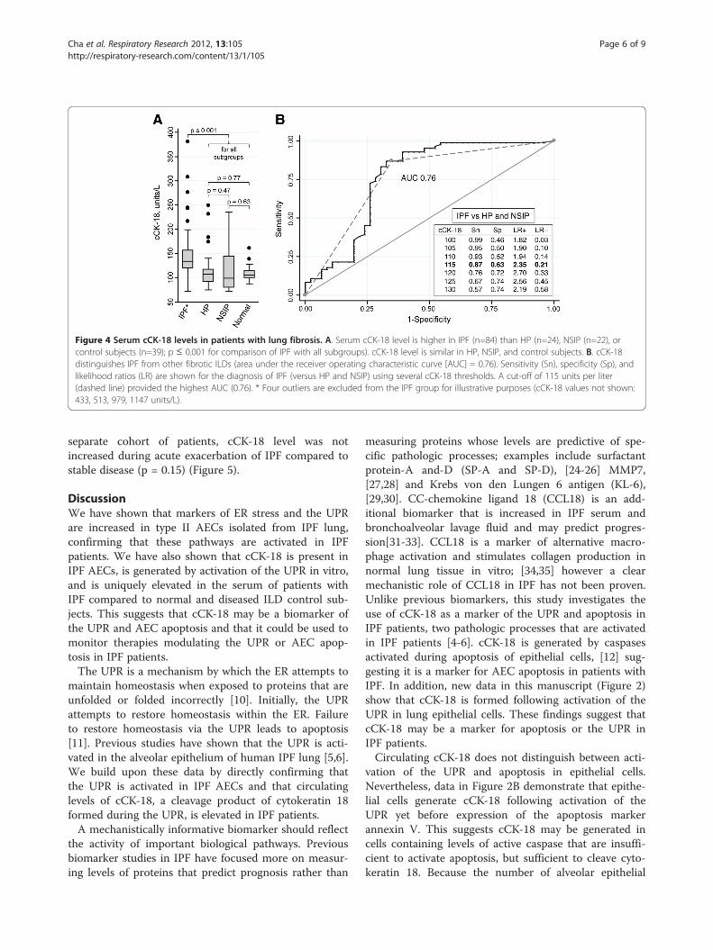
Figure 4 Serum cCK-18 levels in patients with lung fibrosis. A. Serum cCK-18 level is higher in IPF (n=84) than HP (n=24), NSIP (n=22), orcontrol subjects (n=39); p ≤ 0.001 for comparison of IPF with all subgroups). cCK-18 level is similar in HP, NSIP, and control subjects. B. cCK-18distinguishes IPF from other fibrotic ILDs (area under the receiver operating characteristic curve [AUC] = 0.76). Sensitivity (Sn), specificity (Sp), andlikelihood ratios (LR) are shown for the diagnosis of IPF (versus HP and NSIP) using several cCK-18 thresholds. A cut-off of 115 units per liter(dashed line) provided the highest AUC (0.76). * Four outliers are excluded from the IPF group for illustrative purposes (cCK-18 values not shown:433, 513, 979, 1147 units/L).
Cha et al. Respiratory Research 2012, 13:105 Page 6 of 9http://respiratory-research.com/content/13/1/105
separate cohort of patients, cCK-18 level was notincreased during acute exacerbation of IPF compared tostable disease (p = 0.15) (Figure 5).
DiscussionWe have shown that markers of ER stress and the UPRare increased in type II AECs isolated from IPF lung,confirming that these pathways are activated in IPFpatients. We have also shown that cCK-18 is present inIPF AECs, is generated by activation of the UPR in vitro,and is uniquely elevated in the serum of patients withIPF compared to normal and diseased ILD control sub-jects. This suggests that cCK-18 may be a biomarker ofthe UPR and AEC apoptosis and that it could be used tomonitor therapies modulating the UPR or AEC apop-tosis in IPF patients.The UPR is a mechanism by which the ER attempts to
maintain homeostasis when exposed to proteins that areunfolded or folded incorrectly [10]. Initially, the UPRattempts to restore homeostasis within the ER. Failureto restore homeostasis via the UPR leads to apoptosis[11]. Previous studies have shown that the UPR is acti-vated in the alveolar epithelium of human IPF lung [5,6].We build upon these data by directly confirming thatthe UPR is activated in IPF AECs and that circulatinglevels of cCK-18, a cleavage product of cytokeratin 18formed during the UPR, is elevated in IPF patients.A mechanistically informative biomarker should reflect
the activity of important biological pathways. Previousbiomarker studies in IPF have focused more on measur-ing levels of proteins that predict prognosis rather than
measuring proteins whose levels are predictive of spe-cific pathologic processes; examples include surfactantprotein-A and-D (SP-A and SP-D), [24-26] MMP7,[27,28] and Krebs von den Lungen 6 antigen (KL-6),[29,30]. CC-chemokine ligand 18 (CCL18) is an add-itional biomarker that is increased in IPF serum andbronchoalveolar lavage fluid and may predict progres-sion[31-33]. CCL18 is a marker of alternative macro-phage activation and stimulates collagen production innormal lung tissue in vitro; [34,35] however a clearmechanistic role of CCL18 in IPF has not been proven.Unlike previous biomarkers, this study investigates theuse of cCK-18 as a marker of the UPR and apoptosis inIPF patients, two pathologic processes that are activatedin IPF patients [4-6]. cCK-18 is generated by caspasesactivated during apoptosis of epithelial cells, [12] sug-gesting it is a marker for AEC apoptosis in patients withIPF. In addition, new data in this manuscript (Figure 2)show that cCK-18 is formed following activation of theUPR in lung epithelial cells. These findings suggest thatcCK-18 may be a marker for apoptosis or the UPR inIPF patients.Circulating cCK-18 does not distinguish between acti-
vation of the UPR and apoptosis in epithelial cells.Nevertheless, data in Figure 2B demonstrate that epithe-lial cells generate cCK-18 following activation of theUPR yet before expression of the apoptosis markerannexin V. This suggests cCK-18 may be generated incells containing levels of active caspase that are insuffi-cient to activate apoptosis, but sufficient to cleave cyto-keratin 18. Because the number of alveolar epithelial

Figure 5 Serum cCK-18 is not increased during acuteexacerbation of IPF. Serum cCK-18 levels were measured in theserum of 34 patients with stable IPF and 36 different patientssuffering from an acute exacerbation of IPF.
Cha et al. Respiratory Research 2012, 13:105 Page 7 of 9http://respiratory-research.com/content/13/1/105
cells that are immunoreactive for cCK-18 (Figure 3) oractive caspase 3 [8] are far greater than the rare TUNELpositive alveolar epithelial cell found in IPF lungs (datanot shown), this is the most likely scenario in IPF lung.Proving this will require future identification of morespecific biomarkers of the UPR that can be correlated tocCK-18.We show that cCK-18 could also be a useful diagnostic
biomarker that distinguishes IPF from chronic HP andNSIP, independent of age, gender, smoking, disease se-verity, and other baseline variables. A recent small studyfound that cCK-18 may also be elevated in the serum of
patients with organizing pneumonia [36]. The clinicalimportance of this finding is uncertain, given that IPFand organizing pneumonia have unique clinical andradiological features [19]. We used chronic HP and idio-pathic fibrotic NSIP as disease controls because the clin-ical, radiologic, and pathologic features of chronic HPand fibrotic NSIP often have substantial overlap withIPF [15,16,18,19]. Distinguishing IPF from HP and NSIPoften requires a surgical lung biopsy, a procedure thatcarries substantial risk [15,16]. If confirmed in othercohorts, a serum biomarker, such as cCK-18, that distin-guishes IPF from other fibrotic ILDs may reduce theneed for lung biopsy.Serum cCK-18 was not associated with severity or pro-
gression of IPF. This conflicts with a previous study thatshowed cCK-18 correlated with physiologic measures inpatients with a variety of ILDs [36]. Several possibilitiescould explain the lack of association seen our study.First, cCK-18 was measured at a single time point. Thissingle measurement may not be representative of activa-tion of the UPR or apoptosis over the course of an indi-vidual’s disease. Second, circulating levels of cCK-18likely reflect the sum of a complex interplay of physio-logical processes (i.e. production, clearance, metabolism),each of which may impact the cCK-18 level differently inindividual patients. In addition, physiologic progressionwas measured over a time interval of 6-months aftercCK-18 measurement, and cCK-18 level might reflectdisease activity and progression on a more limited timescale (e.g. days or weeks). Third, although data wereadjusted for several clinical and physiologic variables,other confounders may have been present, including oc-cult conditions, unrelated to IPF, that could cause apop-tosis of AECs. cCK-18 was also not elevated in theserum of patients during acute exacerbation of IPF. Thismay suggest that AEC apoptosis is not a prominent fea-ture of acute exacerbation, however this finding requiresfurther study as there was a trend for higher serumlevels of cCK-18 in patients during acute exacerbation ofIPF. Finally, we also were unable to detect cCK-18 inBAL fluid in patients with IPF. The reason for this is un-known, but may relate to dilution that occurs during theBAL procedure or to different metabolism of cCK-18 inthe alveoli compared to the serum.
ConclusionsIn summary, data reported in this manuscript show thatthe UPR is activated in AECs of IPF patients and thatcCK-18 may be a marker of this process. This findingsuggests that cCK-18 could be used in the early-phasedevelopment of drugs targeting the UPR in IPF patients.In addition, serum cCK-18 levels may be clinically in-formative as a diagnostic marker of IPF. Further researchmeasuring serial cCK-18 levels in a longitudinal cohort

Cha et al. Respiratory Research 2012, 13:105 Page 8 of 9http://respiratory-research.com/content/13/1/105
of IPF patients is required to confirm these results anddetermine the impact of temporal changes in serumcCK-18 on its performance as a measure of disease ac-tivity and course.
AbbreviationsAEC: Alveolar epithelial cell; AUC: Area under the receiver operatingcharacteristic curve; BAL: Bronchoalveolar lavage; BSA: Bovine serum albumin;cCK-18: Caspase-cleaved cytokeratin-18; CCL18: CC-chemokine ligand 18;DAB: Diaminobenzidine; DLCO: Diffusing capacity for carbon monoxide;DNA: Deoxyribonucleic acid; ELISA: Enzyme-linked immunosorbent assay;ER: Endoplasmic reticulum; FACS: Fluorescence-activated cell sorting;FVC: Forced vital capacity; HP: Hypersensitivity pneumonitis; HRP: Horseradishperoxidase; ILD: Interstitial lung disease; IPF: Idiopathic pulmonary fibrosis; KL-6: Krebs von den Lungen 6 antigen; M30: Monoclonal antibody to cCK-18;MMP7: Matrix metalloproteinase 7; NSIP: Nonspecific interstitial pneumonia;PBS: Phosphate buffered saline; RNA: Ribonucleic acid; RT-PCR: Reversetranscriptase polymerase chain reaction; SDS-PAGE: Sodium dodecyl sulfatepolyacrylamide gel electrophoresis; SP-A: Surfactant protein A; SP-D: Surfactant protein D; TBS: Tris-buffered saline; TGF-β: Transforming growthfactor-beta; TUNEL: Terminal deoxynucleotidyl transferase dUTP nick endlabeling; UCSF: University of California San Francisco; UPR: Unfolded proteinresponse; XBP-1: X-box binding protein 1.
Competing interestsThe authors have no competing interests related to this manuscript.
Author contributionsSIC performed the experiments and produced the first draft of themanuscript. CJR performed data analysis, contributed to some of theexperiments, and produced the first draft of the manuscript. JSL contributedbronchoalveolar lavage samples. JK contributed tissue samples. SSBperformed experiments. KDJ reviewed pathology for all ILD patients. BMEreviewed radiology for all ILD patients. DSK provided samples for patientswith acute exacerbation of IPF. FRP participated in the study design and dataanalysis. HRC participated in the study design and data analysis. PJWconceived of the study, oversaw the experiments, and produced the firstdraft of the manuscript. All authors read and approved the final manuscript.
AcknowledgementsWe thank Aaron Schroeder for technical assistance, the patients whoparticipated in this study and the providers who referred patients to ourcollective centers. Without the generous donation of biological samples fromthe patients, this study could not have been performed.
FundingThe study was funded in part by grants from the RAP program at UCSF andNIH grants HL104422, HL108794 and HL086516.
Author details1Department of Medicine, University of California, San Francisco School ofMedicine, Box 0111, San Francisco 94143-0111CA, USA. 2Department ofInternal Medicine, Kyungpook National University School of Medicine, Daegu,South Korea. 3Department of Surgery, University of California, San Francisco,CA, USA. 4Department of Pathology, University of California, San Francisco,CA, USA. 5Department of Radiology, University of California, San Francisco,CA, USA. 6Department of Medicine, Asan Medical Center, University of UlsanCollege of Medicine, Seoul, South Korea.
Received: 4 September 2012 Accepted: 16 November 2012Published: 20 November 2012
References1. Bjoraker JA, Ryu JH, Edwin MK, Myers JL, Tazelaar HD, Schroeder DR, Offord
KP: Prognostic significance of histopathologic subsets in idiopathicpulmonary fibrosis. Am J Respir Crit Care Med 1998, 157:199–203.
2. Hubbard R, Johnston I, Britton J: Survival in patients with cryptogenicfibrosing alveolitis: a population-based cohort study. Chest 1998,113:396–400.
3. Barbas-Filho JV, Ferreira MA, Sesso A, Kairalla RA, Carvalho CR, Capelozzi VL:Evidence of type II pneumocyte apoptosis in the pathogenesis ofidiopathic pulmonary fibrosis (IFP)/usual interstitial pneumonia (UIP).J Clin Pathol 2001, 54:132–138.
4. Uhal BD, Joshi I, Hughes WF, Ramos C, Pardo A, Selman M: Alveolarepithelial cell death adjacent to underlying myofibroblasts in advancedfibrotic human lung. Am J Physiol 1998, 275:L1192–L1199.
5. Korfei M, Ruppert C, Mahavadi P, Henneke I, Markart P, Koch M, Lang G, FinkL, Bohle RM, Seeger W, et al: Epithelial endoplasmic reticulum stress andapoptosis in sporadic idiopathic pulmonary fibrosis. Am J Respir Crit CareMed 2008, 178:838–846.
6. Lawson WE, Crossno PF, Polosukhin VV, Roldan J, Cheng DS, Lane KB,Blackwell TR, Xu C, Markin C, Ware LB, et al: Endoplasmic reticulum stressin alveolar epithelial cells is prominent in IPF: association with alteredsurfactant protein processing and herpesvirus infection. Am J PhysiolLung Cell Mol Physiol 2008, 294:L1119–L1126.
7. Sisson TH, Mendez M, Choi K, Subbotina N, Courey A, Cunningham A, DaveA, Engelhardt JF, Liu X, White ES, et al: Targeted injury of type II alveolarepithelial cells induces pulmonary fibrosis. Am J Respir Crit Care Med 2010,181:254–263.
8. Kuwano K, Nakashima N, Inoshima I, Hagimoto N, Fujita M, Yoshimi M,Maeyama T, Hamada N, Watanabe K, Hara N: Oxidative stress in lungepithelial cells from patients with idiopathic interstitial pneumonias.Eur Respir J 2003, 21:232–240.
9. Hagimoto N, Kuwano K, Inoshima I, Yoshimi M, Nakamura N, Fujita M,Maeyama T, Hara N: TGF-beta 1 as an enhancer of Fas-mediatedapoptosis of lung epithelial cells. J Immunol 2002, 168:6470–6478.
10. Merksamer PI, Papa FR: The UPR and cell fate at a glance. J Cell Sci 2010,123:1003–1006.
11. Zhao L, Ackerman SL: Endoplasmic reticulum stress in health and disease.Curr Opin Cell Biol 2006, 18:444–452.
12. Leers MP, Kolgen W, Bjorklund V, Bergman T, Tribbick G, Persson B,Bjorklund P, Ramaekers FC, Bjorklund B, Nap M, et al: Immunocytochemicaldetection and mapping of a cytokeratin 18 neo-epitope exposed duringearly apoptosis. J Pathol 1999, 187:567–572.
13. Ueno T, Toi M, Linder S: Detection of epithelial cell death in the body bycytokeratin 18 measurement. Biomed Pharmacother 2005,59(Suppl 2):S359–S362.
14. Marmai C, Sutherland RE, Kim KK, Dolganov GM, Fang X, Kim SS, Jiang S,Golden JA, Hoopes CW, Matthay MA, et al: Alveolar epithelial cells expressmesenchymal proteins in patients with idiopathic pulmonary fibrosis.Am J Physiol Lung Cell Mol Physiol 2011, 301:L71–L78.
15. American Thoracic Society: Idiopathic pulmonary fibrosis: diagnosis andtreatment. International consensus statement. American Thoracic Society(ATS), and the European Respiratory Society (ERS). Am J Respir Crit CareMed 2000, 161:646–664.
16. Raghu G, Collard HR, Egan JJ, Martinez FJ, Behr J, Brown KK, Colby TV,Cordier JF, Flaherty KR, Lasky JA, et al: An Official ATS/ERS/JRS/ALATStatement: Idiopathic Pulmonary Fibrosis: Evidence-based Guidelines forDiagnosis and Management. Am J Respir Crit Care Med 2011, 183:788–824.
17. Han D, Lerner AG, Vande Walle L, Upton JP, Xu W, Hagen A, Backes BJ,Oakes SA, Papa FR: IRE1alpha kinase activation modes control alternateendoribonuclease outputs to determine divergent cell fates. Cell 2009,138:562–575.
18. Hanak V, Golbin JM, Ryu JH: Causes and presenting features in 85consecutive patients with hypersensitivity pneumonitis. Mayo Clin Proc2007, 82:812–816.
19. American Thoracic Society/European Respiratory Society InternationalMultidisciplinary Consensus Classification of the Idiopathic InterstitialPneumonias: This joint statement of the American Thoracic Society (ATS),and the European Respiratory Society (ERS) was adopted by the ATSboard of directors, June 2001 and by the ERS Executive Committee,June 2001. Am J Respir Crit Care Med 2002, 165:277–304.
20. Travis WD, Hunninghake G, King TE Jr, Lynch DA, Colby TV, Galvin JR, BrownKK, Chung MP, Cordier JF, du Bois RM, et al: Idiopathic nonspecificinterstitial pneumonia: report of an American Thoracic Society project.Am J Respir Crit Care Med 2008, 177:1338–1347.
21. Collard HR, Calfee CS, Wolters PJ, Song JW, Hong SB, Brady S, Ishizaka A,Jones KD, King TE Jr, Matthay MA, Kim DS: Plasma biomarker profiles inacute exacerbation of idiopathic pulmonary fibrosis. Am J Physiol LungCell Mol Physiol, 299:L3–L7.

Cha et al. Respiratory Research 2012, 13:105 Page 9 of 9http://respiratory-research.com/content/13/1/105
22. Collard HR, Moore BB, Flaherty KR, Brown KK, Kaner RJ, King TE Jr, Lasky JA,Loyd JE, Noth I, Olman MA, et al: Acute exacerbations of idiopathicpulmonary fibrosis. Am J Respir Crit Care Med 2007, 176:636–643.
23. Ryerson CJ, Abbritti M, Ley B, Elicker BM, Jones KD, Collard HR: Coughpredicts prognosis in idiopathic pulmonary fibrosis. Respirology 2011,16:969–975.
24. Greene KE, King TE Jr, Kuroki Y, Bucher-Bartelson B, Hunninghake GW,Newman LS, Nagae H, Mason RJ: Serum surfactant proteins-A and -D asbiomarkers in idiopathic pulmonary fibrosis. Eur Respir J 2002, 19:439–446.
25. Takahashi H, Fujishima T, Koba H, Murakami S, Kurokawa K, Shibuya Y,Shiratori M, Kuroki Y, Abe S: Serum surfactant proteins A and D asprognostic factors in idiopathic pulmonary fibrosis and their relationshipto disease extent. Am J Respir Crit Care Med 2000, 162:1109–1114.
26. Kinder BW, Brown KK, McCormack FX, Ix JH, Kervitsky A, Schwarz MI, King TEJr: Serum surfactant protein-A is a strong predictor of early mortality inidiopathic pulmonary fibrosis. Chest 2009, 135:1557–1563.
27. Rosas IO, Richards TJ, Konishi K, Zhang Y, Gibson K, Lokshin AE, Lindell KO,Cisneros J, Macdonald SD, Pardo A, et al: MMP1 and MMP7 as potentialperipheral blood biomarkers in idiopathic pulmonary fibrosis. PLoS Med2008, 5:e93.
28. Richards TJ, Kaminski N, Baribaud F, Flavin S, Brodmerkel C, Horowitz D, Li K,Choi J, Vuga LJ, Lindell KO, et al: Peripheral blood proteins predictmortality in idiopathic pulmonary fibrosis. Am J Respir Crit Care Med 2012,185:67–76.
29. Yokoyama A, Kohno N, Hamada H, Sakatani M, Ueda E, Kondo K, Hirasawa Y,Hiwada K: Circulating KL-6 predicts the outcome of rapidly progressiveidiopathic pulmonary fibrosis. Am J Respir Crit Care Med 1998,158:1680–1684.
30. Yokoyama A, Kondo K, Nakajima M, Matsushima T, Takahashi T, NishimuraM, Bando M, Sugiyama Y, Totani Y, Ishizaki T, et al: Prognostic value ofcirculating KL-6 in idiopathic pulmonary fibrosis. Respirology 2006,11:164–168.
31. Prasse A, Pechkovsky DV, Toews GB, Jungraithmayr W, Kollert F, GoldmannT, Vollmer E, Muller-Quernheim J, Zissel G: A vicious circle of alveolarmacrophages and fibroblasts perpetuates pulmonary fibrosis via CCL18.Am J Respir Crit Care Med 2006, 173:781–792.
32. Prasse A, Pechkovsky DV, Toews GB, Schafer M, Eggeling S, Ludwig C,Germann M, Kollert F, Zissel G, Muller-Quernheim J: CCL18 as an indicatorof pulmonary fibrotic activity in idiopathic interstitial pneumonias andsystemic sclerosis. Arthritis Rheum 2007, 56:1685–1693.
33. Prasse A, Probst C, Bargagli E, Zissel G, Toews GB, Flaherty KR, Olschewski M,Rottoli P, Muller-Quernheim J: Serum CC-chemokine ligand 18concentration predicts outcome in idiopathic pulmonary fibrosis. Am JRespir Crit Care Med 2009, 179:717–723.
34. Kodelja V, Muller C, Politz O, Hakij N, Orfanos CE, Goerdt S: Alternativemacrophage activation-associated CC-chemokine-1, a novel structuralhomologue of macrophage inflammatory protein-1 alpha with aTh2-associated expression pattern. J Immunol 1998, 160:1411–1418.
35. Atamas SP, Luzina IG, Choi J, Tsymbalyuk N, Carbonetti NH, Singh IS,Trojanowska M, Jimenez SA, White B: Pulmonary and activation-regulatedchemokine stimulates collagen production in lung fibroblasts. Am JRespir Cell Mol Biol 2003, 29:743–749.
36. Chung WY, Sun JS, Park JH, Lee HL, Lee KS, Kim YS, Sheen SS, Park KJ,Hwang SC, Lee KB: Epithelial apoptosis as a clinical marker in idiopathicinterstitial pneumonia. Respir Med 2010, 104:1722–1728.
doi:10.1186/1465-9921-13-105Cite this article as: Cha et al.: Cleaved cytokeratin-18 is a mechanisticallyinformative biomarker in idiopathic pulmonary fibrosis. RespiratoryResearch 2012 13:105.
Submit your next manuscript to BioMed Centraland take full advantage of:
• Convenient online submission
• Thorough peer review
• No space constraints or color figure charges
• Immediate publication on acceptance
• Inclusion in PubMed, CAS, Scopus and Google Scholar
• Research which is freely available for redistribution
Submit your manuscript at www.biomedcentral.com/submit