clinical approach to altered serum sodium levels
TRANSCRIPT
S P E C I A L A R T I C L E
Clinical Approach to Altered Serum Sodium levelsAshish K Duggal*, Pushpa Yadav**, AK Agarwal***, BB Rewari****
JIACM 2006; 7(2): 91-103
Disorders of sodium and water balance are very commonand are seen in the emergency department almost everyday worldwide. Sodium is the principal solute in the extra -cellular compartment and hence the plasma osmolalitylargely depends on the serum sodium concentration. Plasmaosmolality in turn is regulated tightly within a narrow rangeof 275 - 290 mosm/kg by various mechanisms. A decreaseor increase in the serum sodium level will have an effecton the plasma osmolality and this can have deleteriouseffects on the whole body – in particular, the central nervoussystem. Severe hypo- and hypernatraemia are associatedwith significantly high mortality and morbidity. Moreover,inappropriate treatment may result in treatment relatedcomplications such as osmotic demyelination syndrome.This article discusses the pathophysiology and managementof both hyponatraemia and hypernatraemia with specialemphasis on preventing treatment related complications.
HyponatraemiaHyponatraemia is defined as a decrease in the serumsodium concentration to a level below 136 mmol/l.Although plasma osmolality is closely related to serumsodium concentration, hyponatraemia can be associatedwith low, normal, or high osmolality1. Osmolality or tonicityrefers to the contribution to osmolality of solutes such assodium, glucose, and urea that cannot freely move acrossthe cell membrane thereby reducing transcellular shifts inwater2. Plasma osmolality can be measured by osmometry,or can be calculated by the following formula3:
Posm = 2 [Na](meq/l)+ [glucose] (mg/dl) + BUN (mg/dl)18 2.8
Plasma osmolality is preserved within the normal range bythe hormone arginine vasopressin, also known as the anti-diuretic hormone (ADH). Osmoreceptors near thehypothalamus sense plasma osmolality and modulatevasopressin release4. Vasopressin functions at the distalcollecting duct of the kidney to increase water reabsorption
* Medical Officer, ** Senior Physician and Associate Professor, *** Consultant, Professor and Head,**** Senior Physician and Assistant Professor, Department of Medicine, Dr RML Hospital, New Delhi - 110 001.
in the otherwise relatively impermeable section of thenephron5. Thirst is another crucial but less sensitivemechanism for maintaining plasma osmolality. Furthermore,because the cell membranes are freely permeable to water,all body fluids are in osmotic equilibrium. As a result, plasmasodium concentration not only reflects the plasmaosmolality but also the intracellular osmolality. Any changein the serum sodium concentration not only changes thetonicity of the extracellular fluid, but also causes water toshift into or out of cells as the tonicity of the twocompartments equilibrates. This shift has importantimplications because the CNS manifestations of hypo- andhypernatraemia are the result of these water fluxes3.
Hyponatraemia is the most common electrolyte abnormalityfound in hospitalised patients3. It has an incidence of around1% and the frequency increases with increasing age6. In acute(< 48 hrs) and symptomatic hyponatraemia, the mortalityrates may be as high as 17.9%7-9. Hyponatraemia is morecommonly caused by an excess free water rather thansodium depletion. There is convincing evidence thathyponatraemia is not only a marker of serious underlyingdisease, but when severe, can itself be the cause of majorneurologic damage and death7, 10.
Classification of hyponatraemiaHyponatraemia can be classified according to the plasmaosmolality into hyperosmolar, iso-osmolar and hypo-osmolar states. Table I lists the various causes ofhyponatraemia according to the classification3, 11.
Table I: Causes of HyponatraemiaHyperosmolar Hyponatraemia
HyperglycemiaHypertonic mannitol
Iso-osmolar HyponatraemiaPseudohyponatraemiaPost-TURP
Hypo-osmolar Hyponatraemia

92 Journal, Indian Academy of Clinical Medicine � Vol. 7, No. 2 � April-June, 2006
Hypovolaemic HyponatraemiaRenal sodium loss
Diuretic agentsOsmotic diuresis (glucose, urea, mannitol)Adrenal insufficiencySalt-wasting nephropathyBicarbonaturia (renal tubular acidosis, disequilibrium stage of vomiting)Ketonuria
Extra-renal sodium lossDiarrhoeaVomitingBlood lossExcessive sweating (e.g., in marathon runners)Fluid sequestration in “third space”, i.e.,– Bowel obstruction– Peritonitis– Pancreatitis– Muscle trauma– Burns
Hypervolaemic HyponatraemiaCongestive heart failureCirrhosisNephrotic syndromeRenal failure (acute or chronic)Pregnancy
Euvolaemic HyponatraemiaThiazide diureticsHypothyroidismAdrenal insufficiencySyndrome of inappropriate secretion of antidiuretic hormoneCancer
Pulmonary tumorsMediastinal tumorsExtrathoracic tumors
Central nervous system disordersAcute psychosisMass lesionsInflammatory and demyelinating diseasesStrokeHaemorrhageTrauma
DrugsDesmopressin, oxytocin, prostaglandin-synthesisinhibitors, nicotine, phenothiazines, tricyclics,serotonin-reuptake inhibitors, opiate derivatives,chlorpropamide, clofibrate, carbamazepine,cyclophosphamide, vincristine
Pulmonary conditionsInfections
Acute respiratory failurePositive-pressure ventilation
MiscellaneousPost-operative statePainSevere nauseaInfection with the human immunodeficiency virusDecreased intake of solutesBeer potomaniaTea-and-toast diet
Excessive water intakePrimary polydipsiaDilute infant formula
Hyperosmolar hyponatraemiaHyponatraemia can occur with increased plasma osmolality(> 290 mosm/kg) as a result of increased concentration ofan effective solute in the extra-cellular fluid compartment.This creates an osmotic gradient that drives water from thecells into the extra cellular space leading to a lower, dilutedsodium concentration. This can be seen in severehyperglycaemia during uncontrolled diabetes. Quantitatively,the measured sodium decreases approximately 1.5 meq/lfor every 100 mg/dl rise in serum glucose concentration12.This formula is based on the presumption that the volumeof distribution of glucose is 45% of the total body water. Butin many patients admitted in the hospital, the bodycomposition may be altered and this formula cannot beused. The following formula is a more generalised formulato predict changes in serum sodium12:
Change in sodium = 5.5 (1 - V)/2 C change in glucosewhere V is the volume of distribution of glucose as a fractionof total body water.
Less common causes of hyperosmolar hyponatraemiainclude hypertonic mannitol, sorbitol, maltose andradiocontrast administration13, 14. This is also known astranslocational hyponatraemia.
Iso-osmolar hyponatraemiaHyponatraemia can occur with a normal plasma osmolality(275 - 290 mosm/kg). This occurs as a result of eitherpseudohyponatraemia, by massive absorption of irrigantsolutions that do not contain sodium, as during transurethralresection of the prostate, and by accumulation of cations inthe extracellular space other than sodium.
Journal, Indian Academy of Clinical Medicine � Vol. 7, No. 2 � April-June, 2006 93
Pseudohyponatraemia is a laboratory artefact and occurs withsevere hypertriglyceridaemia and paraproteinaemia. It occursbecause the large non-aqueous molecules such as lipids orproteins occupy a greater proportion of the serum and thereis a corresponding decrease in total sodium content per unitvolume of serum. This is a laboratory artefact, which is nowfairly obsolete with the use of modern instruments that usesodium ion specific electrodes15.
Irrigating fluids used during TURP and endoscopichysterectomies are hypotonic or isotonic glycine, mannitol,or sorbitol16, 17. Hyponatraemia is caused primarily byretention of near-isotonic fluid in the extracellular space.The plasma osmolality may be normal in case of isotonicsolutions such as 5% mannitol, or hypo-osmolar if thesolution used is hypotonic such as 1.5 percent glycine or3.3% sorbitol11. Whether the symptoms derive from thepresence of retained solutes, the metabolic products ofsuch solutes, hypotonicity, or the low serum sodiumconcentration remains unclear17.
Hypo-osmolar hyponatraemiaMost cases of hyponatraemia are associated with a lowplasma osmolality (< 275 mosm/kg) reflecting a net gain offree water3. Patients can be classified according to the totalbody volume state of the patient.
Hypovolaemic hyponatraemia occurs when there is lossof both water and sodium, but sodium loss exceeds that ofwater. As a result of hypovolaemia, vasopressin release andthe thirst mechanism are activated leading to increasedwater retention, thus further aggravating the hypo-osmolarstate. Causes of water and sodium loss could be:
1. Renal causes, which include:
a. Salt wasting nephropathies such as polycystickidney disease and chronic pyelonephritis in whichthere is loss of sodium in excess of water.
b. Hypoaldosteronism.
c. Use of thiazide diuretics. Although thiazidediuretics are not the most common cause ofhyponatraemia7, 18, 19, they are the most commoncauses of severe symptomatic hyponatraemiacausing neurological sequelae20, 21. Mildasymptomatic hyponatraemia frequently occurs
with diuretic use, but does not require a change intherapy. Few patients, particularly elderly femaleswho are volume depleted are at greater risk ofdeveloping severe hyponatraemia20, 21. It typicallydevelops within three days to three weeks ofinitiation of therapy22. Multiple mechanisms areinvolved in the pathogenesis of diuretic-inducedhyponatraemia. Diuretic-induced volumedepletion impairs renal diluting capacity anddiuretics block reabsorption in the thick ascendinglimb in the loop of Henle21. Diuretic-inducedhypokalaemia and hypomagnesaemia may alsocontribute to hyponatraemia23.
2. Extra-renal causes include: loss of sodium and waterfrom the gut as in diarrhoea and vomiting, or third spacecollection as in severe burns, pancreatitis, and peritonitis.In this case, sodium and water loss causes both waterand sodium reabsorption by the kidneys, but theamount of water reabsorbed is more than the amountof sodium reabsorbed because of the baroreceptor-mediated ADH release and decreased GFR, andincreased proximal tubular reabsorption because ofhypovolaemia. So there is a relative deficiency ofsodium24.
Euvolaemic hyponatraemia occurs when there is freewater gain and negligible sodium loss. These individualsmake up the largest single group of hospitalisedhyponatraemic patients7. In these cases, there is eitherdecreased water excretion or increased water intake.Causes include:
1. Adrenal insufficiency. Hyponatraemia in primary adrenaldeficiency is related both to hypocortism andhypoaldosteronism. The water retention is primarilyADH dependent which is related to reduction in bloodpressure and cardiac output. ADH secretion may alsobe directly increased by CRH secretion, which is alsoincreased in primary adrenal insufficiency25, 26.
2. Hypothyroidism27.
3. Syndrome of inappropriate ADH secretion (SIADH), themost common cause of hyponatraemia in hospitalisedpatients7. Vasopressin is released from the posteriorpituitary or an ectopic site inappropriately resulting indecreased free water excretion. In SIADH, there is no

94 Journal, Indian Academy of Clinical Medicine � Vol. 7, No. 2 � April-June, 2006
overt hypervolaemia inspite of water retention becauseonly one-third of the water is distributed in theextracellular space. However, there is a modestexpansion of the intravascular volume, which results inan increased RPF and GFR. It also results in reducedproximal tubular absorption of sodium, which furthercontributes to hyponatraemia. Once a steady state isreached, urinary sodium excretion becomes equal todietary sodium intake28. Diagnostic criteria of SIADHare3:
a. Hypo-osmolar hyponatraemia
b. Clinical euvolaemia
c. Inappropriately concentrated urine > 100 mosm/kg
d. Normal adrenal, cardiac, thyroid, hepatic and renalfunctions.
Causes of SIADH can be categorised into four majorgroups: malignancy, pulmonary disease, CNS diseaseand pharmacological use (Table I) 3,11.
4. Psychogenic polydipsia or compulsive water drinkingis found predominantly in the psychiatric population,particularly in individuals with schizophrenia. Thesepatients often drink over 15 lit of water a day,overwhelming their kidneys’ maximum capacity toexcrete free water. This leads to dilutional hyponatraemiawith urine osmolality less than 100 mosm/l29.
5. Reset osmostat is a chronic condition where vasopressinosmoreceptors have a lower threshold to triggervasopressin release. This has been associated withquadriplegia, psychosis, tuberculosis, and chronicmalnutrition30.
6. Beer potomania is seen in chronic alcoholism. Chronicalcoholics, who already have low dietary sodium andnutritional stores, ingest large quantities (4 - 5 l) of lowsodium beer with minimal food intake, can havehyponatraemia. Although the kidneys producemaximally dilute urine, the amount of daily dietarysolute intake is so low that the kidneys are unable tomaintain isoosmolality leading to water retention andhyponatraemia31.
Hypervolaemic hyponatraemia: Here free water gainexceeds sodium gain, resulting in hyponatraemia. This is
seen mainly in the setting of increased total body water asoccurs in oedematous states such as CHF, hepatic cirrhosis,nephrotic syndrome, and renal failure. In these conditions,although the total body water is increased, the effectivearterial volume is decreased. Intravascular volume depletiontriggers secretion of ADH, renin-angiotensin,norepinephrine, and thirst. So the intake and retention ofwater exceeds the intake of sodium leading to dilutionalhyponatraemia3.
Clinical featuresThe signs and symptoms of hyponatraemia depend notonly on the absolute serum sodium levels but also on therate of serum sodium decline32. While chronichyponatraemia defined as hyponatraemia for more than48 hrs may be asymptomatic, acute hyponatraemia ofduration < 48 hrs, may result in severe neurologicaldysfunction28. Those at extremes of age are less tolerant tohyponatraemia.
Symptoms of hyponatraemia are listed in Table II32, 33.
Table II: Clinical features of hyponatraemia.Serum sodium > 125 mmol/l:
Usually asymptomaticSerum sodium 120 - 125 mmol/l:
Gastrointestinal:AnorexiaNauseaVomiting
Serum sodium < 120 mmol/l:Neuromuscular:
Muscle crampsGeneralised weaknessSeizures
Neurologic:ConfusionDisorientationAgitationDeliriumLethargyStupor
Serum sodium < 110 mmol/l:SeizuresComa
The neurological manifestations are most likely related to
Journal, Indian Academy of Clinical Medicine � Vol. 7, No. 2 � April-June, 2006 95
diffuse cerebral oedema, which results from movement ofwater from extracellular fluid into brain cells. In severe cases,death can result from tentorial herniation and brain stemcompression32.
Brain adaptation to hyponatraemiaDuring hyponatraemia, the brain cells exposed to hypotonicplasma swell because fluid shifts into the brain cells resultingin cellular swelling. The swelling occurs within the confinesof the rigid skull leading to reduction in cerebral bloodflow. The early adaptation of the brain cells tohyponatraemia is by loss of water into the CSF. This isfollowed by extrusion of sodium and potassium from thebrain cells. This constitutes the early adaptive response34, 35.The late adaptive response occurs with the extrusion oforganic osmolytes such as myoinositol, glycerophosphatyl-choline, glutamate, glutamine, creatinine, and taurine35, 36.Organic osmolytes account for approximately one-third ofthe solute loss in chronic hyponatraemia36. If adaptation ofthe brain is incomplete, raised intracranial tension developswhich may eventually lead to death.
Risk factors for developing acute cerebral oedema duringhyponatraemia are mentioned in Table III28.
result, water is drawn from the brain cells and brain volumeshrinks. Since there is a delay in the reaccumulation oforganic osmolytes, there is a higher concentration ofinorganic ions as compared to the organic osmolytes. Thisprobably has a role in the pathogenesis of myelinolysis35.Table III enlists the various risk factors for development ofODS28, 41. The clinical manifestations of ODS may be delayedfor 2 - 6 days after the elevation of serum sodium andinclude dysarthria, dysphagia, paraparesis or quadriparesis,and rarely even seizures or coma37, 42, 43. Demyelinatinglesions can be detected by MRI and appear as areas ofincreased signal activity on T2-weighted images and asareas of decreased signal intensity on T1-weighted MRIscans28. ODS is associated with a very poor prognosis andthere is no effective therapy, although plasmapheresis andIV immunoglobulins have been tried with variable success44.
Management of hyponatraemiaThe treatement of hyponatraemia requires a properassessment of the patient so as to determine the cause ofhyponatraemia. The two primary goals of therapy are toinitiate the treatment of the underlying condition and torestore the normal serum osmolality without causing aniatrogenic complication. Mild asymptomatic hyponatraemia
Table III: Risk factors for developing acute cerebral oedema during hyponatraemia.Acute cerebral oedema Osmotic demyelination syndromeYoung menstruating women AlcoholicsElderly women on thiazides Malnourished patientsChildren Hypokalaemic patientsPsychiatric polydipsic patients Burn victimsHypoxaemic patients Elderly women on thiazide diuretics
Liver transplant patients
Osmotic demyelination syndrome (ODS) is associatedwith rapid correction of hyponatraemia37-39. The adaptiveresponse of the brain creates a potential problem fortherapy. A rapid increase in the plasma sodiumconcentration can lead to osmotic demyelination syndrome,previously known as central pontine myelinolysis. This rapidcellular dehydration originally was identified in the pons,but now has been identified in other areas of the brain40.Because of the organic solute loss that occurs duringadaptation, the brain cells become hypotonic to theextracellular fluid during correction of hyponatraemia. As a
does not require specific therapy. The management ofsevere symptomatic hyponatraemia is controversial.Although a number of questions remain unanswered, thecurrent evidence is in favour of the following points:
a. Early recognition and correction of severe hyponatraemiais beneficial because, it itself is associated withsignificant morbidity and mortality32, 45, 46.
b. ODS occurs because of rapid correction ofhyponatraemia, and is not due to hyponatraemia itself38.
c. If correction of severe hyponatraemia is carried out

96 Journal, Indian Academy of Clinical Medicine � Vol. 7, No. 2 � April-June, 2006
slowly these patients fare better32.
d. Chronic hyponatraemia alongwith associated riskfactors is associated with permanent neurologicsequelae than acute hyponatraemia32, 45,46.
e. Patients with chronic hyponatraemia are more likely todevelop ODS as compared to those with acutehyponatraemia28.
Based on these evidences, certain guidelines can bemade for treatment of severe hyponatraemia. Hypertonicsaline is clearly indicated in patients who are bothseverely symptomatic and their serum sodiumconcentration is less than 120 meq/l. Patients with mildersymptoms and serum sodium < 105 meq/l may also begiven hypertonic saline22.
Figure 1 shows a suggested algorithm for the diagnosisand management of hyponatraemia.
Hyperosmolar and iso-osmolar hyponatraemia patients donot require immediate correction of hyponatraemia; instead,reversal of the underlying disorder, such as hyperglycaemiais sufficient3.
Hypo-osmolar hyponatraemia: Treatment of hypo-osmolar hyponatraemia depends on the volume status ofthe patient, severity of hyponatraemia, and the duration ofhyponatraemia.
In patients with severe symptomatic hyponatraemia, urgenttreatment is necessary to prevent the complication ofcerebral oedema46.
Isoosmolar Isoosmolar
� Glucose
Rx accordingly
Hypovolaemic
Urinary Na+
< 20meq/l
> 20meq/l
RenalNa loss
+
ExtrarenalNa loss
+
Isotonic IVF
CirrhosisCHF
Nephrotic syndromeRenal failure
Hyponatraemia < 135 meq/l
Plasma osmolality Normal = 275 - 290
Hypoosmolar(most common)
Volume statusassessment
Hypervolaemic Euvolaemic
< 100 mosm/l
Fluid restrictionIsotonic IVF for
Beer potomania
Fluid restriction+
Diuretics
1° polydipsiaReset osmostatBeer potomania
SIADHHyothyroidism
Adrenal insufficiencyDiuretic use
Treat underlying diseaseFluid restriction
Hypertonic IVF for severe cases
> 100 mosm/l
Uosm
PseudohyponatraemiaLipidProtein
Bladder irrigation�
Fig. 1: Algorithm for management of Hyponatraemia.
Journal, Indian Academy of Clinical Medicine � Vol. 7, No. 2 � April-June, 2006 97
In hypervolaemic patients, water and sodium restrictionare usually enough but in some very severe cases(presence of seizures or coma) hypertonic (3%) salinealongwith a loop diuretic may be used47. This helps inremoving the excess of free water. These patients arehowever usually chronically hyponatraemic and careshould be taken so as to avoid rapid or over-correction48.
Euvolaemic patients can be subdivided into 2 groups:those with concentrated urine (Uosm > 100 mosm/kg),and those with a dilute urine (Uosm < 100 mosm/kg).The first group includes mainly patients with SIADH.These patients need to be treated according to theseverity and duration of their symptoms. In severelysymptomatic patients, particularly those with Na+ < 120meq/l, hypertonic saline should be used3. The secondgroup includes psychogenic polydipsia and resetosmostat where simple water restriction is enough3. Inbeer potomania, patients should be treated with isotonicsaline to replenish low sodium stores3.
Hypovolaemic patients with hyponatraemia initially requireisotonic saline for concurrent salt and water repletion.Once the patient has reached a clinically euvolaemic state,there is no longer a stimulus for vasopressin releaseallowing self-correction of hyponatraemia. Therefore, onceeuvolaemia is achieved, hypotonic saline should be usedinstead of isotonic saline3. This will prevent a rapidcorrection of the sodium levels.
Fluid resuscitation rateThere is no consensus about the optimal treatment ofsymptomatic hyponatraemia. Nevertheless, correctionshould be made of a sufficient pace and magnitude toreverse the manifestations of hypotonicity, but not be sorapid and large as to pose a risk of ODS. Most reported casesof ODS have occurred when the rate of correction exceeded12 mmol/l/day, but isolated cases have been reported withcorrections of 9 - 10 mmol/l/ day37, 38, 41, 49. So the currentconsensus is that the rate of correction should not be morethan 8 mmol on any day3, 11. Initially, the rate of correctionmay be rapid, but this also should not be more than 1 - 2mmol/per hour for 3 - 4 hours, or even briefly if thesymptoms improve11. Recommended indications forstopping the rapid correction of symptomatic
hyponatraemia is the cessation of life-threateningmanifestations, moderation of other symptoms, or theachievement of a serum sodium concentration of 125 to130 mmol/l49, 50.
In asymptomatic patients, the rate of correction should benot more than 0.5 to 1.0 mmol/l and less than 8 - 10mmol/l in 24 hours38, 43. It is important to note that theseasymptomatic patients are more likely to develop ODSwith higher infusion rates as the brain in such patients hasundergone adaptation.
The rate of infusion of the selected solution can bedetermined by the following formula3, 11:
Serum Na+ change with 1 l of IVF =Na+ content in IVF- Measured Serum Na+
[Correction factor * weight (kg)] +1
The correction factor is actually the total body water as afraction of the total body weight. The correction factorvaries according to age and sex as shown in Table IV. Theamount of fluid to be infused in l/hr can be calculated asfollows:
Amount of fluid (l/hr) =(total body water +1)* rate of correction (meq/l/hr)
Infusate Na+ – measured Na+
Table IV: Correction factor for calculation of amountof sodium to be infused.
Patient Correction factorPaediatric 0.6Male, non-elderly 0.6Female, non-elderly 0.5Male, elderly 0.5Female, elderly 0.45
For example, in a 60 kg elderly woman who presents witha significant altered mental status and a sodiumconcentration of 110 meq/l, hypertonic saline should beinstituted immediately. The rate of correction in this caseshould initially be 2 meq/l/hr for the first two to three hours.The amount of fluid required is [(0.45*60 = 27) +1]*2/513-110 = 0.139 l or 139 ml in the first hour.
The sodium concentration of various commonly usedsolutions is shown in Table V.

98 Journal, Indian Academy of Clinical Medicine � Vol. 7, No. 2 � April-June, 2006
Table V: Various IV fluids used in the treatment of hyponatraemia and hypernatraemia and the amount ofsodium in the fluid.
Infusate Infusate Na+ Extracellular fluid distribution(mmol per litre) (%)
5% Sodium chloride in water 855 1003% Sodium chloride in water 513 1000.9% Sodium chloride in water 154 100Ringer’s lactate solution 130 970.45% Sodium chloride in water 77 730.2% Sodium chloride in 5% dextrose in water 34 555% Dextrose in water 0 40
Classification of Hypernatraemia
Hypernatraemia is always associated with hypertonicity ofthe plasma. It can therefore be classified according to thevolume status of the patient into the following categories:
1. Hypovolaemic hypernatraemia: Here the fluid lost ishypotonic, so free water loss exceeds sodium loss,resulting in hypernatraemia. Water loss can be extra-renal or renal, and these patients display signs ofhypovolaemia such as tachycardia, poor skin turgor,orthostatic hypotension, and dry mucous membranes.
a. Extra-renal loss can occur through the skin, as withprofuse sweating. Sweat is hypotonic and patientswho have fever, or who are exposed to very hightemperatures and have limited access to water ordecreased thirst, can become hypernatraemic.Similarly, patients with burns or skin diseases suchas pemphigus vulgaris can also havehypernatraemia. Alternatively, extra-renal loss canoccur through the GIT as in diarrhoea (especially ifhypertonic fluid is used for volume replacement,or there is inadequate volume replacement), naso-gastric suctioning, vomiting, and third spacecollection (pancreatitis or bowel obstruction). Inall these cases, the urinary sodium concentrationis low (< 10 meq/l) and the urine osmolality is high(> 700 mosm/kg)3, 22.
b. Renal loss can occur with diuretic use and severeosmotic diuresis resulting from hypertonicmannitol administration, severe glycosuria indiabetics, or elevated urea in the setting of post-
Other therapeutic options include:
1. Urea: it induces water loss by providing an osmotic load.Moreover, urea diffuses into the brain and itsaccumulation prevents excessive brain dehydration. Itcould also trigger the accumulation of organicosmolytes in brain51.
2. Demeclocycline and lithium: these act on the collectingtubule, reducing its responsiveness to ADH. They canbe considered for patients with persistenthyponatraemia unresponsive to water restriction, highsalt intake, and a loop diuretic52.
3. Vasopressin receptor antagonists selective for theV2 receptor may be beneficial in patients with SIADHand in hyponatraemic patients with CHF andcirrhosis53.
HypernatraemiaHypernatraemia is defined as serum or plasma sodiumconcentration > 145 meq/l. Hypernatraemia represents adeficit of water in relation to the body’s sodium stores. Itcan result from net water loss or hypertonic sodium gain.Sustained hypernatraemia can occur only when thirst oraccess to water is impaired1. Those at highest risk arepatients with altered mental status, intubated patients,infants, and elderly patients1, 3. The incidence ofhypernatraemia ranges from 0.3% to 1%54. In adults, acutehypernatraemia has been associated with a mortality rateas high as 75% and chronic hypernatraemia is associatedwith a mortality rate of around 60%55. Even in survivors,severe morbidity in the form of permanent neurologicalsequelae is quite common.
Journal, Indian Academy of Clinical Medicine � Vol. 7, No. 2 � April-June, 2006 99
obstructive diuresis. In these cases, urinary Na+ iselevated (> 20 meq/l) and urine is isotonic orhypotonic (< 700 mosm/kg)3.
2. Euvolaemic hypernatraemia: In this case, there is purewater loss without the signs of hypovolaemia.Patients appear euvolaemic despite free water lossbecause most of the water is lost from intracellularspace. The water loss can result from extra-renal orrenal causes.
a. Extra-renal causes: Extra-renal loss of water canoccur through skin or respiratory tract. Healthyadults average 500 ml/day insensible loss of waterthrough the skin – even in the absence ofdiscernible sweating22. The normal insensiblerespiratory loss is also 500 ml/day22. This canincrease greatly in the presence of fever,respiratory infection, high altitude, orhyperventilation during mechanical ventilation.Skin and respiratory losses may reach upto severallitres/day, but this is easily replaced by mostpeople. Hypernatraemia results when people donot have access to water or fail to drink. Relativehypodipsia is common in elderly patients withnormal mental functions and neurological status.Primary hypodipsia is another cause of extra-renalloss of free water, which results from destructionof thirst centres in the hypothalamus, caused bymultiple disorders such as hypothalamic tumours,granulomatous diseases, vascular abnormalities,and trauma56. Essential hypernatraemia is a variantof primary hypodipsia in which there is an upwardresetting of the osmotic thresholds for thirst andvasopressin release, but their response tohaemodynamic stimuli remains normal57.
b. Renal causes: Renal loss of free water is due todiabetes insipidus (DI). DI is a disorder characterisedby either an absence of ADH secretion (central orneurogenic DI) or a failure of the kidney to respondto ADH (nephrogenic DI)58. In diabetes insipidus,lack of ADH or its effect results in loss of largevolumes of dilute urine and a consequent rise inplasma osmolality and serum Na+ concentration.Thirst is stimulated and alert patients are able tomaintain their plasma osmolality within the normal
range. Hypernatraemia does not occur unless thereis a concomitant defect in the thirst mechanism orrestricted access to water. These patients areeuvolaemic because water is lost both from theextracellular as well as intracellular space. Causesof DI are enlisted in Table VI3, 58, 59.
2. Hypervolaemic hypernatraemia: It results from pure Na+
overload. It is rare and frequently iatrogenic. This is seenin excessive sodium bicarbonate administration duringcardio-pulmonary resuscitation, over-correction ofhyponatraemia, hypertonic dialysate in peritoneal andhaemodialysis, and hypertonic enteral and parenteralhyperalimentation. Non-iatrogenic causes includemineralocorticoid deficiency and near drowning in saltywater.
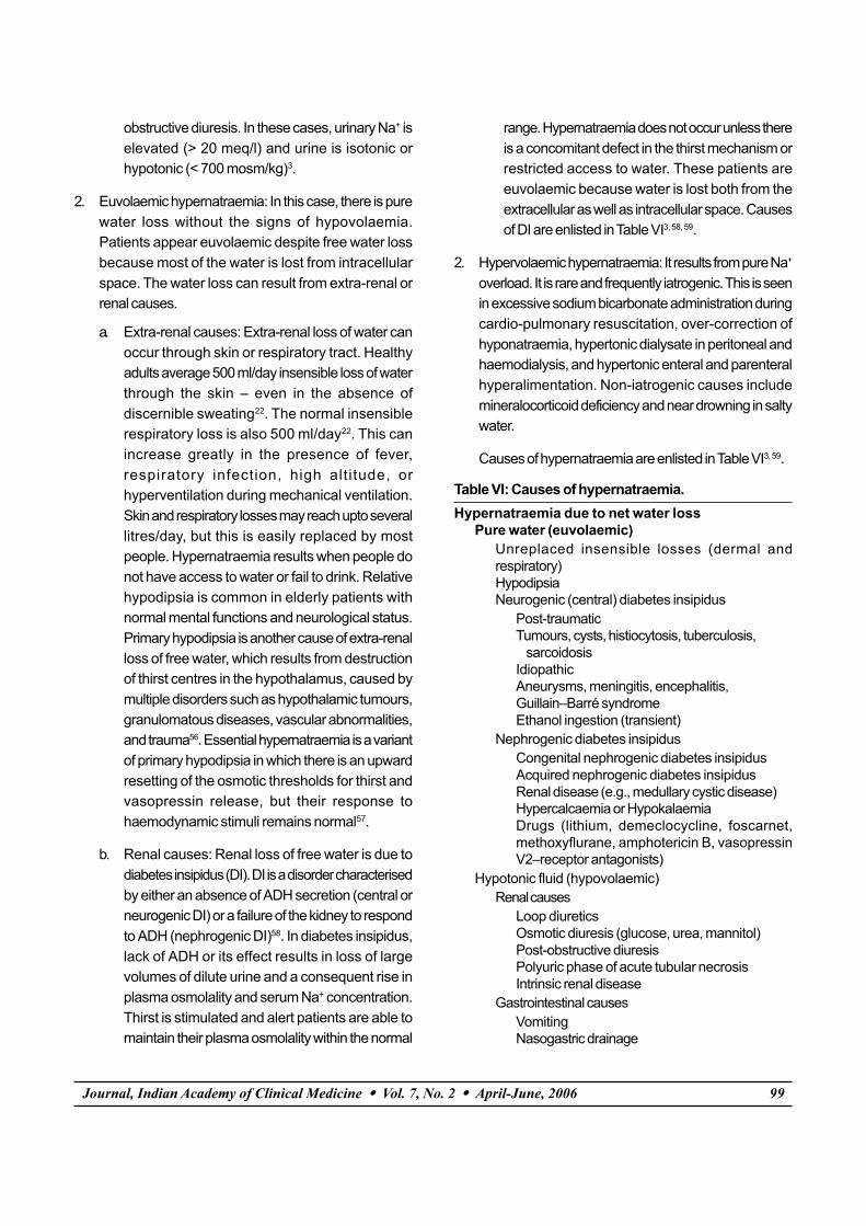
Causes of hypernatraemia are enlisted in Table VI3, 59.
Table VI: Causes of hypernatraemia.Hypernatraemia due to net water loss
Pure water (euvolaemic)Unreplaced insensible losses (dermal andrespiratory)HypodipsiaNeurogenic (central) diabetes insipidus
Post-traumaticTumours, cysts, histiocytosis, tuberculosis, sarcoidosisIdiopathicAneurysms, meningitis, encephalitis,Guillain–Barré syndromeEthanol ingestion (transient)
Nephrogenic diabetes insipidusCongenital nephrogenic diabetes insipidusAcquired nephrogenic diabetes insipidusRenal disease (e.g., medullary cystic disease)Hypercalcaemia or HypokalaemiaDrugs (lithium, demeclocycline, foscarnet,methoxyflurane, amphotericin B, vasopressinV2–receptor antagonists)
Hypotonic fluid (hypovolaemic)Renal causes
Loop diureticsOsmotic diuresis (glucose, urea, mannitol)Post-obstructive diuresisPolyuric phase of acute tubular necrosisIntrinsic renal disease
Gastrointestinal causesVomitingNasogastric drainage

100 Journal, Indian Academy of Clinical Medicine � Vol. 7, No. 2 � April-June, 2006
Enterocutaneous fistulaDiarrhoeaUse of osmotic cathartic agents (e.g., lactulose)
Cutaneous causesBurnsExcessive sweatingPemphigus vulgaris
Hypernatraemia due to hypertonic sodium gain(hypervolaemic)
Hypertonic sodium bicarbonate infusionHypertonic feeding preparationIngestion of sodium chlorideIngestion of sea waterSodium chloride-rich emeticsHypertonic saline enemasIntrauterine injection of hypertonic salineHypertonic sodium chloride infusionHypertonic dialysisPrimary hyperaldosteronismCushing’s syndrome
Clinical features: The principal manifestations ofhypernatraemia largely reflect CNS dysfunction and are
prominent when the increase in serum Na+ is large oroccurs rapidly32. Neurological symptoms occur as aconsequence of cellular dehydration in the brain32, 59. Lossof brain volume results in traction on the cerebral vessels,which may then tear. It is also associated with cerebralbleeding, subarachnoid haemorrhage, and venous sinusthrombosis33, 62. So the threshold for getting a CT scan in apatient with hypernatraemia and altered mental statusshould be low. Convulsions are usually absent and are morecommonly seen in cases of inadvertent sodium loading oraggressive rehydration63, 64. Signs and symptoms ofhypernatraemia are listed in Table VII32.
Table VII: Clinical features of hypernatraemia.Anorexia, nausea, and vomitingAltered mental status, agitation, irritabilityStupor, lethargy, and comaSigns of neuromuscular hyperactivity: hyperreflexia, spasticity, tremor, asterixis, chorea, and ataxia.Focal neurological deficits: hemiparesis and extensor plantars.
Hypovolaemic
Urine osm.
Urinary Na+
Hypotonic IVFisotonic IVG if BP�
Diuretic and waterreplacement
Urinary Na> 1,000 mmol/lit
+
> 700mosm/kg
< 700mosm/kg
Management of hypernatraemia
Hypernatraemia (Na > 145 meq/l)+
Plasma osmolality > 290 mosm/kg
HypotonicIVF
Hypotonic IVF± vasopressin
> 700
Extra-renal loss– Skin loss– Resp tract loss– 1° Hypodipsia– GIT loss
DIRenal loss
< 700
Euvolaemic
Urine osmolatity
Hypervolaemic
Na overload usually iatrogenic+
Extra-renalloss
< 10 > 10
Renalloss
Fig. 2: Algorithm for management of Hypernatraemia.

Journal, Indian Academy of Clinical Medicine � Vol. 7, No. 2 � April-June, 2006 101
ManagementProper management of hypernatraemia requires a two-pronged approach: addressing the underlying cause andcorrecting the prevailing hypertonicity. Figure 2 shows analgorithm for diagnosis and treatment of hypernatraemia.
The treatment of hypernatraemia depends on the volumestatus of the patient. Patients with hypovolaemichypernatraemia should be treated with isotonic salineparticularly if there is evidence of circulatory collapse3.Otherwise, if the volume depletion is mild without evidenceof circulatory failure, hypotonic fluids such as one-quartersaline (0.2% saline), half isotonic saline (0.45%), pure water,or 5% dextrose should be used59. Even in cases of circulatoryfailure, once haemodynamic stability is achieved, hypotonicfluids should be used3. Euvolaemic patients also require purewater replacement with hypotonic saline or free water. Whenadministering dextrose containing solutions, blood glucoseshould be closely monitored, because hyperglycaemia willworsen the hypertonic state and may lead to osmoticdiuresis66. Patients with central DI can be given 5 - 10 units ofaqueous vasopressin subcutaneously every 3 - 4 hours67.Serum sodium concentration and urinary specific gravityshould be monitored every 2 - 4 hours to prevent over-correction. Vasopressin is preferred over desmopressinbecause vasopressin has a shorter duration of action3. Thiswill however be ineffective in nephrogenic DI. Hypervolaemicpatients have sodium overload, and they require natriuresiswith a loop diuretic and free water replacement. Dialysismay be required in patients with severe renal failure toachieve natriuresis3.
Rate of correction of hypernatraemiaThe rate of correction of hypernatraemia depends on therate of development of hypernatraemia. In cases of acutehypernatraemia (< 48 hrs), rapid correction improves theprognosis without increasing the risk of cerebral oedema,because accumulated electrolytes are rapidly extrudedfrom the brain cells. In such patients, the serum sodiumcan be reduced by 1 meq/l/hour65. In chronichypernatraemia, a slower pace is required, as full dissipationof accumulated brain solutes during adaptation occursover a few days. In these patients the appropriate rate ofcorrection is 0.5 meq/l per hour66, 67. The target should beto decrease the serum sodium by 10 meq/l in one day,
except in those in whom the disorder has developed overa matter of hours. The goal of treatment is to achieve asodium concentration of 145 meq/l59. Lowering the plasmaosmolality with hypotonic fluids too rapidly can lead toiatrogenic cerebral oedema because of osmotic shift ofwater into the brain cells68. Allowances must be made forongoing hypotonic fluid loss (obligatory or incidental)while calculating the fluid requirement. The amount offluid required can be calculated using the followingformula:
Serum Na+ change with 1 l of IVF =Na+ content in IVF – measured serum Na+
[Correction factor * weight (kg)] +1
This formula gives the amount of change in serum sodiumwith one litre of the given fluid.
The amount of hypotonic fluid required can be calculatedby the formula:
Amount of fluid (l/hr) =(total body water +1)* rate of correction (meq/l/hr)
Infusate Na+ – measured Na+
For example, in an elderly 80 kg male with a serum sodiumconcentration of 160 meq/l, the rate of decrease should be0.5 meq/l/hr. The amount of 0.45% saline required will be[(0.5*80)+1]*0.5/77-160 = .246 l or 246 ml/hr.
Table V lists the characteristics of various solutions used inmanagement of hypernatraemia.
Both hypernatraemia and hyponatraemia initially presentwith non-specific signs and symptoms and can only bediagnosed if a high index of suspicion is maintained forthese conditions. The treatment of both these conditions istricky, as over-treatment can lead to potentially dangerouscomplications and under-treatment is associated withsignificant mortality and morbidity. It is therefore essentialto monitor the serum sodium concentration every 2 - 4hours to prevent treatment related complications. Frequentneurological examination should also be performed duringtreatment as it can help in early detection of ODS andcerebral oedema.
References1. Genanri FJ. Hypo-hypernatraemia: disorders of water

102 Journal, Indian Academy of Clinical Medicine � Vol. 7, No. 2 � April-June, 2006
balance. In: Davison AM, Cameron JS, Grunfeld J-P, KerrDNS, Ritz E, Winearls CG, eds. Oxford textbook of clinicalnephrology. 2nd ed. Vol 1. Oxford, England: OxfordUniversity Press, 1998; 175-200.
2. Genanri FJ. Serum osmolality: uses and limitations. N EnglJ Med 1984; 310: 102-5.
3. Lin M, Liu SJ, Lim IT. Disorders of water imbalance. EmergMed Clin North Am 2005; 23 (3): 749-70.
4. McKinley MJ, Mathai ML, McAllen RM et al. Vasopressinsecretion: osmotic and hormonal regulation by the laminaterminalis. J Neuroendocrinol 2004 ; 16 (4): 340-7.
5. Berliner RW, Levinsky NG, Davidson DG, Eden M. Dilutionand concentration of the urine and the action ofantidiuretic hormone. Am J Med 1958; 24 (5): 730-44.
6. Al-Salman J, Kemp D, Randall D. Hyponatremia. West J Med2002; 176 (3): 173-6.
7. Anderson RJ, Chung HM, Kluge R, Schrier RW. Hyponatremia:a prospective analysis of its epidemiology and thepathogenetic role of vasopressin. Ann Intern Med 1985; 102(2): 164-8.
8. Tierney WM, Martin DK, Greenlee MC et al. The prognosisof hyponatremia at hospital admission. J Gen Intern Med1986; 1 (6): 380-5.
9. Lee CT, Guo HR, Chen JB.Hyponatremia in the emergencydepartment. Am J Emerg Med 2000; 18 (3): 264-8.
10. Arieff AI. Hyponatremia, convulsions, respiratory arrest,and permanent brain damage after elective surgery inhealthy women. N Engl J Med 1986; 314 (24): 1529-35.
11. Adrogue HJ, Madias NE. Hyponatremia. N Engl J Med 2000;342 (21): 1581-9.
12. Oh MS. Pathogenesis and diagnosis of hyponatremia.Nephron 2002; 92 (Suppl 1): 2-8.
13. Aviram A, Pfau A, Czaczkes JW, Ullmann TD. Hyperosmolalitywith hyponatremia, caused by inappropriateadministration of mannitol. Am J Med 1967; 42 (4): 648-50.
14. Palevsky PM, Rendulic D, Diven WF. Maltose-inducedhyponatremia. Ann Intern Med 1993; 118 (7): 526-8.
15. Maas AH, Siggaard-Andersen O, Weisberg HF, Zijlstra WG.Ion-selective electrodes for sodium and potassium: a newproblem of what is measured and what should bereported. Clin Chem 1985; 31 (3): 482-5.
16. Gonzales R, Brensilver JM, Rovinsky JJ. Post-hysteroscopichyponatremia. Am J Kidney Dis 1994; 23:735-8.
17. Agarwal R, Emmett M. The post-transurethral resection ofprostate Syndrome: therapeutic proposals. Am J Kidney Dis1994; 24:108-11.
18. Chung HM, Kluge R, Schrier RW, Anderson RJ. Clinicalassessment of extracellular fluid volume in hyponatremia.Am J Med 1987; 83 (5): 905-8.
19. Kennedy PG, Mitchell DM, Hoffbrand BI. Severehyponatraemia in hospital inpatients. Br Med J 1978; 2 (6147):1251-3.
20. Abramow M, Cogan E. Clinical aspects andpathophysiology of diuretic-induced hyponatremia. Adv
Nephrol Necker Hosp 1984; 13: 1-28.21. Ashraf N, Locksley R, Arieff AI. Thiazide-induced
hyponatremia associated with death or neurologicdamage in outpatients. Am J Med 1981; 70 (6): 1163-8.
22. Votey SR, Peters AL, Hoffman JR. Disorders of watermetabolism: hyponatremia and hypernatremia. Emerg MedClin North Am 1989; 7 (4): 749-69.
23. Fichman MP, Vorherr H, Kleeman CR, Telfer N. Diuretic-induced hyponatremia. Ann Intern Med 1971; 75 (6): 853-63.
24. Lee CT, Guo HR, Chen JB. Hyponatremia in the emergencydepartment. Am J Emerg Med 2000; 18 (3): 264-8.
25. Linas SL, Berl T, Robertson GL et al. Role of vasopressin inthe impaired water excretion of glucocorticoid deficiency.Kidney Int 1980; 18 (1): 58-67.
26. Schrier RW, Linas SL. Mechanisms of defect in waterexcretion in adrenal insufficiency. Miner Electrolyte Metab1980; 4: 1-18.
27. Derubertis FR Jr, Michelis MF, Bloom ME et al. Impairedwater excretion in myxedema. Am J Med 1971; 51 (1): 41-53.
28. Abraham A, Jacob CK. Severe hyponatraemia: currentconcepts on pathogenesis and treatment. Natl Med J India2001; 14 (5): 277-83.
29. Riggs AT, Dysken MW, Kim SW, Opsahl JA. A review ofdisorders of water homeostasis in psychiatric patients.Psychosomatics 1991; 32 (2): 133-48.
30. Robertson GL, Aycinena P, Zerbe RL. Neurogenic disordersof osmoregulation. Am J Med 1982; 72 (2): 339-53.
31. Fenves AZ, Thomas S, Knochel JP. Beer potomania: twocases and review of the literature. Clin Nephrol 1996; 45 (1):61-4.
32. Arieff AI. Central nervous system manifestations ofdisordered sodium metabolism. Clin Endocrinol Metab 1984;13 (2): 269-94.
33. Schrier RW, Berl T. Disorders of water metabolism. In: SchrierRW ed: Renal and Electrolyte Disorders. Ed 2. Boston, LittleBrown and Co, 1986; 1.
34. Melton JE, Nattie EE. Brain and CSF water and ions duringdilutional and isosmotic hyponatremia in the rat. Am JPhysiol 1983; 244 (5): R724-32.
35. Verbalis JG, Gullans SR. Hyponatraemia causes largesustained reductions in brain content of multiple organicosmolytes in rats. Brain Res 1991; 20; 567 (2): 274-82.
36. Paredes A, McManus M, Kwon HM, Strange K.Osmoregulation of Na(+)-inositol co-transporter activityand mRNA levels in brain glial cells. Am J Physiol 1992; 263(6 Pt 1): C1282-8.
37. Karp BI, Laureno R. Pontine and extrapontine myelinolysis:a neurologic disorder following rapid correction ofhyponatremia. Medicine (Baltimore) 1993; 72: 359-73.
38. Sterns RH, Riggs JE, Schochet SS Jr. Osmotic demyelinationsyndrome following correction of hyponatremia. N Engl JMed 1986; 314: 1535-42.
39. Tanneau RS, Henry A, Rouhart F et al. High incidence ofneurologic complications following rapid correction of

Journal, Indian Academy of Clinical Medicine � Vol. 7, No. 2 � April-June, 2006 103
severe hyponatremia in polydipsic patients. J Clin Psychiatry1994; 55: 349-54.
40. Gocht A, Colmant HJ. Central pontine and extrapontinemyelinolysis: a report of 58 cases. Clin Neuropathol 1987; 6(6): 262-70.
41. Sterns RH, Cappuccio JD, Silver SM, Cohen EP.Neurologic sequelae after treatment of severehyponatremia: a multicenter perspective. J Am SocNephrol 1994; 4 (8): 1522-30.
42. Laureno R. Central pontine myelinolysis followingrapid correction of hyponatremia. Ann Neurol 1983; 13(3): 232-42.
43. Srivastava T, Singh P, Sharma B. Pontine and extrapontinemyelinolysis following rapid correction of hyponatremia.Neurol India 2000; 48 (1): 97.
44. Finsterer J, Engelmayer E, Trnka E, Stiskal M.Immunoglobulins are effective in pontine myelinolysis.Clin Neuropharmacol 2000; 23 (2): 110-3.
45. Arieff AI. Hyponatremia, convulsions, respiratory arrest,and permanent brain damage after elective surgery inhealthy women. N Engl J Med 1986; 314 (24): 1529-35.
46. Ayus JC, Krothapalli RK, Arieff AI. Changing concepts intreatment of severe symptomatic hyponatremia. Rapidcorrection and possible relation to central pontinemyelinolysis. Am J Med 1985; 78 (6 Pt 1): 897-902.
47. Schrier RW. Pathogenesis of sodium and water retentionin high-output and low-output cardiac failure, nephroticsyndrome, cirrhosis, and pregnancy. N Engl J Med 1988; 319(16): 1065-72.
48. Paterna S, Di Pasquale P, Parrinello G et al. Effects of high-dose furosemide and small-volume hypertonic salinesolution infusion in comparison with a high dose offurosemide as a bolus, in refractory congestive heartfailure. Eur J Heart Fail 2000; 2 (3): 305-13.
49. Oh MS, Kim HJ, Carroll HJ. Recommendations fortreatment of symptomatic hyponatremia. Nephron 1995;70:143-50.
50. Berl T. Treating hyponatremia: damned if we do anddamned if we don’t. Kidney Int 1990; 37: 1006-18.
51. Van Reeth O, Decaux G. Rapid correction ofhyponatraemia with urea may protect against braindamage in rats. Clin Sci (Lond) 1989; 77: 351-5.
52. Forrest JN Jr, Cox M, Hong C et al. Superiority ofdemeclocycline over lithium in the treatment of chronicsyndrome of inappropriate secretion of antidiuretichormone. N Engl J Med 1978; 298 (4): 173-7.
53. Gross P, Palm C. The treatment of hyponatraemia usingvasopressin antagonists. Exp Physiol 2000; 85: S253-257.
54. Long CA, Marin P, Bayer AJ et al. Hypernatraemia in anadult in-patient population. Postgrad Med J 1991; 67 (789):643-5.
55. Janz T. Sodium. Emerg Med Clin North America 1986; 4 (1):115-30.
56. Robertson GL. Antidiuretic hormone. Normal anddisordered function. Endocrinol Metab Clin North Am 2001;30 (3): 671-94.
57. DeRubertis FR, Michelis MF, Davis BB. Essentialhypernatremia. Report of three cases and review of theliterature. Arch Intern Med 1974; 134 (5): 889-95.
58. Robertson GL. Posterior Pituatry. In: Felig P, Baxter JD,Broadus AE et al (eds): Endocrinology and Metabolism. Ed2. New York. McGraw Hill.1987; 338.
59. Adrogue HJ, Madias NE. Hypernatremia. N Engl J Med 2000;342 (20): 1493-9.
60. Mattar JA, Weil MH, Shubin H, Stein L. Cardiac arrest in thecritically ill. II. Hyperosmolal states following cardiac arrest.Am J Med 1974; 56 (2): 162-8.
61. Snyder NA, Feigal DW, Arieff AI. Hypernatremia in elderlypatients. A heterogeneous, morbid, and iatrogenic entity.Ann Intern Med 1987; 107 (3): 309-19.
62. Macaulay D, Watson M. Hypernatraemia in infants as acause of brain damage. Arch Dis Child 1967; 42 (225):485-91.
63. Morris-Jones PH, Houston IB, Evans RC. Prognosis of theneurological complications of acute hypernatraemia.Lancet 1967; 2: 1385-9.
64. Hogan GR, Dodge PR, Gill SR et al. Pathogenesis of seizuresoccurring during restoration of plasma tonicity to normalin animals previously chronically hypernatremic. Pediatrics1969; 43: 54-64.
65. Palevsky PM. Hypernatremia. In: Greenberg A, ed. Primeron kidney diseases. 2nd ed. San Diego, Calif.: AcademicPress, 1998; 64-71.
66. Kahn A, Brachet E, Blum D. Controlled fall in natremia andrisk of seizures in hypertonic dehydration. Intensive CareMed 1979; 5: 27-31.
67. Blum D, Brasseur D, Kahn A, Brachet E. Safe oral rehydrationof hypertonic dehydration. J Pediatr Gastroenterol Nutr 1986;5: 232-5.
68. Arieff AI, Guisado R. Effects on the central nervous systemof hypernatremic and hyponatremic states. Kidney Int 1976;10 (1): 104-16.