clinical diagnosis and oral rehabilitation of a patient
TRANSCRIPT
1The Journal of Contemporary Dental Practice, Volume 9, No. 4, May 1, 2008
Clinical Diagnosis and Oral Rehabilitation of a Patient with Amelogenesis imperfecta:
A Case Report
Aim: This clinical report describes the oral rehabilitation of a young female patient diagnosed with the hypocalcified, autosomal recessive type of Amelogenesis imperfecta (AI). A brief discussion on diagnosis of AIis also included.
Background: AI has been defined as a group of hereditary enamel defects not associated with evidence of systemic disease. It can be characterized by enamel hypoplasia and/or hypomaturation or hypocalcification of the existing teeth. Restoration for patients with this condition should be oriented toward the functional and esthetic rehabilitation and the protection of these teeth.
Report: A 31-year-old female patient presented with concerns including extreme sensitivity; dissatisfactionwith size, shape, and shade of teeth; and poor masticatory efficiency. She was very conscious about theappearance of her teeth and reported that her primary dentition was affected in the same manner. The specificobjectives of this treatment were to eliminate tooth sensitivity, enhance esthetics, and restore masticatoryfunction. Treatment included crown lengthening procedures and placement of anterior and posterior metal-ceramic crowns. A 12-month follow-up with clinical and radiographic examinations revealed no evidence of anyuntoward effects of the treatment on the restored teeth or their supporting structures.
Summary: Management of a patient with AI is a challenge for the clinician. The treatment options varyconsiderably depending on several factors such as the age of the patient, socio-economic status, periodontal
Abstract
© Seer Publishing

2The Journal of Contemporary Dental Practice, Volume 9, No. 4, May 1, 2008
IntroductionAmelogenesis imperfecta (AI) has been defined as a complex group of hereditary enamel defects not associated with evidence of systemic disease1,2
affecting both primary and permanent dentitions.1
It is a rare enamel mineralization defect describedby Spokes3 in 1890 as “hereditary brown teeth”with a reported incidence of 1:14,000.4
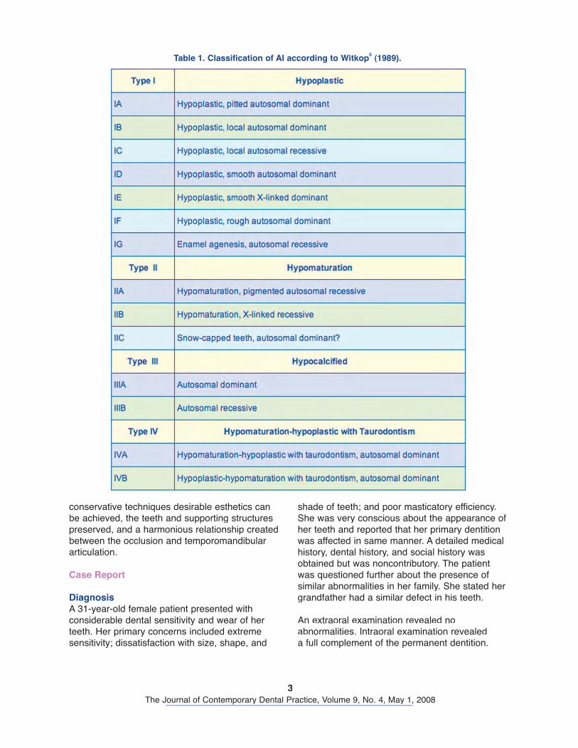
Phenotypically AI is categorized into four broad groups: hypoplastic, hypomaturation, hypocalcified, and a hypomaturation-hypoplastic variety. Fifteen subtypes of AI exist phenotypically and based on modes of inheritance. Thisclassification has been proposed by Witkop5
(Table 1).
The types are characterized as follows:• Type-I: Lesions may appear as pin-point
to pinhead sized pits scattered across the surfaces of teeth. The distribution of lesions may be generalized or localized, and the alteration of the enamel is a result ofinadequate deposition of enamel matrix.
• Type-II: Also known as the hypomaturationtype is associated with abnormalities inthe maturation stages of enamel formationresulting in the enamel being opaque and chalky in appearance. The enamel layer isnormal in thickness but softer than normal and can be easily detached from the underlyingdentin.
• Type-III: The teeth have enamel that isinsufficiently mineralized and clinicallyappears as severely worn teeth. It results from detachment of the enamel from dentin withina short period after tooth eruption. Teeth are very sensitive to thermal changes and appeardark brown in color.
• Type-IV: AI exhibits enamel hypoplasia incombination with hypomaturation. This variety
is associated with taurodontism. The most common form of AI is the autosomal dominant hypocalcified type, followed by hypomaturation,and the hypoplastic type.6
Other associated findings in patients with AIinclude delayed eruption of teeth, taurodontism, congenitally missing teeth, crown and root resorption, and pulp calcification.7 Radiographically the density of enamel layer is lower than normalenamel. Hypoplastic enamel shows great variationin density and it may be difficult to distinguish it radiographically from underlying dentin.
AI is caused by mutations in a variety of genesthat are critical for normal enamel formation. A total of about five genes [AMELX, ENAM, KLK4, MMP20, and DLX3]8,9 are known to be involved inenamel formation. Mutations of the amelogenin gene (AMELX) cause X-linked AI, while mutationsof the enamelin (ENAM) gene causes autosomal inherited forms of AI. Other genes like Kallikrein – 4 (KLK4), MMP–20, and DLX3 genes contribute tothe etiologies of some other varieties of AI which is still under investigation.
Various treatment methods or strategies were initially instituted for AI patients such as theextraction of the compromised teeth and placement of a removable prosthesis or implant supported fixed or removable prosthesis.10 However, these procedures are very invasive and have greaterincidence of complications. Numerous treatmentmodalities have been described for rehabilitation ofpatients with AI.10-17 Rehabilitation of patients with AI requires meticulous oral hygiene maintenanceand patient cooperation.
This rare dental abnormality poses a majorrestorative challenge for the dentist. Using
condition, loss of tooth structure, severity of the disorder, and, most importantly, the patient’s cooperation. The clinician has to consider the long-term prognosis of the treatment outcome. This clinical report describes thefabrication of metal ceramic and all metal crowns for the restoration of severely worn teeth in a patient with AIwhich requires meticulous maintenance of oral hygiene and patient co-operation.
Keywords: Amelogenesis imperfecta, AI, hereditary enamel defects, clinical diagnosis, full-mouth rehabilitation
Citation: Sholapurkar AA, Joseph RM, Varghese JM, Neelagiri K, Acharya SRR, Hegde V, Pai KM, Bhat M. Clinical Diagnosis and Oral Rehabilitation of a Patient with Amelogenesis imperfecta: A Case Report. J Contemp Dent Pract 2008 May; (9)4:092-098.
3The Journal of Contemporary Dental Practice, Volume 9, No. 4, May 1, 2008
shade of teeth; and poor masticatory efficiency.She was very conscious about the appearance of her teeth and reported that her primary dentition was affected in same manner. A detailed medicalhistory, dental history, and social history was obtained but was noncontributory. The patient was questioned further about the presence ofsimilar abnormalities in her family. She stated hergrandfather had a similar defect in his teeth.
An extraoral examination revealed no abnormalities. Intraoral examination revealeda full complement of the permanent dentition.
conservative techniques desirable esthetics canbe achieved, the teeth and supporting structurespreserved, and a harmonious relationship created between the occlusion and temporomandibular articulation.
Case Report
DiagnosisA 31-year-old female patient presented withconsiderable dental sensitivity and wear of herteeth. Her primary concerns included extremesensitivity; dissatisfaction with size, shape, and
Table 1. Classification of AI according to Witkop5 (1989).
4The Journal of Contemporary Dental Practice, Volume 9, No. 4, May 1, 2008
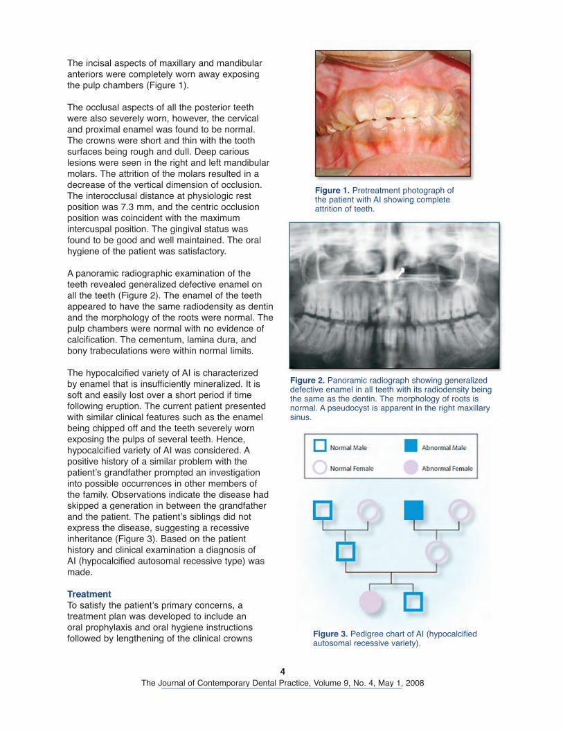
The incisal aspects of maxillary and mandibular anteriors were completely worn away exposing the pulp chambers (Figure 1).
The occlusal aspects of all the posterior teethwere also severely worn, however, the cervicaland proximal enamel was found to be normal. The crowns were short and thin with the toothsurfaces being rough and dull. Deep cariouslesions were seen in the right and left mandibular molars. The attrition of the molars resulted in a decrease of the vertical dimension of occlusion.The interocclusal distance at physiologic rest position was 7.3 mm, and the centric occlusionposition was coincident with the maximumintercuspal position. The gingival status was found to be good and well maintained. The oral hygiene of the patient was satisfactory.
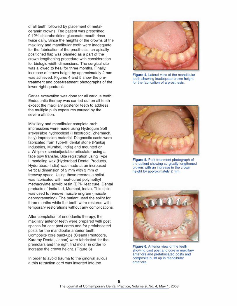
A panoramic radiographic examination of theteeth revealed generalized defective enamel on all the teeth (Figure 2). The enamel of the teeth appeared to have the same radiodensity as dentinand the morphology of the roots were normal. The pulp chambers were normal with no evidence ofcalcification. The cementum, lamina dura, andbony trabeculations were within normal limits.
The hypocalcified variety of AI is characterizedby enamel that is insufficiently mineralized. It is soft and easily lost over a short period if time following eruption. The current patient presented with similar clinical features such as the enamelbeing chipped off and the teeth severely wornexposing the pulps of several teeth. Hence,hypocalcified variety of AI was considered. A positive history of a similar problem with thepatient’s grandfather prompted an investigation into possible occurrences in other members ofthe family. Observations indicate the disease had skipped a generation in between the grandfather and the patient. The patient’s siblings did not express the disease, suggesting a recessive inheritance (Figure 3). Based on the patienthistory and clinical examination a diagnosis of AI (hypocalcified autosomal recessive type) was made.
TreatmentTo satisfy the patient’s primary concerns, a treatment plan was developed to include anoral prophylaxis and oral hygiene instructionsfollowed by lengthening of the clinical crowns
Figure 1. Pretreatment photograph of the patient with AI showing complete attrition of teeth.
Figure 2. Panoramic radiograph showing generalized defective enamel in all teeth with its radiodensity being the same as the dentin. The morphology of roots is normal. A pseudocyst is apparent in the right maxillary sinus.
Figure 3. Pedigree chart of AI (hypocalcified autosomal recessive variety).
5The Journal of Contemporary Dental Practice, Volume 9, No. 4, May 1, 2008
of all teeth followed by placement of metal-ceramic crowns. The patient was prescribed0.12% chlorohexidine gluconate mouth rinsetwice daily. Since the heights of the crowns of themaxillary and mandibular teeth were inadequate for the fabrication of the prosthesis, an apicallypositioned flap was planned as a part of the crown lengthening procedure with consideration for biologic width dimensions. The surgical site was allowed to heal for three months. Finally,increase of crown height by approximately 2 mmwas achieved. Figures 4 and 5 show the pre-treatment and post-treatment photographs of the lower right quadrant.
Caries excavation was done for all carious teeth. Endodontic therapy was carried out on all teeth except the maxillary posterior teeth to address the multiple pulp exposures caused by the severe attrition.
Maxillary and mandibular complete-arch impressions were made using Hydrogum Softirreversible hydrocolloid (Thixotropic, Zhermach, Italy) impression material. Diagnostic casts were fabricated from Type-III dental stone (Pankaj Industries, Mumbai, India) and mounted on a Whipmix semiadjustable articulator using a face bow transfer. Bite registration using Type II modeling wax (Hyderabad Dental Products,Hyderabad, India) was made at an increased vertical dimension of 5 mm with 3 mm offreeway space. Using these records a splintwas fabricated with heat-cured polymethyl methacrylate acrylic resin (DPI-Heat cure, Dentalproducts of India Ltd, Mumbai, India). This splintwas used to remove muscle engram (muscle deprogramming). The patient used the splint forthree months while the teeth were restored withtemporary restorations without any complications.
After completion of endodontic therapy, the maxillary anterior teeth were prepared with postspaces for cast post cores and for prefabricatedposts for the mandibular anterior teeth.Composite core build-ups (Clearfil Photocore,Kuraray Dental, Japan) were fabricated for thepremolars and the right first molar in order to increase the crown height. (Figure 6)
In order to avoid trauma to the gingival sulcus a thin retraction cord was inserted into the
Figure 4. Lateral view of the mandibular teeth showing inadequate crown height for the fabrication of a prosthesis.
Figure 5. Post treatment photograph of the patient showing surgically lengthened crowns with an increase in the crown height by approximately 2 mm.
Figure 6. Anterior view of the teeth showing cast post and core in maxillary anteriors and prefabricated posts and composite build up in mandibular anteriors.
6The Journal of Contemporary Dental Practice, Volume 9, No. 4, May 1, 2008
sulcus prior to preparation. Crown preparationswere done for porcelain-fused-to-metal (PFM)restorations for the maxillary and mandibularanteriors, premolars, and maxillary first molars;on the remaining teeth all-metal restorations wereused. Gingival displacement prior to impression making was done using a non-hemostatic gingival magic foam cord retraction system (Ultradent,South Jordan, UT, USA).
Impressions were made with addition polyvinylsiloxane material (Reprosil, Dentsply/Caulk; Milford, DE, USA) using the putty wash technique (Panasil Putty Soft, Dentsply Kettenbach,Germany). Full-mouth, heat-cured provisionalrestorations (Figure 7) were fabricated at thedesired vertical dimension (with 3 mm freewayspace) using methyl methacrylate acrylic resin. The provisional restorations were temporarilycemented using Provicol, eugenol free Ca(OH)2
cement (Voco, Cuxhaven, Germany).
The patient wore the provisional restorations atthe newly established occlusal vertical dimension for three months. Final impressions were made using the putty-wash technique and casts wereprepared using Type IV stone in conjunctionwith the Pindex system (Confident, Bangalore,India) to create removable dies. The workingcasts were mounted onto the Hanau Widevuesemiadjustable articulator (Waterpik, Ft Collins, CO, USA) using Type I rigid tray material withthe interocclusal records (Take 1, Kerr, Romulus,MI, USA). Wax patterns were made using inlay wax (Harward, Harward, Germany) and thencasting was done using Metal ceramic alloy (Remanium CSe, Dentaurum J.P. Winkelstroeter KG, Ispringen, Germany). During the cast metal try-in marginal fit and passivity were evaluated(Figure 8).
The appropriate shade was then selectedusing the VITA shade guide (Vita Zahnfabrik, Badsackingen, Germany) and porcelain firing was done.
The PFM restorations for the maxillary and mandibular anteriors, premolars, and maxillaryfirst molars were cemented temporarily with Provicol for two weeks then permanently cemented with GIC Fugi II cement ( GC, Tokyo,Japan) (Figure 9).
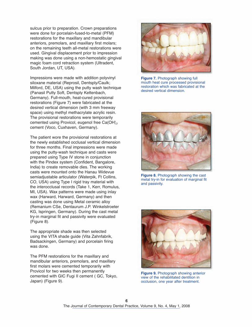
Figure 7. Photograph showing full mouth heat cure processed provisional restoration which was fabricated at the desired vertical dimension.
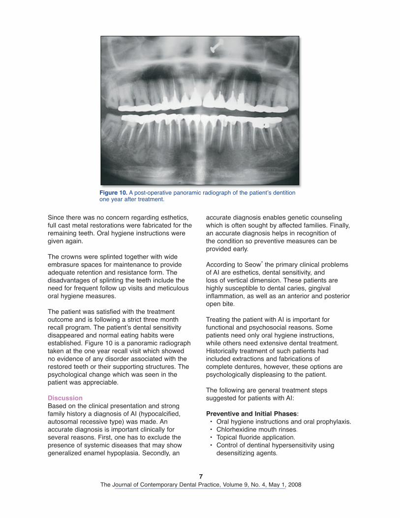
Figure 8. Photograph showing the cast metal try-in for evaluation of marginal fit and passivity.
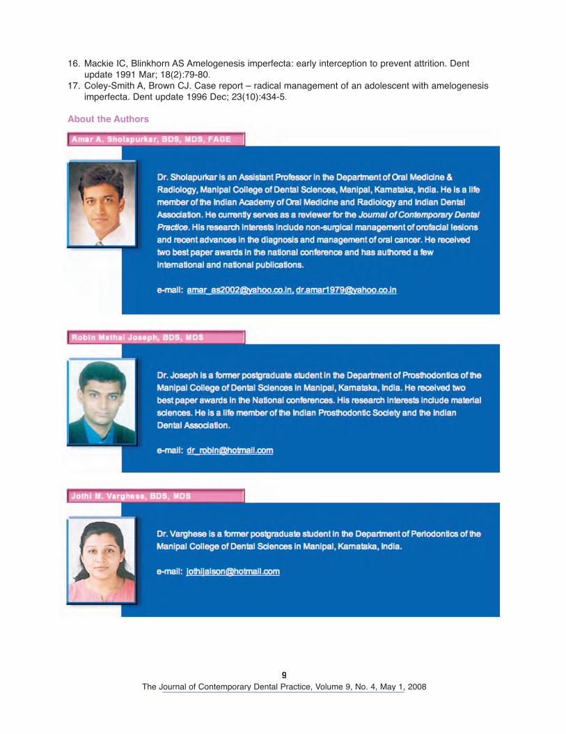
Figure 9. Photograph showing anterior view of the rehabilitated dentition in occlusion, one year after treatment.
7The Journal of Contemporary Dental Practice, Volume 9, No. 4, May 1, 2008
accurate diagnosis enables genetic counselingwhich is often sought by affected families. Finally,an accurate diagnosis helps in recognition of the condition so preventive measures can be provided early.
According to Seow2 the primary clinical problemsof AI are esthetics, dental sensitivity, and loss of vertical dimension. These patients are highly susceptible to dental caries, gingivalinflammation, as well as an anterior and posterior open bite.
Treating the patient with AI is important for functional and psychosocial reasons. Somepatients need only oral hygiene instructions, while others need extensive dental treatment.Historically treatment of such patients had included extractions and fabrications of complete dentures, however, these options are psychologically displeasing to the patient.
The following are general treatment steps suggested for patients with AI:
Preventive and Initial Phases:• Oral hygiene instructions and oral prophylaxis.• Chlorhexidine mouth rinses.• Topical fluoride application.• Control of dentinal hypersensitivity using
desensitizing agents.
Since there was no concern regarding esthetics,full cast metal restorations were fabricated for theremaining teeth. Oral hygiene instructions weregiven again.
The crowns were splinted together with wide embrasure spaces for maintenance to provideadequate retention and resistance form. Thedisadvantages of splinting the teeth include theneed for frequent follow up visits and meticulousoral hygiene measures.
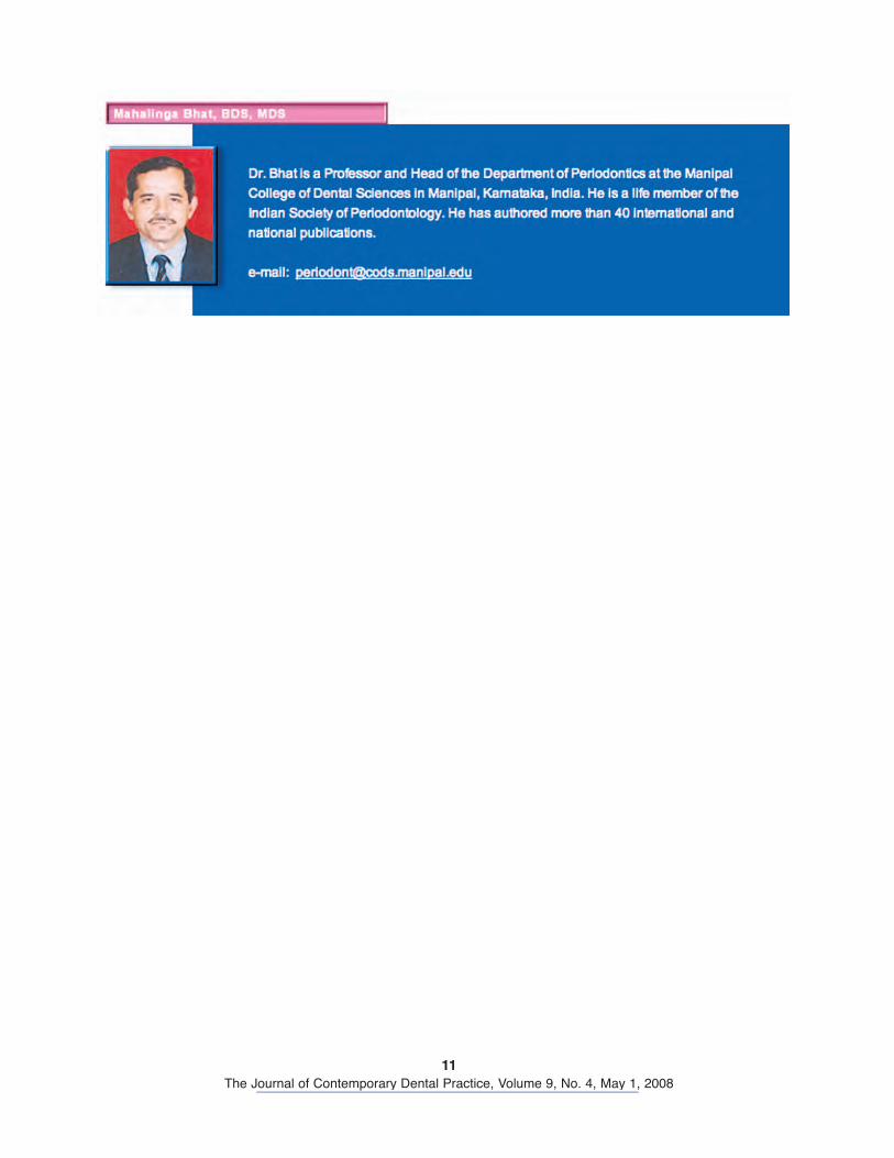
The patient was satisfied with the treatmentoutcome and is following a strict three monthrecall program. The patient’s dental sensitivitydisappeared and normal eating habits were established. Figure 10 is a panoramic radiographtaken at the one year recall visit which showedno evidence of any disorder associated with therestored teeth or their supporting structures. The psychological change which was seen in the patient was appreciable.
DiscussionBased on the clinical presentation and strongfamily history a diagnosis of AI (hypocalcified,autosomal recessive type) was made. An accurate diagnosis is important clinically for several reasons. First, one has to exclude thepresence of systemic diseases that may show generalized enamel hypoplasia. Secondly, an
Figure 10. A post-operative panoramic radiograph of the patient’s dentition one year after treatment.

8The Journal of Contemporary Dental Practice, Volume 9, No. 4, May 1, 2008
Because of the recent advances in the field of esthetics and prosthetic dentistry, it is possible to restore the function and esthetics to anacceptable level in severe AI cases.
SummaryManagement of a patient with AI is a challengefor the clinician. The treatment options varyconsiderably depending on several factors such as the age of the patient, socio-economic status,periodontal condition, loss of tooth structure,severity of the disorder, and, most importantly,the patient’s cooperation. The clinician hasto consider the long-term prognosis of thetreatment outcome. This clinical report describes the fabrication of metal ceramic and all metal crowns for the restoration of severely worn teeth in a patient with AI which requires meticulous maintenance of oral hygiene and patient co-operation.
• Extraction of teeth which have a poorprognosis.
• The initial provisional stage of treatment should be performed as soon as AI is diagnosed when the patient is cooperative.
Restorative Phase:• Establish a favorable occlusal vertical
dimension using a provisional occlusal splint.• Composite build up of the teeth with severe
loss of tooth structure.• Fabricate thin gold crowns for posterior teeth.• Lengthen the crowns of the worn teeth.• Fabricate metal-ceramic crowns, all ceramic
crowns, or porcelain veneers if the enamel is suitable for bonding for teeth where estheticsis a concern.
Maintenance Phase:• Monitor oral hygiene, periodontal, and pulpal
status.
References1. Robinson FG, Haubenreich JE. Oral rehabilitation of a young adult with hypoplastic amelogenesis
imperfecta: a clinical report. J Prosthet Dent 2006 Jan;95(1):10-3.2. Sari T, Usumez A. Restoring function and esthetics in a patient with amelogenesis imperfecta: a
clinical report. J Prosthet Dent 2003 Dec; 90(6):522-5.3. Lindunger A, Smedberg JI. A retrospective study of the prosthodontic management of patients with
amelogenesis imperfecta. Int J Prosthodon, 2005 May-June; 18(3):189-94.4. Bouvier D, Duprez JP, Pirel C, Vincent B. Amelogenesis imperfecta – a prosthetic rehabilitation:
A clinical report J Prosthet Dent 1999 Aug; 82(2):130-1.5. Witkop CJ Jr. Amelogenesis imperfecta, Dentinogenesis imperfecta and dentin dysplasia revisited:
problems in classification. J Oral Pathol 1988: 17;547-53.6. Kostoulas I, Kourtis S, Andritsakis D, Doukoudakis A. Functional and esthetic rehabilitation in
amelogenesis imperfecta with all-ceramic restorations: a case report. Quintessence Int 2005 May; 36(5):329-38.
7. Yip HK, Smales RJ. Oral Rehabilitation of young adults with amelogenesis imperfecta. Int JProsthodont 2003 July-Aug 16(4):345-9.
8. Wright JT. The molecular etiologies and associated Phenotypes. Am J Med Genet A, 2006 Dec 1; 140(23):2547-55.
9. Stephanopoulos G, Garefalaki ME, Lyroudia K. Genes and related proteins involved in amelogenesisimperfecta. J Dent Res 2005 Dec; 84(12):1117-26.
10. Light EI, Rakow B, Fraze RL. An esthetic transitional treatment for amelogenesis imperfecta: report of two cases. J Am Dent Assoc 1975 Jan; 90(1):166-70.
11. Rosenblum SH. Restorative & Orthodontic treatment of an adolescent patient with amelogenesisimperfecta. Pediatr Dent 1999 Jul-Aug; 21(4):289-92.
12. Sengun A, Ozer F. Restoring function and esthetics in a patient with amelogenesis imperfecta:a case report. Quintessence Int 2002 Mar; 33(3):199-204.
13. Lamb DJ The treatment of amelogenesis imperfecta. J Prosthet Dent 1976 Sep; 36(3):286-91.14. Bauvier D, Duprez JP, Bois D. Rehabilitation of young patients with amelogenesis imperfecta:
a report of two cases. ASDC J Dent Child 1996 Nov-Dec; 63(6):443-7.15. Storic DQ, Cheatham JL. Management of amelogenesis imperfecta by Periodontal & Prosthetic
Therapy. J Prosthet Dent 1970 Dec; 24(6):608-15.
9The Journal of Contemporary Dental Practice, Volume 9, No. 4, May 1, 2008
16. Mackie IC, Blinkhorn AS Amelogenesis imperfecta: early interception to prevent attrition. Dentupdate 1991 Mar; 18(2):79-80.
17. Coley-Smith A, Brown CJ. Case report – radical management of an adolescent with amelogenesisimperfecta. Dent update 1996 Dec; 23(10):434-5.
About the Authors

10The Journal of Contemporary Dental Practice, Volume 9, No. 4, May 1, 2008
11The Journal of Contemporary Dental Practice, Volume 9, No. 4, May 1, 2008