romanian journal of oral rehabilitation journal of oral rehabilitation vol. 2, no. 4, october 2010 1...
TRANSCRIPT

Romanian Journal of Oral Rehabilitation
Vol. 2, No. 4, October 2010
1
Romanian Journal
of Oral Rehabilitation
Vol. 2, No. 4, October 2010
Editor in Chief
Norina Consuela Forna, Iaşi, România
Vice-Editor
Viorel Păun, Bucharest, România
Senior Associate Editors
Pierre Lafforgue, Paris, France
Sammi Sandhaus, Lausanne, Switzerland
Robert Sader, Germania
Zhimon Jacobson, Boston, USA
Editorial Board
Corneliu Amariei, Constanţa, România
Vasile Astărăstoae, Iaşi, România
Mihai Augustin, Bucharest, România
Grigore Băciuţ, Cluj-Napoca, România
Constantin Bălăceanu-Stolnici, Bucharest,
România
Marc Bolla, Nice, France
Dorin Bratu, Timişoara, România
Alexandru Bucur, Bucharest, România
Eugen Carasevici, Iaşi, România
Radu Septimiu Câmpean, Cluj-Napoca,
România
Virgil Cârligeriu, Timişoara, România
Costin Cernescu, Bucharest, România
Yves Comissionat, Paris, France
Marysette Folliguet, Paris, France
Cristina Glavce, Bucharest, România
Emilian Hutu, Bucharest, România
Constantin Ionescu-Tîrgoviste, Bucharest,
România
Michel Jourde, Paris, France
Ion Lupan, Chişinău, Republica Moldova
Veronica Mercuţ, Craiova, România
Patrick Missika, Paris, France
Ostin Costin Mungiu, Iaşi, România
Ady Palti, Kraichtal, Germany
Mihaela Păuna, Bucharest, România
Phillipe Pirnay, Paris, France
Constantin Popa, Bucharest, România
Sorin Popşor, Tg. Mureş, România
Dorin Ruse, Vancouver, Canada
Valeriu Rusu, Iaşi, România
Adrian Streinu-Cercel, Bucharest, România
Dragoş Stanciu, Bucharest, România
Mircea Suciu, Tg. Mureş, România
Alin Şerbănescu, Cluj-Napoca, România
General Secretary
Magda Ecaterina Antohe, Iaşi, România
Legislation Committee
Delia Barbu, Bucharest, România
Technical Committee
Andrei Istrate, Iaşi, România
Volum realizat în cadrul Casei Editoriale DEMIURG

Romanian Journal of Oral Rehabilitation
Vol. 2, No. 4, October 2010
2
CUPRINS
FOREWARD (Prof. Univ. Dr. Norina Forna) 3
PATIENTS‘ KNOWLEDGE AND ATTITUDES TOWARDS INFECTION CONTROL IN THE
DENTAL PRACTICE
Lucia Bârlean, I. Dănilă , Cristina Dascălu, C. Meriuţă.
4
THE IMPORTANCE OF FINDING OUT FACTORS THAT CAN TROUBLE THE MUSCULO-
LIGAMENTAR EQUILIBRIUM IN PACIENTS WITH COMPLETE AND EXTENDED PARTIAL
EDENTATION
D.N. Bosînceanu, Silvia Mihaela Silvaş, Dana Gabriela Budală, Norina Consuela Forna
7
THE EFFECTS OF THE PAIN ENDURED DURING DENTAL TREATMENT
Diana Cerghizan, S. Popşor, M. Suciu, A. Kovacs 12
EMDOGAIN AND BIO-OSS BETWEEN HOPE AND REALITY
Laura Cîrligeriu, M. Boariu, A. Marinescu, V. Cârligeriu 17
THE PREVALENCE OF HYPODONTIA IN CHILDREN WITH CLEFT AND NONRELATED
CONTROLS
Claudia Corega, A. Şerbănescu, M.Corega, Mihaela Băciuţ
21
THE FINITE ELEMENT TECHNIQUE IN THE ANALYSYS OF IMPLANT BIOMECHANICS Nicoleta Ioanid, N. Necu, V. Burlui
26
COMPARATIVE STUDY REGARDING THE GUM-PERIODONTAL MANIFESTATIONS DURING
PREGNANCY AND NON-PREGNANCY
Liliana Sachelarie, Adina Simona Bîrgăoanu, Carmen Stadoleanu, Diana Popovici, Florentina Pricop
32
ULTRASOUND EVALUATION IN TEMPOROMANDIBULAR ARTHRITIS IN PATIENTS WITH
CHRONIC JUVENILE ARTHRITIS
Iordache Cristina, Ancuta Codrina, Iordache O., Ancuta E., Pirlia Carmen, Zenaida Surlari, Chirieac
Rodica
36
THE INFLUENCE OF POST MATERIAL ON FRACTURE RESISTANCE AND MODE OF FAILURE
OF THE TEETH RECONSTRUCTED WITH POST SYSTEMS
Anca Mihaela Viţalariu, Monica Silvia Tatarciuc, Diana Diaconu,St.Panaite
40
SURGICAL-ORTHODONTIC TREATMENT OF IMPACTED CANINES
Carmen Stelea, Cristina Popa, Eugenia Popescu, L. Stelea 45
INCIDENCE AND PROGNOSTIC VALUE OF ORAL CANDIDIASIS IN HIV INFECTION
Cristina Popa, Carmen Stelea, Eugenia Popescu 49
AESTHETIC GOALS IN PORCELAIN FUSED TO METAL REHABILITATION
Monica Silvia Tatarciuc, Anca Mihaela Viţalariu, Diana Diaconu, St.Panaite
56

Romanian Journal of Oral Rehabilitation
Vol. 2, No. 4, October 2010
3
FOREWORD
We need effective models to be professional.
Professional performance needs direct access to the European requirements, standards and innovations.
The 16th Edition of the Balkan Society of Stomatology Congress reunites after 10 years the Balkan Elite (represented by the Executive Committee of BASS and the counselors of the Balkan countries: Greece, Cyprus, Turkey, Serbia, Bulgaria, Albania, Croatia, Slovenia, Bosnia and Romania) as well as outstanding names of the world dental medicine. The event is to take
place in Bucharest between April, 28 and May, 1, 2011. This edition novelty is the merging of General Medicine and contemporary
Dental Medicine domains to provide a unique complex approach of the oral pathology patients.
Over 1500 Balkan participants will be actively involved in Conferences, Round Tables, Scientific Works, and they will participate at the Exhibition of Dental Medical Devices and Materials “Ro BaSS Expo 2011.
The organizational support provided by ICOI (the International Congress of Oral Implantologists) and by the Romanian Academy is a warrant of the high practical and academic standards that must characterize a scientific event. The Romanian Society of Oral Rehabilitation and specialized Romanian organizations are also bringing their contribution to the success of the most important Congress of Dental Medicine organized in Romania in the past few years.
Prof. Univ. Dr.Norina Forna President elect of the BASS Congress 2011
Presedent of the Romanian Society of Oral Rehabilitation

Romanian Journal of Oral Rehabilitation
Vol. 2, No. 4, October 2010
4
PATIENTS’ KNOWLEDGE AND ATTITUDES TOWARDS INFECTION
CONTROL IN THE DENTAL PRACTICE Lucia Bârlean, I. Dănilă , Cristina Dascălu, C. Meriuţă.
University of Medicine and Pharmacy ‖Gr.T.Popa‖ Iaşi, România
Faculty of Dental Medicine, Discipline of Preventive Dentistry
Abstract: Objectiv: This study aims to investigate patients concern and knowledge regarding the cross-infection risk and the infection
control methods in the dental practice.
Material and methods: The questionnaire-based survey was conducted among 170 patients aged 16 to 68 years. The
questionnaire included 20 items related to the medical staff protection equipment, dentist professional appearance and safety
protocols in the dental practice. The patients‘ answers were analyzed by gender, age and education level. using the SPSS
15.0 statistical package and levels of statistical significance were set at p<0.05.
Results: The results revealed that 83,6% of the patients have confidence that the medical staff protects them from catching
general illnesses during dental treatment.45,5% of the patients are concerned about the procedures used by the dentist to
control cross-infection. Positive responses were associated with traditional professional clothing as the white coat and the
name tag. 89,0% of the patients want the dentists to wear rubber gloves, 63,6% agree to face masks and 47,2% to protective
eye glasses.
Conclusions: The results of the present study prove that most patients trust the dentist in the matter of infection control
protocols adopted in the dental office but they claim a better approach in this domain. The medical team has the
responsibility to inform the patient on the measures which have been taken to reduce the risk of infection, in order to
increase the public confidence in dental care safety.
Key words: infection control, patient attitude, dentistry.
INTRODUCTION
The complex clinical activity carried on
in the dental practice is associated with a
high risk of transmitting pathogen agents
from blood and saliva directly through
contact with contaminated products,
indirectly through instruments and
equipments, as well as by cross-
infection.(1)(6).
The population concerns regarding their
health status imply a special interest
towards infection control during the dental
treatment, not only concerning the HIV
infection, but also other infectious diseases
such as viral hepatitis, tuberculosis or
respiratory infections.(2) The patients‘
involvement in their own health care
represents a strategy of increasing the
medical staff responsibility for the safety
of the medical act.(4).
MATERIAL AND METHODS
A questionnaire-based study was
conducted among 170 patients in 12 dental
offices in Iasi. The survey lot included
37% men and 63% women with ages
ranging from 16 to 68 years. The
questionnaire comprised 20 questions
regarding the protective equipment,
professional appearance of the medical
team, knowledge concerning diseases that
can be transmitted during dental treatments
and the procedures with high risk of
infection. The data has been analyzed by
educational level, age and gender, using
the SPSS 15.0 statistical package (levels of
statistical significance were set at p<0.05).
RESULTS
The data from the questionnaires
revealed the fact that the majority of the
patients (83,6%) trust the medical staff in
protecting them from contracting general
diseases. Only 10,9% avoid the dental care
because of the risk of getting infected and
5,5% do not think that they could catch a
disease during the dental treatments.
Romanian Journal of Oral Rehabilitation
Vol. 2, No. 4, October 2010
5
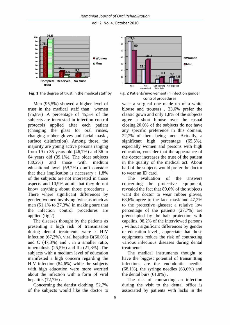
Fig. 1 The degree of trust in the medical staff by Fig. 2 Patients’involvement in infection gender
control procedures
Men (95,5%) showed a higher level of
trust in the medical staff than women
(75,8%) .A percentage of 45,5% of the
subjects are interested in infection control
protocols applied after each patient
(changing the glass for oral rinses,
changing rubber gloves and facial mask ,
surface disinfection). Among those, the
majority are young active persons ranging
from 19 to 35 years old (46,7%) and 36 to
64 years old (39,1%). The older subjects
(80,2%) and those with medium
educational level (69,2%) don‘t consider
that their implication is necessary ; 1,8%
of the subjects are not interested in those
aspects and 10,9% admit that they do not
know anything about those procedures .
There where significant differences by
gender, women involving twice as much as
men (51,1% to 27,3%) in making sure that
the infection control procedures are
applied (fig.2).
The diseases thought by the patients as
presenting a high risk of transmission
during dental treatments were : HIV
infection (67,3%), viral hepatitis B(60,0%)
and C (47,3%) and , in a smaller ratio,
tuberculosis (25,5%) and flu (21,8%). The
subjects with a medium level of education
manifested a high concern regarding the
HIV infection (84,6%) while the subjects
with high education were more worried
about the infection with a form of viral
hepatitis (72,7%) .
Concerning the dentist clothing, 52,7%
of the subjects would like the doctor to
wear a surgical one made up of a white
blouse and trousers , 23,6% prefer the
classic gown and only 1,8% of the subjects
agree a short blouse over the casual
closing.20,0% of the subjects do not have
any specific preference in this domain,
22,7% of them being men. Actually, a
significant high percentage (65,5%),
especially women and persons with high
education, consider that the appearance of
the doctor increases the trust of the patient
in the quality of the medical act. About
half of the subjects would prefer the doctor
to wear an ID card.
The evaluation of the answers
concerning the protective equipment,
revealed the fact that 89,0% of the subjects
want the doctor to wear rubber gloves,
63,6% agree to the face mask and 47,2%
to the protective glasses; a relative low
percentage of the patients (27,7%) are
preoccupied by the hair protection with
capelins. 98,2% of the interviewed persons
, without significant differences by gender
or education level , appreciate that those
equipments reduce the risk of contracting
various infectious diseases during dental
treatments.
The medical instruments thought to
have the biggest potential of transmitting
infections are the endodontic needles
(68,1%), the syringe needles (63,6%) and
the dental burs (61,8%) .
The risk of contracting an infection
during the visit to the dental office is
associated by patients with lacks in the
0
10
20
30
40
50
60
70
80
90
100
Completetrust
Reserves No trust
75,8
15,2 9,1
95,5
4,5 0
Women
Men
0
10
20
30
40
50
60
70
Yes Notcompetent
Not wantingto irritate
Not exposed
63,6
21,2
6,1 9,1
50
36,4
9,1 4,5
Women
Men

Romanian Journal of Oral Rehabilitation
Vol. 2, No. 4, October 2010
6
sterilizing of the instruments (80,0%) and
surfaces and equipments disinfecting
(54,5%).
The procedures considered to be
important for preventing the infection
during dental treatments were: dentists‘
hands washing (78,2%) , the disinfection
of the surfaces in the dental practice after
each patient (56,4%) and handling the
instruments by the doctor in safe
conditions (45,5%).
DISCUSSIONS
The results of the study prove the trust
of the patients in the medical staff and in
the manner of applying the infection
control methods. A low percentage of the
interviewed subjects think that during the
dental treatment they cannot contract a
general disease. This fact demonstrates,
especially in men, the lack of knowledge
concerning the risk of being exposed.
Concernments regarding the procedures
used by the dentists to control the infection
are expressed particularly by young
persons and women, whereas the majority
of the old subjects don‘t have the
necessary knowledge or do not consider
that it is of their competence to interfere
with the doctor acts. Also, the high level of
education inflicts an involvement of the
patient in his own health care, with benefic
effects over the safety level of the dental
treatment.
The majority of the patients want the
doctor to use rubber gloves as an essential
protective equipment for reducing the risk
of infection transmission, the results of our
studies being similar with the ones
reported in the literature (3),(5). The
percentage of the subjects willing to
involve in the dental treatment is low
revealing the trust granted to the dentist
but also the lack of knowledge regarding
the risk of infections and the measures
needed to prevent it.
The way in which the appearance of the
staff influences the perception of the
patients regarding their competence
reflects in the choices of the subjects for a
sober appearance, the classic white gown
and an ID seen as a mean of committing to
the medical act. The subjects with high
education consider that the appearance of
the doctor increases the quality of the
treatment, whereas the majority of the
elderly persons do not asses the
professional merits of the dentist by the
way he is dressed.
CONCLUSIONS
The medical personnel has the
responsibility to inform the patients on the
measures used to reduce the risk of
diseases transmission and to apply them in
an obvious way, in order to reduce the
concerns and the avoidance of the dental
treatment.
The assessment of the patients‘
perception regarding the equipments,
procedures and protective barriers which
are not completely regulated by the law
has to be a decisive factor for the
compliance of the medical staff in using
them in the dental practice according to the
European standards concerning the safety
of the medical act.
REFERENCES 1. Bârlean L., Dănilă I. “Prevenirea transmiterii infecţiei în stomatologie”, Ed. Edict, Iaşi, 2003. 2. Lill M., Wilkinson T.J., Judging a book by its cover: descriptive survey of patients preferences for doctors appearance and mode of address, B.M.J. 2005; (331) : 1524-1527 3. Mousa A.A., Mahmoud N.M., Tag El-Din A.M. Knowledge and attitudes of dental patients towards cross-infection control measures in dental practice , Eastern Mediterranean Health Journal 1997; (3): 263-273. 4. Palenik Ch.J. Strategic Planning for Infection Control The Journal of Contemporary Dental -Practice 2000; 1, (4) : 34-37 5. Shulman E.R., Brehm M.S. Dental clinical attire and infection-control procedures. Patients' attitudes. J.Am.Dent.Assoc. 2001;132 (4):508-516 6. Guidelines for environmental infection control in health-care facilities: recommendations of CDC and the Healthcare Infection Control Practices Advisory Committee, CDC MMWR 2003 ; 52 (nr.RR-10).

Romanian Journal of Oral Rehabilitation
Vol. 2, No. 4, October 2010
7
THE IMPORTANCE OF FINDING OUT FACTORS THAT CAN
TROUBLE THE MUSCULO-LIGAMENTAR EQUILIBRIUM IN
PACIENTS WITH COMPLETE AND EXTENDED PARTIAL
EDENTATION D.N. Bosînceanu, Silvia Mihaela Silvaş, Dana Gabriela Budală, Norina Consuela Forna
University of Medicine and Pharmacy „Gr.T. Popa‖, Iaşi
Faculty of Dental Medicine, ET Clinic and Therapy, EPI Clinic and Therapy
INTRODUCTION
The stomatology of the third millenium
acquired new boundries and dimensions,
upshot of the developement of the
diagnosis and therapy, they themselves
being influenced by the modern and
complex technology and also by the
psycho-social and communication aspects.
The functionality of the stomatognat
system depends on many factors which
can act on it in the direction of equilibrum
and that can anytime be changed, adapting
to new situations and circumstances.
Among the elements that play a role in the
stomatognat system‘stability, a special
place is held by the muscular factor, the
dynamic constituent.
PURPOSE
In this paper, the extended partial
edentation or the complete edentation are
looked in terms of the multiple systemic
conexions, both inner and outer, that are
established between the elements of the
stomatognat system.These conexions are
responsable for the way in which, the
morphological or functional deterioration
of one of the components will drawn the
alteration of all.
Therefore, in this paper we clinicaly
establish the signs of muscular disfunction
and based on these information we get the
incidence of oro-facial muscular
disfunctions, in order to work out a
complete treatment plan, pursuing to get a
complex muscular rehabilitation.
MATERIAL AND METHOD
The patients were chosen from those
who came in our clinic to get
prosthodontic treatment.They were 264,
128 men and 136 women.The average age
was 58,7, the study being held on three
groups of age:40-55years old, 55-70 years
old, 70-85 years old (tabel I).
TABELUL I
Distribution of the patients according to their age and sex Age Men Women %men %women
40-55 39 43 30,46 31,61
55-70 61 78 47,65 57,35
70-85 28 14 21,87 10,29
128 136
The patients we selected were
complete, partial or extended edentated
and they were protheses wearers for 3 to
10 years.All patients were informed about
this study and they consented to it.(tabel
II).

Romanian Journal of Oral Rehabilitation
Vol. 2, No. 4, October 2010
8
TABELUL II
Distribution of the patients according to their type of edentation Age E.P C.E E.P C.E
Men Women Men Women % %
40-55 36 42 3 1 95,12 4,87
55-70 28 37 33 41 46,76 53,23
70-85 6 5 22 9 26,19 73,80
70 84 58 51
To each and every patient was
elaborated a clinical report.They were
throughly examined and so was every
muscular group using the clasic methods
of inspection and palpation.
There were investigated the temporals,
the masseters as muscles of mastication
and the buccinators and the orbiculars as
oro-facial ones.
The palpation was made by pressing
smoothly the muscles‘insertion and
tucking the muscles‘mass, both in
movement and in rest.
During the postural position, the
muscles are characterized by a light
contraction, that can‘t be detected on the
electromyography-muscular tonus of
posture.This can vary depending on many
other factors such as clinical, functional
and morphological ones and it will be
evaluated considering the relation between
the muscles‘ osseous insertions and the
postural tonus that exists.
First we palpated the masseter and the
temporal muscles-the osseous insertions
and the masses and then we palpated the
oro-facials muscles.Every muscle was
examined equably on the right side and on
the left.We assesed and wrote down in
every patient medical report the trophicity
of the muscles and their consitency. The
muscular tonus was examined using
Netter‘s tests.
After the clincal exam of the muscles
we examined the protheses, assesing their
the maintenance and stability usin the
following standards (Tabel III)
0 – Maintenace - Non at all.When it is
inserted in the oral cavity is dislocate itself
.Stability - Non at all.It‘s tipping on the
prothetic field.
1 – Minimal maintenance. It has light
maintenance when pulled vertically and
the same or nothing at all when pull on
side.
Minimal stability. It‘s tipping moderately
on the prothetic field.
2 - Moderately maintenance when pulled
vertically and the same or nothing at all
when pull on side.
Suficient stability. It‘s lightly tipping or
not tipping at all on the prothetic field.
3 – Good maintenance. When pulled
vertically has maximum maintenance and
enough when side forces act. Good
stability, without tipping.
The rating of the protheses was made
likewise:
- minimal stability and maintenance-score
<6
- moderately stability and maintenance-
score 6-8
- good stability and maintenance-score > 8
RESULTS AND DISCUSSIONS
After the clinical exam we found the
following grades of tonicity of the
masticatory muscles:
- at group of age between 40-55 with partial
extended edentation from 78 cases :22 of
them had normal tonicity for masseters, 28
with normal tonicity for the temporals , 33
with normal tonicity for the orbiculars and
29 for the buccinators, 15 with
hipertonicity for the masseters, 17 with
hipertonicity for the temporals, 12 with
hipertonicity for the orbiculars and 8 with
buccinators hipertonics and with
hipotonicity we found 41 cases for
masseters and temporals, 33 for the
buccinators and the orbiculars

Romanian Journal of Oral Rehabilitation
Vol. 2, No. 4, October 2010
9
- at the same group of age but in casese of
complete edentation from 4 cases we
found one case with normal tonicity in
masseters, 2 cases in temporals and
orbiculars and one with normal tonicity in
buccinators
- at group of age between 55-70 with partial
extended edentation from 65 cases : 12 of
them had normal tonicity for masseters, 15
with normal tonicity for the temporals , 21
with normal tonicity for the orbiculars and
14 for the buccinators , 7 with
hipertonicity for the masseters, 8 with
hipertonicity for the temporals, 10 with
hipertonicity for the orbiculars and 6 with
buccinators hipertonics and with
hipotonicity we found 46 cases for
masseters and 42 for temporals, 45 for the
buccinators and 34 for the orbiculars
- at the same group of age but in casese of
complete edentation from 74 cases we
found:8 of them had normal tonicity for
masseters, 10 with normal tonicity for the
temporals, 13 with normal tonicity for the
orbiculars and 8 for the buccinators , 3
with hipertonicity for the masseters, and
the temporals, 4 with hipertonicity for the
orbiculars and 1 with buccinators
hipertonics and with hipotonicity we found
63 cases for masseters and 61 for
temporals, 64 for the buccinators and 57
for the orbiculars.
- at group of age between 70-85 with partial
extended edentation from 11 cases we
found: 2 of them had normal tonicity for
masseters and the temporals , 3 with
normal tonicity for the orbiculars and 2 for
the buccinators , 3 with hipertonicity for
the masseters and the temporals, 2 with
hipertonicity for the orbiculars and 1 with
buccinators hipertonics and with
hipotonicity we found 6 cases for
masseters ,for temporals and for the
orbiculars and 8 for the buccinators
- at the same group of age but in casese of
complete edentation from 31 cases we
found: 3 of them had normal tonicity for
masseters and for the temporals , 2 with
0
20
40
60
80
100
28,2
35,89 42,3
37,17
18,46 23,07
32,3
21,53 18,18 18,18
27,27
18,18
40 - 55 ani
55 - 70 ani
70 - 85 ani
0
20
40
60
80
100
25
50 50
25
10,81 13,51
17,56
10,81 9,67 9,67 6,45 6,45
40 - 55 ani
55 - 70 ani
70 - 85 ani
0
20
40
60
80
100
19,23 21,7 15,38
10,25 10,76 12,3 15,38
9,23
27,27 27,27
18,18
9,09
40 - 55 ani
55 - 70 ani
70 - 85 ani
0
20
40
60
80
100
25 25 25 25
4,05 4,05 5,4 2,7 3,22 3,22
6,45 3,22
40 - 55 ani
55 - 70 ani
70 - 85 ani

Romanian Journal of Oral Rehabilitation
Vol. 2, No. 4, October 2010
10
normal tonicity for the orbiculars and the
buccinators , 1 with hipertonicity for the
masseters, and the temporals and
buccinators, 2 with hipertonicity for the
orbiculars and with hipotonicity we found
27 cases for masseters ,for temporals and
for the orbiculars, 28 for the buccinators
CONCLUSIONS
1. Between the morphological bone
structure and the muscles of the
stomatognat system there is quit
equilibrium, always changing according to
the adaptation of the two systems, the
muscular activity being directly influenced
by the integrity of every element of the
stomatognat system
2. The dishomeostasis of the
stomatognat system as a result of
edentation is just a step on the way of this
complex diseases, the changes that took
place being irreversible.Therefore, the
group of muscles affected can influence
the relationships between the two maxilla
and can also change the mandible‘s
movements in old wearers of protheses.
3. The great variety of the stomatognat
system‘s changes as a result of edentation
and ageing requires a thoroughly
investigation of each and every case, in
order to track down as soon as possible
every muscular disfunction.
4. All patients found during the clinical
exam to have a muscular disorder must be
investigated to set up a complex treatment,
monitorized even after the prothese would
be over
5. As a result of the clinical and
paraclinical investigations, we determined
a high rate of muscular disfunction in the
group of old wearers, with the stability and
maintenance of the protheses affected .The
disfunction was asserted by means of hiper
and hipotonicity of the muscular masses.
In the group of recently edentated patients
the changes were less visible than in the
group of old wearers, in which the body
tried to adjust to the edentation.We also
found muscles that weren‘t yet affected by
the changes of the stomatognat system.
6. The change of the muscular tonus
and of the mandibular movements are
signs and symptoms that lead us to a
diagnosis of muscular disfunction and are
elements that will influence the
prosthodontic treatment.
0
20
40
60
80
100
52,56
42,3 42,3
52,56
70,72 64,61
52,3
69,23
54,54 54,54 54,54
72,72
40 - 55 ani
55 - 70 ani
70 - 85 ani
0
20
40
60
80
100
50
25 25
50
85,13 82,43
77,02
86,48 87,09 87,09 87,09 90,32
40 - 55 ani
55 - 70 ani
70 - 85 ani

Romanian Journal of Oral Rehabilitation
Vol. 2, No. 4, October 2010
11
REFERENCES 1. Ash M. M., Current concepts in the etiology, diagnosis and treatment of TMJ and muscle dysfunction, 1986,
J. of Oral Rehab, 13, 1-20.
2. Burlui V., Forna N., Clinica şi terapia edentaţiei parţial întise, Ed.Apollonia, Iaşi, 2004.
3. Costa E., Sindromul de disfuncţie mandibulară, Ed.Ştiinţifică şi Enciclopedică, Bucureşti, 1987.
4. Korfage J.A., Koolstra J.H., Lagenbach G.E. and Van Eijden T.M., Fiber type composition of the human jaw
muscle-role of hybrid fibres and factors responsible for interindividual variation, J.Dent Rest., 2005:84:9(784-
793).
5. Lejoyeux J., Proteza totală vol I+vol II, Ed.Medicală, Bucureşti, 1968
6. Travell J.G., Simons D.G., Myofascial pain and dysfunction, The Trigger Point Manual, Williams &
Wilkins, 1983
7. Tsai C.M., Chou S.L., Gale E.N., McCall W.D. Jr., Human masticatory activity and jaw position under
experimental stress, J.Oral Reab., 29(1):44-51, 2002
8. Wheeler A.H., Myofascial pain disorder, Theory to therapy, Drugs. 64(1):45-62,2004

Romanian Journal of Oral Rehabilitation
Vol. 2, No. 4, October 2010
12
THE EFFECTS OF THE PAIN ENDURED DURING DENTAL
TREATMENT Diana Cerghizan, S. Popşor, M. Suciu, A. Kovacs
University of Medicine and Pharmacy Tg. Mures
Department of Prosthetic Dentistry and Oral Rehabilitation
INTRODUCTION
It has been acknowledged for many
years that human pain perception is made
up of multiple dimensions, including a
sensory aspect and an emotional/affective
quality aspect (Price, 1988). Researchers
have shown that some ―pain‖ stimuli are
associated with high levels of
emotionality/affect (for example, cancer
pain), whereas other ―pain‖ stimuli can
produce relatively low levels of emotional
distress (for example, labour pain) (Price
et al., 1987). These findings indicate that
people can experience very different
emotional responses to very similar levels
of stimuli intensity, depending on their
perception of the event (Gracely, Kwilosz,
1988). Assessment of clinical pain
response requires the use of measurement
scales designed to capture the different
dimensions of pain perception (Logan,
1995).
The dental treatments usually are
associated by the patient with pain and
anxiety. Is proved that painful therapeutic
procedures are the most important reason
of generating pain and anxiety during a
dental treatment.
An early negative dental experience is
probably the most stated single cause for
dental anxiety (Locker et al., 1996, 1999).
However, a negative dental experience
does not necessarily lead to dental anxiety.
The 'latent inhibition' theory, for instance,
states that a history of positive or neutral
dental experiences may serve as a buffer
against the development of traumatic
associations or experiences (Davey, 1989).
As a consequence, high levels of anxiety
or fear are developed less easily.
Conversely, an early negative dental
experience can serve as a one-shot
conditioner and may leave a patient with
feelings of anxiety. Fear of dental pain is a
highly relevant concept in dental pain
research and, moreover, in dentistry (van
Wijk and Hoogstraten, 2003). Whereas
anxiety and fear can be seen as a state of
distress in anticipation or in the presence
of a perceived danger, respectively, fear of
pain can be seen as a state of distress
related to a very specific type of stimulus,
namely, pain (Gower, 2004). Research
suggests that anxious people tend to
overestimate anticipated pain. Moreover,
individuals tend to overestimate the
intensity of aversive events in general,
including such events as fear. Therefore,
people who are predisposed to respond
fearfully to pain are at an increased risk of
ending up in a vicious circle of anxiety,
fear of pain, and avoidance of dental
treatment (van Wijk, Hoogstraten, 2005).
The target of this study is to prove the
connection between previous pain and
anticipating pain. This study is a part of a
larger research project , and the results
presented here are only preliminary, they
can modify with the advancement of the
study (ex. rising patient number).
MATERIAL AND METHOD
This study is based on a questionnaire
created by us, which includes general
data‘s about the patient (age, sex, studies),
and also contains four questions , which
are helping us to determine, if the patient
had any painful experiences during the
dental treatment, if he‘s anticipating the

Romanian Journal of Oral Rehabilitation
Vol. 2, No. 4, October 2010
13
pain, or if he is avoiding the appointments
because of pain.
At the same time we determined the
patient‘s anxiety level using the Dental
Anxiety Scale (DAS) questionnaire. DAS
contains four questions about different
situations which are occurring during the
dental treatment. Every question is rated
between 1 (no anxiety) and 5 (very
anxious), the final score can alternate
between 4 and 20. A result higher than 15
is the proof for a high level of anxiety.
The patient‘s selection was based on the
next criteria‘s:
1. patients older than 18
2. patients who had contact with one or
more dentist‘s before the start of the study
3. we used only the fully completed
questionnaires
After a selection made using this
criteria‘s it resulted a lot of 247 persons
with age between 18 and 79 (M = 38,03),
179 (72,47 %) female and 69 (27,53%)
male.
Using the DAS we confirmed that the
majority of the patients with painful
experiences in the past are subject of high
or even severe level of anxiety.
RESULTS
The questionnaire carry out by us
presents questions with closed answer
(yes, no), codified by entering them in
statistical analysis charts, done by
GraphPad InStat 3 and NCSS software‘s.
Out of 247 questioned patients 60 %
said that they endured painful dental
treatments in the past and also 60|% said
that they during a dental treatment are
waiting for the appearance of the pain. For
statistical analysis we used the Fisher test
and the results showed that is a very
significant association between pain in the
past and anticipating pain (p< 0,0001) The
association is significant both statistically
and scientifically to (OR = 3.951, CI =
95%, 2,298 – 6,794) (fig. 1).
Fig. 1 Correlation between pain in the past and anticipating pain
After dividing on age groups we
observed a extremely significant positive
association, both from statistically or
scientifically points of view , between pain
in the past and anticipating pain at patients
with ages between 18 -30 (n= 81) years
and 41 – 50 (n= 47) years with p=0,0005
(OR = 5.600 95% CI: 2,088 to 15,017), or
in case of p=0,0006 (OR = 12 95%
CI:2,685 to 53,636). At patients with ages
between 18 and 30 years we could prove
statistically significant correlation between
anticipating pain and avoiding dental
treatment (p=0,0024) (Fig. 2).
durerea din antecedente si anticiparea dureriiColumn Totals
Columns
anticipeaza nu anticipeaza
140
130
120
110
100
90
80
70
60
50
40
30
20
10

Romanian Journal of Oral Rehabilitation
Vol. 2, No. 4, October 2010
14
Fig. 2 Pain anticipation and ages
The statically reading of the results
showed that between pain in the past and
avoiding dental treatments exists a positive
association, but statistical insignificant
(p=0,08 OR = 1,743 95% CI: 0,9635 to
3,151). This helped us to conclude that to
obtain accurate results we need a larger lot
of patients.
CONCLUSIONS
In 1984 Wall and Melzack said ― Pain
always is one-sided. Every individual is
learning the signification of this word by
the experiences he starts to have from his
first years. Without doubts is a sensation
with organic origins, but this sensation
always is apprehended like an unlike one,
which makes from this an emotional
experience‖
For many patients, fear of dental pain
and avoidance of dentistry are
synonymous (Freeman, 1991). Moreover,
clinicians report that managing some
patients‘ pain and distress can be a
frustrating task (Lindsay, Jackson 1995).
From this lot of patients 60% (n=149)
had in the past dental treatments involving
pain. This result has to put the
practitioners to think how they can avoid
pain, because pain could be the starting or
the aggravating factor of the dental
anxiety.
The high number of patients who had a
positive answer to the first question from
our questionnaire shows us that
practitioners are not giving enough
significance to the symptom of pain,
resulting an absence of interest in trying to
challenge the pain. Between pain in the
past and avoiding dental treatments exists
a positive association, but to determine the
statistically and scientifically magnitude
we have to rise the number of questioned
patients. We can claim that any pain
endured during the dental treatment
remains printed in the patients memory,
making them to think on possible pain at
their following appointment. 73% (n=109)
out of the patients who experienced
painful dental treatments , are believing
that at the next appointment pain can show
up again. A number of 40 patients are
waiting for pain to show up at their next
appointment, even they never experienced
painful dental treatments. This situation is
making us to associate pain with the dental
treatment.
It often is assumed that aging results in
loss of pain sensitivity. Although some
efforts have been made to study the effects
of aging on pain perceptions, the results
are not conclusive. Experimental studies of
acute pain responses do not show
significant age-related alteration in the
pain perceptions of healthy elderly
subjects (Harkins et al., 1994). It has been
proposed that differences in acute pain
responses between younger and older
0.0
26.7
53.3
80.0
d
a
nu

Romanian Journal of Oral Rehabilitation
Vol. 2, No. 4, October 2010
15
patients (Lash et al., 1997) may be a result
of changes in pathophysiology (for
example, neural conductivity) rather than
changes in the pain perception itself
(Harkins et al., 1990; Heft et al., 1996). It
is not clear, however, from the literature
whether these changes in pathophysiology
influence both affective pain and sensory
intensity in the elderly.
During our study we observed that
patients with age between 18 – 30 years
are avoiding dental treatments because of
the pain which can show up during the
dental treatment. Patient older than 50
years are not avoiding dental treatments.
One of the main reasons of this can be that
painful experiences are fading during the
years in patients memory.
New evidence suggests that there are
differences in pain perceptions between
men and women (Riley et. al. 1998, Unruh
et al. 1999). Although, most studies
suggest that women have greater pain
sensitivity than men, there are
inconsistencies in the literature (Eli et al.,
1996). These inconsistencies suggest that
the type of pain stimuli may influence
perceived pain differences between men
and women (Fillingim, 1998). In addition,
the influence of aging on these reported
sex differences has yet to be clarified.
In our study because of the lower
number of questioned male patients we
couldn‘t determine a precise correlation
between pain in the past , anticipating pain
and avoiding dental treatments.
Our own experience is showing that the
majority of patients are favoring different
methods to fight pain showing up during
the oral rehabilitation treatments.
Patients avoiding dental treatments
usually presents a poor oral health, and at
the end they will need elaborate oral
rehabilitation treatments.
This work was supported by the
CNCSIS – Ministry of Education and
Research, Romania, Project Td No
417/2007.
REFERENCES 1. Davey GC, Dental phobias and anxieties: evidence for conditioning processes in the acquisition and
modulation of a learned fear. Behav Res Ther 1989, 27:51–58
2. Eli I, Bar-Tal Y, Zvu F, Korff E., Effect of biological sex differences on the perception of acute pain
stimulation in the dental setting. Pain Res Manage 1996;1(4):201–6.
3. Fillingim RB, Maixner W, Kincaid S, Silva S., Sex differences in temporal summation but not sensory-
discriminative processing of thermal pain. Pain 1998;75:121–7.
4. Freeman R., The role of memory on the dentally anxious patient‘s response to dental treatment. Ir J
Psychological Med 1991;8(2):110–5.
5. Gower PL, editor (2004). Psychology of fear. New York: Nova Science Publishers, Inc
6. Gracely RH, Kwilosz DM, The Descriptor Differential Scale: applying psychophysical principles to clinical
pain assessment. Pain 1988;35:279–88.
7. Harkins SW, Kwentus J, Price DD, Pain and suffering in the elderly. In: Bonica JJ, ed. The management of
pain. 2nd ed. Philadelphia: Lea & Febiger; 1990:552–9.
8. Heft MW, Cooper BY, O‘Brien KK, Hemp E, O‘Brien R., Aging effects on the perception of noxious and
non-noxious thermal stimuli applied to the face. Aging (Milano) 1996;8(1):35–41.
9. Lasch H, Castell DO, Castell JA., Evidence for diminished visceral pain with aging: studies using graded
intraesophageal balloon distension. Am J Physiol 1997;272(1 Pt 1):G1–3.
10. Lindsay SJ, Jackson C, Fear of routine dental treatment in adults: its nature and management. Psychol
Health 1993;8:135–53.
11. Locker D, Liddell A, Dempster L, Shapiro D., Age of onset of dental anxiety. J Dent Res 1999, 78:790–796
12. Locker D, Shapiro D, Liddell A., Negative dental experiences and their relationship to dental anxiety.
Community Dent Health 1996, 13:86–92.
13. Logan HL, Baron RS, Kohout F. Sensory focus as therapeutic treatments for acute pain. Psychosom Med
1995; 57:475–84.
14. Lyndsay C. Bare, B.A.; Lauren Dundes, M.H.S., Sc.D. Strategies for Combating Dental Anxiety J Dent
Educ. 68(11): 1172-1177 2004

Romanian Journal of Oral Rehabilitation
Vol. 2, No. 4, October 2010
16
15. Price DD, Harkins SW, Baker C. Sensory-affective relationships among different types of clinical pain and
experimental pain. Pain 1987;28:297–307.
16. Price DD. Psychological and neural mechanisms of pain. New York: Raven; 1988:2–70.
17. Riley JL III, Robinson ME, Wise EA, Myers CD, Fillingim RB Sex differences in the perception of noxious
experimental stimuli: a meta-analysis. Pain 1998;74:181–7.
18. Unruh AM, Ritchie J, Merskey H. Does gender affect appraisal of pain and pain coping strategies? Clin J
Pain 1999;15:31–40.
19. van Wijk AJ, Hoogstraten J (2003). The fear of dental pain questionnaire; construction and validity. Eur J
Oral Sci 111:12–18
20. van Wijk A.J. and J. Hoogstraten. Experience with Dental Pain and Fear of Dental Pain J Dent Res
84(10):947-950, 2005
21. Wall PD, Melzack R. Geriatric pain. In: Wall PD, Melzack R, eds. Textbook of pain. 3rd ed. London:
Churchill Livingstone; 1994:769–84.

Romanian Journal of Oral Rehabilitation
Vol. 2, No. 4, October 2010
17
EMDOGAIN AND BIO-OSS BETWEEN HOPE AND REALITY Laura Cîrligeriu, M. Boariu, A. Marinescu, V. Cârligeriu
,,Victor Babes,, University of Medecine and Pharmacy Faculty of Dental Medecine
Abstract: In the last years the treatment of periodontal disease has gain new meanings thanks to new developed
materials that helped the surgical treatment. The developers of these materials promise a lot in the field of
guided tissue regeneration, but in order to know how much of these is true we tried some of them in clinical
tests. The most frequently used materials by us was Emdogain and Bio-Oss and made a study (similar studies
was conducted by Sculeanu et.al.) to demonstrate how much of these materials properties was hope and how
much was reality. The aim of the study was to evaluate the clinical and radiographic outcome using two
different materials in the treatment of periodontal bone defects based on guided tissue regeneration techniques
(2). Our study included 40 subjects with generally healthy status with periodontitis ilness in advanced stage.
Depending of the materials used in the treatment our patients were divided into two equal groups. The group
number I, was treated with a bovine-derived hydroxyapatite xenograft (Bio-Oss, ) combined with a resorbable
collagen membrane (COREM, Poneti) was used. In the second group was used enamel matrix derivative
(Emdogain® Gel). Before the surgical treatment a clinical and radiographic examinations were performed, and
the following parameters were evaluated: plaque index, gingival index, probing pocket depth, clinical
attachment level, radiographic defect depth, and defect width. At the patients with bad oral hygiene the plaque
and gingival index was haing high values. All clinical and radiographic parameters (except plaque index) were
significantly reduced after treatment in both groups . No great differences were revealed between the two groups
of patients in examined parameters after treatment. The results demonstrated that the treatment of periodontal
bone defects with both materials leads to similar and significant improvements in clinical and radiographic
parameters. The exception was made by the patients with bad oral hygiene.
Key words: emdogain, Bio-Oss, guided tissue regeneration, osteoregenerative materials.
INTRODUCTION:
In the last years the treatment of
periodontal disease has gain new
meanings thanks to new developed
materials that helped the surgical
treatment. The developers of these
materials promised a lot in the field of
guided tissue regeneration, but in order to
know how much of these is true we tried
some of them in clinical tests. The most
frequently used materials by us was
Emdogain and Bio-Oss and made a study
to demonstrate how much of these
materials properties was hope and how
much was reality. The aim of the study
was to evaluate the clinical and
radiographic outcome using two different
materials in the treatment of periodontal
bone defects.
MATERIAL AND METHODS
The study included 40 generally healthy
subjects with advanced periodontitis (20
women, 20 men), aged 35–60, supra- and
subgingival scaling and root planing were
carried out during a 3-month preoperative
period in all the patients, who also
received oral hygiene instructions.. The
patients were divided into two groups, 20
in each, according to the material used. In
the first group, a resorbable collagen
membrane (COREM, Poneti) and a
bovine-derived xenograft (Bio-Oss®;
Geistlich, Wolhusen, Switzerland, Bioos,
Cerasorb) was used. The other group of
patients was treated with enamel matrix
derivative (Emdogain®; Biora, Malmö,
Sweden).
Clinical and radiographic examinations
were performed prior to, and 1 year after
surgery. A periodontal probe (WHO
Probe, CPITN) was used for clinical
examinations prior and 1 year after
surgery. At the same time the radiographic
examination was made. Clinical were
evaluated the folowing parameters: plaque
index (PI - Sillness& Löe), gingival index
(GI - Löe& Sillness), probing pocket depth

Romanian Journal of Oral Rehabilitation
Vol. 2, No. 4, October 2010
18
(PPD, in mm), clinical attachment level
(CAL, in mm). The measurements were
made on folowing sites mesiovestibular,
vestibular, distovestibular, mesiooral, oral,
distooral.
Radiographic examination used two
intraoral radiographs taken with
customized film holders and long-cone
paralleling technique. The following
parameters were measured; (1) defect
depth (the vertical distance between the
crest of the alveolar process and the
jonction site of the root surface and normal
alveolar bone, in mm), (2) defect width
(the horizontal distance between the root
surface and the most coronal part of the
alveolar process, in mm).
Only patients with deepest site of
gingival pocket was considered for
analysis. This selection was made using
the folowing criteria: PI<1, PPD≥6 mm
and radiographic defect depth≥3 mm.
Bone defects chosen for analysis were
similar in size and shape, and were found
at homonymous teeth in the two groups to
be compared. Both groups of patients have
presented periodontal pockets (22 with two
walls, 8 with three walls, 5 circular bone
defects and the remaining five other
defects).
In both groups of patients, the surgical
procedures were performed under local
anaesthesia (4% Ubistesin). Following an
incision in the gingival pocket, a
mucoperiosteal flap was raised
vestibularly and lingually. Vertical
incisions were placed only if necessary for
adequate access to the surgical site or to
achieve complete coverage of the
membrane with the mucoperiosteal flap
(1). Granulation tissue was removed from
the bone defect, and the root surface was
cleaned and planed using hand and rotative
instruments depending of the site defect.
The surgical area was then rinsed with
physiological saline. In the first group, the
root surface was etched for 2 min with
neutral 24% EDTA to remove the smear
layer. Then the site was rinsed again with
physiological saline and Emdogain was
applied starting at the bottom of the defect.
Finally, the flap was repositioned
coronally and sutured tightly with non-
resorbable sutures. In the second group of
patients, the bone defect was filled with
Bio-Oss and then covered with the
resorbable Bio-Gide membrane. The
membrane was fixed using resorbable
sutures. The sutures were removed 14 days
after the surgery. All patients received
antiobiotics – 1 g amoxiclav twice a day
for 1 wk. Moreover, the patients were
instructed to avoid mechanical tooth
brushing in the region involved for 4 wk,
to chew carefully for 4 wk, and to rinse the
oral cavity with 0.2% chlorhexidine
solution (Plak-Out) twice daily for 6 wk.
Four weeks after the procedure the patients
started gentle tooth cleaning using the roll
technique. Postsurgical appointments were
scheduled at 2, 4 and 6 wk, and then every
2 months. During the first year after
surgery, only professional supragingival
tooth cleaning was carried out.
RESULTS AND DISCUTIONS
In all patients postoperative healing was
uneventful. No inflammatory
complications were observed, except a few
case of patients with bad oral hygiene.
The mean PI values in the Bio-Oss and
the Emdogain groups at baseline were
identical. Following the treatment PI
remained unchanged in both groups. The
mean GI values in the two groups before
treatment were not significantly different.
The mean GI values after treatment were
significantly reduced in both groups, and
the values were not statistically different
between the two groups. Likewise, PPD
and CAL values were initially similar
between the two groups. After surgery, the
mean PPD and CAL values significantly
decreased in both groups.
The differences in radiographic
parameters between the two groups of
patients before treatment was not
significant. The mean radiographic

Romanian Journal of Oral Rehabilitation
Vol. 2, No. 4, October 2010
19
parameters (depth and width of bone
defects) were significantly reduced after
both Bio-Oss and Emdogain treatment.
We will try to exemplify this using the
RX from two patients. First patient was
treated with Emdogain. It presents
periodontal pockets around 1.7 and 1.8
with a probing depth of 8 mm, 4.7 presents
a lvl 4 furcation, and around 4.2 and 4.3
periodontal pockets with a probing depth
of 5 mm (Figure 1). Also around 2.2-2.3
periodontal pockets with a probing depth
of 6 mm and 2.7-2.8 periodontal pockets
with a probing depth of 7 mm. The second
Rx (Figure 2) was taken after 1 year and it
shows a bone gain of 5-6 mm.
Fig. 1 Fig. 2
This patient was treated with Bio-Oss.
It presents periodontal pockets around 1.7-
1.6 1.3, 2.4-2.5 with a probing depth of 6
mm (Figure 3). The second Rx was taken
after 1 year and it shows a bone gain of 5-
6 mm (Figure 4).
Fig. 3 Fig. 4
CONCLUSIONS:
Based on the above data and our own
experience it appears that the use of Bio-
Oss combined with resorbable membrane
and the regenerative tehnique with
Emdogain are comparable in the aspect of
clinical and radiografically assessed
periodontal healing and ofered the bone
regeneration promised by their developer.
The reults of our study are overlaping with
data reported by other authors, who
demonstrated a similar improvement in
radiographic parameters. In some cases we
couldn`t reach the bone regeneration
desired because of the patient bad hygiene
and other healing factors that are not
related to the proprieties of the material
used.
Emdogain aplication caused pocket
depth reduction and improvement in the
periodontal atachement level, which
statistically shows significant reduction in
gingival pocket depth by at least 4 mm
after 1 year period.
This results corespund with those of
others authors (Sculean et al., Heden et al).
In conclusion the results of our study
are overlaping with another study (Sculean
et. al. and Melonig et. al.) (3) and
demonstrated the treatment of periodontal
defect with Bio-Oss and Emdogain leads
to improvement in clinical and
radiographic parameters and are a reality.

Romanian Journal of Oral Rehabilitation
Vol. 2, No. 4, October 2010
20
REFERENCES 1. Carranza F.A., Newman M.G., Clinical periodontology, Philadelphia, W.B. Saunders, 1996
2. Chiantella GC & Sculean A. Intrabony defects: Active principle for periodontal regeneration. Dent Cadmos
1998;1:45–51.
3. Sculean A, Donos N, Blaes A, Lauermann M, Reich E, Brecx M. Comparison of enamel matrix protein and
bioabsorbable membranes in the treatment of intrabony periodontal defects. A split-mouth study. J Periodontol
1999; 70: 255–262.
4. Sculean A, Reich E, Chiantella GC, Brecx M. Treatment of intrabony periodontal defects enamel matrix
protein derivative: a raport of 32 cases. Int J Periodontics Restorative Dent 1999; 19: 157–163.

Romanian Journal of Oral Rehabilitation
Vol. 2, No. 4, October 2010
21
THE PREVALENCE OF HYPODONTIA IN CHILDREN WITH CLEFT
AND NONRELATED CONTROLS Claudia Corega, A. Şerbănescu, M.Corega, Mihaela Băciuţ
University of Medicine and Pharmacy ‖Iuliu Haţieganu‖ Cluj-Napoca, Romania
Abstract: The aim of this study was to compare the occurrence of hypodontia, dental age, and asymmetric dental
development in children with cleft with a non- sibling control group. The study sample consisted of 30 children
with cleft (aged 7.2 to 17.1 years) and 60 controls without cleft (aged between 7 and 18.8 years). Hypodontia,
dental age, and asymmetric dental development were assessed on panoramic radiographs of the children with
cleft and the control children without cleft. The cleft (p.001) group showed a significantly higher frequency of
hypodontia and a significantly higher occurrence (cleft p.01) of asymmetric dental development, compared with
the control group. Only a small, but insignificant delay in dental development could be found in the cleft group.
The cleft subjects showed a significantly higher occurrence of hypodontia and asymmetric dental development
than the non-cleft control group. This may suggest a genetic component for the occurrence of hypodontia and
asymmetric dental development.
Key words: cleft, hypodontia, tooth formation
INTRODUCTION
Some dental traits such as hypodontia,
supernumerary teeth, peg-shaped teeth,
dental delay and dental asymmetry occur
with higher frequency in individuals
affected with cleft lip, cleft palate, or both
(Ranta, 1986). The literature includes a
large number of studies dealing with tooth
formation in patients with cleft with a
range of findings. Several studies report a
delayed formation of the permanent teeth
(Bailit et al., 1968; Ranta, 1972, 1982;
Harris and Hullings, 1990; Brouwers and
Kuijpers-Jagtman, 1991). Other studies
report only a delayed dental development
in boys until the age of 9 years (Prahl-
Andersen, 1978; Prahl-Andersen et al.,
1979). In the study of Loevy and Aduss
(1988), early development in boys with
clefts was observed. Left-to-right
differences in tooth formation are also
greater in children with cleft (Ranta 1973;
Harris and Hullings, 1990). The incidence
of hypodontia away from the cleft area in
individuals is also markedly increased as
compared with the population without cleft
(Haataja et al., 1971; Ranta, 1986;
Jiroutova and Mu¨ llerova, 1994). In
particular, hypodontia most frequently
involves the second premolars in the upper
and lower jaw and the upper lateral incisor
on the noncleft side (Ranta, 1986).
Some studies (Jordan et al., 1966;
Schroeder and Green, 1975) report an
increase in dental aberrations such as
abnormal shape of teeth and
supernumerary or missing teeth in siblings
of children with cleft, compared with the
general population.
However, these studies were only
descriptive with little statistical analysis
and in the meantime the dental age was not
investigated. Investigations of Adams and
Niswander (1967) and Bhatia (1972)
support the idea that the same etiological
factors that cause the formation of the cleft
can affect the development of the
dentition. Significant associations of some
patients with cleft lip and palate with
tranforming growth factor alpha and
retinoic acid receptor loci (Chevenix-
Trench et al., 1992) were found.
Since there are few studies on children
with a cleft, the aim of the present study
was to compare hypodontia, dental delay,
and asymmetric dental development in
children affected with cleft lip or palate
with a group of control children.

Romanian Journal of Oral Rehabilitation
Vol. 2, No. 4, October 2010
22
MATERIALS AND METHODS
Sample Selection
The cleft group consisted of 30 children
(20 girls and 10 boys), aged 7 years 2
months to 17 years 1 month (mean age 10
years 2 months). All were of Caucasian
origin with nonsyndromic clefting. Twenty
of these children had a complete cleft lip
and palate, 6 children showed an isolated
cleftpalate, and only 4 children had a cleft
lip with cleft alveolar process. They were
all enrolled for treatment at the
Department of Orthodontics at the
University of Medicine and Pharmacy
‖Iuliu Hațieganu‖ Cluj-Napoca, Romania.
The nonsibling control group consisted of
60 children (40 girls and 20 boys) whose
age ranged from 7 years to 18 years 9
months (mean age 11 years 3 months). At
the time of the orthopantomogram, none
had been treated orthodontically. The
children of the noncleft sibling and control
groups were of Caucasian origin and were
nonsyndromic.
METHOD
An orthopantomogram was taken of
each child to assess the frequency of
hypodontia and the dental maturation
(dental age). The sample for evaluating the
frequency of hypodontia consisted of 30
children with cleft.
Dental age was calculated using the
method of Demirjian and Goldstein
(1976). A computer system and individual
data sheets were used to train the
evaluators in scoring the stages of
development correctly and consistently.
Individual radiologic appearances of the
seven permanent teeth on the left side of
the mandible were evaluated according to
developmental criteria. Development of
each tooth was categorized into one of
eight stages. These individual scores were
entered into a clinical evaluation program,
which converted them, depending on the
sex of the child, into a maturation and
dental age score. Panoramic X-rays, which
showed a full maturation score, or bilateral
agenesis or extraction of at least one tooth
in the lower jaw were excluded. Thus the
final sample for evaluating the dental
development consisted of 30 children
affected with namely 20 with cleft lip and
palate, 6 with a cleft palate, and 4 with a
cleft lip and alveolus. In order to assess the
reliability of this method, the scores of 30
children were measured twice with an
interval of 1 month by two examiners as a
pilot study.
To investigate the symmetry of
permanent tooth formation, individual
tooth developmental stages of seven left
and right mandibular teeth were compared.
A pair of teeth was regarded as having
undergone asymmetrical development
when the tooth development stage of the
left tooth deviated from that of the
antimeric tooth by at least one
developmental stage.
The panoramic X-rays were also
studied for congenitally missing teeth
outside the cleft region (excluding the
lateral incisor in the upper jaw on the cleft
side). A tooth germ was considered to be
congenitally missing if it was absent on the
X-ray, although the child‘s age would have
supported its being radiographically
detectable (Haavikko, 1970). The presence
of the preceding deciduous tooth was in
most cases a supporting criterion for the
diagnosis of hypodontia. When the
deciduous tooth was missing, the patient‘s
file was reviewed and the patient was
interviewed in order to exclude the
possibility of an extraction.
All data were transferred to Microsoft
Excel 97 (Microsoft Corporation,
Redmond, Washington) for statistical
analysis.
For each patient, missing teeth, the
difference between dental and
chronological age, the dental delay
compared with the controls as well as the
asymmetry of dental development were
assessed.
For each group (cleft group and control
group), the means and the standard

Romanian Journal of Oral Rehabilitation
Vol. 2, No. 4, October 2010
23
deviations of dental age, chronological
age, differences between dental and
chronological age and dental delay of the
cleft compared with the controls were
calculated. Differences between the groups
were analyzed using the unpaired t test and
the F test for equality of variances. The
chi-square test was used in order to test
differences (frequency) in hypodontia and
dental asymmetry among the two groups.
Probabilities less than .05 were
considered to be statistically significant.
RESULTS
Error of Method
No statistically significant differences
were found between the means of the
intra- and interobserver set of
measurements.
The intraobserver measurements
yielded a correlation of 0.988, which was
almost equal to the correlation of the
interobserver measurements: 0.994. The
measurement error for the dental age was
at most one developmental stage.
Hypodontia
In the group of 30 children with cleft,
15 children (50%) showed hypodontia of
one or more teeth outside the cleft
region.A total of 17 teeth were absent
(upper/lower jaw 10/7). In the control
group of 60 children, 6 children (10%)
showed hypodontia of one or more teeth.
A total of 9 teeth were absent (upper/lower
jaw 6/3).Compared with the nonsibling
controls, the cleft group showed a highly
significant increase in frequency of
hypodontia (p .001).
Hypodontia involved mostly the second
premolars of the upper and lower jaw and
the upper lateral incisor on the
contralateral side to the cleft. The most
frequently missing teeth in all the groups
were the second premolars. No significant
difference in hypodontia between the
upper and lower jaw or any significant sex
differences were found.
Comparison of the Dental and
Chronological Age
The cleft group had a mean dental age
of 10.2 years, which was 0.25 years (3
months) greater than the mean
chronological age of 9.11 years of this
group.
The control group showed a mean
dental age of 11.3 years, which was 0.3
years (4 months) older than the mean
chronological age of 10.11 years.
Asymmetric Tooth Formation
In the group of 30 cleft children, 25
(50%) were found to have one or more
asymmetrically developing pair of teeth
and in the control group, 17 of 60 children
(28.33%) showed asymmetric tooth
development. The cleft group showed
significantly more asymmetrical dental
formation, compared with the control
group (chi-square: cleft-control p .01). In
each group, the premolars most frequently
exhibited asymmetric development.
DISCUSSIONS
The aim of this study was to compare
dental development among a cleft and a
control group.
Sample size precluded comparison of
scores for different cleft types, which
would also influence results.
In the cleft group, some of the children
had been treated orthodontically.
According to Fanning (1962), orthodontic
treatment can influence the eruption but
not the root formation of the teeth.
Teeth close to the cleft are likely to
have various malformations because of
some additional environmental factors
(Ranta, 1986). Since this study was
interested in the genetic issues in
hypodontia of children with cleft, we
excluded hypodontia in the cleft area.
The most frequently missing teeth on
the noncleft side were the premolars and
the maxillary lateral incisor. This is in
agreement with Ranta (1986). Our findings
show a certain gradation in frequency of
hypodontia among the two groups: the
cleft group shows the highest frequency of
hypodontia outside the cleft region

Romanian Journal of Oral Rehabilitation
Vol. 2, No. 4, October 2010
24
(34.5%), followed by the control group
(22.6%). This frequency of hypodontia
outside the cleft region is in accordance
with previous studies (Weise and
Erdmann: 1967; 28% in unilateral cleft lip
and palate, 17.9% in bilateral cleft lip and
palate; Ranta 1983: 31.5% in isolated cleft
palate). Concerning the dental
development, we preferred to use the
method of Demirjian and Goldstein
(1976), which uses the teeth of the lower
jaw so that local (environmental) factors
such as surgical trauma are excluded. We
found no significant differences in mean
(dental-chronological) age among the two
groups. Compared with the controls, the
cleft groups show an insignificant mean
relative dental delay. Ranta (1986)
estimated the delay in tooth formation to
var y from 0.3 years to 0.7 years according
to the severity of the cleft and the
hypodontia. Tooth formation was delayed
longer in the more severe cleft cases and in
the subgroups with severe hypodontia.
This is in agreement with the mean dental
delay of 0.2 years reported in this study.
With the method of Demirjian and
Goldstein, however, we were not able to
assess dental age in cases of multiple
missing teeth, which were often severe
cleft cases.
Concerning the dental age assessment, a
consistent overestimation of 3.5 months
was found in all groups using the method
of Demirjian and Goldstein. This confirms
the results of other studies, which found an
overestimation from 6 to 10 months with
Demirjian and Goldstein‘s method (Ha¨ gg
and Matson, 1985; Staaf et al., 1991).
Given this consistent overestimation in all
groups (greater overestimation in the
control group than in the cleft group), one
could wonder whether the cleft group are
really as different as results indicate.
No gender difference could be
discovered with Demirjian and Goldstein‘s
method because the conversion of the
maturity score into a dental age is
dependent on the sex.
Significant differences were found in the
frequency of asymmetric dental
development between the cleft group and
the control group. This agrees with the
results of several other studies that found a
significantly higher frequency of
asymmetric dental development in
children with cleft (Ranta, 1973, 1986).
We should be careful with these results,
given the reliability of the method (useable
within one development stage).
CONCLUSIONS
The cleft group showed findings which
were significantly different from the
control individuals.
The children with cleft demonstrated a
significantly higher frequency of
hypodontia and a significantly higher
frequency of dental asymmetries together
with a small but nonsignificant mean
dental delay relative to controls without
cleft.
The results of this study suggest that
some genetic factors for clefting and tooth
development have some relationship.

Romanian Journal of Oral Rehabilitation
Vol. 2, No. 4, October 2010
25
REFERENCES 1. Adams MS, Niswander JD. Development ‗‗noise‘‘ and a congenital malfor- mation. Genet Res.
1967;10:313–317.
2. Bailit HL, Doykos JD, Swanson LT. Dental development in children with cleft palates. J Dent Res.
1968;47:664.
3. Bhatia SN. Genetics of cleft lip and palate. Br Dent J. 1972;132:95–103.
4. Brouwers HJM, Kuijpers-Jagtman AM. Development of permanent tooth length in patients with unilateral
cleft lip and palate. Am J Orthod Dentofac Orthop. 1991;99:543–549.
5. Chevenix-Trench G, Jones K, Green AC, Duffy DL, Martin NG. Cleft lip with or without cleft palate:
associations with transforming growth factor alpha and retinoic acid receptor loci. Am J Hum Genet.
1992;51:1377–1385.
6. Demirjian A, Goldstein H. New systems for dental maturity based on seven and four teeth. Hum Biol.
1976;3:411– 421.
7. Fanning EA. Effect of extraction of deciduous molars on the formation and eruption of their successors.
Angle Orthod. 1962;32:44 –53.
8. Fraser FC. The genetics of cleft lip and cleft palate. Am J Hum Genet. 1970; 22:336–352.
9. Haataja J, Haavikko K, Ranta R. Hypodontia and supernumerar y teeth in Finnish children affected with
facial clefts. Suom Hammasla¨ ak Toim. 1971;67: 303–311.
10. Haavikko K. The formation and the alveolar and clinical eruption of the permanent teeth. An
orthopantomographic study. Acad. Diss., Helsinki. Suom Hammasla¨ ak Toim. 1970;66:103–170.
11. Ha¨gg U, Mattson L. Dental maturity as indicator of chronological age: the accuracy and precision of three
methods. Eur J Orthod. 1985;7:25–34.
12. Harris EF, Hullings JG. Delayed dental development in children with isolated cleft lip and palate. Arch
Oral Biol. 1990;35:469 – 473.
13. Jiroutova O, Mu¨ llerova Z. The occurrence of hypodontia in patients with cleft lip and/or palate. Acta
Chirurg Plast. 1994;36:53–56.
14. Jordan RE, Kraus BS, Neptune CM. Dental abnormalities in the deciduous and permanent dentition of
individuals with cleft lip and palate. Cleft Palate J. 1966;3:22–55.
15. Loevy HT, Aduss H. Tooth maturation in cleft lip, cleft palate, or both. Cleft Palate J. 1988;25:343–347.
16. Prahl-Andersen B. The dental development in patients with cleft lip and palate. Trans Eur Orthod Soc.
1978;155–161.
17. Prahl-Andersen B, Kowalski CW, Heyendaal PHJ. eds. A Mixed Longitudinal, Interdisciplinary Study of
Growth and Development. New York: Academic Press, 1979.
18. Ranta R. A comparative study of tooth formation in the permanent dentition of Finnish children with cleft
lip and palate. Proc Finn Dent Soc. 1972;68: 58–66.
19. Ranta R. A review of tooth formation in children with cleft lip/palate. Am J Orthod Dentofac Orthop.
1986;90:11–18.
20. Ranta R. Asymmetric tooth formation in the permanent dentition of cleft-af- fected children. Scand J Plast
Reconstr Surg. 1973;7:59 –63.
21. Ranta R. Comparison of tooth formation in non-cleft and cleft-affected children with and without
hypodontia. J Dent Child. 1982;49:197–199.
22. Ranta R. Hypodontia and delayed development of the second premolars in cleft palate children. Cleft
Palate J. 1983;20:163–165.
23. Schroeder DC, Green LJ. Frequency of dental trait anomalies in cleft, sibling and non-cleft groups. J Dent
Res. 1975;54:802–807.
24. Staaf V, Mo¨ rnstad H, Welander U. Age estimation based on tooth development: a test of reliability and
validity. Scand J Dent Res. 1991;99:281–286.
25. Weise W; Erdmann P. Anomalien der Zahnzahl and Zahnform im bleibenden Gebiss bei Lippen-Kiefer-
Gaumen-Spalten. Zahnaerztl Rundsch. 1967;76: 357–372

Romanian Journal of Oral Rehabilitation
Vol. 2, No. 4, October 2010
26
THE FINITE ELEMENT TECHNIQUE IN THE ANALYSYS OF
IMPLANT BIOMECHANICS
Nicoleta Ioanid, N. Necu, V. Burlui
Abstract: The complexity of the forces which act on the dental implant can have miscellaneous consequences on
the periimplantar tissues. In this study we want to reveal the influence of two geometrical parameters, implant
length and diameter, considered critical factors in achieving and maintaining osseointegration in optimal
parameters. The analysis of the mechanical stress distribution over the implant bed was based on the finite
element technique, using three-dimensional computerized models. The simulation was applied for cylindrical,
solid and non-threaded implants made of titan. Three different lengths were chosen for the embedded
endosseous implant, 8 mm, 10 mm. and 12 mm, all of them having the same diameter (3.75 mm.), respectively,
three different diameters: 4 mm, 5 mm. and 6 mm, all of them having the same length (10 mm.). The implant
with 8 mm length and 3.75 diameter (the smallest size used) produced not only a concentration of the strain on
the cortical wall, but maximally values on the stressed area and a minimally strain dissipation on the bone
adjacent to implant. When the length increase, maximally values decrease, being distribute on a smaller zone,
and the residual stress dissipating on a larger area. The increases of the implant diameter significant reduce the
strain on the bone adjacent to implant neck. This influence is more effective comparing with implant length, due
to a more favorable distribution of the forces applied in our study. If the diameter increases with only 1 mm. it
determines a significant strain reduction on the periimplantar bone, almost 50%, and may achieve till 60% (in
case of more than 6 mm. diameter).
Key words: finite element technique, biomechanics, implant length, implant diameter.
INTRODUCTION
The analysis of the biomechanical
behavior of the bone-implant interface is
the most often used application of the
finite element technique in implant
dentistry. In the past two decades, FEA
(Finite Element Analysis) become an
increasingly tool for researching on two
main purposes: the interface studies and
the analysis of implant prosthesis
connection. From the biomechanical
perspective interface studies are to reveal
and identify the role of some physical and
geometrical parameters played in the
interaction between bone tissue and
implant surface, correlated to: 1) stress and
strain characteristics 2) the material
properties, 3) the geometry of the implant,
4) implant surface definition, 5) bone-
implant interface, 6) the quality and the
quantity of the periimplantar bone tissue.
Studying the mechanical actions of the
implant-prosthetic complex, the purpose
was to analyze the stress which acts on
different connection: a) implant-abutment
or fixture abutment, b) prosthesis witch are
supported by a multiimplant system or c)
by a dental implant system. (Keson B.C.
Tan, 2001)1
The biomechanical behavior of the
oral implant was tested both on statically
forces and dynamical ones. The most FEA
applications for simulation of the statically
stress and strain uses vertical and/or
horizontal forces. In the oral environment
there are large variations on forces
magnitude, a lot of studies sustaining that
the impact of strain depends mainly of the
implant location and aliment strength, for
the vertical forces and mainly of cusp
angularity and occlusal width of the
prosthetic crown, for the horizontal one.
One of the goals of our analysis is to
simulate forces applied on three different
directions by using oblique forces, because
this type of action is more appropriate for
the occlusal loads acting in oral
environment. The essential purpose is to
investigate the role of two geometrical
parameters, the length and the implant
diameter and to show which one ensures a

Romanian Journal of Oral Rehabilitation
Vol. 2, No. 4, October 2010
27
more balanced distribution of the stress at
the bone-implant interface.
MATERIALS AND METHODS
The study was realized using the
three dimensional finite element
techniques, commonly used for stress
analysis of nonrigid bodies (Tat‘jana
Dosta´lova, 2004)2 The implant embedded
in maxillary bone was split into a mesh of
elements and for each one of these were
defined physical properties. In three
dimensional models stress is generally
represented by a stress tensor,
characterized by 9 components. Six from
these are independent, being normal
tension on x, y, and z direction and
tangential tension. To determinate the
equivalent tension resulting in the
analyzed bodies we used – so called
„resistant theories―, witch define the
equivalent tension by all components of
stress tensor. Predefined in the software
used (ABAQUS, version 6.5) was von
Mises criteria, commonly applied for the
reprezentation of the strain.
The simulation was realized for solid
and cylindrical titanium implants, without
thread (for example IMZ, Interpore
International, Irvine, California, ITI
Bonefit, Insitute Sraumann, Waldenburg,
Switzerland) and with bioactive coating,
similar to the TPS implants. The influence
of the implant length on the stress around
cylindrical dental implant was analyzed by
selecting three different lengths for the
depth of the implant bone immersion:
8mm, 10mm. and 12mm, all of them
having the same diameter of 3.75 mm. To
study the impact of the implant diameter
there have been chosen three different
diameters: 4mm, 5mm and 6mm, all of the
having same length of 10mm.
The implant was placed in a vertical
position and in a solid parallelepiped
designed like a maxillary bone, with two
layers: cortical on the exterior walls, width
of 1, 4 mm and spongy on the interior. The
dimensions of the bone volume are: the
total height H=15mm, the width
L=11,5mm, and the thickness
l=9,5mm.(fig.1). The interface between
implant and bone was modeled as an
immovable junction, which simulated the
condition of the optimal implant
osseointegration. For this reason, the ―tied
contact‖ option in the software was chosen
(Larsen, 2000)3. The mesial and distal
borders of the end of the modeled
parallelepiped were constrained so that the
displacement of nodes in all directions was
equal to zero (Brink, 2007)4. The
properties of the materials considered in
the analysis are: titan – longitudinal
elasticity module (Young Module)
E=102GPa, density ρ = 4511 kg/m3,
transversal stress constant (Poisson
constant) µ = 0.35 – cortical layer,
longitudinal elasticity module E=13 Gpa
and the transversal stress constant µ = 0.30
–spongy layer, longitudinal elasticity
module E=9.5GPa (for a density of ρ =
1500 kg/m3), transversal stress constant µ
= 0.30.( Teixeira ER, 1998)5
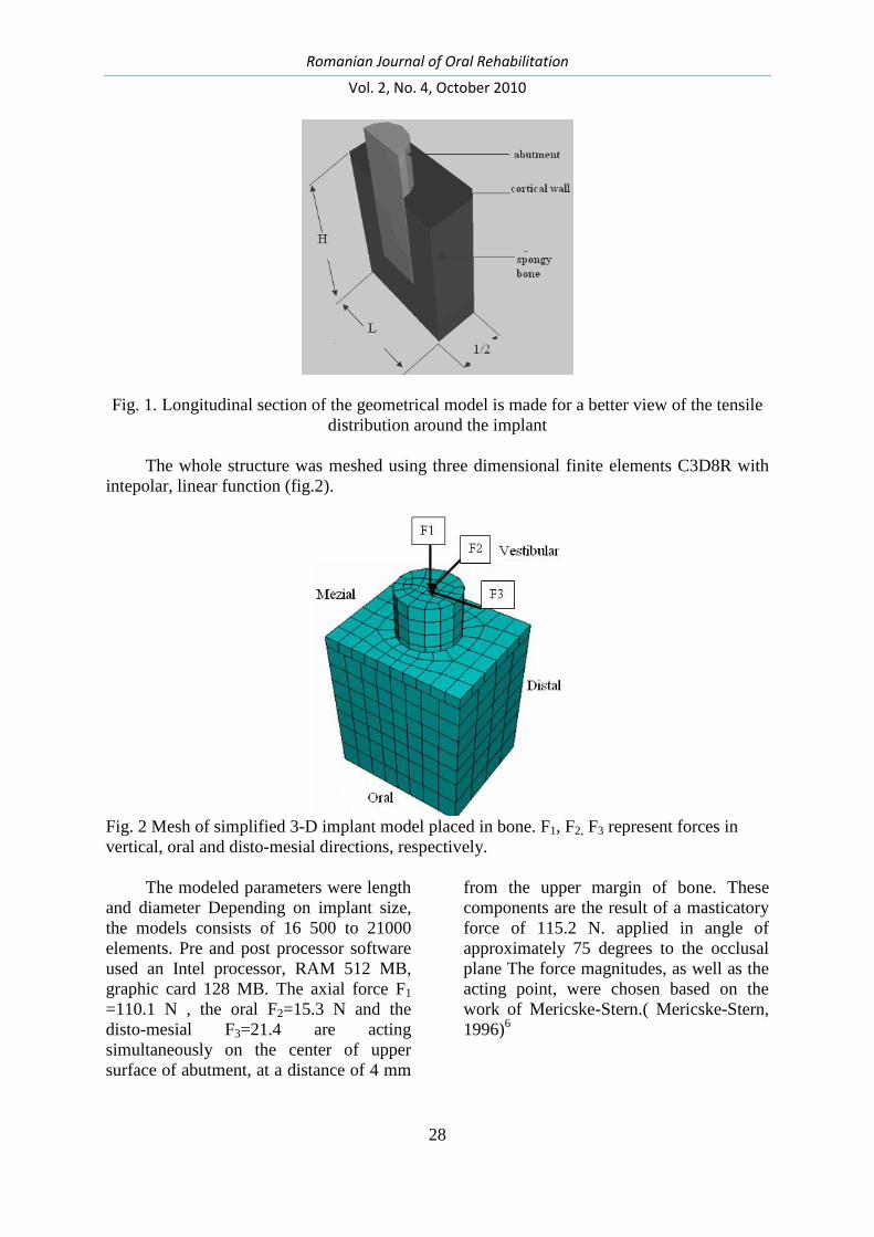
Romanian Journal of Oral Rehabilitation
Vol. 2, No. 4, October 2010
28
Fig. 1. Longitudinal section of the geometrical model is made for a better view of the tensile
distribution around the implant
The whole structure was meshed using three dimensional finite elements C3D8R with
intepolar, linear function (fig.2).
Fig. 2 Mesh of simplified 3-D implant model placed in bone. F1, F2, F3 represent forces in
vertical, oral and disto-mesial directions, respectively.
The modeled parameters were length
and diameter Depending on implant size,
the models consists of 16 500 to 21000
elements. Pre and post processor software
used an Intel processor, RAM 512 MB,
graphic card 128 MB. The axial force F1
=110.1 N , the oral F2=15.3 N and the
disto-mesial F3=21.4 are acting
simultaneously on the center of upper
surface of abutment, at a distance of 4 mm
from the upper margin of bone. These
components are the result of a masticatory
force of 115.2 N. applied in angle of
approximately 75 degrees to the occlusal
plane The force magnitudes, as well as the
acting point, were chosen based on the
work of Mericske-Stern.( Mericske-Stern,
1996)6
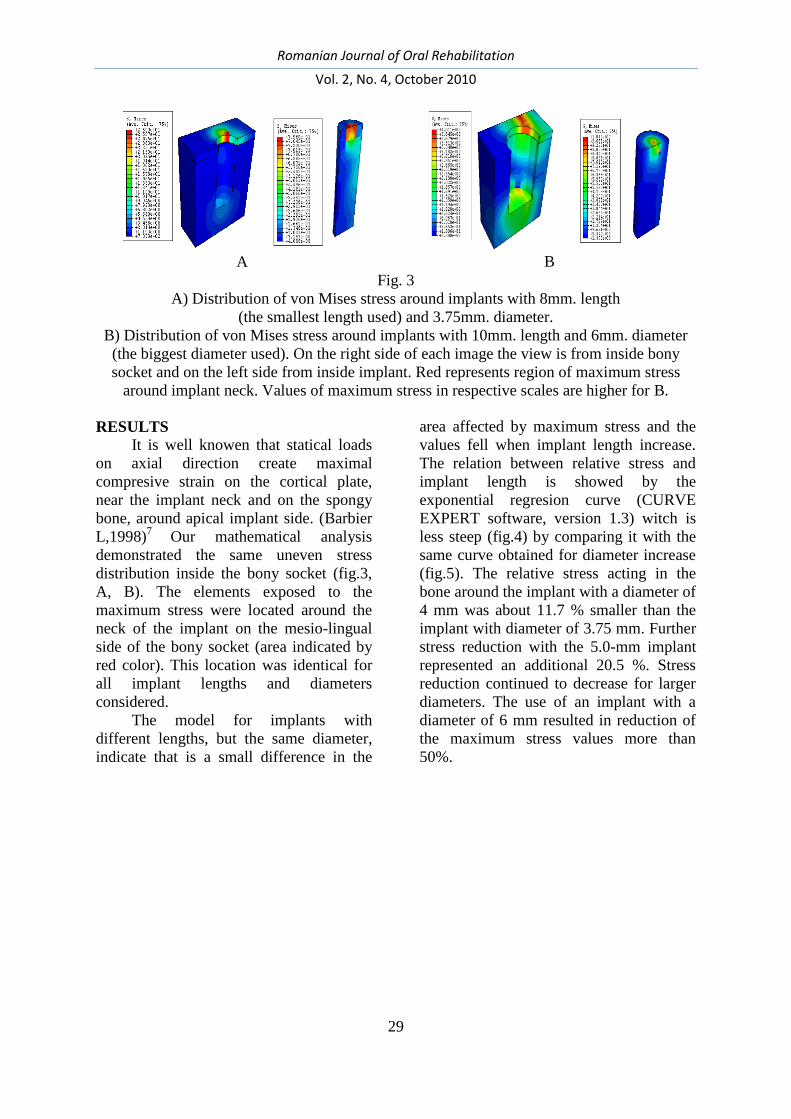
Romanian Journal of Oral Rehabilitation
Vol. 2, No. 4, October 2010
29
A
B
Fig. 3
A) Distribution of von Mises stress around implants with 8mm. length
(the smallest length used) and 3.75mm. diameter.
B) Distribution of von Mises stress around implants with 10mm. length and 6mm. diameter
(the biggest diameter used). On the right side of each image the view is from inside bony
socket and on the left side from inside implant. Red represents region of maximum stress
around implant neck. Values of maximum stress in respective scales are higher for B.
RESULTS
It is well knowen that statical loads
on axial direction create maximal
compresive strain on the cortical plate,
near the implant neck and on the spongy
bone, around apical implant side. (Barbier
L,1998)7
Our mathematical analysis
demonstrated the same uneven stress
distribution inside the bony socket (fig.3,
A, B). The elements exposed to the
maximum stress were located around the
neck of the implant on the mesio-lingual
side of the bony socket (area indicated by
red color). This location was identical for
all implant lengths and diameters
considered.
The model for implants with
different lengths, but the same diameter,
indicate that is a small difference in the
area affected by maximum stress and the
values fell when implant length increase.
The relation between relative stress and
implant length is showed by the
exponential regresion curve (CURVE
EXPERT software, version 1.3) witch is
less steep (fig.4) by comparing it with the
same curve obtained for diameter increase
(fig.5). The relative stress acting in the
bone around the implant with a diameter of
4 mm was about 11.7 % smaller than the
implant with diameter of 3.75 mm. Further
stress reduction with the 5.0-mm implant
represented an additional 20.5 %. Stress
reduction continued to decrease for larger
diameters. The use of an implant with a
diameter of 6 mm resulted in reduction of
the maximum stress values more than
50%.

Romanian Journal of Oral Rehabilitation
Vol. 2, No. 4, October 2010
30
Fig. 4 Exponential regression curve express relations between relative values of stress and
implant length
Fig. 5 Relations between relative values of stress and implant length. Exponential regression
curve less steep than for implant diameter
DISCUSSIONS The results of the FEA computation
depend on many individual factors,
including material properties, boundary
conditions, interface definition and also on
the overall approach to the geometrical
model. (Himmlova, 2004)8 The presented
model was only an approximation of the
clinical situation, but using the
components of an oblique force for loading
and a 3-D model for the dental implant,
resulted in a more satisfactory results
comparing with vertical or horizontal
loading and 2-dimensional geometries.
The model simplification refers only to the
implant shape, designed like cylinder and
not like screw or other shapes commonly
used in clinical practice. Thus made it
possible to reduce the required computer
time without affecting the purpose of this
study, which was to establish only the
influence of the implant length and
diameter.

Romanian Journal of Oral Rehabilitation
Vol. 2, No. 4, October 2010
31
The strain produced by axial and
paraxial loads act in a diferent way on the
bone – implant interface. Barbier and co.
studied the bone remodeling process and a
very important conclusion was made: the
areas with highest equivalent stress has an
accelerated process of remodeling. The
location of zones with higher stress around
the implant neck may indicate a danger of
overloading in this area. The elements
exposed to maximum stress were located
where most of non-axial masticatory
forces were transferred. For example,
forces acting in lingual and disto-mesial
directions are associated with grinding
movements, in comparison with axial
loading during chopping movements. This
situation corresponds to nonparametric
computerized models of loaded dental
implants. Models show utmost strain
acting around the implant neck (Holmgren,
1998, Oosterwyck, 2001)9, 10
CONCLUSIONS This finite element study showed that
implant with large diameter better
dissipated the simulated force and
significant reduce the stress on the bone –
implant interface, while the effect of
implant length was less notable. The
highest decrease in stress, compared to the
implant 3.75mm diameter, was obtained
for the maximal diameter used in the study
(52.7% decreases for 6 mm width). These
results are in contradiction with some
theories witch consider the implant length
a dimensional parameter more important
than implant diameter (Carl E. Misch,
2005)13
Our conclusion is consensual with
majority studies based on FEA: implant
with the maximum possible diameter
(allowed by the clinical condition) is the
optimum choice (Goodacre, 2003, Keller,
2004)11, 12
REFERENCES 1. Jian-Ping Geng, Keson B. C. Tan, Gui-Rong Liu, Application of finite element analysis in implant dentistry.
A review of the literature, J Prosthet Dent 2001;85:585-98
2 Lucie Himmlova´, Tat‘jana Dosta´lova´, Alois Ka´covsky´, Svatava Influence of implant length and diameter
on stress distribution: A finite element analysis J Prosthet Dent 2004;91:20-5.
3. Carr AB, Gerard DA, Larsen PE. Histomorphometric analysis of implant anchorage for 3 types of dental
implants following 6 months of healing in baboon jaws. Int J Oral Maxillofac Implants 2000;15:785-91.
4. Jeff Brink, Stephen J. Meraw, David P. Sarment. Influence of implant diameter on
surrounding bone. Clin. Oral Impl. Res. 18, 2007; 563– 568
5. Teixeira ER, Sato Y, Shindoi N. A comparative evaluation of mandibular finite element models with different
lengths and elements for implant biomechanics. J Oral Rehabil 1998;25:299-303
6. Mericske-Stern R, Piotti M, Sirtes G. 3-D in vivo force measurements on mandibular implants supporting
overdentures: a comparative study. Clin Oral Impl Res 1996;7:387-96.
7. Barbier L, Vander Sloten J, Krzesinski G, Schepers E, Van der Perre G. Finite element analysis of non-axial
versus axial loading of oral implants in the mandible of the dog. J Oral Rehabil 1998;25:847-58.
8. Himmlova, L., Dostalova, T., Kacovsky, A. & Konvickova, S. Influence of implant length and diameter on
stress distribution: a finite element analysis. Journal of Prosthetic Dentistry 2004, 91:20–25.
9. Holmgren EP, Seckinger RJ, Kilgren LM, Mante F. Evaluating parameters of osseointegrated dental implants
using finite element analysis—a two dimensional comparative study examining the effects of implant diameter,
implant shape, and load direction. J Oral Implantol 1998;24:80-8.
10. Duyck, J., Ronold, H.J., Van Oosterwyck, H., Naert,I., Vander Sloten, J. & Ellingsen, J.E. (2001) The
influence of static and dynamic loading on marginal bone reactions around osseointegrated implants: an animal
experimental study. Clinical Oral Implants Research 12: 207–218.
11. Goodacre, C.J., Bernal, G., Rung-Larassaerg, K. & Kanjy, K. Clinical complications with implants and
implant prostheses. Journal of Prosthetic Dentistry 2003, 90: 121–132.
12. Cho, P., Schneider, G.B., Krizan, K. & Keller, J.C. Examination of the bone–implant interface in
experimentally induced osteoporotic bone. Implant Dentistry 13: 2004 79–87.
Books
13.Carl E. Misch, Martha Warren Bidez, A scientific Rationale for Dental Implant Design in: Dental Implant
Prosthetics, Ed. Elsevier Mosby, Inc., Canada, 2005, 332 – 349

Romanian Journal of Oral Rehabilitation
Vol. 2, No. 4, October 2010
32
COMPARATIVE STUDY REGARDING THE GUM-PERIODONTAL
MANIFESTATIONS DURING PREGNANCY AND NON-PREGNANCY Liliana Sachelarie
1, Adina Simona Bîrgăoanu
2, Carmen Stadoleanu
3, Diana
Popovici4, Florentina Pricop
5
1,2,3 ―Petre Andrei‖ University, Faculty of Dental Medicine, Iaşi
4,5 University Of Medicine and Pharmacy Gr.T.Popa, Iaşi
Abstract: The pregnancy, by itself is not able to produce the taint of the limit paradont but the hormonal change
together with the vitamin deficit which occures during this periode may affect the local reaction of tissues
concerning the bacterial plack, the real cause of this problem. In case of pregnancy woman the bacterial flora
inside protection against it decreases, so the oral hygiena and the alimentation hold the main role in the
evolution of the gingivo – paradontal diseases.
A comparative study involving 25 pregnancy woman and 25 unpregnant over shows that the appearance of
gingivo –parodontal diseases is bigger in the first lot that the second one. We also concluded that, in case of
pluri –even pregnant woman, the paradontal teritory is more affected because of the previous pregnancies.
Key words: gingiva-parodontal diseases, pregnancy
INTRODUCTION
The pregnancy, by itself, does not produce
the disease of the marginal parodont, but
the hormonal modifications together with
the vitamin coefficients which appear
during this period influence the local
reaction of the tissues compared to the
bacterial plate-the real determinant
factor.therefore, the gravity constitutes a
secondary ethiopathogenic factor.
The pregnancy, through the modification
of the level of estrogens and/or
progesterone, determines the intense
increase of the vascularization degree of
the oral mucous, which determines the
appearance of some intense desquamation
lesions of the corion and of the gingival
epithelium, accompanied by a gingival
congestive edema.Although, the studies
carried out proved that these modifications
are transitory and do not appear in all the
pregnant women leading to the conclusion
that the area factor has an important role in
the oral pathology.
The main cause of the gum-periodontal
disorders in general and in the pregnancy
in particular, is represented by the local
factor, the bacterial plack, that organo-
bacterian pellicle which adheres both to
the mucosal surfacesand to the dental ones.
During the pregnancy, the bacterial plate
contained in the plate increases and is
modified, resulting a greater
disequilibrium that leads to the
development of the inflammatory process.
The purpose of the study consisted in
determining the incidence of the gum-
periodontal disorders during the pregnancy
and non-pregnancy, emphasizing the
correlative aspects between the dental
clinical entities and the general status,
aspects that will be at the basis of a well-
led therapy.
MATERIAL AND METHOD
The clinical statistic study was carried out
on a lot of 50 patients, 50% pregnant
(primipar and multipar) found in different
stages of pregnancy and 50% non-
pregnant. They had ages comprised
between 20 and 30 and came from the
urban environment.
We compared the two lots analyzing the
following parameters : the aspect of the
gum, the number of births, the personal
gum pathological aspects, the dental
hygiene state and the alimentation type.
With this purpose, we used the data
supplied by the :

Romanian Journal of Oral Rehabilitation
Vol. 2, No. 4, October 2010
33
1. Anamnesis: it comprised a series of
questions through which we found out
from the patients‘ representations data
about the diet, the periodontal disease
historic and the heredo-colateral
antecedents. In addition, we picked
information about their vicious habits
since the specialized studies carried out
until the present have proven that the
smokers are 2-7 times more affected by the
gum-periodontal disorders than the non-
smokers. With the purpose to centralize
the data obtained, we drew out an
individual sheet, according to a dental
sheet model.
2. The plate index (IP ). For emphasizing
the bacterial plate with the purpose to
establish the oral hygiene state, we used
plate revelators (basic fuchsine tablets or
0,03 brome-cresol green). The results
obtained after the examination of the two
lots are presented in the table below.
Tabelul I
Oral hygiene state
Type of persons tested No. of persons
studied
Satisfactory oral
hygiene
Non-satisfactory
oral hygiene
Pregnant 25 6 19
Non- pregnant 25 10 15
3. The oral microbial degree was
established with the help of the Snyder
test, which measures the rapidity of the
acid formation from a saliva sample (1-2
ml) on a sample medium with a pH
indicator. Its color changed in the
following manner:
- light-blue: pH 5.4-5.5;
- green: pH 4.5-4.6;
- yellow: 4.2-3.8.
The results obtained after examining
the two lots are presented in the table
below (tab. 2):
Tabelul II
Oral Ph
Type of persons tested No. of persons studied Light-blue Green Yellow
Pregnant 25 12 9 4
Non- pregnant 25 15 7 3
4. The bleeding index. For establishing
the gum inflammation degree we used the
test proposed by Silnes –Loe, using with
this purpose a small condenser, spherical,
that we introduced in the gum sulcus until
its bottom but without pressure, in an
angle of 25-30 degrees from the dental
surface.
We calculated the gum bleeding index
from the sum of values for four areas of
gum exploration: disto-vestibular, mesio-
vestibular and oral divided in 4, and the
notation was carried out according to the
scheme proposed by the author:
0- normal gum;
1- light inflammation with color
modification,edema, the absence of
provoked bleeding;
2- moderate inflammation with
congestion, edema and provoked bleeding;
3- severe inflammation with
congestion, edema and tendency of
spontaneous bleeding.
The results obtained after examining the
two lots are presented in the table below
(tab. 3):

Romanian Journal of Oral Rehabilitation
Vol. 2, No. 4, October 2010
34
Tabelul 3
The gum inflammation degree
Type of persons
tested
Normal
gum
Light
inflammation
Moderate
inflammation
Severe
inflammation
Total pregnant
women
Pregnant 6 6 12 1 25
Non- pregnant 12 8 4 1 25
RESULTS
Wy mention that of the 25 pregnant
women studied, two did not present gum-
periodontal modifications.
After the centralization of the data
obtained through the methods above, we
processed it statistically, and namely:
Tabelul 4
The gum aspect
Normal
gum
Congestive
gum
Congestive-hypertrophic
gum
Number
of cases
Pregnant 2 14 9 25
% 8 56 36 100
Non- pregnant 12 12 1 25
% 48 48 4 100
Tabelul 5
Number of births
Parity
No. of
pregnant
women
%
parity
No. of pregnant
Women with
gum-periodontal pathology
%
according
to the
parity
% according to the
total no. of pregnant
women with gum-
periodontal
pathology
Primipar 4 20 2 40 8
Multipar 19 80 15 75 60
According to the anamnesis data, we obtained the following statistical numbers:
Tabelul 6
Personal pathological gum antecedents
Antecedents
No. of
pregnant
women
% according
to the
antecedents
No. of
pregnant
women with
gingival
pathology
% according
to the
antecedents
% according
to the no.of
pregnant
women with
gingival
modifications
Existent before
the current
pregnancy
17 74 13 76 56
No antecedents 6 26 2 24 9

Romanian Journal of Oral Rehabilitation
Vol. 2, No. 4, October 2010
35
Tabelul 7
Types of alimentation
Types of
alimentation
No. of pregnant
women with
gum-periodontal
pathology
% according to the
no. of pregnant
women with
gingival modifications
Non-pregnant
women with
gum-
periodontal
pathology
% according
to the no.of
non-pregnant
women with
gingival
modifications
Meat 2 8,7 0 0
Vegetarian 3 13 1 7,69
Lactate 3 13 3 23,07
Hydrocarbonate 10 43,5 5 38,46
Mix 5 21,8 4 30,76
CONCLUSIONS
1. The deficit hygiene of the oral cavity
associated with the state of pregnancy
constitutes the main cause of the
appearance of gum-periodontal diseases
during the pregnancy.
2. We noticed an increased incidence of
the gingival phatology during the
pregnancy or non- pregnancy
state, in the patients whose diet mainly
consists of the hydrocarbonate aliments
and less in the patients who consume
lactate or vegetarian aliments.
3. The periodontal status indicates us a
majority of pregnant women with gum-
periodontal modifications.
4. We noticed an increased incidence of
the gum-periodontal pathology in the
multipar pregnant women both compared
to the primipar patients and compared to
the non-pregnant ones, which proves the
existence of pathological states, appeared
during the pregnancy and previous births
with their exacerbation in the current
pregnancy.
5. The graphical representation of the
incidence of gum-periodontal diseases in
the 50 patients shows that the greatest
percentage was encountered in the case of
pregnant women.
6. The interdisciplinary, gynecology and
dental exam, the illustration of the
incidences of gum-periodontal diseases,
suggests the existence of a determinism
between the gum-periodontal lesions and
gravity state.
7. The increase of the addressability to the
dentist of the pregnant women could
contribute to the improvement of their oral
health state.
REFERENCES: 1. Abraham Inpijn L. et al. –The significance of endocrine factors and microorganisms in the development of
gingivitis in pregnant women. Stomatologica 75: 15-18, 1996.
2. Lang NP, Nyman S, Seen C, et al. Bleeding on probing as it relates to probing pressure and gingival health.
J. Clin. Periodontol 1991; 18:257.
3. Offenbacher S,Lieff S, Borggess KA, Murtha AP, Madianos PN, Champagne CM, et al. Maternal
periodontitis and prematurity. Part I: Obstetric outcome of prematurity and growth restriction. Ann Periodontol
2001;6: 164-74.
4. Aldea MJ. Obstetrica, Ed. Apollonia, Iaşi 1999.
5. Mârţu S, Mocanu C. Parodontologie clinică. Ed. Apollonia Iaşi 2000
6. Academy Report. The Pathogenesis of Periodontal Diseases,J. Periodontol 1999;70: 457-470

Romanian Journal of Oral Rehabilitation
Vol. 2, No. 4, October 2010
36
ULTRASOUND EVALUATION IN TEMPOROMANDIBULAR
ARTHRITIS IN PATIENTS WITH CHRONIC JUVENILE ARTHRITIS Iordache Cristina, Ancuta Codrina, Iordache O., Ancuta E., Pirlia Carmen,
Zenaida Surlari, Chirieac Rodica
Disciplina Ergonomie, Facultatea de Medicina Dentara, Universitatea de Medicina si
Farmacie ―Gr.T.Popa‖ Iasi, Romania, Disciplina de Reumatologie, Facultatea de Medicina
Universitatea de Medicina si Farmacie ―Gr.T.Popa‖ Iasi, Romania, Disciplina de Edentatie
Partial Redusa, Facultatea de Medicina Dentara, Universitatea de Medicina si Farmacie
―Gr.T.Popa‖ Iasi, Romania
Abstract: The EULAR classification defines the CJA as a systemic manifestation affecting both sexes before 16
years old and registers 3 clinical forms: the systemic disease (Still‘s disease), oligo- and poly-articular.
Ultrasonography has remarkable contribution in the diagnosis of musculoskeletal apparatus‘ lesions. Due to
the advantages (non-invasive, low cost, no negative effects and contraindications), this method allows the static
and dynamic evaluation of the hard and soft structures.
The purpose of the paper: to assess TMJ involvement in CJA patients by ultrasonography and to define
characteristic patterns of the disease.
Material and method: the research was conducted on 2 patients with CJA, hospitalized in Rheumatology
Department, Rehabilitation Hospital of Iasi during 01.01.2005-31.07.2006. Assessment was done according to a
standard protocol in all patients and included: inflammatory and immune parameters and a complex, both static
and dynamic, ultrasound evaluation of TMJ. Descriptive statistical analysis was done using SPSS-11.
Results and discussions 86.36% patients featured either inflammatory-destructive or degenerative types
ultrasound lesions, the majority of them (59.09%) being inflammatory and destructive.
Conclusions a non-invasive, easy, low cost and easily reproducible method, ultrasonography, reconfirmed its
diagnostic function in the small joints evaluation, including TMJ.
Key words: tmd, ultrasonography, tmj, cja
INTRODUCTION
The temporomandibular joint or
temporomaxilar joint, the main component
of the stomatognat system, is the only
mobile joint in the cephalic extremity, the
most developed and the most complex
body joint, so that Gouppille Philippe
(1988) used to name it also the most
challenging joint of the human body.
The EULAR classification defines CJA as
a systemic disease affecting both sexes
before 16 years old; 3 major clinical forms
are actually known including the systemic
form of the disease (Still‘s disease), the
olygo-articular and the poly-articular form.
About 14-65% of CJA present with TMJ
involvement as reported by Goupille in
1995; in 17% of cases (Grosfeld, 1973
and Mayne, 1969) and 63 % according to
Ronnig, 1981, distinct types of lesions are
described because the inflammatory
synovitis leads to both destruction of the
articular components (especially the
condyle) and deformation of the region.
In the last two decades the imagistic
diagnostic of the TMJ recorded important
progresses assuring by this both the
differentiation of the entities causing the
chronic pain and disability in the oro-facial
area and the results‘ evaluation of the
conservatory and surgical therapy.
The ultrasonography has a remarkable
contribution in the diagnosis of the
musculo-skeletal lesions. Through its main
advantages including non-invasive, low
cost, no negative effects and
contraindications, this method allows the
static and dynamic evaluation of the hard
and soft structures.
Objectives: to assess the TMJ
involvement in CJA patients by ultrasound

Romanian Journal of Oral Rehabilitation
Vol. 2, No. 4, October 2010
37
and to define characteristic patterns of the
disease.
MATERIAL AND METHOD 22 patients diagnosed with CJA as defined
by EULAR criteria were enrolled in the
present research. All patients were
admitted in Rheumatology Department,
Rehabilitation Hospital of Iasi during
01.01.2005-31.07.2006.
The main inclusion criteria were: both
static and dynamic pain in the TMJ area
(during the examination or in the history);
either morning or after rest TMJ stiffness;
difficulties in mouth opening or closing;
trismus; articular noises (including
crepitations, cracments).
The following exclusion criteria were
taken into account: cranio-facial traumas;
tumors and infections of the facial and
cranial area; patients with neoplasia with
other localizations. 32 patients with CJA,
defined according to EULAR criteria,
were included in the present study. The
selected patients were clinically,
biologically and echographically assessed
using a 10MHz probe.
RESULTS AND DISCUSSIONS
TMJ ultrasound was based on a standard
protocol allowing imaging in the antero-
superior compartment, sagital and frontal
planes.
The patient was supine, the transducer
being placed directly on the TMJ area,
parallel with the long axis of the
mandibular ram. The transducer was
oriented also parallel to Camper line (that
connects the nose with the tragus). The
movement in this way allowed the
identification and the measurement of a
hypoecogenic space, homogenous situated
between the mandibular condyle and the
lateral part of the articular eminence
(patient with shout mouth), that
corresponds to the disk.
Both static and dynamic ultrasound TMJ
examination were done, with Doppler
color SAOT ultrasound device with a
10MHz probe. The following ultrasonic
evaluation score was used:
- condyle shape: N (normal) = 0; flat (F) =
1, extended lysis (L) = 2;
- articular space: N ( >0.5) = 0; <0.5 = 1;
- synovial fluid: the presence of the articular
liquid in a high amount (hypoechogenic):
LA = 2
- condyle mobility (trajectory, ampleness):
normal (M0) = 0; moderately affected
(M1) = 1, severely affected (M2) = 2;
- condyle contour: linear (CL) = 0; sclerous
(CSc) = 2; jagged (CD)=3;
- temporal bone contour: linear (CL)=0;
sclerous (CSc)=1; jagged (CD)=2;
- erosions: one (E1) = 1; > 1 (E2) = 2;
- geodes: one (G1)=1; >1 (G2) =2;
- osteoporosis: OP =1;
- discal anomalies: the displacement (DD) =
1; morphological changes (MMD) = 2;
- osteophytes: bone = 2;
Both inflammatory–destructive and
degenerative lesions have been reported by
ultrasound evaluation of TMJ in more than
86.36% cases; among them, 59.09% were
of inflammatory –destructive types as
shown in graphic no 1 and table 1.

Romanian Journal of Oral Rehabilitation
Vol. 2, No. 4, October 2010
38
Table 1: Ultrasound examination of TMJ involvement
Results N %
Normal 3 13.64
Inflammatory lesions 10 45.45
Destructive lesions 3 13.64
Destructive lesions + osteoarthritis 2 9.09
Osteoarthritis 4 18.18
Graphic no.1: The frequency of the affections detected in ultrasound
CONCLUSIONS
1. Sonography, an easy, non-invasive, low
cost and easily reproducible method has
reconfirmed its diagnostic place in the
evaluation of the small joints relieving the
TMJ lesions;
2. Ultrasound examination used in this
study to a significant number of patients,
can be considered as an useful diagnostic
means for the TMJ pathology; we
appreciate that the method must be
indicated even of first and unique
intention in the actual exploration, due to
the advantages it has;
3. The technique has relieved pathological
modifications of inflammatory, destructive
and degenerative type.
REFERENCES 1. Aldescu C: Radiologie pentru studenţi şi medici stomatologi, Ed. Polirom, Iasi, 1998
2. Brown A K, O‘Connor J P, Roberts T E, Emery P, si colab: Ultrasonography for rheumatologist: the,
development of specific competency based educational outcomes, Ann rheum Dis, 2006, 65, (5): 629637
3. Burlibaşa C, Nimigean V: „Anatomia funcţională a ATM”, Editura Cernea, Bucuresti, 1993
4. Emshoff R, Bertram S, Rudisch A si colab: The diagnostic value of ultrasonography to determine the
temporomandibular joint disc position, Oral Surg Oral Med Oral Pathol Oral Radiol Endod, 1997, 84: 688- 696
5. Filipppuci E, Iagnoco A, Salaffi F, Cerioni A, Valesini G, Grassi W: Power Doppler sonography
monitoring of synovial perfusionat the wrist joints in patients with rheumatoid arthritis treated with
adalimumab, Ann Rheum Dis, 65, 11: 1433- 1438
6. Kuseler A, Pedersen TK, Herlin T si colab: Contrast enhanced magnetic residence imaging as a method
to diagnose early inflammatory changes in tha temporomandibular joint in children with juvenile chronic
arthritis, J Rheumatol 1998, 25: 1406-1412
7. Arthographic findings in the temporomandibular joint in patients with rheumatic disease, J Oral
Maxillofac Surg, 1989, 47: 780-784
8. Larheim T: Current trends in temporomandibular joint imaging, Oral Surgery Oral Medicine Oral
Pathology, 1995, 80 (5): 555-576.
9. Larheim T, Haanes HR, Ruud AF: Mandibular growth temporomandibular joint changes, and dental
occlusion in juvenil rheumatoid arthritis: a 17-year followup study, Scand J Rheumatol 1981, 10: 225-233
0
5
10
15
20
25
30
35
40
45
50
Normal Infl.les. Destr.les. Destr.les.+arth Arthrosis
13,64
45,45
13,64
9,09
18,18
Romanian Journal of Oral Rehabilitation
Vol. 2, No. 4, October 2010
39
10. Martini G, Bacciliero U, Tregnaghi A, si colab: Insolated temporomandibular synovitis as unique
presentation of juvenile idiopathic arthritis, J Rheumatol 2001, 28: 1689-1692
11. Mock D: The differential diagnosis of temporomandibular disorders, Journal of Orofacial Pain, 1999,
13 (4): 246-250
12. Ogus H, Toller P : Larticulation temporo-mandibulaire, Editura Masson, Paris, 1985
13. Petty R E, Southwood TR, Manners P, Baum J Glass DN si colab: International League of Assotiations
for Rheumatology Classification of Juvenile Idiopathic Arthritis; second revision, Edmonton, 2001, J
Rehumatol, 2004, 31: 390- 392
14. Scheunemann I, Dannecker G E, Roth J : Phalangeal bone ultrasound is of limited value in patients with
juvenile idipathic arthritis, Rheumatology, 2006, 45, (8): 1125- 1129
15. Schramm I, Teuscher T, Witt E : Involvement of the temporomandibular joint in juvenile idiopatic
arthritis, Arthritis & Rheum 2001, vol 44: 453-458
16. Schanberg L, Gil K, Antony K, Yow E, Rochon J: Pain, stiffness and fatigue in juvenile polyarticular
Arthritis: Contemporaneus stressful events and mood as predictors, Arthritis & Rheumatism, 2005, 52, 94):
1196-1205
17. Stone M, Bergin D, Whelan B si colab: Power Doppler ultrasound assessment of rheumatoid hand
synovitis, J Rheum, 2001, 29:1979-1982
18. Strunk J, Stube K, Muller- Ladner U, Lange U: Three dimensional power Doppler ultrasonogrphy
confirms early reduction of synovial perfusion after intrarticular steroid injection, Ann Rheum Dis, 2006, 65
(3): 411- 413
19. Svensson B, Adell R, Kopp S: Temporomandibular disorders in juvenile chronic arthritis patients,
Swed Dent J, 2000, 24: 83-92
20. Twilt M, Schulten A J M, Nicolas P, Dulger si colab: Facioskeletal changes in children with juvenile
idiopathic arthritis, Ann Rheum Dis, 2006, 65, (6): 823- 826

Romanian Journal of Oral Rehabilitation
Vol. 2, No. 4, October 2010
40
THE INFLUENCE OF POST MATERIAL ON FRACTURE
RESISTANCE AND MODE OF FAILURE OF THE TEETH
RECONSTRUCTED WITH POST SYSTEMS Anca Mihaela Viţalariu, Monica Silvia Tatarciuc, Diana Diaconu,St.Panaite
University of Medicine and Pharmacy ”Gr.T.Popa”of Iasi, Romania
Abstract: The purpose of this study was to evaluate the fracture strength and mode of failure of endodontically
treated teeth reconstructed with different posts. Forty maxillary central incisors were reconstructed with
different posts and divided in 4 groups: carbon fibre posts, glass fibre posts, titan posts and control group
(without posts). The force was applied at an angle of 45 degrees, until the failure of specimens. The failure loads
were recorded and analyzed with student t method. The mode of failure (reparable or irreparable) was also
evaluated. Regarding the mean value of resistance to fracture there were no significant differences between
groups, but differences in the mode of failure were recorded. Metal posts caused a bigger number of irreparable
root fractures, while non-metallic posts produced in most cases reparable root fractures. In conclusion, the fibre
reinforced posts have a protective effect on the dental tissues comparing with metallic posts and are best suited
for the restoration of endodontically treated teeth.
Key words: fracture resistance, carbon fibre post, glass fibre post, metallic post
INTRODUCTION
Despite the steady evolution of post and
core materials and techniques, the failure
of post-retained crowns is relative common.
It is well known that endodontically
treated teeth present a higher risk of failure
than vital teeth (Caputo 1987). Some
authors (Freedman 1996) consider that is
the result of the loss of tooth structure,
whereas others (Gutmann 1992) reported
the effect of decreased moisture content
and subsequent brittleness of
endodontically treated teeth as causes of
fracture. There is no consensus about the
techniques and materials that are best
suited for the restoration of endodontically
treated teeth. The cast post and core has
some disadvantages that may jeopardize
long-term success: tooth weakened related
to the removal of root structure to
accommodate the necessary post length,
lack of cement retention, corrosion risks,
unevenly stress distribution leading to root
fracture, difficulties in removal of the post,
increased working time and laboratory
costs (Purton 1996).
The restoration of endodontically treated
teeth with metal-free materials that have
physical properties similar to those of the
dentin has become a major objective in
dentistry. In the late 1980s were
introduced fibre-reinforced post systems:
carbon fibre, glass fibre and quartz fibre
posts (Mannocci 1999). The carbon fibre
post have a modulus of elasticity very
close to that of dentin, so it causes less
tooth stress resulting in fewer root
fractures (Dean 1998). Glass fibre-
reinforced posts are composed of
unidirectional glass fibres in a resin matrix
that strengthened the structure of the post
without compromising the modulus of
elasticity. These posts have the same good
mechanical properties as the carbon fibre
posts and are tooth-coloured, allowing to
achieve better aesthetics (Rosentritt 2000).
This ―in vitro‖ study aimed to evaluate the
fracture strength and the mode of failure of
endodontically treated teeth reconstructed
with different posts: carbon fibre post
(Carbopost/Carbotech), glass fibre post
(Snowlight/ Carbotech) and titan post
(Ancorextra/Svedia International SA) (Fig.
1).

Romanian Journal of Oral Rehabilitation
Vol. 2, No. 4, October 2010
41
Fig. 1. The posts used in this study (from left to right): Carbopost, Snowlight, Ancorextra
MATERIAL AND METHOD
Forty sound maxillary central incisors,
recently extracted, were divided in four
groups, according to the type of post used
for restoration: gr. C: carbon fibre posts
(Carbopost), gr. S: glass fibre posts
(Snowlight), gr. T: titan posts and gr. M:
control (endodontically treated teeth, but
without posts).
All roots were filled with gutta-percha and
a resin sealer. The crowns of teeth were
cut perpendicular to their long axes, 2mm
above the cemento-enamel junction (CEJ)
using a diamond disk. After 48 hours
gutta-percha was removed, leaving 3mm
of canal filling in the apical third to ensure
a good endodontic seal. The posts were cut
4 mm coronal to tooth section and
cemented with dual-cure resin cement
(Panavia F). Composite cores were built
up with a light-curing composite to yield
an abutment height of 6 mm from the CEJ.
All preparations were covered with metal
crowns, cemented with Panavia F and
embedded into metallic cylinders filled
with self-polymerizing resin
Fig. 2. Preparing the samples for the compressive test
On the palatal surface of the crowns, a
0.3mm deep notch was made, in order to
place there the compressive load during
the test. The load was applied using a
chisel-shaped steel pin, corresponding to
the shape and dimension of the incisal
edge of a lower incisor. The metallic
cylinders were screwed into a mounting
device, directing the force at an angle of 45
degrees to the long axis of the tooth. This
angle reproduces the position and loading
characteristics on anterior teeth in a Class I
occlusion (Fig. 3).

Romanian Journal of Oral Rehabilitation
Vol. 2, No. 4, October 2010
42
Fig. 3. Load is applied on the palatal surface, at an angle of 45 degrees to the long axis of the
tooth
Each specimen was subjected to
progressive compression load in a
universal load-testing machine with
nominal forces of 25 kN, at a crosshead
speed of 19 mm/min and a monotonously
increasing force until the specimens failed.
The testing machine was connected to a
data acquisition board. LabVIEW 6.0
National Instruments software was used to
perform a data acquisition virtual
instrument for measurement with a scan
rate of 1000scan/sec. The compressive loads
at failure recorded were compared with the
Student t test. The mode of failure of the
specimens was also evaluated.
RESULTS
Fracture resistance
All specimens failed because of teeth
fracture. The range of load resistance of
the teeth to the point of fracture was about
31-35daN. The mean values for each
group were: gr.C: 31,8 ’ 32,2, gr.S: 31,3 ’
35,1, gr.T: 31,75 ’ 35,25, gr. M:31 ’ 34,9
The values of fracture resistance obtained
for endodontically treated teeth restored
with post-and-cores are close to that
recorded in the control group
(endodontically treated teeth but without
post and core) and are higher than
maximal values of the physiological
occlusal forces recorded clinically in
anterior teeth (20 daN).The statistical
analysis (Student t test) of the values
obtained showed no statistically significant
differences between the groups (α = 0.05).
Mode of failure
According to their localization and
complexity, root fractures were classified as
favourable (reparable) if they appeared in
the cervical third and catastrophic
(irreparable) if they were in the median or
apical third of the root. There were recorded
three different patterns of fracture: a) vertical
fracture, b) cervical (horizontal) fracture, c)
complex root fracture (vertical and
horizontal)
In group C (Carbopost) all three modes of
root fracture were recorded: five of them
were cervical (horizontal), three vertical
and two complex root fractures (Fig. 4).
In group S (Snowlight) were found only
two types of root fracture, most of them
being reparable (eight cervical) and only
two irreparable (complex root fractures)
(Fig. 5). There were also recorded two post
fractures, one associated with a cervical
fracture and one with a complex root
fracture. The posts fractured before the
roots did and this is a clinical major
advantage, because if the post fractures,
the whole restoration fails and has to be
immediately replaced. In this manner, the
risk of root overloading and fracture is
eliminated.

Romanian Journal of Oral Rehabilitation
Vol. 2, No. 4, October 2010
43
The patterns of fractures recorded in titan
post group are typical for teeth restored
with metal post: most occurred fractures
were irreparable (five vertical fractures,
three complex fractures) and only two
cervical (reparable). In one root with a
complex fracture we noticed that the titan
post was bent (Fig. 6). Because of its
higher elastic modulus, rigid metal post
absorbs forces without rupture but
transmits load to the tooth walls, which
fracture on exceeding the elastic limit of
the dentin.
The behaviour of the teeth in the control
group (endodontically treated but without
post) wasn‘t different compared with the
test groups, the fractures recorded being
50% cervical (restorable) and 50%
complex root fracture (irreparable)
(Fig.7).These results show that the
presence of the post have no significant
influence on the mode of root fracture, the
same types of fractures being observed in
the test groups (with posts) and in the
control group (without post) as well.
Comparing the test groups (with different
posts) between them, it can be observed
that post material has a certain influence
on the mode of fracture. We found
important differences between the three
experimental groups one from each other
regarding the mode of fracture. So, metal
posts produced a bigger number of
irreparable root fractures, while the non-
metallic posts produced in most cases
reparable root fractures.
DISCUSSION AND CONCLUSIONS
The results of this investigation, in
concordance with those obtained by
Mannocci, Mollersten, Rosentritt, showed
Fig. 4. Fractures in the group C (from
left to right): vertical, complex and
cervical fracture
Fig. 5. Modes of fracture in group S: few
complex root fracture (irreparable) and most
of them cervical fracture (reparable)
Fig. 7. Fractures in the control group were
cervical and complex root fractures Fig. 6. Fractures in the group T: cervical,
vertical and complex root fracture,

Romanian Journal of Oral Rehabilitation
Vol. 2, No. 4, October 2010
44
that the post material hasn‘t a significant
influence on the fracture resistance of the
endodontically treated teeth restored with
post-and-core systems. There is a general
consensus between specialists on the fact
that the post material influences the mode
of failure of the teeth with post and core.
Regarding metallic posts, the main cause
of failure is the vertical catastrophic root
fracture (irreparable). Because of their
rigidity, they can‘t follow the elastic
deformation of the tooth structures and
bend, transforming the contact area
between post and dentinal walls in contact
points. These points become zone of stress
concentration inside the root that will lead
to root fractures in certain unfavourable
conditions.
The results of the in vitro studies show that
non-metallic posts reinforced with carbon
and glass fibres have a protective effect on
the dental tissues by reducing to minimum
the risk of root fracture and if these
fractures occur, they are mostly restorable,
comparing with the metallic posts that
produce mostly vertical irreparable root
fractures (Akkayan 2002, Mannocci 1999,
Rosentritt 2000, Butz 2001).
The explanation for this behaviour results
from the fact that their low Young
modulus is closed to that of the dentin‘s
and of the resin-cement used for fixing the
posts. In addition, the chemical bond
between all these elements (fibre
reinforced post, dentin and resin-cement)
is very strong and create a complex that
behave like a unitary block under the
occlusal forces.
Within the limitation of this in vitro study,
the following conclusions can be drawn:
1. The post material don‘t have a
significant influence on the fracture
resistance of the teeth reconstructed with
post-and-core systems but influence
significantly the mode of fracture of these
teeth.
2. Root fractures caused by metallic posts
are mostly catastrophic (irreparable) and
lead to the tooth extraction.
3. The non-metallic posts made from resin
composite reinforced with carbon and
glass fibres have a protective effect on the
dental supporting tissues, the mode of
failure recorded being more favourable to
the root when compared with the metallic
posts and therefore increase longevity of
the restoration.
4. Fibre reinforced posts are the best
choice for reconstruct endodontically
treated teeth.
REFERENCES 1. Akkayan B, Gülmez T, Resistance to fracture of endodontically treated teeth restored with diffent post
systems, J Prosthet Dent , 2002, 87, 431-437
2. Butz F, Lennon A, G. Heydecke, J.R. Strub, Survival rate an fracture strength of endodontically treated
maxillary incisors restored with different post systems: an in vitro study, J Prosthodont, 2001,14, 58-64
3. Caputo A, Standlee J, Biomechanics in clinical dentistry, Quintessence, Chicago 1987, 185-203
4. Dean JP, Jeansone B, N. Sarkar, In vitro evaluation of a carbon fiber post, J Endod, 1998, 24, 807-810
5. Freedman G, The carbon fibre post: metal-free post-endodontic rehabilitation, Oral Health 1996, 86, 29-30
6. Gutmann JL, Restoring endodontically treted teeth, Tex Dent J, 1997, 114, 14-23
7. Mannocci F, Three-point bending test of fiber posts, J Endod 2001,27(12) : 758-761
8. Möllersten L, A comparison of strengths of five post-and-core systems, Quintessence Int. 2002, 33(2),140-149
9. Purton DG, Comparison of carbon fiber and stainless steel posts, Quintessence Int. 1996,27,93-97
10. Rosentritt M, Comparison of fracture strength of metallic and toth-colored posts, J Oral Rehabil 2000,27, 595-601
`
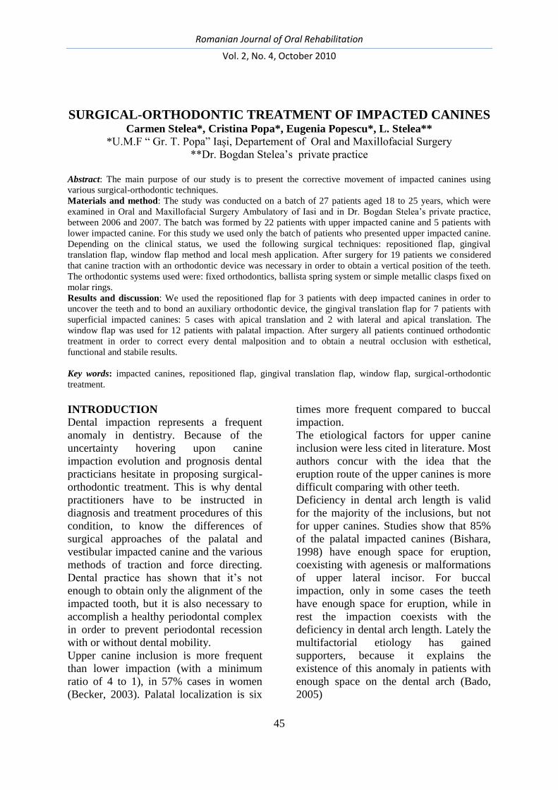
Romanian Journal of Oral Rehabilitation
Vol. 2, No. 4, October 2010
45
SURGICAL-ORTHODONTIC TREATMENT OF IMPACTED CANINES Carmen Stelea*, Cristina Popa*, Eugenia Popescu*, L. Stelea**
*U.M.F ― Gr. T. Popa‖ Iaşi, Departement of Oral and Maxillofacial Surgery
**Dr. Bogdan Stelea‘s private practice
Abstract: The main purpose of our study is to present the corrective movement of impacted canines using
various surgical-orthodontic techniques.
Materials and method: The study was conducted on a batch of 27 patients aged 18 to 25 years, which were
examined in Oral and Maxillofacial Surgery Ambulatory of Iasi and in Dr. Bogdan Stelea‘s private practice,
between 2006 and 2007. The batch was formed by 22 patients with upper impacted canine and 5 patients with
lower impacted canine. For this study we used only the batch of patients who presented upper impacted canine.
Depending on the clinical status, we used the following surgical techniques: repositioned flap, gingival
translation flap, window flap method and local mesh application. After surgery for 19 patients we considered
that canine traction with an orthodontic device was necessary in order to obtain a vertical position of the teeth.
The orthodontic systems used were: fixed orthodontics, ballista spring system or simple metallic clasps fixed on
molar rings.
Results and discussion: We used the repositioned flap for 3 patients with deep impacted canines in order to
uncover the teeth and to bond an auxiliary orthodontic device, the gingival translation flap for 7 patients with
superficial impacted canines: 5 cases with apical translation and 2 with lateral and apical translation. The
window flap was used for 12 patients with palatal impaction. After surgery all patients continued orthodontic
treatment in order to correct every dental malposition and to obtain a neutral occlusion with esthetical,
functional and stabile results.
Key words: impacted canines, repositioned flap, gingival translation flap, window flap, surgical-orthodontic
treatment.
INTRODUCTION
Dental impaction represents a frequent
anomaly in dentistry. Because of the
uncertainty hovering upon canine
impaction evolution and prognosis dental
practicians hesitate in proposing surgical-
orthodontic treatment. This is why dental
practitioners have to be instructed in
diagnosis and treatment procedures of this
condition, to know the differences of
surgical approaches of the palatal and
vestibular impacted canine and the various
methods of traction and force directing.
Dental practice has shown that it‘s not
enough to obtain only the alignment of the
impacted tooth, but it is also necessary to
accomplish a healthy periodontal complex
in order to prevent periodontal recession
with or without dental mobility.
Upper canine inclusion is more frequent
than lower impaction (with a minimum
ratio of 4 to 1), in 57% cases in women
(Becker, 2003). Palatal localization is six
times more frequent compared to buccal
impaction.
The etiological factors for upper canine
inclusion were less cited in literature. Most
authors concur with the idea that the
eruption route of the upper canines is more
difficult comparing with other teeth.
Deficiency in dental arch length is valid
for the majority of the inclusions, but not
for upper canines. Studies show that 85%
of the palatal impacted canines (Bishara,
1998) have enough space for eruption,
coexisting with agenesis or malformations
of upper lateral incisor. For buccal
impaction, only in some cases the teeth
have enough space for eruption, while in
rest the impaction coexists with the
deficiency in dental arch length. Lately the
multifactorial etiology has gained
supporters, because it explains the
existence of this anomaly in patients with
enough space on the dental arch (Bado,
2005)
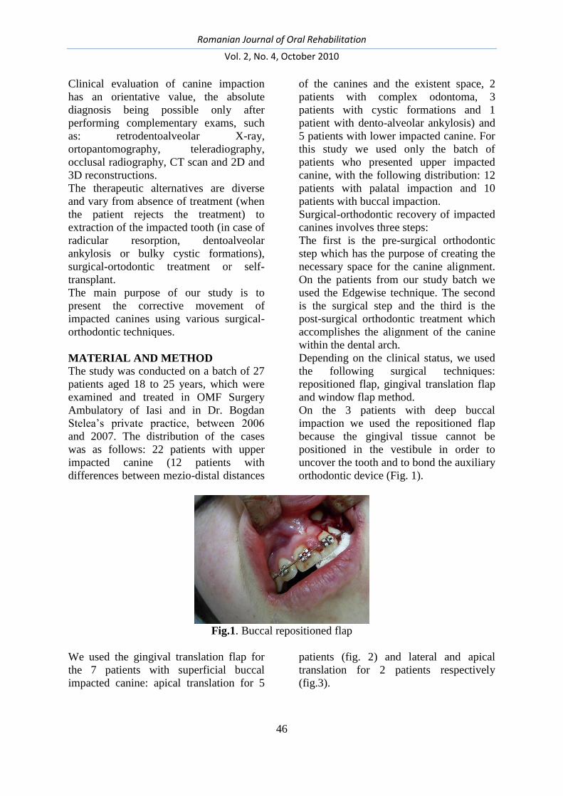
Romanian Journal of Oral Rehabilitation
Vol. 2, No. 4, October 2010
46
Clinical evaluation of canine impaction
has an orientative value, the absolute
diagnosis being possible only after
performing complementary exams, such
as: retrodentoalveolar X-ray,
ortopantomography, teleradiography,
occlusal radiography, CT scan and 2D and
3D reconstructions.
The therapeutic alternatives are diverse
and vary from absence of treatment (when
the patient rejects the treatment) to
extraction of the impacted tooth (in case of
radicular resorption, dentoalveolar
ankylosis or bulky cystic formations),
surgical-ortodontic treatment or self-
transplant.
The main purpose of our study is to
present the corrective movement of
impacted canines using various surgical-
orthodontic techniques.
MATERIAL AND METHOD
The study was conducted on a batch of 27
patients aged 18 to 25 years, which were
examined and treated in OMF Surgery
Ambulatory of Iasi and in Dr. Bogdan
Stelea‘s private practice, between 2006
and 2007. The distribution of the cases
was as follows: 22 patients with upper
impacted canine (12 patients with
differences between mezio-distal distances
of the canines and the existent space, 2
patients with complex odontoma, 3
patients with cystic formations and 1
patient with dento-alveolar ankylosis) and
5 patients with lower impacted canine. For
this study we used only the batch of
patients who presented upper impacted
canine, with the following distribution: 12
patients with palatal impaction and 10
patients with buccal impaction.
Surgical-orthodontic recovery of impacted
canines involves three steps:
The first is the pre-surgical orthodontic
step which has the purpose of creating the
necessary space for the canine alignment.
On the patients from our study batch we
used the Edgewise technique. The second
is the surgical step and the third is the
post-surgical orthodontic treatment which
accomplishes the alignment of the canine
within the dental arch.
Depending on the clinical status, we used
the following surgical techniques:
repositioned flap, gingival translation flap
and window flap method.
On the 3 patients with deep buccal
impaction we used the repositioned flap
because the gingival tissue cannot be
positioned in the vestibule in order to
uncover the tooth and to bond the auxiliary
orthodontic device (Fig. 1).
Fig.1. Buccal repositioned flap
We used the gingival translation flap for
the 7 patients with superficial buccal
impacted canine: apical translation for 5
patients (fig. 2) and lateral and apical
translation for 2 patients respectively
(fig.3).
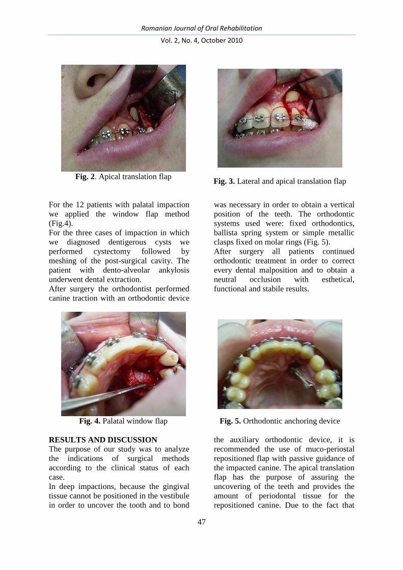
Romanian Journal of Oral Rehabilitation
Vol. 2, No. 4, October 2010
47
Fig. 2. Apical translation flap
Fig. 3. Lateral and apical translation flap
For the 12 patients with palatal impaction
we applied the window flap method
(Fig.4).
For the three cases of impaction in which
we diagnosed dentigerous cysts we
performed cystectomy followed by
meshing of the post-surgical cavity. The
patient with dento-alveolar ankylosis
underwent dental extraction.
After surgery the orthodontist performed
canine traction with an orthodontic device
was necessary in order to obtain a vertical
position of the teeth. The orthodontic
systems used were: fixed orthodontics,
ballista spring system or simple metallic
clasps fixed on molar rings (Fig. 5).
After surgery all patients continued
orthodontic treatment in order to correct
every dental malposition and to obtain a
neutral occlusion with esthetical,
functional and stabile results.
Fig. 4. Palatal window flap
Fig. 5. Orthodontic anchoring device
RESULTS AND DISCUSSION
The purpose of our study was to analyze
the indications of surgical methods
according to the clinical status of each
case.
In deep impactions, because the gingival
tissue cannot be positioned in the vestibule
in order to uncover the tooth and to bond
the auxiliary orthodontic device, it is
recommended the use of muco-periostal
repositioned flap with passive guidance of
the impacted canine. The apical translation
flap has the purpose of assuring the
uncovering of the teeth and provides the
amount of periodontal tissue for the
repositioned canine. Due to the fact that

Romanian Journal of Oral Rehabilitation
Vol. 2, No. 4, October 2010
48
the lower margin of the flap is positioned
in direct contact with the tooth, this
method contributes to periodontal
restoration.
The window flap used in palatal impaction
avoids extensive decollation of the
mucosa, allows the attachment of
orthodontic devices and minimizes the
trauma to the marginal periodontal tissue.
Periodontal follow-up 6 month after
surgery shows that in the cases in which
we used the repositioned flap, apical
translation flap and window flap there
were no periodontal recessions or dental
mobility which could compromise the
treatment. Conversely, in the cases in
which we used the lateral and apical
translation method and meshing, the
periodontal tissue was damaged and it
needed surgical restoration.
CONCLUSIONS
We recommend conservatory surgical
orthodontic treatment due to its role in
alveolar bone formation during the
movement of an impacted tooth, with
restoration of the periodontium with
esthetical and functional implications.
REFRENCES 1. Bado- Silviera F, Recoing J. Disengagement and traction of included canines. Rev. Chir. Maxillofac.2005
Jun.;106(3):166-70
2. Becker A, Chaushu S, Success rate and duration of orthodontic treatment for adult patients with palatally
impacted maxillary canines. Am J Orthod Dentofacial Orthop. 2003 Nov; 124(5):509-14
3. Bishara SE., Clinical menagement of impacted maxillary canines. Semin.Orthod. 1998.Jun; 4(2):87-98.
4. Kokich VG,Surgical and orthodontic management of impacted maxillary canines. Am. J Orthod. Dentofacial
Orthop. 2004 Sept;126(3):278-83

Romanian Journal of Oral Rehabilitation
Vol. 2, No. 4, October 2010
49
INCIDENCE AND PROGNOSTIC VALUE OF ORAL CANDIDIASIS IN
HIV INFECTION Cristina Popa
1, Carmen Stelea
2, Eugenia Popescu
3
1, 2 , 3 ― Gr. T.Popa‖ Iaşi University of Medicine and Pharmacy
1, 2 , 3 Department of Oral and Maxilo-Facial Surgery
Abstract: Oral candidiasis is the most frequent lesion encountered in HIV infected patients and is detected
before any other clinic symptoms of oral pathology. Its growth is a complex process of changes in the oral
cavity, and sometimes disorders involving the entire body. Its risk is increased by serious factors as follows:
autoimmune deficiencies (illness of the primary cells which acts as modulators of the immune response),
xerostomia, malignant neoplasm, different therapies (chemotherapy, antibiotic therapy, therapy with steroids) or
iron deficiency anaemia.
Key words: oral candidiasis, HIV infection, prognostic value.
INTRODUCTION
The oro-maxilo-cervico-facial territories
are areas where manifestations of HIV
infection are most often observed. The vast
majority of infected patients present at
least one oral lesion, which sometimes can
be the only expression of the disease. In
this context, it is very important for
dentists to be well informed of the oral
signs of HIV infection. Affection of the
oral mucosa is constant, appears early in
the disease and it is a clinical indicator of
the prognosis, which usually indicates an
ominous evolution. Oral candidiasis,
frequently observed in HIV infected
patients, appears almost always before
other clinical oral manifestations. It
represents an indicator of
imunosuppression, but it can also imply an
increase in viral load associated with
AIDS or resistance to anti-retroviral
medication. The development of
candidiasis is a complex process which is
influenced by changes that may take place
in the oral cavity; sometimes it implies
disorders involving the entire body.
Increased risk of developing oral
candidiasis is associated with several
factors including: established
imunosuppression (disease of the primary
cells implicated in modulation of the
immune response), xerostomia, malignant
tumours and therapy (chemotherapy,
antibiotherapy, steroid therapy) or iron
deficiency anaemia.
MATERIAL AND METHODS Our study covers a period of 6 years and
analyses a number of 117 patients
diagnosed with HIV infection or AIDS
associated with various oral pathology.
The study aims to identify characteristics
related to the incidence of the disease and
biological, clinical and therapeutic aspects
which could offer information towards the
overall prognosis, therapeutic response and
evolution of the disease.
Over the period of study, we followed:
-particularities of the patient group
studied, analysis of the early clinical
symptoms, the correlation to the disease
stage with reference to specific oral
symptomatollogy.
-the relation between specific clinical and
haemato-immunological particularities in
different forms and stages of the disease.
-the identification of clinical and
therapeutic factors with prognostic value
in a unitary analysis of the evolution and
response to therapy.
Patients considered eligible for the study:
- serological diagnosis (ELISA confirmed
WESTERN-BLOT) was infection with
HIV
- absence of history and/or clinical signs of
oral, oesophageal candidiasis and absence

Romanian Journal of Oral Rehabilitation
Vol. 2, No. 4, October 2010
50
of anti-fungal treatment at least 3 months
prior to present examination.
-with oral pathology encompassing HIV
infection
Patients excluded from the study:
-HIV infection without associated oral
pathology
-which were associated with other
immunological and/or haematological
pathology
-with high risk of death
Evaluation of initial diagnosis was
followed by compulsory complete
anamnesis, motives for presentation,
complete objective clinical examinations,
and biological and imagistic paraclinical
examinations. Immunological markers
(total lymphocyte and beta 2 micro-
globulin) or viral markers (HIV p24 core
antigen) were also used to monitor HIV
infection and response to antiretroviral
therapy.
The associated oral pathology was
monitored by an examination of all
patients keeping in line with strict clinical
protocols of classification and
identification of diagnostic criteria for oral
lesions based on recommendations of ―EC-
Clearinghouse on Oral Problems Related
to HIV Infection and WHO Collaborating
Centre on Oral Manifestations of the
Immunodeficiency Virus‖.
RESULTS
From the total number of patients studied
(117 HIV infected), 71 presented signs of
low grade or moderate disease (stage A
and B-table I), while 46 were classified in
final stage of HIV infection (AIDS) (fig.
1)
Table I
Clinical category Nr. of cases
A1 5
A2 0
A3 10
B1 37
B2 5
B3 14
C1 23
C2 16
C3 7
A number of 18 patients were diagnosed as HIV positive on presentation in the out-patient
department for various oral affections (5 of whom were in final stage AIDS) (fig.2)
Oropharyngian candidiasis was the most frequent lesion found in HIV infected patients. The
diagnosis (isolated or recurrent) was made in a number of 88 patients (75,2%). Systemic
candidiasis (Candidemia) and fungemia was found in one single patient only.
In approximately 50% of the cases (over a median of 3-5 years) one or several episodes of
oral candidiasis were observed. The presence of oral fungal lesions represented the early
symptoms that lead to initial diagnosis of AIDS. Although it is not a pathognomic symptom,
oral candidiasis, especially in chronic variation, multifocality, or when associated with
esophagian candidiasis, was suggestive of AIDS diagnosis, with completion of the clinical
pattern developing in the following months.
A1 4%
A2 0%
A3 8%
B1 32% B2
4%
B3 12%
C1 20%
C2 14%
C3 6%
Fig. 1. Retrospective quantification of the patients on the first apointment

Romanian Journal of Oral Rehabilitation
Vol. 2, No. 4, October 2010
51
The following anatomic-clinical forms of
candidiasis were encountered:
erythematous, pseudomembranous,
hyperplasic and angular cheilitis. Table II
and fig. 3 present the incidence of cases
with candidiasis:
Table 1I
Incidence and forms of oral candidiasis reported in the investigated patients
Erythematous Candidiasis (atrophic) 16
Angular cheilitis 21
Pseudomembranous Candidiasis 38
Chronic hyperplasic Candidiasis 13
0
10
20
30
40
50
60
70
80
known
58
41
13
5
Fig.2. HIV infection known or detected in clinic
HIV Infection SIDA

Romanian Journal of Oral Rehabilitation
Vol. 2, No. 4, October 2010
52
Patient inclusion criteria followed CDC
1994 protocols, with the mention that once
a patient was classified as being HIV
positive, he can no longer be reclassified
in any other clinic-immunological
category, even if his clinical or
immunological status is modified.
Prevalence of oral candidiasis depended on
the clinical stage of the patient as follows:
-stage I (without immunosuppression) –
predominantly angular cheilitis registered
in 19,4% of the cases
-stage II (moderate suppression) –
predominantly erythematous candidiasis -
30,4% of the patients
-stage III (severe imunosuppression) –
predominantly pseudomembranous form -
77% patients.(fig.4)
Table III presents the relation between the
immune status and oral candidiasis in 84
patients (immunological status was
monitored over a period of +/- 3 months
after initial diagnosis was made).
Table III
Oral mucosa LyT
CD4/mmc
LyT
CD8/mmc
CD4/
CD8
Normal mucous (24 cases) 540 1100 0,50
Erythematous candidiasis (40 cases) 320 790 0,40
Pseudomembranous candidiasis (20
cases)
220 840 0,26
1) Acute erythematous candidiasis
(atrophic)
All patients presented a mild sensation of
discomfort or burning; in 20 cases it was
not reported as subjective complaints. The
lesions presented in the form of an
erythematous area (with varying colour
intensity), having a preference to appear at
the palate or dorsal part of the tongue.
Occasionally, lesion of the tongue and the
internal aspect of the lips were covered
with a fine white-creamy non-adherent
film.
Considering all these aspects the clinician
should carefully examine the oral mucosa
of the infected patient in order to diagnose
and treat different forms of oral
candidiasis, starting in the early stages
when possible. In 28 cases, erythematous
candidiasis preceded the

Romanian Journal of Oral Rehabilitation
Vol. 2, No. 4, October 2010
53
pseudomembranous form. Diagnosis was
confirmed in 15 cases, by the presence of
Candida spp in cultures with Saburaund
media; another criterion was positive
response to antimicotic treatment.
The clinical form described in literature
„median rhomboid glossitis‖ was observed
in 7 cases, as an area of soft, red
depapilation, followed by transformation
into a rusty, lobulated induration. Form
and dimensions varied, but the most
frequently encountered was an area of well
demarked, 1/1, 25 cm oval or rhomboid
shaped lesion on the posterior 1/3 and
dorsal aspect of the tongue.
2) Pseudomembranous Candidiasis
Subjectively, patients complained of dry
mouth, burning sensation, dysphagia and
hyper-salivation. Other complaints
included pain, sensation of tension in the
affected mucosa, and altered gustation.
Clinical examination showed the presence
of papules or yellowish/white plaques (of a
creamy consistency, non-adherent and
easily removed by minimal pressure),
covering the erythematous surface as an
isolated or multifocal layer. Petechial or
isolated erythematous puntiform elements
were observed occasionally after
debridgement of the white deposits. In
general, the localisation of these lesions
did not present a tropism towards a
specific area of the oropharynx (oral
mucosa, in the oropharynx, margins of the
ventral aspect of the tongue, palatine
mucosa).
Some of the lesions had a sudden debut
(acute pseudomembranous candidiasis),
usually after antibiotherapy; others had a
slow, delayed evolution (chronic
pseudomembranous candidiasis) which
was observed in the majority of HIV
positive patients.
Current diagnostic criteria include clinical
aspect and positive response to anti-fungal
therapy. Histological examination was
possible only in a limited number of cases
(35 patients). Microscopic examination,
with Gram staining, showed the presence
of fungi (pseudohypha, spores) in
pseudomembranous deposits made up
from desquamated keratinocytes, keratin,
inflammatory cells, bacteria and fibrin.
Biopsy examination revealed a hyperplasic
epithelium, inflamed, infiltrated with
polynuclear neutrophils, fungal hypha
penetrating epithelial basal cells from the
surface inwards deeply. In the lamina
propria, a lymphoplasmocytic infiltrate
was found. In specific stains - H-E, PAS
and silver impregnation, fungal hyphae
were remarked, with typical „bamboo
stick‖ aspect. From the 98, oral gavaj
before and after antimicotic treatment,
from the 35 patients, the following
biotypes of Candida were isolated:C.
albicans (66%), C. krusei (12%), C.
torulopsis glabrata (16, 5%), C. tropicalis
(4%) and Geotrichum candidum (1, 5%).
C. albicans was isolated in over 75% of
the patients after treatment with
ketoconazol. In 10 patients with recurrent
oral candidiasis, several biotypes were
isolated.
There was no correlation observed
between the different biotypes of C.
albicans and the clinical picture of the oral
lesions, stages of HIV infection or the
number of CD4 cells. In reference to the
different biotypes identified between
different episodes, a conclusion could not
be drawn as to the reappearance of the
lesion as an effect of exogenous
reinfection or relapse (through
modification of the same stem).
Data obtained from literature reports the
last possibility without having justified the
pathogenic mechanism; alternation of the
different biotypes could be in fact a
different phenotype expression of the same
fungal genotype. These phenotype
variations could possibly be due to
modifications in the local immune
response, physiochemical variations
induced by therapy or even ecological oral
variations. The differential diagnosis was
made with all possible white lesions of the
oral mucosa.

Romanian Journal of Oral Rehabilitation
Vol. 2, No. 4, October 2010
54
3) Chronic Hyperplasic Candidiasis
Also named Candida leukolpakia,it is the
rarest form and also the most
controversial. Some authors consider it as
being a candidiasis superimposed over a
pre-existing leucoplasic plaque, but it has
been demonstrated that a fungus can on its
own induce hyperplasic lesions at the level
of the oral mucosa.
It was represented in 5 patients under the
form of a plaque with variable thickness,
an irregular aspect, surrounded by an
erythematous area, situated on the dorsal
surface of the tongue or on the superior or
inferior labial mucosa. Their removal by
debridgement was not possible, persisting
up to 1-2 years. In 3 patients this was
associated with angular cheilitis.In 12
patients, multiple Candida lesions were
discovered as being chronic multifocal
candidiasis.In one case, the leucoplasic
area was in intimate contact with zones of
red colouring, constituting spotted
leucoplasia, a situation where it was
clinically differentiated from
erythroleucoplasia, and which, from the
microscopic point of view, presented
frequent displasic lesions. The diagnosis
was confirmed microscopically,
demonstrating fungal hypha infiltrating
epithelial hyperplasia and also by
therapeutic tests- complete resolution
under antifungal treatment.
4) Angular cheilitis
Subjective symptoms frequently
encountered were sensation of dryness and
burning at the level of the lips, associated
with impossibility to consume hot or
spiced foodstuff.
It has been described under the form of an
erythematous area, fissured and covered
by crusts, situated at the level of the labial
commisures. The surrounding tissue is
sheared. The majority of the patients have
bilateral lesions, training discomfort and
pain on opening the mouth, thus limiting
normal oral function.Diagnosis is
confirmed by presence of fungus (hypha
and blastosporea) from oral prelevations
and favourable results to antifungal
therapy.
In the study group were isolated C.
albicans (20% of cases), or associated with
Staphylococcus aureus (60% of cases),
while the rest 20% had just
Staphylococcus aureus. Pathogenesis of
the affection is not sufficiently known and
is probably due to the frequent damping of
the labial mucosa (due to xerostomia)
which permits the micro-organism access
to superficial plans of the labial
epithelium, accompanied by their
desquamation.
In the majority of the patients, the process
of localized inflammation at the labial
commisures was associated with atrophic
or membranous candidiasis, with different
localizations.
In the absence of suggestive lesions, and in
conditions of suspected candidiasis, the
use of quantitative culture from undiluted
saliva or from oral gavaj was implemented
DISCUSSIONS
Oral Candidiasis is one of the clinical
indicators of the development and
progression of infection with the human
immunodeficiency virus. The development
of this pathology, without a local cause
(xerostomia, antibiotherapy,
corticotherapy) has to suggest an HIV
infection. Our research has proven a close
correlation between oral candidiasis after
T lymphocyte depletion, with the report of
CD4/CD8 and with laboratory and clinical
markers of HIV progression.The study has
proven that the presence of oral lesions has
been in close concordance with the value
of biological test results (absolute number
of lymphocytes, beta 2 microglobulin,
IgA). This correlation (even in the absence
of determining CD4 lymphocytes) has
imposed an attitude of prophylaxis of HIV
infection in asymptomatic patients, with
modified biological tests only.Atrophic
Candidiasis precedes the
pseudomembranous form in the case of
HIV infection and appears early in the
Romanian Journal of Oral Rehabilitation
Vol. 2, No. 4, October 2010
55
disease. No significant statistical
correlations were found between the
presence of atrophic candidiasis and the
low CD4 count (p=0, 68) or AIDS (p=0,
81). The pseudomembranous form is most
frequently encountered in patients with
AIDS and had a significant statistical
association with CD4 lymphocyte count
under 200 cells/mm3, appearing frequently
in the final stages B3, C1, 2, 3 of HIV
infection. Angular cheilitis constitutes a
candidic associated manifestation of HIV
infection which, in our study, demonstrates
a severe immunodeficiency.Keeping in
mind the possibility of an asymptomatic
evolution of candidiasis, the clinician
should carefully examine the oral mucosa
of the infected patient and thus be able to
diagnose and treat the different forms of
candidiasis in its early stages. Patients
without oral candidiasis have had a better
prognosis as compared to those with
clinical forms of candidiasis. Thus we
have established the prognostic value of
oral manifestations of candidiasis, as a
marker of evolution in AIDS. Esophageal
candidiasis appeared later, in a case with
advanced immune deficiency representing
a criterion for diagnosing AIDS (over 20%
of cases).There were no observations made
between AIDS related deaths for different
types of oral candidiasis.
CONCLUSIONS
Oral manifestations should be considered
as clinical signs of HIV infection and as an
objective indicator of disease progression.
In this context, oral candidiasis is one of
the early clinical indicators of infection
and its severity. The pseudomembranous
form and the atrophic form of candidiasis
are markers of HIV, highly predictive for
the further development of AIDS.
Oropharyngian candidiasis has been
associated with other markers of
prognosis: reduction of CD4 lymphocytes,
therapy with anti-retroviral agents and
evolution of the disease AIDS.
Furthermore, for patients with AIDS, oral
candidiasis has been a pre-monitor marker
for esophageal candidiasis.
The ability to recognise the predictors of
HIV progression may step up the decision
in administration of early protection for
HIV infected individuals. Dentists can
play an important role in the detection of
symptoms associated with HIV/AIDS and
can considerably improve the quality of
life for these patients.
REFERENCES 1. Barr C.E., Lopez M.R., Rua-Dobles A., Heller L.K. et al. (1992): HIV associated oral lesions; immunologic,
virulogic and salivary parameters, J. Oral Pathol. Med., 21: 295-298.
2. Cousteau C., Bigot-Massoni D., Vittecoq D. et al (1987): Lesions stomatologique observee au cours de
l'infection per le virus HIV, Revue de Stomatologie et de Chirurgie Maxillo-faciale, 88: 85-92.
3. Dios Diz P., Ocampo A., Miralles C. et. all (1999); Frequency of oropharyngeal candidiasis in HIV-infected
patients on protease inhibitor therapy, Oral Surg. Oral Med. Oral Pathol,87(4):437-440
4. Dodd Caroline, Greenspan D, Katz M. et al. (1991): Oral candidiasis in HIV infection: pseudomembranous
and erythematous candidiasis show similar rates of progression to AIDS, AIDS, 5: 1339-1343.
5. Glick M., Muzyka B., Lurie D., Salkin L. (1994): Oral manifestation associated with HIV-related diseases as
markers for immune suppression in AIDS, Oral Surg. Oral Med. Oral Pathol., 77: 344-349

Romanian Journal of Oral Rehabilitation
Vol. 2, No. 4, October 2010
56
AESTHETIC GOALS IN PORCELAIN FUSED TO METAL
REHABILITATION Monica Silvia Tatarciuc, Anca Mihaela Viţalariu, Diana Diaconu, St.Panaite
University of Medicine and Pharmacy ‖Gr.T.Popa‖of Iasi, Romania
Abstract:
Purpose. The ultimate goal for ceramic fused to metal restoration is the creation of a perfect aesthetic
integration of the restoration with healthy tissues.
Material and method. The study included a group of twenty-one patients. Sixteen of them were partial
edentulous patients and five presented dental lesions or chromatic modifications of the dental tissues. The
treatment plan was represented by metal-ceramic crowns and bridges. The ceramic system used was Vintage
Halo (Shofu).
Results. According to the patient's age, to the topography of the missing teeth or to the colour modification, we
especially surveyed the aesthetic rehabilitation creating the specific morphological elements, in order to realise
an individual natural aspect with a similar behaviour for the reflection of the light as the dental enamel.
Conclusions. The current popularity of metal-ceramic restorations is primarily related to their predictable
strength achieved with reasonable aesthetics. The main parameters that are responsible for a high-quality result
are represented by: non-invasive, reversible procedures, conservative preparations, individual function offered
by an individualized reconstruction, gnathological concepts into the occlusal rehabilitation, biocompatible
materials, longevity of the reconstruction and comfort for the patient.
Key words: aesthetic rehabilitation, shade determination, metal-ceramic restorations
INTRODUCTION
The aesthetic rehabilitation represents an
important clinical goal of the prosthetic
therapy. The aesthetic reasons often are the
main factor for the treatment demands. To
get a perfectly integrated rehabilitation, the
dentist and the dental technician have to
respect the specific conditions of the
clinical case, to realize an appropriate
design, to make a gnathologycal
preparation of the abutment teeth, to
preserve and to rehabilitate the occlusion
and to use biocompatible dental materials,
corresponding from the aesthetic point of
view (Patrascu 2002, Dailey 2003).
MATERIAL AND METHOD
The study included a group of twenty-one
patients. Sixteen of them were partial
edentulous patients and five presented
dental lesions or chromatic modifications
of the dental tissues.
The treatment plan was represented by
metal-ceramic crowns and bridges. The
amount of tooth reduction should never be
based on the existing tooth surface, but
rather on the final volume of the
restoration. The final preparation should
not have undercuts, so that a precise
impression can be taken and the
restoration can be correctly placed during
cementation. A precision impression with
polyether or a polyvinyl-siloxane material
was taken.
The spatial orientation and architectural
dimensions of a wax-up were used to pre-
design and validate the intended
preparations for the teeth involved. The
properly made diagnostic provisional
provides a means of transferring the three-
dimensional changes-planned with the
patient through the aesthetic and functional
analysis and captured in the diagnostic
wax-up into the patient‘s mouth. After the
wax pattern realisation, the metallic
framework was cast from a non-precious
chrome-cobalt dental alloy (Remanium
CS/Dentaurum/Germany containing Ni
61%, Cr 26%, Si 5% Mo11%, S-Fe-Co-Al
lower than 1%), selected for its strength,
rigidity and high flexural strength. To
enhance the link between metallic

Romanian Journal of Oral Rehabilitation
Vol. 2, No. 4, October 2010
57
infrastructure and the aesthetic component,
the framework was sandblasted with 50μm
Al2O3.
The opaque layer was applied with the
spray-on technique according to the
manufacturer‘s directions. The layer
thickness must be equal to 0,2mm and it
will be burned at 970-980°C, during 6
minutes, in vacuum. The main advantages
of this technique are represented by a
shorter working time, low cost and a
homogenous, thin opaque layer. After the
application of the opaque layer, the
porcelain element was realized using the
Vintage Halo Kit.
Achieving a shade match with a ceramic
restoration continues to be problematic for
the restorative dentist, even with increased
knowledge of colour science.
Shade determination and communication
are made difficult and demanding because
of the limitations imposed by conventional
manufactured shade guides. Conventional
guides do not cover the shade range of
natural teeth, are inconsistent and do not
correspond to the materials or thickness
used in to the actual restorations. Many
concepts attempt to improve the colour
communication, however, and various
colour standards are available from market
leaders such as Vita Classic, Vita
3DMaster, Chromascop and VITA
Easyshade (fig.1)
Fig.1. Classic and modern shade system determination
The standard for visual shade
determination was established by Sproull
as a result of his investigations:
- shade guides present a different
stratification than do natural teeth
- the material of which shade guides are
made does not correspond to the
restorative material. This leads to
metamerism when reproducing colour
- there is no logical organization of the
hues of natural teeth in the current shade
guides
The ability to visualize the hue, chroma
and value of an unfired ceramic element
allows the clinician more precise control
of shade determination, communication
and realization.
The corresponding build up during the
ceramic layering procedure requires
appropriately colour-intensive ceramic
powders that do not lose their effect when
covered later with enamel and transparent
ceramics. The incisal and transparent
powders have to avoid a high grey value.
Final contouring is accomplished with
diamond rotary instruments and the
restoration is glazed in a furnace. The low
backing temperature of the opaque dentin
and cervical powders provides a
particularly periodontium-friendly,
homogenous surface. Areas that will be in
contact with the opposing natural dentition
and by–passing areas between lobes are
controlled by polishing with rubber
wheels.
The main parameters that are responsible
for a high-quality result are represented
by: non-invasive, reversible procedures,
conservative preparations, individual
function offered by an individualized
reconstruction, gnathological concepts into
the occlusal rehabilitation, biocompatible

Romanian Journal of Oral Rehabilitation
Vol. 2, No. 4, October 2010
58
materials, longevity of the reconstruction
and comfort for the patient.
RESULTS AND DISCUSSIONS
The transition from the preoperative
condition to an ideal and acceptable
restorative outcome is complex. The active
participation of two partners - the clinician
and the patient, inevitably compounds
subjectivity by requiring mutual agreement
on the nature of the aesthetic problem and
the approach to treating it.
According to the patient's age, to the
topography of the missing teeth or to the
colour modification, we especially surveyed
the aesthetic rehabilitation creating the
specific morphological elements, in order to
realise an individual natural aspect with a
similar behaviour for the reflection of the
light as the dental enamel.
A particular aspect is looking to the
embrasure spaces adjacent to the abutment
teeth, that should be open to allow room
for interproximal tissue and access for oral
hygiene [Kataoke] (fig.2). Conversely, the
embrasure space between two adjacent
elements of the pontic should be close
enough to reduce food and plaque
accumulation as long as it is aesthetically
feasible.
Fig.2. Cervical embrasures should be open to allow space for interdental papilla
The aesthetic rehabilitation with porcelain
fused to metal crowns and bridges is
conditioned by the natural aspect
rehabilitation that depends on the
perception, a concept that is not strictly
defined, and that is conditioned by the
internal dynamic of the interaction
between the fluorescence and opalescence,
by the conditions of illumination and the
position of the observer (Dancy 2003,
Gebhardt 2003). When we realize a
ceramic fused to metal rehabilitation we
have to survey the specific colour and the
translucency of the ceramic layers
according to the shape and relief, in order
to get an optimal equilibrated prosthetic
appliance with excellent aesthetics (Gurel
2003, Adolfi 2003). The focused light
conduction of the ceramic layers that
belong to the Vintage Halo system forms
the basis for colour interplay of an
aesthetic, high–value prosthetic
rehabilitation. The optimal distribution of
impacting light from the crown to the
surrounding tissues assures the luminosity
and vitality of the prosthetic appliance
(Rufenacht 2000) The layering technique
make possible to achieve the desired shade
by additive applications of the powders.
The opaque dentin and the dentin powders
are provided to match the relevant shade
tone (fig.3).
The translucence of the dentin powders
promotes the colour effect from deeper
layers as in the natural teeth. Under
various spectra of light, natural tooth
surfaces show a broad range of colours
from pure blue to diffuse red-brown tones.
The strong guiding effect of light becomes
visible at the incisal edges and the enamel
of natural teeth. To get a suitable
strengthening depth effect on the cervical
and proximal regions translucent cervical
powders have to be used (fig.4).

Romanian Journal of Oral Rehabilitation
Vol. 2, No. 4, October 2010
59
Fig.3. Application of the dentin powder Fig.4. Final aspect of the aesthetic restoration
The use of pressed-to–metal technology
for fabrication of conventional porcelain
margins required a material of sufficient
opacity to hide the opaque substructure,
yet translucent enough to blend with the
tooth margins. Sometimes a situation of
diametrically opposing needs is created. If
the restoration was pressed out of a
material that was translucent enough to
fulfill the marginal blending requirements,
there was not enough opacity to hide the
metal framework, and the opaque layer
reflected through. On the other hand, if a
material of sufficient opacity is used a
slight lack of illumination it will be present
at the margin (Muia 1994). An important
evolutionary step consisted in the
reduction of the amount of metal coverage
located in the facial area of the restoration
and the extension of the porcelain margins.
This eliminates the need to hide the
opaqued metal and allowed the use of
more translucent pressing materials. The
result will consists in a greatly improved
gingival illumination, an increased vitality
of the restored tooth and a better natural
appearance (Ubassy 1994).
CONCLUSIONS
Aesthetic and functional parameters that
have been identified during diagnosis may
be verified, tested and adapted as needed
in the definitive restoration. The
restoration of the anterior arch is a
continuous challenge between dentist and
nature, the great potential of the new
ceramics being very satisfactory.
The ultimate goal for ceramic fused to
metal restoration is the creation of a
perfect aesthetic integration of the
restoration with healthy tissues.
Fabricating a restoration that is in harmony
with the remaining teeth and that express
the coronal morphology and natural
aspect, requires an understanding of the
necessity of achieving this harmony with
the adjacent teeth. Metal-ceramic bridges
are suitable for long-span situation and can
be used when higher stress resistance is
required. However, their fabrication
requires more tooth reduction and it may
be more difficult to achieve a natural-
appearing restoration. Replacing metal
margins with porcelain ones greatly
improves gingival illumination and
enhances aesthetic results, but the amount
of metal that can be removed up the axial
wall of the prepared tooth is limited to the
amount of powdered ceramic that can be
accurately lifted from the die during the
fabrication process.
The evolution of the patient‘s aesthetic
expectations created new type of
restoration that selectively uses metal
where needed for strength and that
eliminates it from the areas where it
compromises aesthetics (metal
encapsulated pressed-ceramic bridges), so
that a large amount of clinical situations
can be treated with high aesthetic results.
The numerous choices increase the
dentist‘s responsibilities, as it is not only
the diagnosis and treatment planning that
is important but also the communication
between the members of the team involved
as well as their performance.

Romanian Journal of Oral Rehabilitation
Vol. 2, No. 4, October 2010
60
REFERENCES 1. Adolfi D, Natural aesthetics, Ed. Quintessence, Chicago 2003
2. Dailey B, S. Katabi, Restauration des incisives maxillaires, Les cahiers de prothese,2003,127, 27-32
3. Dancy W, Color measurements as quality criteria for clinical shade matching of porcelain crowns, J. Esthet
Restor Dent, 2003, 15, 114-122
4. Gebhardt W, A comprehensive approach for restoring esthetics and function in fixed prosthodontics,
QDT,2003, 26,21-47
5. Gurel G, The science and art of porcelain laminate veneers, QDT, 2003, 26, 99-111
6. Kataoka S, Nishimura y, Nature‘s morphology, Ed. Quintessence, Chicago 2003
7. Muia P, Esthetic restorations: Improved dentist-laboratory Communication, Ed.Quintessence, Chicago 1994
8. Patrascu I, Materiale dentare, Ed. Horanda Press, Bucuresti, 2002
9. Rufenacht C, Principles of esthetic integration, Ed.Quintessence, Chicago 2000
10. Ubassy G, Shape and color: The key to successful ceramic restorations, Ed. Quintessence, Chicago 1994

Romanian Journal of Oral Rehabilitation
Vol. 2, No. 4, October 2010
61