clinical investigation plan safeboosc - rigshospitalet · pdf fileclinical investigation plan...
TRANSCRIPT

SafeBoosC CIP– Phase II Version: 3.0 – 16.03.2012 Reference number: SB010512
Page 1 of 67
Clinical Investigation Plan
SafeBoosC
Safeguarding the brain of our smallest children
– an investigator-initiated randomised, blinded, multinational, phase II
feasibility clinical trial on near-infrared spectroscopy monitoring
combined with defined treatment guidelines versus
standard monitoring and treatment as usual in premature infants
CIP number: SB010512
Trial phase Phase II
ClinicalTrials.gov: xxxx
CTU number: SafeBoosC-DP-202
CIP date (version): 16.03.12 (Version 3.0)
Sponsor: Region Hovedstaden Department of Neonatology 5024, Rigshospitalet, Blegdamsvej 9, 2100 Copenhagen Ø, Denmark Phone: +45 3545 1326, Fax: +45 3545 5025 Email: [email protected]
Study site: Multicentre; international

SafeBoosC CIP – Phase II Version: 3.0 - 16.03.2012 Reference number: SB010512
Page 2 of 67
Summary
Background
25,000 infants are born extremely preterm every year in Europe. This group of infants carries a
high risk of death and subsequent cerebral impairment for the infant, especially in the first 72
hours of life. Mortality is about 20%, and about 25% of survivors live with either cerebral palsy
or low intelligence quotient. Preventative measures are keys to reducing mortality and morbidity
in this population. There is evidence that the cerebral oxygenation time spent out of range (time
with hypoxia or hyperoxia) is associated with poor outcome in infants. Near-infrared
spectroscopy (NIRS) has been used to monitor tissue oxygenation since the mid-1980s, and
quantification of oxygenation (rStO2) in a percentage from 0 to 100% has been possible for 10
years. From almost 400 preterm infants normal ranges of rStO2 has been determined to be from
55% to 85%. Still, there are no clinical trials and thus no solid evidence of the clinical utility of
NIRS in preterm infants. Thus, research on the benefits and harms of cerebral monitoring using
NIRS as a part of clinical management of premature infants is much needed.
Objectives
The primary objective of the SafeBoosC trial is to examine if it is possible to stabilise the
cerebral oxygenation of extremely preterm infants during the first 72 hours of life through the
application of cerebral NIRS oximetry and implementation of an rStO2-specific clinical treatment
guideline. We hypothesise that by using the specified treatment guideline to respond to cerebral
monitoring readings outside the target range, we would reduce the burden of hypo- and
hyperoxia and consequently reduce brain injury.
Trial design
This is an investigator-initiated randomised, blinded, multinational, phase II feasibility clinical
trial involving preterm infants from 12 European countries.
Inclusion criteria
The inclusion criteria are: neonates born more than 12 weeks preterm (gestational age up to 27
weeks and 6 days); decision to conduct full life support; parental informed consent; and cerebral
NIRS oximeter placed within 3 hours after birth.
Sample size
With a 50% reduction of the area outside the normal range of oxygenation in %hours in the
experimental group compared to the control group as the minimal clinically significant
difference, a standard deviation of the area outside the normal range of 83.2 %hours, a type I
error (alpha) of 5%, and a type II error of 0.05 (power of 95%) inclusion of 75 preterm infants in
the experimental group and 75 preterm infants in the control group is required..
Intervention
The premature infants will be randomised into one of two groups (experimental or control).
Common is that both groups will have a cerebral oximeter monitoring device placed within three
hours after birth. In the experimental group, the cerebral oxygenation reading is visible, and the

SafeBoosC CIP – Phase II Version: 3.0 - 16.03.2012 Reference number: SB010512
Page 3 of 67
infant will be treated accordingly using a defined treatment guideline. In the control group, the
cerebral oxygenation reading is NOT visible, and the infant will be treated as usual.
Trial duration
Monitoring by cerebral oximeter will be started as soon as possible and within 3 hours after birth
and the intervention will last for 72 hours. Thereafter, each neonate will be followed up at term
date (approximately three months after birth) and at 24 months after term date.
Outcome measures
The primary outcome is the burden of hypo- and hyperoxia in %hours during the first 72 hours
after birth. The secondary outcomes are brain activity on an amplitude-integrated
electroencephalogram (aEEG), blood biomarkers (brain fatty acid binding protein (BFABP),
neuroketal, and S100β), serious adverse reactions (SARs), severe brain injury, and all cause
mortality at term date (approximately three months after birth). The exploratory outcomes are
burden of hypoxia, burden of hyperoxia, neonatal morbidities, brain injury score on magnetic
resonance imaging (MRI), number of therapies implemented during the intervention,
physiological variables (mean blood pressure (BP), pulse oximeter oxygen saturation (SpO2),
and partial pressure of carbon dioxide (pCO2)), and psychomotor impairment according to
neurodevelopmental scales at 24 months after term date.
Safety
Predefined serious adverse reactions, and suspected unexpected serious adverse reactions
(SUSARs) will be recorded and reported to the appropriate competent authorities and ethics
committees.
Ethical considerations
The approval from the relevant ethics committees will be sought. Parental informed consent will
be obtained prior to randomisation. The trial will be conducted in compliance with the guidelines
of the Declaration of Helsinki in its latest form and the International Conference on
Harmonisation good clinical practice guidelines (ICH GCP). Procedures will be established to
prevent and/or minimise risk of complication for participants, such as complications related to
the device and the treatment guideline includes only interventions that are commonly used
during intensive care in this population.

SafeBoosC CIP – Phase II Version: 3.0 - 16.03.2012 Reference number: SB010512
Page 4 of 67
SafeBoosC trial flow chart
Assess eligibility
Exclusion criteria
Number of participants: Followed-up completely (n=?)
Lost to follow-up (n=?, reasons) Discontinued intervention (n=?,
reasons)
Experimental group NIRS oximeter reading visible
+ Clinical treatment according to guideline based on oximeter
reading
Control group
NIRS oximeter reading NOT visible +
Treatment as usual.
Final analysis (according to ITT) Excluded from analysis (n=?,
reasons)
A L L O C A T I O N
FOL L O W - UP / A N A L Y S I S
Randomisation
E N R O L L M E N T
SafeBoosC-Phase II trial
Inclusion criteria
Obtain informed consent
Intervention time: 72 hours
1st follow-up
(at term date)
2nd
follow-up (24 months after term date)
N=150
n=75 n=75
Source: adapted from the CONSORT Statement, 2010

SafeBoosC CIP – Phase II Version: 3.0 - 16.03.2012 Reference number: SB010512
Page 5 of 67
Participating centres
Sponsor-investigator
Region Hovedstaden Department of Neonatology 5024, Rigshospitalet, Blegdamsvej 9, 2100 Copenhagen Ø, Denmark Phone: +45 3545 1326, Fax: +45 3545 5025 Email: [email protected] Gorm Greisen
DK
National coordinators
Medical University of Graz Gerhard Pichler
AT
Katholieke Universiteit Leuven, Gunnar Naulaers
BE
University Hospital Zurich Cornelia Hagmann
CH
Vest Children's Hospital, Datteln Claudia Roll
DE
University of Copenhagen, Gorm Greisen
DK
La Paz University Hospital (HULP) Adelina Pellicer
ES
Hospices Civils de Lyon, Service de Neonatologie, Femme-mere-enfant Hospital Olivier Claris
FR
University College Cork Gene Dempsey
IE
Università di Milano Monica Fumagalli
IT
Universitair Medisch Centrum Utrecht Frank van Bel
NL
Uppsala Universitet, Lena Hellström-Westas
SE
Cambridge University Hospitals NHS Foundation Trust Topun Austin
UK

SafeBoosC CIP – Phase II Version: 3.0 - 16.03.2012 Reference number: SB010512
Page 6 of 67
Steering Committee Members Gerhard Pichler Department of Pediatrics, Medical University of Graz Auenbruggerplatz 30, Graz, Austria Phone: +0316 38580520 E-mail: [email protected]
AT Olivier Claris Hospices Civils de Lyon, Service de Neonatologie, Femme-mere-enfant Hospital, Department of Neonatology Hospital Femme Mere Enfants, 59 Boulvard Pinel 69500 Bron France Phone: +33 (0) 427855283 E-mail: [email protected]
FR
Gunnar Naulaers Katholieke Universiteit Leuven Herestraat 49, 3000 Leuven, Belgium Phone: tel. +321843213 of +32 16 343211, fax +32 16 343209 E-mail: [email protected]
BE Gene Dempsey University College Cork University College Cork, College Road, Cork, Ireland Phone: +353 21 492 0525 E-mail: [email protected]
IE
Cornelia Hagmann Clinic of Neonatology University of Zurich 8091 Zurich, Switzerland Phone: +41 44 255 53 98 E-mail: [email protected]
CH Monica Fumagalli Department of Maternal and Pediatric Sciences, Università di Milano Fondazione IRCCS Ca‟ Granda Ospedale Maggiore Policlinico Milan – Università degli Studi di Milano, Via della Commenda 12, IT-20122 Milan, Italy Phone: +39 02 55 032 951 E-mail: [email protected]
IT
Martin Wolf Biomedical Optics Research Laboratory Clinic of Neonatology, University Hospital Zurich Frauenklinikstr 10, 8091 Zurich, Switzerland Phone: +41 44 255 5346, Fax: +41 44 255 4442 E-mail: [email protected]
CH Frank van Bel Universitair Medisch Centrum Utrecht Wilhelmina Children‟s Hospital KE 04.123.1 PO Box 85090, 3508 AB Utrecht, The Netherlands Phone: +31-88-7554545 E-mail: [email protected]
NL
Claudia Roll Department of Neonatology and Paediatric Intensive Care Vest Children‟s Hospital Datteln University Witten-Herdecke Dr.-Friedrich-Steiner-Str. 5 45711 Datteln, Germany Phone: +49 (0)2363-975 852 E-mail: [email protected]
DE Wim van Oeveren Haemoscan B.V. Stavangerweg 23 Groningen 9723 JC The Netherlands Phone : +31(0)646181604 Fax nr +31 (0)847269747 E-mail: [email protected]
NL
Gorm Greisen Department of Neonatology 5024, Rigshospitalet, Blegdamsvej 9, 2100 Copenhagen Ø, Denmark Phone: +45 3545 1326, E-mail: [email protected]
DK Lena Hellström-Westas Dept of Women's and Children's Health Uppsala Universitet,Dept of Neonatology, University Hospital, 751 85 Uppsala, Sweden Phone: +46 18 6114877, mobile: +46 73 3916330 E-mail: [email protected]
SE
Christian Gluud Copenhagen Trial Unit Centre for Clinical Intervention Research Department 3344, Rigshospitalet Blegdamsvej 9, DK-2100 Copenhagen Phone: +45 3545 7171, Fax: +45 3545 7101 E-mail: [email protected]
DK Topun Austin Rosie Maternity Hospital, Cambridge University Hospitals NHS Foundation Trust Hills Road Cambridge, CB2 0SW, United kingdom Phone: E-mail: [email protected]
UK
Adelina Pellicer La Paz University Hospital (HULP) Department of Neonatology, La Paz University Hospital, Paseo de la Castellana 261, 28046 Madrid, Spain Phone +34 917277416, Fax: +34 917277362 e-mail: [email protected]
ES

SafeBoosC CIP– Phase II Version: 3.0 – 16.03.2012 Reference number: SB010512
Page 7 of 67
Executive Committee Members
Martin Wolf Biomedical Optics Research Laboratory Clinic of Neonatology University of Zurich 8091 Zurich, Switzerland Phone: +41 44 255 5346, Fax: +41 44 255 4442 E-mail: [email protected]
CH Adelina Pellicer La Paz University hospital (HULP) Department of Neonatology, La Paz University Hospital, Paseo de la Castellana 261, 28046 Madrid, Spain Phone +34 917277416, Fax: +34 917277362 e-mail: [email protected]
ES
Gorm Greisen Department of Neonatology 5024, Rigshospitalet, Blegdamsvej 9, 2100 Copenhagen Ø, Denmark Phone: +45 3545 1326, Email: [email protected]
DK Frank van Bel Universitair Medisch Centrum Utrecht Wilhelmina Children‟s hospital KE 04.123.1 PO Box 85090, 3508 AB Utrecht, The Netherlands Phone: +31-88-7554545 E-mail: [email protected]
NL
Christian Gluud Copenhagen Trial Unit Centre for Clinical Intervention Research Department 3344, Rigshospitalet Blegdamsvej 9, DK-2100 Copenhagen, Denmark Phone: +45 3545 7171, Fax: +45 3545 7101 E-mail: [email protected]
DK Lena Hellström-Westas Dept of Women's and Children's Health Uppsala Universitet Dept of Neonatology, University Hospital, SE-751 85 Uppsala, Sweden Phone: +46 18 6114877, mobile: +46 73 3916330 E-mail: [email protected]
SE
Data Monitoring and Safety Committee
Jan Miletin Department of Paediatrics and Newborn Medicine, Coombe Women and Infants University Hospital, Dublin, Ireland. Phone: +353-(0)87 9819668 E-mail: [email protected]
IE
Cuno Uiterwaal Julius Centre for Health Sciences and Primary Care, University Hospital for Children and Youth 'Wilhelmina Children's Hospital', University Medical Center Utrecht, Utrecht, The Netherlands Phone: +31-(0)88-75-59369 E-mail: [email protected]
NL
Heike Rabe Trevor Mann Baby Unit, Brighton and Sussex University Hospitals NHS Trust, United Kingdom Phone +44-(0)1273-696955 ext 4195/4296, fax +44-(0)1273-664435 E-mail [email protected]
UK

SafeBoosC CIP – Phase II Version: 3.0 - 16.03.2012 Reference number: SB010512
Page 8 of 67
Clinical laboratories and other collaborators Manon Benders and Cornelia Hagmann MRI and CUS Department of Neonatology, KE 04.123.1 Universitair Medisch Centrum Utrecht Wilhelmina Children‟s Hospital KE 04.123.1 PO Box 85090, 3508 AB Utrecht, The Netherlands Phone: +31-88-7554545 E-mail: [email protected]
NL Wim van Oeveren Haemoscan B.V. Stavangerweg 23 Groningen 9723 JC The Netherlands Phone : +31(0)646181604 Fax nr +31 (0)847269747 E-mail: [email protected]
NL
Martin Wolf Oximetry Biomedical Optics Research Laboratory Clinic of Neonatology, University Hospital Zurich Frauenklinikstr 10, 8091 Zurich, Switzerland Phone: ++41 44 2555346, Fax: ++41 44 2554442 Email: [email protected]
CH Lena Westas aEEG Dept of Women's and Children's Health, Uppsala Universitet Dept of Neonatology, University Hospital, SE-751 85 Uppsala, Sweden Phone +46-18-611 00 00 or 611 48 77 Phone: +46 18 6114877, mobile: +46 73 3916330 E-mail: [email protected]
SE
Per Winkel Biostatistician Copenhagen Trial Unit Centre for Clinical Intervention Research Department 3344, Rigshospitalet Blegdamsvej 9, DK-2100 Copenhagen, Denmark Phone: +45 3545 7171, Fax: +45 3545 7101 E-mail: [email protected]
DK Christian Gluud Data Management Copenhagen Trial Unit Centre for Clinical Intervention Research Department 3344, Rigshospitalet Blegdamsvej 9, DK-2100 Copenhagen,Denmark Phone: +45 3545 7171, Fax: +45 3545 7101 E-mail: [email protected]
DK
Trial monitors The GCP-unit at the Copenhagen University Hospital Bispebjerg Hospital, Building 51, 3.sal Bispebjerg Bakke 23, DK-2400 Copenhagen NV Tel: +45 3531 3890 E-mail: [email protected], http://gcp-enhed.dk/kbh
DK TBA IT
TBA NL TBA AT
TBA BE TBA UK
TBA ES TBA DE

SafeBoosC CIP – Phase II Version: 3.0 - 16.03.2012 Reference number: SB010512
Page 9 of 67
TBA CH TBA FR
TBA SE TBA IE
SafeBoosC CIP – Phase II Version: 3.0 - 16.03.2012 Reference number: SB010512
Page 10 of 67
Table of contents
SUMMARY ................................................................................................................................................ 2
SAFEBOOSC TRIAL FLOW CHART ..................................................................................................................... 4
PARTICIPATING CENTRES .............................................................................................................................. 5
STEERING COMMITTEE MEMBERS .................................................................................................................. 6
EXECUTIVE COMMITTEE MEMBERS ................................................................................................................. 7
DATA MONITORING AND SAFETY COMMITTEE .................................................................................................. 7
CLINICAL LABORATORIES AND OTHER COLLABORATORS ........................................................................................ 8
TRIAL MONITORS ........................................................................................................................................ 8
TABLE OF CONTENTS .................................................................................................................................. 10
1. INTRODUCTION AND BACKGROUND ...................................................................................................... 15 1.1 THE POPULATION AND CONDITION ................................................................................................... 15 1.2 PATHOPHYSIOLOGY ....................................................................................................................... 15
1.2.1 THE TRANSITION FROM FOETAL TO INFANT CIRCULATION .................................................................. 15 1.2.2 CEREBRAL AUTOREGULATION ..................................................................................................... 15 1.2.3 THE VULNERABLE BRAIN ............................................................................................................ 16 1.2.4 MECHANISMS OF BRAIN DAMAGE IN PRETERM INFANTS ................................................................... 16
1.3 CURRENT CLINICAL MANAGEMENT ................................................................................................... 16 1.4 ASSESSMENT OF BRAIN INJURY AND NEURODEVELOPMENTAL DEFICIT ...................................................... 17
1.4.1 ULTRASOUND ......................................................................................................................... 17 1.4.2 AEEG .................................................................................................................................... 17 1.4.3 MRI ..................................................................................................................................... 18 1.4.4 BIOMARKERS .......................................................................................................................... 18 1.4.5 NEUROLOGICAL DEVELOPMENTAL ASSESSMENT TOOL ...................................................................... 19
1.5 CEREBRAL OXIMETRY MONITORING .................................................................................................. 19 1.5.1 NIRS DEVICES ......................................................................................................................... 20
1.6 REGIONAL OXYGENATION SATURATION IN PRETERM INFANTS ................................................................. 21 1.7 PREVIOUS TRIALS ON SIMILAR RESEARCH QUESTION ............................................................................. 21 1.8 TRIAL RATIONALE .......................................................................................................................... 22 1.9 JUSTIFICATION FOR THE DESIGN OF THE CLINICAL INVESTIGATION ............................................................ 22
1.9.1 RATIONALE FOR USE OF INVOS ADULT SOMASENSORS® AS REFERENCE STANDARD FOR MEAN VALUES IN
NEONATES IN THE STUDY .......................................................................................................................... 22 1.9.2 RATIONALE FOR THE EVALUATION OF THE INVESTIGATIONAL DEVICES .................................................. 22
2. TRIAL OBJECTIVE AND HYPOTHESIS ....................................................................................................... 23
3. TRIAL DESIGN ................................................................................................................................. 23 3.1 RANDOMISATION ......................................................................................................................... 24 3.2 TRIAL INTERVENTIONS ................................................................................................................... 24 3.3 DURATION .................................................................................................................................. 24 3.4 BLINDING .................................................................................................................................... 24
4. PARTICIPANTS ................................................................................................................................. 25 4.1 INCLUSION CRITERIA ...................................................................................................................... 25 4.2 EXCLUSION CRITERIA ..................................................................................................................... 25 4.3 PARTICIPANT DISCONTINUATION AND WITHDRAWAL ............................................................................ 25 4.4 RECRUITMENT FEASIBILITY .............................................................................................................. 26
5. INTERVENTIONS .............................................................................................................................. 26 5.1 COMMON TO BOTH GROUPS ........................................................................................................... 26 5.2 EXPERIMENTAL GROUP .................................................................................................................. 26

SafeBoosC CIP – Phase II Version: 3.0 - 16.03.2012 Reference number: SB010512
Page 11 of 67
5.3 CONTROL GROUP.......................................................................................................................... 26 5.4 CONCOMITANT MEDICATION/TREATMENT ......................................................................................... 26 5.5 MEDICAL CARE AFTER END OF CLINICAL INVESTIGATION ........................................................................ 27
6. OUTCOME MEASURES ....................................................................................................................... 27 6.1 PRIMARY .................................................................................................................................... 27 6.2 SECONDARY ................................................................................................................................ 27 6.3 EXPLORATORY .............................................................................................................................. 27 6.4 OUTCOME ASSESSMENT TOOLS ....................................................................................................... 27 6.5 OUTCOME ASSESSMENT COMMITTEE ................................................................................................ 28 6.6 COMPLIANCE WITH THE CIP ........................................................................................................... 28
7. DATA COLLECTION AND TRIAL ASSESSMENT SCHEDULE .............................................................................. 28 7.1 COLLECTION OF TRIAL DATA ............................................................................................................ 28
7.1.1 NIRS DEVICE .......................................................................................................................... 28 7.1.2 CASE RECORD FORM ................................................................................................................. 28
7.2 TRIAL ASSESSMENT SCHEDULE ......................................................................................................... 29
7.3 RESEARCH BIOBANK ..................................................................................................................... 29
8. ASSESSMENT OF SAFETY .................................................................................................................... 32 8.1 ADVERSE EVENTS AND REACTIONS .................................................................................................... 32
8.1.1 DEFINITIONS ........................................................................................................................... 32 8.1.2 CLASSIFICATION OF CAUSALITY .................................................................................................... 33 8.1.3 RECORDING AND REPORTING OF ADVERSE EVENTS AND REACTIONS AND DEVICE DEFICIENCIES .................. 33 8.1.4 JUSTIFICATION FOR RECORDING AND REPORTING PERIODICITY AND MODALITY ...................................... 35 8.1.5 TIMELINES FOR RECORDING AND REPORTING ................................................................................. 35
8.2 CEREBRAL NIRS MONITORING DEVICE .............................................................................................. 36 8.3 DATA MONITORING AND SAFETY COMMITTEE ................................................................................... 36 8.4 SUSPENSION OR PREMATURE TERMINATION OF THE CLINICAL INVESTIGATION ........................................... 36
9. ETHICAL CONSIDERATIONS ................................................................................................................. 37 9.1 INFORMED CONSENT PROCEDURE .................................................................................................... 37 9.2 RISK OF COMPLICATION FOR PARTICIPANTS ........................................................................................ 38 9.3 BENEFIT FOR PARTICIPANTS ............................................................................................................ 38
10. STATISTICAL PLAN AND DATA ANALYSIS ............................................................................................ 38 10.1 SAMPLE SIZE ESTIMATION ............................................................................................................... 38 10.2 DATA ANALYSIS AND STATISTICAL METHODS ....................................................................................... 39
10.2.1 STATISTICAL MODELS ........................................................................................................... 39 10.2.2 DEALING WITH MISSING VALUES ............................................................................................. 40 10.2.4 DEALING WITH MULTIPLICITY ................................................................................................ 41
11. DATA MANAGEMENT ................................................................................................................... 44 11.1 DATA HANDLING AND ARCHIVING .................................................................................................... 44 11.2 MEDICAL CODING ......................................................................................................................... 45
12. QUALITY ASSURANCE ................................................................................................................... 45 12.1 MONITORING .............................................................................................................................. 45 12.2 DEVICE QUALITY CONTROL .............................................................................................................. 45
13. TRIAL AND FUNDING TIMEFRAME .................................................................................................... 45
14. LEGAL ASPECTS ........................................................................................................................... 46 14.1 FINANCE ..................................................................................................................................... 46 14.2 PARTICIPANT INSURANCE ............................................................................................................... 46 14.3 PUBLICATION PLAN ....................................................................................................................... 46 14.4 STATEMENTS OF COMPLIANCE ......................................................................................................... 46
15. APPENDICES .............................................................................................................................. 48

SafeBoosC CIP – Phase II Version: 3.0 - 16.03.2012 Reference number: SB010512
Page 12 of 67
15.1 APPENDIX A: TREATMENT GUIDELINES AND JUSTIFICATIONS .................................................................. 48 15.2 APPENDIX B: PROCEDURE FOR ASSESSMENT OF CHEMICAL BIOMARKERS .................................................. 51 15.3 APPENDIX C: PROCEDURES FOR ASSESSMENT OF AEEG/EEG ................................................................ 53 15.4 APPENDIX D: PROCEDURES FOR MRI EXAMINATION ........................................................................... 54 15.5 APPENDIX E: PROCEDURE FOR ASSESSMENT OF CRANIAL ULTRASOUND ................................................... 57 15.6 APPENDIX F: PARENTAL INFORMATION ............................................................................................. 59 15.7 APPENDIX G: INFORMED CONSENT – THE SAFEBOOSC PHASE II TRIAL ..................................................... 59 15.8 APPENDIX H: PARENTAL INFORMATION - THE SAFEBOOSC TRIAL -MRI ................................................... 59 15.9 APPENDIX I: INFORMED CONSENT- THE SAFEBOOSC TRIAL -MRI ........................................................... 59 15.10 APPENDIX J: SAFEBOOSC - DATA FLOW ............................................................................................ 59
16. REFERENCES .............................................................................................................................. 61

SafeBoosC CIP – Phase II Version: 3.0 - 16.03.2012 Reference number: SB010512
Page 13 of 67
List of abbreviations
AE Adverse events
aEEG Amplitude-integrated electroencephalogram
AR Adverse reaction
ASQ Ages & Stages Questionnaires
AUC Area under the curve
BFABP Brain fatty acid binding protein
BP Blood pressure
BPD Broncopulmonary dysplasia
BSID-III Bayley‟s scale of infant development, third version
cPVL Cystic periventricular leucomalacia
CIP Clinical Investigation Plan
CRF Case record form
CTU Copenhagen Trial Unit
cUS Cerebral ultrasound
Da Dalton
EAR Expected adverse reaction
eCRF electronic case record form
EEG Electroencephalogram
EDTA Ethylenediaminetetraacetic acid
ELISA Enzymelinked immunosorbent assay
ESAR Expected serious adverse reaction
GA Gestational age
GCP Good clinical practice
H Hydrogen
Hb Haemoglobin
Hz Hertz
IB Investigator's Brochure
ICH International Conference on Harmonization
ITT Intention-to-treat
IVH Intraventricular haemorrhage
LLT Lowest level term

SafeBoosC CIP – Phase II Version: 3.0 - 16.03.2012 Reference number: SB010512
Page 14 of 67
MAR Missing at random
MedDRA Medical Dictionary for Regulatory Activities – a medical coding system
MI Multiple imputation
MMRM Mixed model with repeated measures
MNAR Missing not at random
MRI Magnetic resonance imaging
NEC Necrotizing enterocolitis
NICU Neonatal intensive care unit
NIRS Near-infrared spectroscopy
O2 Oxygen
pCO2 Partial pressure of carbon dioxide
PDA Patent ductus arteriosus
PPV Positive predictive value
PT Preferred term
PV-IVH Periventricular-intraventricular haemorrhage
PVL Periventricular leucomalacia
RCT Randomised clinical trial
ROP Retinopathy of prematurity
rStO2 Regional tissue oxygen saturation
S100β Acidic calcium binding protein found in the nervous system
SAR Serious adverse reactions
SAE Serious adverse event
SafeBoosC Safeguarding the brain of our smallest children
SD Standard deviation
SOC System organ class
SpO2 Pulse oximeter oxygen saturation
SUSAR Suspected unexpected serious adverse reaction
Term date Is defined as gestational age of 40 weeks. The term date will be approximately three months after birth for this extremely preterm population.
UAR Unexpected adverse reaction
3D-MRI Three-dimensional magnetic resonance imaging

SafeBoosC CIP – Phase II Version: 3.0 - 16.03.2012 Reference number: SB010512
Page 15 of 67
1. Introduction and background
1.1 The population and condition
Infants born more than 12 weeks preterm (extremely preterm) carry a high risk of death or long-
term cerebral impairment. Currently, mortality is about 20%, and about 25% live with either
cerebral palsy or low intelligence quotient (1). Every year 25,000 extremely preterm infants are
born in Europe. Psychomotor impairment is the major cause of reduced quality of life and
increased costs of medical care, rehabilitation, and special education in this population.
Because of the long life expectancy of children, this is an important problem.
Unfortunately, prevention of preterm birth and its consequences has not been successful; the
rate of extremely preterm birth is stable or even increasing. Although there are risk factors, such
as multiple pregnancy, and previous preterm birth, most extremely preterm births occur in
otherwise normal and healthy women.
1.2 Pathophysiology
1.2.1 The transition from foetal to infant circulation
The transition from foetal to neonatal life is a particular problem in the extremely preterm infant.
In foetal life, blood circulation includes only minimal perfusion of the lungs due to a large right-
to-left shunt through the foramen ovale to the left side of the heart and through the arterial
ductus from the pulmonary artery to the descending aorta. At birth, increased oxygenation of the
body results in a systemic vasoconstriction and increasing arterial blood pressure. This may
become a problem since the immature myocardium is intolerant to increased afterload. Also, as
the lung function improves, the resistance of the pulmonary vessels drops causing left-to-right
shunting over the arterial duct, which increases the need for left ventricular output. The
immature myocardium‟s ability to increase stroke volume is limited due to poor diastolic
function. The increased afterload and possible left-to-right shunting may cause low systemic
blood flow (2)
1.2.2 Cerebral autoregulation
Autoregulation is the ability to keep the organ blood flow constant despite fluctuations in
perfusion pressure. It is accomplished by regulation of the arterial tone so that low perfusion
pressure results in vasodilation and high perfusion results in vasoconstriction. On the systemic
level, organs such as the brain, heart, and adrenals are vital and autoregulation maintains
normal organ blood flow when systemic blood flow is low, while non-vital organs (e.g., skin and
kidney) vasoconstricts to direct the circulating blood to the vital organs.
Cerebral autoregulation has limited capacity and is thought to be particularly fragile in the
immature brain (1). Pressure passive flow is a state where the blood flow follows the blood
pressure. It is hypothesized that the potential large fluctuations in flow that this entails is a
cause of cerebral haemorrhages in premature infant due to rupture of the immature blood
vessels. It is a problem that, at the current state, it is not possible to identify the individual
threshold systemic blood pressure below which cerebral blood flow begins to fall (3).
SafeBoosC CIP – Phase II Version: 3.0 - 16.03.2012 Reference number: SB010512
Page 16 of 67
1.2.3 The vulnerable brain
All the organs are immature when an infant is born more than 12 weeks before term. The
immaturity and functional limitations of the lungs, heart, intestine, kidneys, liver, and endocrine
system all contributes to the acute problems of extremely preterm birth. The brain is special,
however, in the sense that brain damage results in death or in neuropsychological deficits such
as cerebral palsy, cognitive deficit, attention deficit disorder, and major psychiatric disorder.
These damages result in long-term consequences for children after extremely preterm birth.
The most easily identifiable type of brain damage in extremely preterm birth is periventricular-
intraventricular brain haemorrhage (PV-IVH). Its severity varies: in the mildest form, the
haemorrhage is limited to the subependymal germinal matrix – possibly with a small
intraventricular clot. The most severe form is a large periventricular haemorrhagic infarction
primarily located in the central white matter in one or both hemispheres. This predicts a high
probability of death or cerebral palsy and may result in hydrocephalus (4). Hydrocephalus
needing surgical treatment carries a poor neurodevelopmental prognosis. Periventricular
leucomalacia (PVL) is a non-haemorrhagic white matter damage. In the mildest form, the
condition is non-cystic and predicts poor psychomotor development. The most severe form of
PVL is when the condition becomes cystic (cPVL) 2-5 weeks after the damage is induced and is
a strong predictor of cerebral palsy (5).
1.2.4 Mechanisms of brain damage in preterm infants
The mechanisms of the brain damage in preterm infants are complex. Some of the mechanisms
are evoked before birth or even before the start of delivery such as a foetal inflammatory
response induced by infection ascending to the foetal membranes. Also, late effects such as
insufficient nutrition and poor growth during the first months of life may play a role.
The days after birth, however, are likely to be of particular importance. This is the period of
change from a state of low oxygen pressure ('Mount Everest in-utero') to a state of
'normoxaemia'. Moreover the circulatory adaption to birth is as described problematic in the
preterm infant. Thus fluctuations in systemic blood flow are common during the first days of life.
The following postnatal factors have been shown or are thought to be associated with brain
injury: respiratory distress syndrome (6), hypocapnia due to inadvertent hyperventilation (7), low
blood pressure (8), perturbations in arterial and venous pressure (9), and also low cerebral
blood flow (10). In addition, clinical and experimental evidence is suggesting that
hyperoxygenation is dangerous due to lack of a developed antioxidant defence system (11).
An important common mechanism for these associations is disturbance of cerebral blood flow
partly due to impaired cerebral autoregulation.
1.3 Current clinical management
Current standard of care of the extremely premature infants during their first 72 hours involves a
number of different parallel interventions:

SafeBoosC CIP – Phase II Version: 3.0 - 16.03.2012 Reference number: SB010512
Page 17 of 67
Respiratory support: continuous positive airway pressure or mechanical ventilation is
almost universal and surfactant is usually administered within the first 24 hours.
Haemodynamic support: Before diagnosing a patent ductus arteriosus (PDA),
prophylactic use of indomethacin can be used. Either indomethacin or ibuprofen can be
used for the closure of a PDA. Fluid boluses, inotropics, or vasopressors are used to treat
hypotension, although the level or targeted blood pressure is controversial (12).
Fluid balance/nutrition: Close observation of hourly and daily estimations of in- and
output, scheduled fluid administration, and blood sugar monitoring. Most infants initially
receive full parenteral nutrition and will slowly be introduced to breast milk.
Monitoring: Invasive/non-invasive blood pressure monitoring, continuous pulse oximetry,
transcutaneous partial pressure of carbon dioxide (pCO2), and electrocardiographic
monitoring with frequent measurements of arterial blood gases, electrolytes and
temperature.
Treatment of the extremely premature infants has certainly improved over the last three
decades despite great areas of unknown territory. However, the treatment of hypotension, the
optimal arterial oxygen content, the optimal pCO2 level, and many other possible interventions
are dealt with on more or less loose grounds. And while still more comprehensive monitoring is
implemented in the intensive care of premature infants, an end-organ monitoring with sufficient
high time resolution to guide evidence-based treatment interventions is lacking. Near infrared
spectroscopy has the potential to become that monitor of the brain.
1.4 Assessment of brain injury and neurodevelopmental deficit
1.4.1 Ultrasound
Cerebral ultrasound (cUS) is a standard tool for diagnosing conditions such as haemorrhage
and hypoxic-ischaemic lesions. Furthermore, signs of brain atrophy at term equivalent age are
associated with neurodevelopmental outcome in preterm infants (13). The pooled probability for
a normal neuromotor outcome of a normal ultrasound was 94% (95% confidence interval (CI)
92% to 86%) and 82% (95% CI 79% to 85%) for a normal cognitive outcome. Additionally, for
IVH Grade I-II, the probability of abnormal outcome was 9%, and for IVH Grade III 26% (95% CI
13% to 45%) (14). Parenchymal haemorrhagic infarction predicted an abnormal
neurodevelopmental outcome with an increased risk positive predictive value (PPV) of 47%
(95% CI 31% to 64%) (15). Cystic PVL was predictive of cerebral palsy with a PPV of 77% (95%
CI 59% to 89%) (15). Cerebellar haemorrhage predicted abnormal outcome with a PPV of 71%
(95% CI 42% to 90%) (16).
1.4.2 aEEG
Amplitude-integrated electroencephalography (aEEG) is a technique where the EEG signal from
bilaterally placed electrodes is recorded and amplified. The signal is then passed through an
asymmetrical band pass filter, which minimises noise and artefacts before it is displayed
bedside. The aEEG is widely used in term infants with asphyxia. It has shown good predictive
value (17) and is suitable for detection of seizure activity (18), cerebral haemorrhages (19) as
well as the effects of variation in pCO2 and blood sugar in the first day of life in extremely
preterm infants (20).

SafeBoosC CIP – Phase II Version: 3.0 - 16.03.2012 Reference number: SB010512
Page 18 of 67
1.4.3 MRI
Three-dimensional magnetic resonance imaging (3D-MRI) of the newborn brain is helpful in
identifying extra information about brain injury and maturation, which cannot always be
visualised on cranial ultrasound. Furthermore, when using advanced post-processing
techniques, it is possible to measure the volume of the total brain volume and of various brain
structures, which might be helpful in identifying non-visual brain injury or
differences. Differences in volume of different cerebral tissue classes and cortical folding and
other brain maturation measurements can be precisely measured in a quantitative way to
assess cerebral growth and maturation as well as in relation to sulci formation that has been
described to be related to cognitive outcome. Thus, 3D-MRI provides us valuable information
about brain growth/development and neurodevelopmental outcome.
1.4.4 Biomarkers
Earlier research demonstrates that several neuro-biomarkers are released into body fluids in
response to perinatal asphyxia or hypoxic-ischaemic brain injury. Some of them have also been
correlated to severity of hypoxic ischaemia and long-term neurological outcome in neonates
within the first day of life. Moreover, the course of these biological markers could correlate with
the process of hypoxic ischaemic injury and thus have a diagnostic value for brain injury in
preterm neonates with hypoxic ischaemic injury. Serum can be analysed for the rise in the
following chemical biomarkers: brain fatty acid binding protein (BFABP), neuroketal, and
S100β.
Brain fatty acid binding protein (BFABP) is a 15 kDa protein, which is specific for brain
tissue. It is released from asterocytes after mechanical damage, ischaemia and oxidative
brain damage. BFABP is determined by means of enzyme-linked immunosorbent assay
(ELISA) with BFABP specific monoclonal capture antibodies and polyclonal detection
antibody in plasma/serum as well as urine. Due to its low concentrations, 100 µl is
needed for a BFABP ELISA.
Neuroketal is formed in the brain via the neuroprostane pathway during oxidation of
docohexaenoic acid. Neuroketals rapidly adduct to lysine, and these crosslinks induce
neurodegenerative disease. Neuroketal thus contributes to the injurious effects of
oxidative pathologies in the brain. Determination of neuroketals is performed by
competitive enzyme immunoassay in 100 µl of plasma, serum or urine.
S100β is a marker for brain damage. S100 β is an acidic calcium-binding protein found in
the nervous system of vertebrates. It is a dimer of α and β subunits. As such, S100β,
which consists of two β subunits, is present in high concentration in glial and Schwann
cells. It leaks from the cerebrospinal fluid into blood after cerebral damage and is an
indicator of brain injury as well as increased permeability of the blood brain barrier. The
determination of S100β concentrations in serum is performed with an ELISA. It can be
detected in 50 µl samples of serum, heparin plasma or urine.

SafeBoosC CIP – Phase II Version: 3.0 - 16.03.2012 Reference number: SB010512
Page 19 of 67
1.4.5 Neurological developmental assessment tool
Neurodevelopmental outcome should be evaluated by standardised tests which are
psychometric measures designed to inventory an infant‟s skills against a ‟normal‟ population
within a specific assessment. The standardisation is designed to reduce measurement error by
precluding subjective interpretation of the child‟s responses. Bayley Scales of Infant
Development (BSID) is such a standardised test (21). BSID was revised and re-standardised in
1993 to form the BSID-II and in 2005 to form the BSD-III and is widely used worldwide as
outcome measure in clinical and research practice. BSID-III provides a multi-domain
assessment of children aged 1 to 42 months. It reveals five major developmental domains:
cognitive, language, motor, adaptive behaviour, and social-emotional. The scale also includes a
parent report form, the Adaptive Behaviour Questionnaires that provides scores based on
parents‟ perceptions of the child‟s level of function. In this trial, the BSID-III will be performed at
24 months after term date, using the cognitive, language, and motor domains.
The Ages & Stages Questionnaire (ASQ) is a parental survey that comprises questionnaires for
children age 4 months to 5 years. It consists of 6 questions in each of 5 developmental domains
covering motor as well as mental development. The agreement between ASQ and with
concurrent assessment by BSID-III is good and ASQ can be a substitute for the BSID-III in
cases or at centres where the BSID-III is not possible (22).
1.5 Cerebral oximetry monitoring
Near-infrared spectroscopy (NIRS) is a non-invasive technology that has been utilised to assess
the adequacy of peripheral and cerebral oxygenation in the preterm infant (23). Near-infrared
light penetrates deep into the tissue, and through spectroscopy, it is possible to monitor tissue
oxygenation. NIRS uses the relative transparency of human tissue to light in the near-infrared
region of the spectrum. The oxygen-dependent absorption of light by haemoglobin enables the
calculation of relative changes in the oxygenated and deoxygenated haemoglobin (24). The
NIRS has been used in newborns since 1985 (25) and it is particularly suitable for the neonatal
population due to their thin scalp and skull. Newer generations of oximeters provide an absolute
value of tissue oxygenation (rStO2). This is most often done by spatially resolved spectroscopy.
The assumption behind is that light propagates in a diffusional manner in a highly scattering
media such as human tissue, and that the light attenuation by scattering is constant with light
distances over 3 centimetres. The rStO2 can then be expressed as (k x O2HB)/(k x O2HB + k x
HHB), where k is the scattering component which cancels out (26). Examples of some of the
commercialised NIRS devices are INVOS® System, NIRO Series, and Casmed Foresight.
NIRS is based on the same principles as the widely used pulse oximetry, but where pulse
oximetry uses the pulsating signal and thereby selectively measures arterial blood, NIRS
measures the light attenuation of the tissue as a whole. This means that venous blood
contributes more to the attenuation than arterial blood simply because venous blood has a
greater volume. The ratio of venous:arterial contribution is generally considered to be 75:25,
although this has been found to differ between and within infants (27). It is thus not surprising
that cerebral tissue oxygenation has shown a fair correlation with the saturation in cerebral
venous blood drawn from the jugular bulb (28). The Bland-Altman limit of agreement is ± 15-
SafeBoosC CIP – Phase II Version: 3.0 - 16.03.2012 Reference number: SB010512
Page 20 of 67
20% (29,30). It has to be remembered that rStO2 is volume weighted, whereas jugular bulb
saturation is flow weighted. Good agreement cannot be expected. It could be that during
conditions where the microcirculation is compromised such as sepsis, rStO2 may give a better
picture of the cerebral oxygen balance than jugular bulb saturation (31).
1.5.1 NIRS devices
Different NIRS devices differ in absolute values and in dynamic ranges (32).There is no
„reference standard‟ for tissue oximetry. Therefore, comparison of devices can be done in a
standardised setup in the forearm of healthy adults. Reproducibility is assessed by re-siting of
the sensors during steady state and a test of dynamic range is done by arm exercise and
subsequent arterial occlusion by a cuff. Four devices have been compared in a preliminary
study:
NIRO 200 NX;
NIRO 300;
INVOS 5100c; and
OxyPrem (a prototype).
The reproducibility of the NIRO 200 NX, NIRO 300, and the INVOS 5100c was similar (within-
subject standard deviation 4.35%, 4.10% and 5.46%, respectively). In addition, these devices
showed similar absolute oxygenation (rStO2) values (63% to 70%) and similar dynamic range.
The OxyPrem showed significantly better reproducibility of 2.7%, but a somewhat lower
absolute oxygenation (rStO2) value (60%) and dynamic range only two-thirds of the other three
instruments (33) The results are comparable to earlier studies on the head regarding
reproducibility and inter-instrument differences (32,34-37)
All eligible candidate NIRS devices for use in the SafeBoosC trial will be tested by the same
procedure and should be within five percentage points in mean values and dynamic range in
comparison with the above devices, and have a reproducibility of 6% or better. NIRS devices
and sensors are non-sterile.
The SafeBoosC trial is a clinical investigation of benefits and harms of a range of different NIRS
devices in combination with a treatment guideline. The trial is not designed to assess the
performance of each NIRS device, since no reference standard is available in the preterm
population. It will however evaluate whether each NIRS device is suitable for the trial population
by careful collection of all adverse device effects.
Each participating centre is required to provide NIRS device(s) for the SafeBoosC trial. Each
country shall apply with the regulations regarding the use of their device(s) in a clinical trial
(e.g., a CE-mark that indicates use for the SafeBoosC trial population, as laid down by the
national competent authority).
The investigational NIRS device at each participating trial site is identified and described,
including instructions for installation and use, in the site-specific investigator's brochure (IB). All
material in the IB are provided by the sponsor. The investigator at each trial site is responsible

SafeBoosC CIP – Phase II Version: 3.0 - 16.03.2012 Reference number: SB010512
Page 21 of 67
for education and training in cerebral NIRS oximetry and the relevant NIRS device. The required
training before use of each device is described in the relevant IB.
1.6 Regional oxygenation saturation in preterm infants
A study conducted by McNeil et al, 2011, characterises the baseline cerebral rStO2 in 12 stable
preterm infants (gestational age (GA) 29-34 weeks) during the first weeks of life to be between
66-83% (38). In the study conducted by Zhou et al., 2009, where they enrolled a total of 223 full-
term newborns, the „normal‟ cerebral rStO2 was determined to be between 62 ± 2% and
cerebral hypoxia was defined as rStO2 less than 58% (39). Derived from data, collected over
time, from about 390 preterm babies (born at GA<32wks) during the first 3 days of life, van Bel
et al. (unpublished data) concluded the rStO2 baseline target range in this population to be 55-
85% (±2SD) with the mean value of 71%. The data was collected with the INVOS 4100/5100
with the Adult Somasenor® are constitutes the basis of the normal ranges in SafeBoosC.
1.7 Previous trials on similar research question
There is a variety of observational studies that document, that cerebral oximetry in preterm
infants gives meaningful physiological data. Wolf and Greisen systematically reviewed 36
studies in neonates that all contribute to an understanding of oxygen delivery-consumption
balance in this population (24). Based on their study, however, we are able to conclude that
evidence of the clinical benefits and harms of cerebral oximetry in preterm infants does not
exist.
We systematically searched in four medical literature databases on November 29, 2011:
Cochrane Library, MEDLINE, EMBASE, and Science Citation Index Expanded. Using the
keywords „near-infrared spectroscopy‟, „NIRS‟ „preterm‟, „infant‟, „neonatal‟ and „newborn‟, the
searches yielded a total 1, 797, 534, and 372 hits, respectively. The search found no published
randomised clinical trials (RCTs) focusing on the effect of cerebral monitoring in preterm infants
in combination of clinical treatment guidelines based on the rStO2 produced by the NIRS
oximeter.
A search on ClinicalTrials.gov (www.ClinicialTrials.gov) on November 29, 2011 using the
keyword ‟NIRS‟ and 'Near Infrared spectroscopy' on age group birth to 17 years gave 40 and 44
hits, respectively. Only one trial on preterm infants, Treatment of Hypotension of Prematurity
(TOHOP) (NCT01434251), involves cerebral NIRS oximetry as part of the trial intervention.
NIRS is part of an assessment of tissue perfusion and end-organ function. If StO2 is normal a
low blood pressure will be tolerated in the intervention group. It is thus not the benefits of NIRS
that are evaluated, but the possible benefits of allowing a low blood pressure when the tissue
perfusion is not compromised. The NIRS readings will be visible in both groups. The trial is
currently recruiting participants (Sept. 2011).
Hence, no study investigates the possible benefits and harms of cerebral NIRS oximetry out-of-
range readings as an indication to intervene in the preterm infant.

SafeBoosC CIP – Phase II Version: 3.0 - 16.03.2012 Reference number: SB010512
Page 22 of 67
The evidence of cerebral oximetry in adults is also limited. To date, there is only one published
systematic review of evidence of clinical utility of cerebral oximetry in adults during coronary
surgery concluding that, with data from 47 trials including more than 5,000 participants, the
methodological quality of the trials was low and therefore clinical benefits and harms are
uncertain (40).
1.8 Trial rationale
There is accumulating evidence that hypoxia and hyperoxia is associated with the risk of brain
injury and death in prematurely born infants. Thus, the monitoring of cerebral oxygen saturation
levels in the first hours after birth and subsequent treatment according to pre-specified
guidelines has a great potential for prevention of brain injury and maybe death. Despite this,
there are no randomised clinical trials on the benefit and harms of NIRS in preterm infants. Yet,
the technology is increasingly implemented in clinical care. To obtain evidence-based
knowledge on the benefits and harms of cerebral monitoring using NIRS as a part of clinical
management of premature infants, a large-scale RCT is needed.
1.9 Justification for the design of the clinical investigation
This clinical investigation of NIRS devices is investigator initiated and funded on public
donations. It will include several different investigational devices. The different device
manufacturers have not been involved in any part of the design or funding of the investigation.
All pre-clinical and clinical data are from published studies. Clinical evaluation of each specific
investigational device is provided in the site-specific IB.
1.9.1 Rationale for use of INVOS Adult SomaSensors® as reference standard for mean
values in neonates in the study
This clinical investigation will use the INVOS 5100c with the adult SomaSensors® as reference
standard for NIRS device eligibility, because the normal material of 390 infants that defines the
target thresholds of rStO2, 55 and 85%, comes from the group of Petra Lemmers and Frank Van
Bel using that sensor. The application of the Adult SomaSensor in this population is considered
safe as no serious adverse device effects, such as skin burns etc., were encountered during the
monitoring of these infants (41-45). Data collected with the Adult SomaSensor indicates that a
high rStO2 could possibly predict poor outcome (46). Clinical data suggests that the INVOS
5100c neonatal sensor OxyAlert™ NIRSensors gives values that are about 10 percentage
points higher that what found with the Adult SomaSensors® and NIRO 300 (32,47,48). This
means that rStO2 above 85% as defined by the adult SomaSensors® cannot be monitored with
the OxyAlert™ NIRSensors as the device cannot give values above 95%,, values that moreover
are physiologically unreasonable. As described above other device-sensor combinations are
eligible provided fulfilment of the specified criteria.
1.9.2 Rationale for the evaluation of the investigational devices
Cerebral oximetry by NIRS in extremely preterm infants has no reference standard, and a study
of device performance in this population is thus no possible. The design of this clinical

SafeBoosC CIP – Phase II Version: 3.0 - 16.03.2012 Reference number: SB010512
Page 23 of 67
investigation is targeted at assessment of the clinical utility of NIRS by assessing whether rStO2
can be kept within normal ranges by pre-specified medical interventions. Whether each
investigational device (CE or non-CE marked) is suitable for the trial population, will be
assessed be careful collection of all adverse device effects.
2. Trial objective and hypothesis The objectives of this phase II trial are to:
examine whether it is possible to reduce the burden of cerebral hypoxia and hyperoxia
through the application of NIRS and the implementation of a set of defined clinical
treatment guidelines.
evaluate the feasibility of a large-scale randomised clinical trial of complex interventions
in European neonatal units;
explore the relationships between cerebral hypo- and hyperoxia, clinical interventions,
and markers of brain injury in extremely preterm infants.
We hypothesise that by using a specified treatment guideline to cerebral monitoring readings
outside the target range of 55-85% we would reduce the burden of cerebral hypo- and
hyperoxia in order to reduce brain injury in preterm infants.
3. Trial design This is an investigator-initiated randomised, blinded, multinational, phase II feasibility clinical
trial that will enrol 150 preterm infants from European countries (Figure 1).
Figure 1: Trial design
N=150
n=75
NIRS oximeter reading NOT visible + Treatment as usual n=75
Experimental
Control
NIRS oximeter reading visible + Clinical treatment guideline based on reading

SafeBoosC CIP – Phase II Version: 3.0 - 16.03.2012 Reference number: SB010512
Page 24 of 67
3.1 Randomisation
Participants will be randomised into either the experimental group or the control group. The
allocation sequence will be computer-generated with a varying block size, and is kept concealed
for all investigators. The ratio of allocation is 1:1. The randomisation will be stratified by the
variable gestational age (low gestational age ( 26 weeks) vs. high gestational age ( 26
weeks)). Randomisation will be centralised at the Copenhagen Trial Unit and web-based.
Singleton infants will be randomised individually. Multiple birth infants will be randomised as a
„pair‟ or a „group‟, i.e., all siblings will be allocated to the same treatment group. Randomisation
of multiple birth infants will count as „one randomisation‟ in the total sample size of 150 infants.
In centres, where only one or two cerebral monitoring devices are available, it may not be
feasible to include infants from multiple births.
3.2 Trial interventions
The premature infants will be randomised into one of the two groups. Common is that both the
experimental and the control group will have a cerebral NIRS oximeter monitoring device placed
within three hours after birth.
Experimental: The cerebral oxygenation reading is visible, and the infant will be treated
according to a defined treatment guideline (see appendix A).
Control: The cerebral oxygenation reading is NOT visible, and the infant will be treated
according to standard clinical practice („treatment as usual).
3.3 Duration
Cerebral monitoring will start within three hours of age and the intervention will last until 72
hours after birth, as these are the most critical. Each neonate will be followed up at the term
date (approximately after 3 months) and 24 months after the term date (Figure 2).
Figure 2: SafeBoosC Trial timeline
3.4 Blinding
Due to the nature of the intervention, the intervention cannot be blinded for the clinical staff and
the parents. However, blinding will be used in all other aspects of the trial. Firstly, the allocation
Trial timeline
Randomisation End of follow-up
1st follow-up at term date
0-month 72-hr 2nd follow-up at 24 months
after the term date
End of monitoring/treatment

SafeBoosC CIP – Phase II Version: 3.0 - 16.03.2012 Reference number: SB010512
Page 25 of 67
sequence will be concealed for the investigators and all other trial personnel. Secondly, some
outcomes, including the primary, will be assessed by an outcome assessment committee
(centrally), blinded to the allocation of the participants. Thirdly, the statistical data analyses will
performed with the two intervention groups concealed as, e.g., X and Y. Finally, two conclusions
of the results of the trial will be drawn – one presuming X is the experimental group and Y the
control, and one assuming the opposite. Hereafter, the blinding will be broken.
4. Participants
4.1 Inclusion criteria
Neonates meeting the following criteria will be included:
Neonates born more than 12 weeks preterm (gestational age up to 27 weeks and 6
days).
Decision to conduct full life support.
Cerebral NIRS oximeter placed within 3 hours after birth.
Obtained parental signed written informed consent.
4.2 Exclusion criteria
Neonates meeting the following criteria will be excluded:
A clinical decision not to provide full life support.
No possibility to place the cerebral NIRS oximeter within 3 hours after birth.
Lack of parental signed written informed consent.
4.3 Participant discontinuation and withdrawal
The participant‟s parents are free to withdraw the participant from the intervention or from the
SafeBoosC trial entirely at any time, and this will not have any consequences for the
participant‟s further treatment. The reason(s) will be recorded. When possible, the parents will
be asked if they will allow their child to participate in the remaining follow-up assessments, and
allow their child‟s already collected data to be used in a database, a registry, and/or a
publication. If withdrawal is due to problems related to the investigational device the parent's will
be asked for permission to follow the status/condition outside the clinical investigation. The
follow-up will be individualised.
The attending clinician can withdraw the participant from the trial at any time. The reasons shall
be documented. There are no pre-specified criteria for discontinuation of participants from trial.
The discontinuation of participants in the trial will not result in replacement with new
participants.

SafeBoosC CIP – Phase II Version: 3.0 - 16.03.2012 Reference number: SB010512
Page 26 of 67
4.4 Recruitment feasibility
Neonates are expected to be included from 12 neonatal intensive care units (NICUs) in 12
European countries. The units have estimated the likely rate of recruitment between 10 and 40
infants per year. The total is about 145-310 infants per year. The trial should therefore have
good chance to be able to recruit 150 participants within a year. There are no defined minimum
or maximum number of subjects to be included in each centre, since this is a phase 2 trial not
powered to detect clinical outcome differences, but well-powered to detect the minimal clinical
significant difference in rStO2.
5. Interventions
5.1 Common to both groups
Both groups will have a cerebral NIRS oximeter placed within three hours after birth. Cerebral
oximeters meeting the following validity and reliability criteria will be allowed in the trial:
precision better than 6%, accuracy as well as dynamic range within 5% points, sensor type
appropriate for the oximeters meeting the aforementioned criteria. In centres that have more
than one NIRS device, a ranking list shall be designed and applied.
5.2 Experimental group
In the experimental group, the reading of the cerebral oxygenation will be visible. A clinical
treatment guideline recommending adjustments of respiratory and cardiovascular support will
be followed to keep cerebral oxygenation within defined target range of 55% to 85%. The
treatment guideline is detailed in Appendix A and will be used for all participants allocated to the
experimental group regardless of countries where the treatment is given.
5.3 Control group
In the control group, the reading of the cerebral oxygenation will NOT be visible. The infants will
be given the best standard treatment – „treatment us usual‟.
A locked box will be used to cover the cerebral NIRS oximeter used in the control group to
ensure that the reading of the oximeter is not visible to the physician. The signal quality
indicator of the cerebral oximeter will be visible to allow the clinical staff to replace the sensor as
needed.
5.4 Concomitant medication/treatment
There is no specified „per-protocol‟ concomitant medication or treatment, as any other aspects
of treatment not defined in the treatment guideline is per choice of the treating physician and the
treating team.

SafeBoosC CIP – Phase II Version: 3.0 - 16.03.2012 Reference number: SB010512
Page 27 of 67
5.5 Medical care after end of clinical investigation
It is unlikely that the trial participants will need medical care at the end of this clinical
investigation at 2 years corrected age. If needed, it will be according to local guidelines.
6. Outcome measures The outcome measures are listed below. Please see more details in table 1 and in appendices
B-E.
6.1 Primary
The primary outcome is:
Burden of hypo- and hyperoxia in %hours during the first 72 hours after birth.
6.2 Secondary
The secondary outcomes are:
Brain activities on aEEG as assessed by the interburst interval.
Blood and urine biomarkers (brain fatty acid binding protein, neuroketal, and S100β).
Serious adverse reactions.
Brain injury by cerebral ultrasound.
All-cause mortality.
6.3 Exploratory
The exploratory outcomes are:
Burden of hypoxia.
Burden of hyperoxia.
Neonatal morbidities (necrotizing enterocolitis (NEC) stage 2-3, bronchopulmonary
dysplasia (BPD) defined as oxygen requirement at 36 weeks, and retinopathy of
prematurity (ROP) stage 3+ and above).
aEEG band power and EEG patterns.
Brain injury score according to Woodward and quantitative measurements of brain
maturity and brain volumes on MRI.
Number of therapies implemented during the intervention (mechanical ventilation, volume
substitution, blood transfusion, inotrope, vasopressor, ductal closure).
Physiological parameters (mean BP, SpO2, and pCO2)
BSID-III (cognitive score, verbal score, and motor score).
ASQ (score between 0-60 from 5 domains).
6.4 Outcome assessment tools
The outcome assessment tools used in the trials are the aEEG, cUS, biomarkers, MRI, and the
psychomotor scales (BSID-III and ASQ). Table 1 lists the outcome measures to be assessed
and the time points of the assessments.
SafeBoosC CIP – Phase II Version: 3.0 - 16.03.2012 Reference number: SB010512
Page 28 of 67
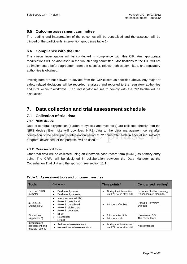
6.5 Outcome assessment committee
The reading and interpretation of the outcomes will be centralised and the assessor will be
blinded of the participants‟ intervention group (see table 1).
6.6 Compliance with the CIP
The clinical investigation will be conducted in compliance with this CIP. Any appropriate
modifications will be discussed in the trial steering committee. Modifications to the CIP will not
be implemented before agreement from the sponsor, relevant ethics committee, and regulatory
authorities is obtained.
Investigators are not allowed to deviate from the CIP except as specified above. Any major or
safety related deviations will be recorded, analysed and reported to the regulatory authorities
and ECs within 7 workdays. If an investigator refuses to comply with the CIP he/she will be
disqualified.
7. Data collection and trial assessment schedule
7.1 Collection of trial data
7.1.1 NIRS device
Data of cerebral oxygenation (burden of hypoxia and hyperoxia) are collected directly from the
NIRS device. Each site will download NIRS data to the data management centre after
completion of the participant‟s intervention period at 72 hours after birth. A specialised software
program, developed for the purpose, will be used.
7.1.2 Case record form
Other trial data will be collected using an electronic case record form (eCRF) as primary entry
point. The CRFs will be designed in collaboration between the Data Manager at the
Copenhagen Trial Unit and the sponsor (see section 11.1).
Table 1: Assessment tools and outcome measures
Tools Outcomes Time points* Centralised reading#
Cerebral NIRS oximeter
Burden of hypoxia
Burden of hyperoxia
During the intervention until 72 hours after birth
Department of Neonatology, Rigshospitalet, Denmark
aEEG/EEG (Appendix C)
Interburst interval (IBI)
Power in delta band
Power in theta band
Power in alpha band
Power in beta band
64 hours after birth Uppsala University, Sweden
Biomarkers (Appendix B)
BFBP
Neuroketal
S100β
6 hours after birth
64 hours birth
Haemoscan B.V., The Netherlands
Investigator‟s assessment and medical records
Serious adverse reactions
Non-serious adverse reactions
During the intervention until 72 hours after birth
Not centralised

SafeBoosC CIP – Phase II Version: 3.0 - 16.03.2012 Reference number: SB010512
Page 29 of 67
Tools Outcomes Time points* Centralised reading#
cUS (Appendix E)
IVH grade (Papille)
Cerebellar haemorrhage
cPVL
Cerebral atrophy
Post-haemorrhagic hydrocephalus
During the intervention and follow-up period, according to these assessment points:
At 1-4 days after birth
At 7 days after birth
At 14 days after birth
At 35 days after birth
At term date
Department of Neonatology, Wilhelmina Children‟s hospital, UMC Utrecht, The Netherlands
Investigator‟s assessment and medical records
All-cause mortality Term date
24 months after term date
Not centralised
Investigator‟s assessment and medical records
Neonatal morbidities:
NEC stage 2-3
ROP stage 3+ and above Term date Not centralised
Investigator‟s assessment and medical records
Neonatal morbidities:
Oxygen requirement At 36 weeks Not centralised
MRI (Appendix D)
Brain injury score (Woodward)
Volumetric
Cortical folding Diffusion tensor imaging
At term date (approximately three months after birth)
Department of Neonatology, Wilhelmina Children‟s hospital, UMC Utrecht, The Netherlands
Investigator‟s assessment and medical records
Therapies implemented:
Mechanical ventilation
Volume substitution
Blood transfusion
Inotrope
Vasopressor
Ductal closure
During the intervention until 72 hours after birth
Not centralised
CRF
Physiological parameters
Mean BP
Mean SpO2
Mean pCO2
During the intervention until 72 hours after birth
Not centralised
BSID-III
Cognitive score
Verbal score
Motor score
24 months after term date
Not centralised
ASQ
Communication
Gross motor
Fine motor
Problem solving
Personal-social
24 months after term date
Not centralised
* If assessment is not possible at the specified time, the assessment shall still be conducted, and the time will appear in the eCRF and the deviations from the CIP can be assessed. # Central assessment is conducted by a blinded outcome assessment committee.
7.2 Trial assessment schedule
Trial data to be collected and the specified time points are detailed in Table 2 - Trial assessment
schedule.
7.3 Research biobank Blood and urine samples will be stored at the individual clinical sites according to national
regulations and guidelines and used for assessment of biomarkers BFABP, neuroketal, and
S100β.

SafeBoosC CIP – Phase II Version: 3.0 - 16.03.2012 Reference number: SB010512
Page 30 of 67
Blood
If a central vascular catheter is in place one ml of blood will be collected in heparin. After
processing for obtaining serum this must be stored frozen (-20 ºC for a maximum of one week,
thereafter at -80ºC for prolonged storage). Blood will be withdrawn 2 times, and in total 1 ml
serum will be stored per participant.
Urine
If a urinary catheter is present, 1 ml of urine shall be collected and stored frozen (-20 ºC for a
maximum of one week, thereafter at -80ºC for prolonged storage) without further treatment.
Urine samples will be collected 2 times, and in total 2 ml will be stored per participant. There are
no immediate risks with obtaining the urine samples.
Storing
Serum and urine samples are labelled with unique identification (as outlined in appendix B).
Each principal investigator shall keep record of the participant identity linked to the participant
number. All samples should be stored locally at the site until last participants samples has been
collected, then all samples will be shipped to Haemoscan, Netherlands (refer to appendix B).
After analysis - expected to be in 2014 - all remaining samples after final analysis will be
discarded.

SafeBoosC CIP– Phase II Version: 3.0 – 16.03.2012 Reference number: SB010512
Page 31 of 67
Table 2: Trial assessment schedule
Visit Number 0 1 2 3 4 5 6 7
Visit description Screening Randomisatio
n
Intervention period
(Cerebral monitoring visibility and treatment according to guideline or ‘us usual’)
Follow-up
Visit code V0 V1
V2a V2b V2c V2d V3 V4 V5 V6 V7
Time period 0-hour 0-3 hours after birth
3 hours after birth
6 hours after birth
24 hours after birth
64 hours after birth
7 days after birth
14 days after birth
35 days after birth
Term date 24 months after term
date
Informed consent can be obtained before birth
X
In- and exclusion criteria X
Maternal history X
Maternal antenatal concomitant medication
X
Infant concomitant medication X X X X X
Placement of cerebral NIRS oximeter between 0-3 hours after birth
X
Blood/urine (biomarker)
X X
Serious adverse reaction X X X X X
Non-serious adverse reactions X X X X
Neonatal comorbidities X
aEEG
X
cUS X X X X X X
MRI X
BSID-III X
ASQ X
All-cause mortality X X

SafeBoosC CIP– Phase II Version: 3.0 – 16.03.2012 Reference number: SB010512
Page 32 of 67
8. Assessment of safety
8.1 Adverse events and reactions
8.1.1 Definitions
Adverse events (AE): any undesirable event occurring to a participant during a clinical trial,
whether or not considered related to the trial intervention.
Serious adverse event (SAE): any adverse event that results in death, is life-threatening,
requires prolongation of existing hospitalisation, result in persistent or significant disability or
incapacity, or requires intervention to prevent permanent impairment or damage.
Adverse reactions (AR) / Adverse device effects (ADE): all untoward and unintended
responses related to the interventions of application of the treatment guideline and/or cerebral
oximeter.
Expected adverse reactions (EAR) / Expected adverse device effect (EADE): adverse
reactions, we expect to be related to the interventions cerebral NIRS oximeter and/or the
application of the treatment guideline are:
Local skin reactions (rashes, burns, etc.).
Reactions related to the manipulation of the patient during positing and re-positing of the
cerebral NIRS oximeter sensors.
o Accidental displacements of the endotracheal tube or extubation.
o Accidental displacement of venous or arterial catheters,
o Severe hypoxia or bradycardia otherwise unexplained.
Other expected adverse reactions encompass reactions to any interventions directed at
correcting an out-of-range oximeter reading by improving respiratory status,
cardiovascular status, oxygen transport, and blood glucose level (see appendix A).
Unexpected adverse reactions (UAR): all untoward and unintended responses, that not is an
expected reaction, related to the interventions cerebral oximeter and/or application of the
treatment guideline.
Serious adverse reactions (SAR): any adverse reaction that results in death, is life-
threatening, requires prolongation of existing hospitalisation, results in persistent or significant
disability or incapacity, or requires intervention to prevent permanent impairment or damage.
This definition includes Serious adverse device effect (SADE): adverse device effect that has
resulted in any of the consequences characteristic of a SAE, or are due to imprecise or
incomplete results from diagnostic equipment e.g. incorrect or delayed diagnosis or incorrect or
delayed treatment and Serious adverse effect-near-incidents (SAE-NI): device deficiencies
that might have led to a SAE if a suitable action had not been taken, or intervention had not
been made, or if circumstances had been less fortunate.
Expected serious adverse reactions (ESAR): any of the expected adverse reactions (listed
above) that results in death, is life-threatening, requires prolongation of existing hospitalisation,

SafeBoosC CIP – Phase II Version: 3.0 - 16.03.2012 Reference number: SB010512
Page 33 of 67
results in persistent or significant disability or incapacity, or requires intervention to prevent
permanent impairment or damage.
Suspected unexpected serious adverse reaction (SUSAR) / Unanticipated serious
adverse device effect (USADE): an adverse reaction which is both serious and unexpected,
i.e., not identified in the current risk analysis listed above (in expected adverse reactions).
Included in this definition are the events with imminent risk of death, serious injury, or serious
illness that requires prompt remedial action for other participants, users, or persons (including
events that are of significant and unexpected nature such that they become alarming as a
potential public health hazard or the possibility of multiple deaths occurring at short intervals) or
a new finding to the event.
Device deficiencies: malfunction, misuse, or use error, e.g. rStO2 values displayed despite
displaced sensor, sensor positioned with light source facing away from skin surface, sudden
malfunction of device that result in no or false rStO2-values.
Figure 3: Relation among adverse events and reactions
8.1.2 Classification of causality
Classification of causality of adverse reactions are outlined in table 3.
8.1.3 Recording and reporting of adverse events and reactions and device deficiencies
Adverse events and device deficiencies are recorded and reported to the competent authority in
all countries in which the trial is being conducted according to national guidelines, Directive
90/42/ECC, as amended by the Directive 2007/47/EC and the EU Commission guidelines on
medical devices, Meddev 2.7/3 “Clinical investigations: serious adverse event reporting”.
AEs
SAEs
EARs
ARs
UARs
SARs ESARs SUSARs
Neonatal mortality
and morbidities
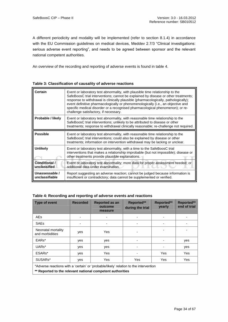
SafeBoosC CIP – Phase II Version: 3.0 - 16.03.2012 Reference number: SB010512
Page 34 of 67
A different periodicity and modality will be implemented (refer to section 8.1.4) in accordance
with the EU Commission guidelines on medical devices, Meddev 2.7/3 “Clinical investigations:
serious adverse event reporting”, and needs to be agreed between sponsor and the relevant
national competent authorities.
An overview of the recording and reporting of adverse events is found in table 4.
Table 3: Classification of causality of adverse reactions
Certain Event or laboratory test abnormality, with plausible time relationship to the SafeBoosC trial interventions; cannot be explained by disease or other treatments; response to withdrawal is clinically plausible (pharmacologically, pathologically); event definitive pharmacologically or phenomenologically (i.e., an objective and specific medical disorder or a recognised pharmacological phenomenon); or re-challenge satisfactory, if necessary.
Probable / likely Event or laboratory test abnormality, with reasonable time relationship to the SafeBoosC trial interventions; unlikely to be attributed to disease or other treatments; response to withdrawal clinically reasonable; re-challenge not required.
Possible Event or laboratory test abnormality, with reasonable time relationship to the SafeBoosC trial interventions; could also be explained by disease or other treatments; information on intervention withdrawal may be lacking or unclear.
Unlikely Event or laboratory test abnormality, with a time to the SafeBoosC trial interventions that makes a relationship improbable (but not impossible); disease or other treatments provide plausible explanations.
Conditional / unclassified
Event or laboratory test abnormality; more data for proper assessment needed; or additional data under examination.
Unassessable / unclassifiable
Report suggesting an adverse reaction; cannot be judged because information is insufficient or contradictory; data cannot be supplemented or verified.
Table 4: Recording and reporting of adverse events and reactions
Type of event Recorded Reported as an outcome measure
Reported**
during the trial
Reported** yearly
Reported** end of trial
AEs - - - - -
SAEs - - - - -
Neonatal mortality and morbidities
yes Yes - - -
EARs* yes yes - - yes
UARs* yes yes - - yes
ESARs* yes Yes - Yes Yes
SUSARs* yes Yes Yes Yes Yes
*Adverse reactions with a „certain‟ or „probable/likely‟ relation to the intervention
** Reported to the relevant national competent authorities
SafeBoosC CIP – Phase II Version: 3.0 - 16.03.2012 Reference number: SB010512
Page 35 of 67
8.1.4 Justification for recording and reporting periodicity and modality
The preterm patient population is a very seriously ill group. Most adverse events may be of a
serious nature with or without the SafeBoosC trial intervention, and both intervention groups are
expected to have a very high proportion of serious adverse events. It is therefore not possible,
nor meaningful to record and report all adverse events. The following will be recorded and
reported (for overview refer to table 4):
Adverse reactions (i.e. both expected (EAR) and unexpected (UAR) with a „certain‟ or
„probable/likely‟ relation (see table 3) to the interventions will be recorded together with an
assessment and reported with the trial results, The final report clinical investigation report to the
competent authorities will summarize the ARs, including a discussion of causal relationship with
investigational device, severity, and treatment needed.
Serious adverse events (SAE) to be recorded are: mortality; the mortality is high and will thus
be analysed and reported as a secondary outcome measure and thus will not be reported to the
authorities, and neonatal morbidities; these are believed to be the most important predictors for
later development and health, and are recorded and reported as an outcome measure
(necrotising enterocolitis stage 2-3, oxygen requirement, and retinopathy of prematurity stage
3+ and above, as well as severe intraventricular haemorrhage and periventricular leucomalacia)
thus not reported to the competent authorities.
Serious adverse reactions (i.e., both expected (ESAR) and unexpected (SUSAR) related to the
cerebral oximeter and/or the treatment guideline will be recorded and analysed as a secondary
outcome measure, but only SUSARs will subject to expedited reporting. Due to the nature of the
population and the intervention, the near-incidents (SAE-NI) will be recorded, but not reported
to the authorities.
8.1.5 Timelines for recording and reporting
The site investigators will report the SAEs (mortality and neonatal morbidities) and the EARs
and UARs through the eCRF periodically according to the assessment schedule (refer to
section 7.2).
The site investigators will report all SARs (ESARs and SUSARs) and device deficiencies with
potential severe effects to the sponsor through the eCRF or by fax if necessary and by phone
(+4535451326) immediately and within 24 hours of knowledge. The ESARs, or new information
regarding already reported events will be reported by the sponsor to the relevant competent
authorities in the yearly report. The sponsor will immediately inform all investigators of any
SUSAR as soon as s/he is made aware of the event. The sponsor will submit an expedited
report of all SUSARs or new information in relation with an already reported event, to the
relevant competent authorities within 2 calendar days of knowledge. The sponsor will review all
SUSARs. If the judgement of sponsor differs from the investigator, both assessments of the
SUSAR shall be included in the SUSAR report to the authorities.

SafeBoosC CIP – Phase II Version: 3.0 - 16.03.2012 Reference number: SB010512
Page 36 of 67
As the trial is estimated to be finalised within one year, the annual report will be omitted and
replaced by the end of trial report in the case where the reporting timelines are overlapping with
3 months.
8.2 Cerebral NIRS monitoring device
A summary of the risk analysis for each investigational NIRS device is provided in the relevant
IB. To ensure safe application of the device,investigators and the involved staff will be trained
on how to place the device (see section 9.2). To minimise skin irritation related to the device,
the oximeter will be moved to different location every 4th hour, further, if skin irritation is
occurring, it will be treated according to local clinical practice. Interaction with the drugs applied
in this population is not possible.
The requirements specified in ISO14155:2011 "6.9 Investigational device accountability" does
not apply to this clinical investigation. All devices are owned by the local institution and not
provided by the sponsor.
8.3 Data Monitoring and Safety Committee
An independent Data Monitoring and Safety Committee (DMSC) is established to, during the
trial, monitor SUSARs with „certain‟ or „probably/likely‟ relationship with the cerebral NIRS
oximeter or the application of the treatment guideline. The charter for the DMSC will be written
prior to inclusion of participants and prior to any analysis. The members of the DSMC are: Jan
Miletin, Cuno Uiterwaal, and Heike Rabe.
8.4 Suspension or premature termination of the clinical investigation
The sponsor/principal investigator, the ECs, and the regulatory authorities can decide about trial
continuation. The clinical investigation can be suspended or prematurely terminated if the
serious adverse device effects are considered disproportionately large compared to the possible
benefits of the intervention. If the trial is terminated or suspended the parents of all trial
participants will be informed and appropriate follow-up will be assured. If sponsor/principal
investigator terminates or suspends the trial the relevant ECs and regulatory authorities will be
provided with a detailed written explanation of the termination or suspension.
The sponsor/principal investigator can upon completion of the analysis of the reason(s) for a
suspension decide to lift the suspension, when the necessary corrective actions have been
implemented. The investigators, ECs, and relevant regulatory authorities will be notified and
provided with the relevant data supporting the decision.
Breaking of blinding will not be relevant in this trial, since group allocation is visible and the
assessment of the adverse device effects in the control group are independent of rStO2
readings of the device.
SafeBoosC CIP – Phase II Version: 3.0 - 16.03.2012 Reference number: SB010512
Page 37 of 67
9. Ethical Considerations The research question; can monitoring of cerebral oxygen saturation levels in the first hours
after birth and subsequent treatment according to pre-specified guidelines prevent brain injury
and maybe death, can only be answered in the specified population, and there is a clear benefit
to the premature patient population as a whole, should the research gain a positive outcome
(research in the Research Ethics Committee group c) As there are no randomised clinical trials
(RCT), thus no conclusive evidence, on the benefit and harms of NIRS monitoring in preterm
infants, we find it ethically suitable that the control group receives „treatment as usual‟. All
interventions proposed in the treatment guideline are commonly used in this patient group.
There is clinical equipoise, which means that there is genuine uncertainty over whether the
cerebral oximeter and subsequent treatment will be beneficial or may even be harmful to the
participants. Due to the clinical nature of the trial population, the premature infants, some stress
reactions related to the manipulation during positing and re-positing of the cerebral NIRS
oximeter sensors can occur. These are however not clinically evaluated to give substantially
more risk or discomfort as compared to no intervention. To obtain evidence-based knowledge
on the benefit and harms of cerebral monitoring using NIRS as part of clinical management of
premature infants, a large-scale RCT is needed. The SafeBoosC phase II trial serves as a
feasibility trial for such a large phase III trial.
The SafeBoosC trial will only start the randomisation of participants after approvals from the
relevant ethics committees and competent authorities have been received and parental
informed consent is obtained. All parents will receive written and oral information about the trial
before they are asked for their written consent (se section 9.2 for further details). They will only
enrol their new-born in the trial by their own free will and can withdraw their consent for
participation at any time. If a parent wishes to withdraw the participation, the patient will be
treated according to the respective hospital‟s standard procedures. The trial will be conducted in
compliance with the guidelines of the Declaration of Helsinki in its latest form and the
International Conference on Harmonization of Good Clinical Practice Guidelines (49) To avoid
concern it has been decided that multiple births will be randomised together and undergo
allocation to the same intervention.
9.1 Informed consent procedure
Parents of potential participants will be invited to enrol their preterm newborn, when possible,
before delivery. Due to the nature of the intervention, if potential participants are found after
their delivery, informed consent procedures must take place within 3 hrs. Surrogate informed
consent is required from both parents, unless one parent has power of attorney. The
sponsor/sponsor delegate (qualified physician or nurse connected to the trial) will make contact
and parents will be informed of the trial, given the Parent information sheet (Appendix F) for the
trial, and told about the possibility of the assessor. The information consultation will be held in
an undisturbed setting, preferably the department meeting facilities. The parents will be given
24 hours to ask questions and make a decision, before a written informed consent can be

SafeBoosC CIP – Phase II Version: 3.0 - 16.03.2012 Reference number: SB010512
Page 38 of 67
obtained from both parents. Parents will be given a copy of the informed consent. If the birth is
sudden the time to make a decision will be shorter.
9.2 Risk of complication for participants
The following procedure will be implemented to prevent and/or minimise risk of complication for
participants.
Related to devices
Correct reading of the monitor (mainly to prevent false reading which may lead to wrong
treatment) is critical. The investigators will train local staff as appropriate and trained staff will
closely supervise all participants during the intervention.
To minimise skin irritation related to the device, the oximeter will be moved to different location
every 4 hours.
Related to application of the treatment guideline
All investigators have been involved in the development of the guideline and have approved the
final version. The treatment guideline do not contradict any national current clinical practices.
The investigators will train local staff as appropriate and will be available for consultation as
needed.
9.3 Benefit for participants
The participants in both groups will receive careful attention from qualified physicians and
hospital staff during the trial and a closer follow-up after hospital discharge compared to infants
not enrolled into the trial.
10. Statistical plan and data analysis
10.1 Sample size estimation
An unpublished dataset of cerebral NIRS oximetry in 23 extremely preterm infants monitored
from the first hours after birth until 72 hours after birth by the INVOS 4100 NIRS device in
Utrecht during the period January 2004 until January 2008 was used to estimate the sample
size (unpublished data). The cerebral saturation was recorded in 5 second-values. The data
were censored by the proprietary Signal Base software. The recording was reliable 79% of the
time, ranging from 48% to 97%. The time spent with a cerebral saturation below 55% and above
85% was calculated and the burden of hypo- and hyperoxia was calculated by summing the
percentage deviation over time area under the curve – AUC). The burden of hypo- and
hyperoxia was corrected for the loss of recording time and expressed as %hours per 72 hours.
There was a relatively high correlation between time spent in hypoxia and the burden of
hypoxia. Overall, hyperoxia was only about 10% of hypoxia. There was a trend towards inverse
correlation between hypoxia and hyperoxia. The distribution of AUC outside the range was
skewed towards the right (higher values). The mean was 76.0%hours ± SD of 83.2. After a log-

SafeBoosC CIP – Phase II Version: 3.0 - 16.03.2012 Reference number: SB010512
Page 39 of 67
transformation, the distribution was not significantly different from normal. The mean was 1.64 ±
SD of 0.50 (Figure 4).
As a result, the statistical distribution has to be normalised by logarithmic transformation.
Reducing the duration of time with hypoxia or hyperoxia by 0.30 (corresponding to a 50%
reduction of the time with hypoxia or hyperoxia) with a type I error (alpha) of 5% and a type II
error of 0.05 (power of 95%) requires randomisation of a total of 150 preterm infants (or „pair‟ or
„groups of infants‟ in case of multiple births): 75 preterm infants in the experimental group and
75 preterm infants in the control group.
Figure 4: The distribution of the primary outcome burden of hypo-and hyperoxia
expressed as %hours and transformed logarithmically
10.2 Data analysis and statistical methods
All analyses will be performed blinded with the two intervention groups concealed as, e.g., X
and Y. All analyses are intention-to-treat analyses (se section 10.2.2 for modification). Table 5
shows each class of outcome, its category and priority, the mathematical type of measure,
value of optimistically imputed missing value, value of pessimistically imputed missing value,
time at which or period during which it was measured and type of statistical methodology
applied to measure the effect of the intervention. The following describes the statistical models
that will be applied and how missing values and multiplicity issues are dealt with.
10.2.1 Statistical models
Depending on the specific type of outcome measure, one of the four types of regression
analyses will be applied. The indicator variable I (1 if X and 0 if Y) is included as a covariate and
the outcome measure as the dependent variable.

SafeBoosC CIP – Phase II Version: 3.0 - 16.03.2012 Reference number: SB010512
Page 40 of 67
Type 1 includes a continuous outcome measure. The general linear univariate model is
used. As a sensitivity analysis a non-parametric test is conducted and the result is
discussed (Mann Whitney).
Type 2 includes a binary outcome measure and logistic regression analysis is used. In
case of lack of convergence fit or non-estimable odds ratios Fisher‟s exact test will be
used and the protocol specified covariates thus disregarded.
Type 3 includes an ordinal quantity (three or four categories) as outcome measure. The
proportional odds model will be used. As a sensitivity analysis the linear by linear model
and a Chi-square test of independence will be applied.
For type 4 (mixed-model analyses), each biochemical outcome measure (y) measured at
6 and 72 hours after birth is viewed as a linear function of time (t). The model is. y = a +
b*I + c*t + d*I*t which includes the impact of the intervention on the intercept and slope of
the linear function. I is the intervention indicator and a, b, c and d are the coefficients of
the regression equation. A mixed-model with repeated measures (MMRM) is used. An
unstructured covariance matrix will be used. In case of lack of convergence a compound
symmetric covariance matrix and random intercept will be used.
All of the above analyses will include the two protocol specified variables (gestational age
category and site of study indicator) as co-variates. These analyses adjusted as appropriate for
any impact of data missing at random (see below) are the primary analyses. The analyses will
be repeated without co-variates included and major discrepancies between the results of the
two analyses will be discussed. For each co-variate an explorative analysis including the
interaction between this and the intervention indicator will be conducted.
In the case of multiple births (randomised as a „pair‟ or a „group‟) analyses will be
1. Based on one in which one of the doublets or triplets are chosen randomly without
knowledge of their outcome to be included in the analysis. This analysis is considered
the primary on which the main conclusion is to be drawn.
2. Thereafter sensitivity analyses are conducted in which first doublet a and then doublet b
is included. The triplets are represented by the one chosen randomly.
3. Thereafter sensitivity analyses are conducted in which first triplet a, and then triplet b,
and then triplet c is included. The doublets are represented by the one chosen randomly.
10.2.2 Dealing with missing values
To include all patients in analyses of type 1 to type 3 the data are first subjected to multiple
imputation and then the analyses are done using the data sets generated by the multiple
imputation. Thus the results based on multiple imputations (see below), represent the primary
analyses.

SafeBoosC CIP – Phase II Version: 3.0 - 16.03.2012 Reference number: SB010512
Page 41 of 67
For the results of the mixed-model analysis (type 4), the primary results are based on multiple
imputations of missing covariate values (see below)..
Using multiple imputations respectively the MMRM secures that missing values will not create
any bias as long as the values are missing at random (MAR), a rather mild restriction implying
that the probability of missing data on Y is unrelated to the value of Y, after controlling for other
variables in the analysis. However, if covariate values are missing, these values are first
imputed using the method of multiple imputations (MI) and then the MMRM analysis is
conducted.
In the MI analysis, the model variables and additional variables significantly related to the
variables with missing values and/or the missingness of these variables will be used. The
method to be used is the fully conditional specification method of SPSS (version 17.0). This is
an iterative Markov chain Monte Carlo method that can be used when the pattern of missing
data is arbitrary (monotone or non-monotone). The default number of iterations will be used
initially and then increased if the Markov chain has not converged. Prior to the MI, the
distributions of the continuous variables are inspected to see if serious deviations from the
normal distribution that need transformations are present. Constraints are set to restrict the
range of imputed values of continuous variables so that they are plausible. Ten imputed data
sets are produced.
To assess the potential impact of values missing not at random (MNAR), two sensitivity
analyses will be conducted.
1. Sensitivity analysis 1. Parameter estimates obtained by complete participant analysis (in
the MMRM analysis only participants without missing values are included) are compared
to those obtained by MI or MMRM analysis where the latter includes all participants.
2. Sensitivity analysis 2. Let BEST be the intervention group in which during the experiment
a specified outcome measure (X) is beneficially and significantly changed as compared
with the other intervention group. Two types of values are used for imputation of missing
values of X, an optimistic one (x-op) supporting the findings of the complete participant
analysis and a pessimistic one (x-pes) not supporting the findings. Four analyses are
conducted. In the first, all missing values are imputed by x-op, in the second by x-pes, in
the third all missing values in group BEST are imputed by x-op while those in the other
group are imputed by x-pes and in the fourth analysis all missing values in group BEST
are imputed by x-pes while those in the other group are imputed by x-op (worst case
analysis). Table 3 (columns 4 and 5) shows the values chosen for x-op and x-pes
respectively.
10.2.4 Dealing with multiplicity
The null hypotheses corresponding to the outcome measures are gathered into three families
(see table 5) according to priority and the gate keeping method of Dmitrienko et al. (50). Parallel

SafeBoosC CIP – Phase II Version: 3.0 - 16.03.2012 Reference number: SB010512
Page 42 of 67
gate keeping will be used to adjust the observed P values. The originally computed P values
and the corresponding adjusted ones obtained using the gate keeping method will both be
reported and the problems arising from the fact that the sample size calculation is based on the
primary outcome measure only will be discussed.

SafeBoosC CIP– Phase II Version: 3.0 – 16.03.2012 Reference number: SB010512
Page 43 of 67
Table 5: Statistical analyses
a) Family of null hypotheses. b) P stands for primary outcome. S for secondary outcome and E for outcome subject to explorative hypothesis generating analysis. No stands for number of this category of outcome. c) The 5 %centile in the distribution of the whole material. d) The 95%centile in the distribution of the whole material.
Statistical type of analysis
(family)a)
Category of outcome
(priority/No)b)
Mathematical type of outcome measure
Values imputed for sensitivity analysis
Times of measurements
X-op X-pes During intervention
Measured at 6 and 72 hours
Measured at 72 hours
Measured at term date (approx. three months after birth)
Measured at 24 months after term date
1 (1) Hypoxia or hyperoxia burden (P/1)
Continuous Minc) Maxd) yes
1 ( 2)
aEEG (S/1) Continuous n.a. n.a. Yes
4 ( 2)
Biomarkers (S/3) Continuous Maxd) Minc) yes
1
Hypoxia-burden (E/1) Continuous Minc) Maxd) Yes
1 Hyperoxia-burden (E/1)
Continuous Minc) Maxd) yes
2 (3) Severe adverse reactions (S/1)
Binary 0 1 yes
2 (3) Mortality (S/1) Binary 0 1 Yes
2
Morbidities (E/5) Binary 0 1 Yes
3(2) Brain injury (cUS) (S/2)
Categorical 1 3 Yes
3 Brain injury score (E/1) (MRI)
Categorical or continuous
0 3 Yes
2
Therapy (E/6) Binary 0 1 yes
1 Physiological quantities (E/3)
Continuous Maxd) Minc) yes
2 Mortality between 3 month and 24 months (E/1)
Binary 0 1 yes
1 BSID-III (E/3) Continuous Maxd) Minc) yes

SafeBoosC CIP– Phase II Version: 3.0 – 16.03.2012 Reference number: SB010512
Page 44 of 67
11. Data management
11.1 Data handling and archiving
All participant data are protected in accordance with the Danish Act on processing of personal
data and the Danish Health Act. Source data will be registered in the participant‟s medical
records or directly into the eCRF. A common web-based electronic case report form (eCRF) will
be devised to enable a central database. Data entry into the central database handling of
medical records is the responsibility of the investigators. Data will be stored in accordance with
guidelines issued by the Danish Data Protection Agency, with which the trial will also be
registered. After the establishment of a ‟clean file‟, the database will be locked, data will be send
for statistical analysis at the Copenhagen Trial Unit. The trial database will hereafter be kept
according to the respective national laws. After end of trial, the data will be archived for five
years according to good clinical practice guideline (appendix I).
At each trial-site the data flow (figure 5) will be monitored according to the GCP principles by a
locally appointed external monitoring committee.
Figure 5: The data flow in SafeBoosC
After completion of statistical data analysis, data will be pseudo-anonymised and deposited at
the Danish Data Archive. Also, data will be made available for other researchers by uploading a
completely anonymised dataset onto ClinicalTrials.gov.

SafeBoosC CIP – Phase II Version: 3.0 - 16.03.2012 Reference number: SB010512
Page 45 of 67
The investigator(s) permits trial-related monitoring, audits, regulatory inspection(s) by providing
direct access to the source data and other relevant documents. Trial data will be handled
according to regulations of data protection agency in the respective countries.
11.2 Medical coding
Adverse reactions (ARs) will be coded using MedDRA coding system by lowest level term
(LLT), preterm term (PT), and system organ class (SOC). Concomitant medications will not be
coded.
12. Quality assurance The trial will be carried out in accordance with the Declaration of Helsinki in its latest form and
the ICH GCP guidelines as well as national laws and regulations.
12.1 Monitoring
The trial will be monitored according to the ICH GCP guidelines, and a detailed monitoring plan
will be developed. Centrally eCRFs will be checked, and as a minimum the following will be
monitored locally:
All patients for existence
All patients for documented informed consent.
12.2 Device quality control
Each site is responsible for adhering to the quality control measures described in the oximeter
manufacturer‟s users guidelines.
13. Trial and funding timeframe
Trial stages Timeframe
CIP development Spring 2011
CIP finalised (first draft) May 31, 2011
Site selection Ongoing – depend on finance
Recruitment phase May 2012 – May 2013
Assessment phase Last participant recruited (Primary outcome May
2013)
Final analysis 2015

SafeBoosC CIP – Phase II Version: 3.0 - 16.03.2012 Reference number: SB010512
Page 46 of 67
Trial stages Timeframe
Publication Early 2014 on primary and secondary outcomes
Funding Timeframe
START December 2010, awarded 183,848 DKK
Danish Strategic
Research Council
November 2011, awarded 11,100.000 DKK
14. Legal aspects
14.1 Finance
The SafeBoosC project has received 183,848.00 DKK from the Danish Council for Strategic
Research (DSF) under the Preparation of International Application (START) theme in December
2010. An application to the DSF seeking for 10,320,640.00 DKK under the Strategic Research
in Individuals, Disease and Society theme, and was awarded 11,100,000.00 DKK November
2011. The sponsor/principal investigator Gorm Greisen is the initiator of the SafeBoosC project.
He has no financial interest in the results of the trial, nor the NIRS-instruments. He is not
financially or in any other way involved in the Danish Council for Strategic Research.
14.2 Participant insurance
The participants will be insured in accordance with existing legislation of their respective
country.
14.3 Publication plan
The trial will be registered on ClinicalTrials.gov prior to the randomisation of the first participant.
Attempts will be sought to publish all results, positive, neutral, as well as negative, in a peer-
reviewed international journals. Authorship will be determined according to the International
Committee of Medical Journal Editors. Attempts will be made to publish a list of all investigators
with their contributions in all publications.
14.4 Statements of compliance
The clinical investigation will be conducted in accordance with the ethical principles that have
their origin in the Declaration of Helsinki.

SafeBoosC CIP – Phase II Version: 3.0 - 16.03.2012 Reference number: SB010512
Page 47 of 67
The clinical investigation will comply with ISO14155 and the relevant national regulations of
each participating medical centre, and will not begin until required approvals from ECs and
regulatory authorities have been obtained.
Additional requirements imposed by the ECs or regulatory authorities will be followed.

SafeBoosC CIP – Phase II Version: 3.0 - 16.03.2012 Reference number: SB010512
Page 48 of 67
15. Appendices
15.1 Appendix A: Treatment guidelines and justifications
SafeBoosC Clinical Guidelines Assessment of cerebral oxygen saturation Regional cerebral tissue oxygen saturation (rStO2) is a composite measure of tissue oxygen saturation across arterial, capillary and venous beds and reflects a balance between cerebral oxygen delivery (CDO2) and cerebral metabolic rate (CMRO2). In preterm infants, the CMRO2 is unlikely to vary much and a change in rStO2 largely reflects changes in CDO2. The factors which influence CDO2 are arterial oxygen saturation (SaO2), haemoglobin concentration ([Hb]), and cerebral blood flow (CBF). Establishment of monitoring of cerebral oxygenation As soon as possible and within 3 hours of age Period of monitoring of cerebral oxygenation Until 72 hours after birth Instruction Document the intervention chosen and any changes in rStO2 following intervention. Recommendation for clinical interventions The rStO2 target normal range is 55% to 85%. If the rStO2 is out of normal range and there is no reason to believe that it will normalise without intervention, consider one of the interventions
(identified in „ ‟) listed below and reassess 30 to 60 minutes after the intervention. Generally, only one intervention should be chosen at a time. All the interventions proposed here are commonly used in this patient group. After each intervention, the level of evidence for each intervention (I-III) and recommendation (A-E) are given (defined in table 1 and 2). For further explanation, see below. rStO2 < 55% Aim of intervention: A low rStO2 reflects a low CDO2. The interventions should be directed to increasing SaO2, [Hb], and/or CBF. Assess cardiovascular status: Blood pressure low in normal range, consider:
Vasopressor-inotropes (I/B) (59,60)
Fluid bolus (normal saline) (I/C) (61,62)
Decrease mean airway pressure (III/B) (54,63-65)
Poor systemic circulation, consider if: Echocardiography shows low cardiac output and/or low SVC flow
Inotropes (I/B) (62,66-70)
Fluid bolus (normal saline) (I/C) (61,62)
Decrease mean airway pressure (III/B) (54,63-65)
Reduce vasopressor (III/ B) (71)
Echocardiography not available but has at least 2 of the following signs:
Lactate > 3.5 mmol/l CRT > 3 seconds Urine output < 1 ml/kg/hour
consider:

SafeBoosC CIP – Phase II Version: 3.0 - 16.03.2012 Reference number: SB010512
Page 49 of 67
Inotropes (I/B) (62,66-70)
Fluid bolus (normal saline) (I/C) (61,62)
Decrease mean airway pressure (III/B) ) (54,63-65)
Reduce vasopressor (III/B) (71)
Patent ductus arteriosus, consider:
Medical treatment (II-2/B) (44,63,64,72) Assess oxygen transport: Haemoglobin low in the normal range, consider: Red blood cell transfusion (I/B) (55,73-75)
Assess respiratory status: SaO2 low in normal range, consider:
Increase FiO2 (II-1/A) (51)
(ATTENTION: be careful not to exceed the upper target threshold of
SpO2)
Increase mean airway pressure (III/B) (52-54)
PCO2 low in normal range, consider:
Decrease minute ventilation (II/A) (55-58)
rStO2 > 85% Aim of intervention: A high rStO2 reflects impaired oxygen utilisation and/or disturbed cerebral autoregulation (hyperaemia) and interventions should be directed at identifying and treating the underlying cause. Assess respiratory status: SaO2 high in normal range, consider: Decrease FiO2 (II-2/A) (76-80)
Decrease mean airway pressure (III/B) (52,53)
PCO2 high in normal range, consider: Increase minute ventilation (II/A) (55-58)
Assess blood glucose level: Blood glucose < 2.5 mmol/l, consider to: Increase glucose intake (II/A)
(81,82)
Level of evidence and recommendation of intervention The level of evidence (Table 1) and recommendation for a given intervention (in brackets and Table 2)) were graded according to the U.S. Preventive Services Task Force system
(83)
.
Table 1: Hierarchy of research design and level of evidence
Level of evidence Type of study
I Evidence obtained from at least one properly randomized controlled trial
II-1 Evidence obtained from well-designed controlled trials without randomization
II-2 Evidence obtained from well-designed cohort or case-control analytic studies, preferably from more than one centre or research group
II-3 Evidence obtained from multiple time series with or without intervention. Dramatic results in uncontrolled experiments (such as the results of the introduction of penicillin treatment in the 1940s) could also be regarded as this type of evidence
III Opinions of respected authorities, based on clinical experience,

SafeBoosC CIP – Phase II Version: 3.0 - 16.03.2012 Reference number: SB010512
Page 50 of 67
descriptive studies and case reports, or reports of expert committees
Table 2: Recommendation grid
Net benefit
Quality of evidence
substantial moderate small zero/negative
Good A B C D
Fair B B C D
Poor E E E E
Standard recommendation language
A= Strongly recommended (good evidence that the intervention improves important health outcomes and benefits substantially outweigh harms). B= Recommended (at least fair evidence that the intervention improves important health outcomes and benefits substantially outweigh harms). C= No recommendation for or against routine provision of the intervention (fair evidence that the service can improve health outcomes but the balance of the benefits and harms is too close to justify a general recommendation). D= Recommends against routinely providing the intervention (at least fair evidence that the service is ineffective or that harms outweigh benefits). E= Insufficient to recommend for or against routinely providing the intervention (evidence that the intervention is effective is lacking, of poor quality, or conflicting and the balance of benefits and harms cannot be determined).
Abbreviations SVC Superior vena cava CRT Capillary refill time FiO2 Fraction of inspired oxygen rStO2 Regional cerebral tissue oxygen saturation SaO2 Saturation of Oxygen (arterial blood) PCO2 Partial Pressure of Carbon Dioxide CDO2 Cerebral oxygen delivery CMRO2 Cerebral metabolic rate [Hb] Haemoglobin concentration CBF Cerebral blood flow

SafeBoosC CIP – Phase II Version: 3.0 - 16.03.2012 Reference number: SB010512
Page 51 of 67
15.2 Appendix B: Procedure for assessment of chemical biomarkers
OUTCOME MEASURES Outcome measure 1: S100β Outcome measure 2: BFABP Outcome measure 3: Neuroketal MEDIUM Blood If a central blood catheter is in place one ml of blood will be collected in heparin. Commercially available tubes containing lithium or sodium heparin are available to collect 2 ml of blood. These tubes can be used without vacuum and filled with 1 ml from a syringe. The higher final heparin concentration is no problem. EDTA or citrate plasma cannot be used for measurement of S100β. Blood must be kept cold after collection on ice or in the fridge at 4ºC until it is centrifuged (1200xg for 12 minutes in a normal centrifuge or 13000 rpm in a microcentrifuge) to obtain platelet poor plasma. As much as possible plasma is then collected, transferred to small screw-cap tubes and stored frozen (-20 ºC for a maximum of one week, thereafter at -80ºC for prolonged storage). Urine If possible, 1 ml of urine shall be collected and stored frozen (-20 ºC for a maximum of one week, thereafter at -80ºC for prolonged storage) without further treatment.
TIME OF COLLECTION Blood:
6 hours after birth, and
64 hours after birth Urine (if possible) - special instruction (e.g., can be +/-2 hours of specified time points):
6 hours after birth, and
64 hours after birth.
STORING Samples should be labeled with two country letters, identification (randomisation number), Plasma or Urine, time points (urine samples may extent to 4 or 5). Example: DK 5 P 3 = Rigshospitalet participant 5, plasma collected after 64 hrs. The principal investigator shall keep record of the participant identity linked to the participant number. All samples should be stored at the site until last participants samples has been collected and recorded. SHIPPING: When last participant has been enrolled, samples should, immediatedly hereafter, be shipped in batches on dry ice, using DHL as a courier. Shipping will always be Monday or Tuesday. To the following address: Haemoscan B.V. Stavangerweg 23, Groningen 9723 JC, The Netherlands Att: Wim van Oeveren Phone : +31(0)646181604 Fax nr +31 (0)847269747 Material transfer agreements between Haemoscan and the sites will be issued by each institutions legal department. METHODS OF ASSESSMENT
SafeBoosC CIP – Phase II Version: 3.0 - 16.03.2012 Reference number: SB010512
Page 52 of 67
All assessments are conducted without knowledge of allocation group (experimental or control). S100β The S100β serum-concentration will be assesed by ELISA. It can be detected in heparin plasma, heparin serum or urine in 50 µl samples. BFABP Brain fatty acid binding protein (BFABP) is determined by means of ELISA with BFABP specific monoclonal capture antibodies and polyclonal detection antibody in plasma/serum as well as urine. 100 µl is needed for a BFABP ELISA, due to its low concentrations. Neuroketal Determination of neuroketal is performed by competitive enzyme immunoassay in 100 µl of plasma/serum or urine.
Abbreviations S100β Acidic calcium binding protein found in the nervous system BFABP Brain fatty acid binding protein ELISA Enzyme linked immunosorbent assay ml Millilitre

SafeBoosC CIP – Phase II Version: 3.0 - 16.03.2012 Reference number: SB010512
Page 53 of 67
15.3 Appendix C: Procedures for assessment of aEEG/EEG
OUTCOME MEASURES Outcome measure 1: Interburst interval (IBI) Outcome measure 2: Power in delta band Outcome measure 3: Power in theta band Outcome measure 4: Power in alpha band Outcome measure 5: Power in beta band Outcome measure 6: Background pattern Outcome measure 7: Sleep wake cycling Outcome measure 8: Seizures MEDIUM Raw EEG-data TIME OF COLLECTION Single-channel EEG is applied for at least 120 minutes at 48-72 hours after birth. This should be at least 3 hours after administration of bolus morphine, if possible. Hydrogel electrodes or needle electrodes may be used. P3 and P4 positions according to the international 10–20 system; a frontal reference electrode is also applied. METHOD OF ASSESSMENT OR SCORING The EEG is divided in 10-minutes epochs. The quality of all EEG epochs is visually assessed without knowledge of allocation group (experimental or control). Epochs showing artifacts that could affect the quantitative analysis are discarded. IBIs are measured by using an automated algorithm and averaged in each 10-minute epoch. The IBI detector algorithm is based on a non-linear energy operator that reflects both amplitude and frequency content of the EEG. Power spectral analysis is performed using fast Fourier transformation with a time base of 10 seconds. For every epoch, total and relative (%) band power is calculated within the following frequency bands: delta (0.5–4 Hz), theta (4–8 Hz), alpha (8 –13 Hz), and beta (13–30 Hz). If more than one 10 minutes epoch is analysed, the simple average of all measures is used for further analysis
Abbreviations aEEG Amplitude-integrated electroencephalogram
EEG Electroencephalogram
IBI Interburst interval
Hz Hertz

SafeBoosC CIP – Phase II Version: 3.0 - 16.03.2012 Reference number: SB010512
Page 54 of 67
15.4 Appendix D: Procedures for MRI examination OUTCOME MEASURES Outcome measure 1: Brain injury score according to Woodward with cerebellum score Outcome measture 2: Volumetric measurements: Outcome measure 3: Cortical folding with formation of the sulci: Outcome measure 4: Diffusion tensor imaging: MEDIUM Cerebral MRI scan TIME OF COLLECTION At term date (approx. 3 months after birth) ± 2 weeks METHOD OF ASSESSMENT of brain injury scoring and quantitative measurements The MRI protocol consists of anatomical scans preferable on 3Tesla MRI (T1 3D weighted image and T2 weighted image), DWI and DTI. The T1 and T2 weighted images are performed in coronal slices and the T1-3D weighted image at around 100-110 slices. The DTI images are performed in axial slices covering the whole brain. The focus is on the following features: brain tissue volumes, including myelinisation, cortical folding with formation of the sulci, in relation to DWI/DTI. All assessments are conducted without knowledge of allocation group (experimental or control). Brain injury score according to Woodward plus cerebellum: The scoring system for the composite white matter and gray matter scores consists of eight assessments each using a 3-point scale. White matter abnormality is graded using five areas of assessment within the white matter with a score of 1 (normal), 2 (mild abnormality) and 3 (moderate-severe abnormality) for each area of assessment. The five areas of white matter abnormality that are scored include: 1. The nature and extent of white matter signal abnormality - This is scored based on the T1- and T2-weighted signal abnormalities in the white matter best observed in the axial imaging. Grade 1: normal T1- and T2-weighted signal throughout the white matter. Grade 2: focal regions of high T1-or T2-weighted signal (2 or fewer regions per hemisphere). Grade 3: multiple regions of high T1- or T2-weighted signal (more than 2 regions per hemisphere). 2. Periventricular white matter volume loss – This is also scored based on the combined T1- and T2-weighted imaging analyzing the ratio of periventricular white matter to ventricular volume in both the axial and sagittal imaging. Grade 1: normal periventricular white matter volume usually associated with small ventricles Grade 2: mild reduction in white matter volume with mild to moderate increased ventricular size. Grade 3: marked reduction in white matter volume often occurring with marked increase in the size of the ventricle and/or extra-axial space. 3. Cystic abnormalities – This is easily assessed on any imaging view or modality but occurs rarely. Grade 1: normal, with no cystic abnormality. Grade 2: less than 2mm single focal cyst. Grade 3: multiple cysts or a single larger (2mm or greater) single focal cyst is rare. 4. Ventricular dilatation – This is also scored based on the combined T1- and T2-weighted imaging in both the axial and sagittal imaging. Grade 1: normal with no evidence of ventricular dilatation. Grade 2: moderate enlargement resulting in mild rounding of the frontal horns, minimal enlargement of the temporal horns and moderate enlargement of the occipital horns. Grade 3: more global enlargement of a moderate to severe nature including significant enlargement of the frontal, temporal and occipital horns. 5. Thinning of the corpus callosum – This is scored based on the sagittal and coronal T1-

SafeBoosC CIP – Phase II Version: 3.0 - 16.03.2012 Reference number: SB010512
Page 55 of 67
weighted MR imaging. Grade 1: normal, with thick corpus callosum visible in all views. Grade 2 :focal thinning in the corpus callosum often visible in the mid region of the body of the corpus on sagittal images. Grade 3 :global thinning across the entire corpus callosum. 5 assessments of the cerebral white matter are then combined to give an overall white abnormality score which is categorized as:
no abnormality - Total score 5 to 6
mild abnormality – Total score 7 to 9
moderate abnormality – Total score 10 to 12
severe abnormality – Total score 13 to 15. Gray matter abnormality is graded using three scales assessing: 1) Presence of gray matter cortical signal abnormality . This is very rarely detected and thus does not contribute significantly to the overall scoring. It is scored on high signal intensity in the cortex on axial T1 and/or loss of cortical ribbon signal on axial T2-weighted MR imaging. 2) Quality of gyral maturation – Cortical gyration maturation is rated by standard gyral model (3,4) using representative MRI images and cerebral models of gyral development at 34 weeks, 36 weeks, 38 weeks and 40-42 weeks on axial T1- and T2-weighted MR imaging. The gyral pattern assessment in these maturational assignments includes at 34-36 weeks the presence of marginal sulcus, paracentral gyrus with secondary sulci present in the frontal lobes, superior and middle temporal, rolandic and occipital regions. By 36-38 weeks the gyral pattern should include additional secondary gyri in the transverse and inferior temporal and cingulated gyri. At 40 weeks‟ gestation there would be tertiary inferior temporal and inferior occipital gyri and sulci Grade 1: normal for 40 weeks. Grade 2: 2 to 4 weeks delay in gyral development (i.e. consistent with 36 to 40 weeks). Grade 3: more than 4 weeks delay in gyral development. 3) Size of the subarachnoid space - This is scored based on the sagittal T1 and coronal T1- or T2-weighted MR images for the size of the subarachnoid space in relation to the cerebral mantle. Grade 1: small subarachnoid space which is barely visible. Grade 2:mildly enlarged CSF space with visible enlargement of the space between the major sulci and the interhemispheric space in addition to the extracerebral space. Grade 3: more substantially enlarged global subarachnoid space with visible cerebrospinal fuid between many gyri as well as interhemispheric and extracerebral. The total gray matter score was then calculated and categorized as (a) normal - score 3 to 5 (b) abnormal score 6 to 9. Cerebellar lesions:
(a) normal: no lesions (b) mild: < 6 punctate lesions (c) moderate /severe: > 6 punctate lesions/cerebellar hemorrhage
Volumetric measurements: The segmentation is performed on the MR images of individual participants. The segmented tissue types include: unmyelinated and myelinated white matter, cortical and central gray matter (basal ganglia and thalamus), cerebro-spinal fluid around the brain, ventricles, cerebellum and brainstem. This segmentation method was developed for neonates (84). Quantitative measurements of the cerebral tissue volumes are performed by the knn-approach segmentation method which is based on the probability of each tissue classes from signal intensities in T1- and T2-weighted images and the anatomic location. Thereafter, the volume of each tissue is quantified. Cortical folding with formation of the sulci: For cortical folding, the inner cortical surface is segmented and reconstructed in 3D from the T2-weighted images. The surface is detected

SafeBoosC CIP – Phase II Version: 3.0 - 16.03.2012 Reference number: SB010512
Page 56 of 67
between the developing cortex and white matter zone. The global area of this inner cortical surface is then computed. The segmentation of the inner surface is used as it highlights the cortical sulcation pattern more precisely than the outer surface, particularly where sulci are still not deeply folded. Finally, the local surface curvature is estimated from the mesh local geometry: positive curvatures correspond to the gyri top, and negative curvatures to the folds bottom. So the 3D image is then created. The sulci are defined as connected components of negative curvature and labelled manually according to post-mortem and MRI atlases and prenatal images. To characterize the sulci maturation according to gestational age, their sulci areas will be calculated. Thereafter, for each infant, the sulcation index with total cortical surface and thickness can be computed. Sulcation index is defined as the ratio between the areas of sulci and the surface of the total cortex. This surface increases with the age-related brain growth. The sulcation index, thus, characterizes the proportion of sulci according to the whole brain size, and is a measure of maturation of the cortex. Diffusion tensor imaging: (DTI) scans are obtained to perform analyses of the diffusion coefficients on the brain averages, as well as on individual participants. It visualizes the movement of fluid in the brain, and can be used to identify white matter tracts. We will study the maturation of white matter fiber tracks in relation to cortical development.
Abbreviations DWI Diffusion weighted imaging
DTI Diffusion tensor imaging
MRI Magnetic resonance imaging
T1 T1-weighted
T2 T2-weighted
3D Three-dimensional

SafeBoosC CIP – Phase II Version: 3.0 - 16.03.2012 Reference number: SB010512
Page 57 of 67
15.5 Appendix E: Procedure for assessment of cranial ultrasound
OUTCOME MEASURES Outcome measure 1: Parenchymal/periventricular haemorrhagic infarction / inhomogeneous flaring Outcome measure 2: IVH Grade III Outcome measure 3: Cerebellar haemorrhage Outcome measure 4: Posthaemorrhagic hydrocephalus Outcome measure 5: cPVL Outcome measure 6: Cerebral atrophy MEDIUM Sonogram Coronal: C1 frontal lobes C2 foramen Monro C3 lateral ventricles with largest size of VI C4 cerebellum largest diameter left-right C5 posterior horns with plexus C6 periventricular Sagital S1 midline S2 left lateral ventricle S3 left periventricular through fissura Sylvi S4 right lateral ventricle S5 right periventricular throuh fissura Sylvi Cerebellum via mastoid with tranducer in coronal position such as coronal view via anterior fontanel TIME OF COLLECTION
Early scans (day 1,3):
Late scans (day 7, 14, 35)
Term scan: METHOD OF ASSESSMENT OR SCORING The assessment classification is: 1. Normal brain scan
No cysts
No ventricular dilatation
No enlargement of extra-cerebral spaces
Normal cortical grey matter 2. Mild brain injury:
Grade 1-2 IVH (including GLH)
Persistent pathologic non-decreasing inhomogeneous flaring at day 7 and 14
Thinning of the corpus callosum
Ventriculomegaly at term, ventricular index < p97 3. Severe brain injury will be defined as
Intraventricular haemorrhage grade III (ventricular index > p97 during the acute phase)
Posthaemorrhagic ventricular dilatation
Parenchymal/periventricular haemorrhagic infarction
Local cystic lesions (unilateral)
Cystic periventricular leukomalacia (bilateral)]

SafeBoosC CIP – Phase II Version: 3.0 - 16.03.2012 Reference number: SB010512
Page 58 of 67
Cerebellar haemorrhage
Cerebral atrophy at term age (ventricular dilation and/or increased extra-cerebral spaces All assessments are conducted without knowledge of allocation group (experimental or control).
Abbreviations IVH Intraventricular haemorrhage
cPVL Cystic periventricular leucomalacia
cUS Cranial ultrasound

SafeBoosC CIP – Phase II Version: 3.0 - 16.03.2012 Reference number: SB010512
Page 59 of 67
15.6 Appendix F: Parental Information
For Danish version please separate document: SafeBoosC_deltagerinfomation_ dansk_Appendix F (vs.1.1)
15.7 Appendix G: Informed consent – the SafeBoosC phase II trial
For Danish version please separate document: SafeboosC_ samtykke_dansk_ Appendix G (vs.1.1)
15.8 Appendix H: Parental information - the SafeBoosC trial -MRI
For Danish version please separate document: SafeBoosC_deltagerinformaton_ MRI_ dansk_Appendix H (vs.1.1)
15.9 Appendix I: Informed consent- the SafeBoosC trial -MRI
For Danish version please separate document: SafeBoosC _samtykke_ MRI_dansk_Appendix I (vs.1.1)
15.10 Appendix J: SafeBoosC - Data flow
SafeBoosC - Data flow
Web-based Case Report Form The CRF will be a web-based solution in the open source clinical trials software OpenClinica
®.
This will handle the inclusion procedure, the documentation of the stratification and randomisation process, the adverse reaction and the relevant clinical data from the patient apart from rStO2 and interventions plus physiological parameters. The data will be entered into the CRF by keying by the medical staff. A form for the inclusion, the randomisation, the NIRS monitoring period, the term follow up, and the two-year follow up will be created. Serious adverse reactions will be reported directly to the sponsor within 24 hours. Regional tissue oxygen saturation rStO2 Each oximeter will be connected to a laptop computer through serial port that will display or blind the values of rStO2. A dedicated software solution designed to SafeBoosC data acquisition will handle the NIRS data. The 0.2 Hz rStO2 numeric values will be stored continuously in CSV file with a data and time code on the laptop throughout the monitoring period of 72 hours. If the oximeter provides information on signal quality this will be recorded simultaneously. The CSV file will be labelled with the patient trial identification number (trial-ID). At the end of the monitoring period the CSV file of 5-10 MB will be uploaded to Copenhagen Trial Unit (CTU) that will store the data and perform the data analysis. If the file is not uploaded within 2 workdays the relevant investigator will be contacted. Treatment guideline interventions If interventions from the treatment guideline are initiated due to an out-of-range rStO2 the type of intervention, the current values of rStO2, SpO2, MABP, and pCO2 will by entered by the

SafeBoosC CIP – Phase II Version: 3.0 - 16.03.2012 Reference number: SB010512
Page 60 of 67
attending medical staff by simple keying into a software solution designed to SafeBoosC data acquisition. Depending on the intervention the data acquisition software will prompt the staff to enter the values again 30 and 60 minutes or 60 and 240 minutes afterwards. Each possible intervention will be given specific number. The interventions and values will be recorded and stored in the same CSV file as the rStO2 values and uploaded to Copenhagen Trial Unit at the end of monitoring period. Cerebral Ultrasound Cerebral ultrasound will be done at 1-4 days after birth, and again at 7, 14, and 35 days, and finally at term date. 11 pre-specified views are to be recorded and stored. After each cerebral ultrasound examination the digital pictures 20 - 100 MB will be labelled with the trial-ID and uploaded to the CTU for storage. If the pictures are not uploaded within 3 workdays or are insufficient the relevant investigator will be contacted. The files' ID will be recoded before transfer to Wilhelmina Children's Hospital in Utrecht, Holland, for centralised final analysis. Blood biomarkers One ml of blood will be collected in heparin at time points 6 hours after birth and 64 hours after birth. Blood will be kept cold after collection on ice or in the fridge at 4ºC until it is centrifuged (1200xg for 12 minutes in a normal centrifuge or 13000 rpm in a micro-centrifuge) to obtain platelet poor plasma. As much as possible plasma is then collected, transferred to small screw-cap tubes, and stored frozen (-20 ºC for a maximum of one week, thereafter at -80ºC for prolonged storage). Samples will be labelled with trial ID, and time points. All samples will be stored at the site until last participants samples has been collected and recorded. When last participant has been enrolled, samples will be shipped in batches on dry ice, using DHL as a courier to the Haemoscan B.V., Groningen, Holland. The numeric results of the biomarkers concentration in blood will be transferred to Rigshospitalet, Copenhagen, Denmark, for final, centralised analysis. Electroencephalogram Single-channel EEG is applied for at least 120 minutes at 48-72 hours after birth. Electrodes are placed at positions P3 and P4 according to the international 10–20 system; a frontal reference electrode is also applied. The raw data file (5-10 MB) is labelled with the trial-ID and up-loaded to the CTU. The ID of the files will be Ied before transfer to Dept. of Women's and Children's Health, Uppsala University, Sweden, for centralised final analysis. Magnetic resonance imaging Magnetic resonance imaging (MRI) will be done at term age. The raw data (100-400 MB) will be stored on a cd-rom, labelled with the trial-ID and sent to CTU. CTU will recode the ID before shipment to Wilhelmina Children's Hospital in Utrecht, Holland, for centralised final analysis. Flow chart of SafeBoosC data flow:

SafeBoosC CIP – Phase II Version: 3.0 - 16.03.2012 Reference number: SB010512
Page 61 of 67
16. References
1. Volpe JJ. Brain Injury in the Premature Infant: Neuropathology, Clinical Aspects, and Pathogenesis. Ment Retard Dev Disabil Res Rev. 1997 Jun. 5;(3):3–12.
2. Kluckow M. Low systemic blood flow and pathophysiology of the preterm transitional circulation. Early Hum Dev. 2005 May 1;81(5):429–437.
3. Cayabyab R, Mclean CW, Seri I. Definition of hypotension and assessment of hemodynamics in the preterm neonate. Journal of perinatology : official journal of the California Perinatal Association. 2009 May 1;29 Suppl 2:S58–62.
4. Guzzetta F, Shackelford GD, Volpe S, Perlman JM, Volpe JJ. Periventricular intraparenchymal echodensities in the premature newborn: critical determinant of neurologic outcome. Pediatrics. 1986 Dec.;78(6):995–1006.

SafeBoosC CIP – Phase II Version: 3.0 - 16.03.2012 Reference number: SB010512
Page 62 of 67
5. Volpe JJ. Brain injury in premature infants: a complex amalgam of destructive and developmental disturbances. Lancet Neurol. 2009 Jan. 1;8(1):110–124.
6. Perlman JM. White matter injury in the preterm infant: an important determination of abnormal neurodevelopment outcome. Early Hum Dev. 1998 Dec.;53(2):99–120.
7. Greisen G, Vannucci RC. Is periventricular leucomalacia a result of hypoxic-ischaemic injury? Hypocapnia and the preterm brain. Biol Neonate. 2001;79(3-4):194–200.
8. Miall-Allen VM, de Vries LS, Whitelaw AG. Mean arterial blood pressure and neonatal cerebral lesions. Arch Dis Child. 1987 Oct. 1;62(10):1068–1069.
9. Perlman JM, Volpe JJ. Are venous circulatory abnormalities important in the pathogenesis of hemorrhagic and/or ischemic cerebral injury? Pediatrics. 1987 Nov.;80(5):705–711.
10. van Bel F, Ouden den L, van de Bor M, Stijnen T, Baan J, Ruys JH. Cerebral blood-flow velocity during the first week of life of preterm infants and neurodevelopment at two years. Dev Med Child Neurol. 1989 Jun.;31(3):320–328.
11. Toet MC, Lemmers PMA, van Schelven LJ, van Bel F. Cerebral oxygenation and electrical activity after birth asphyxia: their relation to outcome. Pediatrics. 2006 Feb.;117(2):333–339.
12. Dempsey EM, Barrington KJ. Treating hypotension in the preterm infant: when and with what: a critical and systematic review. Journal of perinatology : official journal of the California Perinatal Association. 2007 Aug. 1;27(8):469–478.
13. Horsch S, Muentjes C, Franz A, Roll C. Ultrasound diagnosis of brain atrophy is related to neurodevelopmental outcome in preterm infants. Acta Paediatr. 2005 Dec.;94(12):1815–1821.
14. Nongena P, Ederies A, Azzopardi DV, Edwards AD. Confidence in the prediction of neurodevelopmental outcome by cranial ultrasound and MRI in preterm infants. Arch Dis Child Fetal Neonatal Ed. 2010 Nov.;95(6):F388–90.
15. de Vries LS, Van Haastert I-LC, Rademaker KJ, Koopman C, Groenendaal F. Ultrasound abnormalities preceding cerebral palsy in high-risk preterm infants. The Journal of Pediatrics. 2004 Jun.;144(6):815–820.
16. O'Shea TM, Kuban KCK, Allred EN, Paneth N, Pagano M, Dammann O, et al. Neonatal cranial ultrasound lesions and developmental delays at 2 years of age among extremely low gestational age children. Pediatrics. 2008 Sep.;122(3):e662–9.
17. Hellström-Westas L, Rosén I, Svenningsen NW. Predictive value of early continuous amplitude integrated EEG recordings on outcome after severe birth asphyxia in full term infants. Arch Dis Child Fetal Neonatal Ed. 1995 Jan.;72(1):F34–8.
18. Toet MC, der Meij van W, de Vries LS, Uiterwaal CSPM, van Huffelen KC. Comparison between simultaneously recorded amplitude integrated electroencephalogram (cerebral function monitor) and standard electroencephalogram in neonates. Pediatrics. 2002 May;109(5):772–779.
19. Hellström-Westas L, Klette H, Thorngren-Jerneck K, Rosén I. Early prediction of

SafeBoosC CIP – Phase II Version: 3.0 - 16.03.2012 Reference number: SB010512
Page 63 of 67
outcome with aEEG in preterm infants with large intraventricular hemorrhages. Neuropediatrics. 2001 Dec.;32(6):319–324.
20. Wikstrom S, Lundin F, Ley D, Pupp IH, Fellman V, Rosén I, et al. Carbon Dioxide and Glucose Affect Electrocortical Background in Extremely Preterm Infants. Pediatrics. 2011 Apr. 1;127(4):e1028–e1034.
21. Bayley N. Manual for the Bayley Scales of Infant Developmen. 1st ed. Psychological Corp; 1969.
22. Gollenberg AL, Lynch CD, Jackson LW, McGuinness BM, Msall ME. Concurrent validity of the parent-completed Ages and Stages Questionnaires, 2nd Ed. with the Bayley Scales of Infant Development II in a low-risk sample. Child Care Health Dev. 2010 Jul.;36(4):485–490.
23. Menke J, Voss U, Möller G, Jorch G. Reproducibility of cerebral near infrared spectroscopy in neonates. Biol Neonate. 2003;83(1):6–11.
24. Wolf M, Greisen G. Advances in near-infrared spectroscopy to study the brain of the preterm and term neonate. Clin Perinatol. 2009 Dec. 1;36(4):807–34, vi.
25. Brazy JE, Lewis DV, Mitnick MH, Jöbsis vander Vliet FF. Noninvasive monitoring of cerebral oxygenation in preterm infants: preliminary observations. Pediatrics. 1985 Feb.;75(2):217–225.
26. Suzuki S, Takasaki S, Ozaki T, Kobayashi Y. A tissue oxygenation monitor using NIR spatially resolved spectroscopy. Proc SPIE. 1999;3597:582–592.
27. Watzman HM, Kurth CD, Montenegro LM, Rome J, Steven JM, Nicolson SC. Arterial and venous contributions to near-infrared cerebral oximetry. Anesthesiology. 2000 Oct. 1;93(4):947–953.
28. Shah N, Trivedi NK, Clack SL, Shah M, Shah PP, Barker S. Impact of hypoxemia on the performance of cerebral oximeter in volunteer subjects. J Neurosurg Anesthesiol. 2000 Jul. 1;12(3):201–209.
29. Kim MB, Ward DS, Cartwright CR, Kolano J, Chlebowski S, Henson LC. Estimation of jugular venous O2 saturation from cerebral oximetry or arterial O2 saturation during isocapnic hypoxia. J Clin Monit Comput. 2000;16(3):191–199.
30. Daubeney PE, Pilkington SN, Janke E, Charlton GA, Smith DC, Webber SA. Cerebral oxygenation measured by near-infrared spectroscopy: comparison with jugular bulb oximetry. ATS. 1996 Mar. 1;61(3):930–934.
31. de Backer D, Ospina-Tascon G, Salgado D, Favory R, Creteur J, Vincent J-L. Monitoring the microcirculation in the critically ill patient: current methods and future approaches. Intensive Care Med. 2010 Nov.;36(11):1813–1825.
32. Dullenkopf A, Frey B, Baenziger O, Gerber A, Weiss M. Measurement of cerebral oxygenation state in anaesthetized children using the INVOS 5100 cerebral oximeter. Paediatr Anaesth. 2003 Jun.;13(5):384–391.
33. Hyttel-Sørensen S, Sorensen L, Riera J, Greisen G. Tissue oximetry: a comparison of mean values of regional tissue saturation, reproducibility and dynamic range of four NIRS-instruments on the human forearm

SafeBoosC CIP – Phase II Version: 3.0 - 16.03.2012 Reference number: SB010512
Page 64 of 67
34. Yoshitani K, Kawaguchi M, Tatsumi K, Kitaguchi K, Furuya H. A comparison of the INVOS 4100 and the NIRO 300 near-infrared spectrophotometers. Anesth Analg. 2002 Mar. 1;94(3):586–90; table of contents.
35. Thavasothy M, Broadhead M, Elwell C, Peters M, Smith M. A comparison of cerebral oxygenation as measured by the NIRO 300 and the INVOS 5100 Near-Infrared Spectrophotometers. Anaesthesia. 2002 Oct.;57(10):999–1006.
36. Nagdyman N, Ewert P, Peters B, Miera O, Fleck T, Berger F. Comparison of different near-infrared spectroscopic cerebral oxygenation indices with central venous and jugular venous oxygenation saturation in children. Paediatr Anaesth. 2008 Feb.;18(2):160–166.
37. Jenny C, Biallas M, Trajkovic I, Fauchere J-C, Bucher H-U, Wolf M. Reproducibility of cerebral tissue oxygen saturation measurements by near-infrared spectroscopy in newborn infants. J Biomed Opt. 2011;16(9):097004.
38. Mcneill S, Gatenby JC, Mcelroy S, Engelhardt B. Normal cerebral, renal and abdominal regional oxygen saturations using near-infrared spectroscopy in preterm infants. Journal of Perinatology. 2011 Jan.;31(1):51–57.
39. Zhou CL, Liu Y-F, Zhang J-J, Xie L-J, Li Z-G, Wang D-H, et al. [Measurement of brain regional oxygen saturation in neonates in China: a multicenter randomized clinical trial]. Zhonghua Er Ke Za Zhi. 2009 Jul.;47(7):517–522.
40. Taillefer M-C, Denault AY. Cerebral near-infrared spectroscopy in adult heart surgery: systematic review of its clinical efficacy. Can J Anaesth. 2005 Jan. 1;52(1):79–87.
41. Lemmers PMA, van Bel F. Left-to-right differences of regional cerebral oxygen saturation and oxygen extraction in preterm infants during the first days of life. Pediatr Res. 2009 Feb.;65(2):226–230.
42. Lemmers PMA, Toet M, van Schelven LJ, van Bel F. Cerebral oxygenation and cerebral oxygen extraction in the preterm infant: the impact of respiratory distress syndrome. Exp Brain Res. 2006 Aug.;173(3):458–467.
43. van den Berg E, Lemmers PMA, Toet MC, Klaessens JHG, van Bel F. Effect of the “InSurE” procedure on cerebral oxygenation and electrical brain activity of the preterm infant. Arch Dis Child Fetal Neonatal Ed. 2009 Dec. 17;95(1):F53–F58.
44. Lemmers PMA, Toet MC, van Bel F. Impact of patent ductus arteriosus and subsequent therapy with indomethacin on cerebral oxygenation in preterm infants. Pediatrics. 2008 Jan.;121(1):142–147.
45. Lemmers PMA, Molenschot MC, Evens J, Toet MC, van Bel F. Is cerebral oxygen supply compromised in preterm infants undergoing surgical closure for patent ductus arteriosus? Arch Dis Child Fetal Neonatal Ed. 2010 Nov.;95(6):F429–34.
46. Lemmers P. The clinical use of near infrared spectroscopy-monitored cerebral oxygen saturation and extraction in the preterm infant. Self-published thesis; 2010.
47. Wijbenga RG, Lemmers PMA, van Bel F. Cerebral oxygenation during the first days of life in preterm and term neonates: differences between different brain regions. Pediatr Res. 2011 Oct.;70(4):389–394.
48. Pocivalnik M, Pichler G, Zotter H, Tax N, Muller W, Urlesberger B. Regional tissue

SafeBoosC CIP – Phase II Version: 3.0 - 16.03.2012 Reference number: SB010512
Page 65 of 67
oxygen saturation: comparability and reproducibility of different devices. J Biomed Opt. 2011 May;16(5):057004.
49. E6 (R1) Guideline for Good Clinical Practice. The International Conference on Harmonisation [Internet]. 2009 Jul. 13;:1–48. Available from: http://www.ema.europa.eu/docs/en_GB/document_library/Scientific_guideline/2009/09/WC500002874.pdf
50. Dmitrienko A, Wiens BL, Tamhane AC, Wang X. Tree-structured gatekeeping tests in clinical trials with hierarchically ordered multiple objectives. Stat Med. 2007 May 30;26(12):2465–2478.
51. Schulze A, Whyte RK, Way RC, Sinclair JC. Effect of the arterial oxygenation level on cardiac output, oxygen extraction, and oxygen consumption in low birth weight infants receiving mechanical ventilation. The Journal of Pediatrics. 1995 May;126(5 Pt 1):777–784.
52. Cantagrel S, Cloarec S, Suc AL, Chamboux C, Tessier V, Saliba E, et al. Consequences of pulmonary inflations (sighs) on cerebral haemodynamics in neonates ventilated by high-frequency oscillation. Acta Paediatr. 1999 Sep.;88(9):1004–1008.
53. de Waal KA, Evans N, Osborn DA, Kluckow M. Cardiorespiratory effects of changes in end expiratory pressure in ventilated newborns. Arch Dis Child Fetal Neonatal Ed. 2007 Nov.;92(6):F444–8.
54. Trang TT, Tibballs J, Mercier JC, Beaufils F. Optimization of oxygen transport in mechanically ventilated newborns using oximetry and pulsed Doppler-derived cardiac output. Crit Care Med. 1988 Nov.;16(11):1094–1097.
55. Wardle SP, Yoxall CW, Weindling AM. Determinants of cerebral fractional oxygen extraction using near infrared spectroscopy in preterm neonates. J Cereb Blood Flow Metab. 2000 Feb.;20(2):272–279.
56. Vanderhaegen J, Naulaers G, Vanhole C, de Smet D, van Huffel S, Vanhaesebrouck S, et al. The effect of changes in tPCO2 on the fractional tissue oxygen extraction--as measured by near-infrared spectroscopy--in neonates during the first days of life. Eur. J. Paediatr. Neurol. 2009 Mar.;13(2):128–134.
57. Dietz V, Wolf M, Keel M, Siebenthal von K, Baenziger O, Bucher H. CO2 reactivity of the cerebral hemoglobin concentration in healthy term newborns measured by near infrared spectrophotometry. Biol Neonate. 1999 Jan. 1;75(2):85–90.
58. Pryds O, Greisen G, Skov LL, Friis-Hansen B. Carbon dioxide-related changes in cerebral blood volume and cerebral blood flow in mechanically ventilated preterm neonates: comparison of near infrared spectrophotometry and 133Xenon clearance. Pediatr Res. 1990 May;27(5):445–449.
59. Pellicer A, Valverde E, Elorza MD, Madero R, Gayá F, Quero J, et al. Cardiovascular support for low birth weight infants and cerebral hemodynamics: a randomized, blinded, clinical trial. Pediatrics. 2005 Jun.;115(6):1501–1512.
60. Pellicer A, Bravo MDC, Madero R, Salas S, Quero J, Cabañas F. Early systemic hypotension and vasopressor support in low birth weight infants: impact on neurodevelopment. Pediatrics. 2009 May;123(5):1369–1376.
61. Lundstrøm K, Pryds O, Greisen G. The haemodynamic effects of dopamine and
SafeBoosC CIP – Phase II Version: 3.0 - 16.03.2012 Reference number: SB010512
Page 66 of 67
volume expansion in sick preterm infants. Early Hum Dev. 2000 Feb.;57(2):157–163.
62. Osborn D, Evans N, Kluckow M. Randomized trial of dobutamine versus dopamine in preterm infants with low systemic blood flow. The Journal of Pediatrics. 2002 Feb.;140(2):183–191.
63. Skinner JR, Boys RJ, Hunter S, Hey EN. Pulmonary and systemic arterial pressure in hyaline membrane disease. Arch Dis Child. 1992 Apr.;67(4 Spec No):366–373.
64. Evans N, Kluckow M. Early determinants of right and left ventricular output in ventilated preterm infants. Arch Dis Child Fetal Neonatal Ed. 1996 Mar.;74(2):F88–94.
65. Kluckow M, Evans N. Relationship between blood pressure and cardiac output in preterm infants requiring mechanical ventilation. The Journal of Pediatrics. 1996 Oct.;129(4):506–512.
66. Osborn DA, Evans N, Kluckow M, Bowen JR, Rieger I. Low superior vena cava flow and effect of inotropes on neurodevelopment to 3 years in preterm infants. Pediatrics. 2007 Aug.;120(2):372–380.
67. Osborn DA, Paradisis M, Evans N. The effect of inotropes on morbidity and mortality in preterm infants with low systemic or organ blood flow. Cochrane Database Syst Rev. 2007;(1):CD005090.
68. Takami T, Sunohara D, Kondo A, Mizukaki N, Suganami Y, Takei Y, et al. Changes in cerebral perfusion in extremely LBW infants during the first 72 h after birth. Pediatr Res. 2010 Nov.;68(5):435–439.
69. Moran M, Miletin J, Pichova K, Dempsey EM. Cerebral tissue oxygenation index and superior vena cava blood flow in the very low birth weight infant. Acta Paediatr. 2009 Jan.;98(1):43–46.
70. Kissack CM, Garr R, Wardle SP, Weindling AM. Cerebral fractional oxygen extraction in very low birth weight infants is high when there is low left ventricular output and hypocarbia but is unaffected by hypotension. Pediatr Res. 2004 Mar.;55(3):400–405.
71. Kleinman CS, Seri I. Hemodynamics and Cardiology. 1st ed. Polin RA, editor. Saunders. Elsevier; 2008.
72. Jim W-T, Chiu N-C, Chen M-R, Hung H-Y, Kao H-A, Hsu C-H, et al. Cerebral hemodynamic change and intraventricular hemorrhage in very low birth weight infants with patent ductus arteriosus. Ultrasound Med Biol. 2005 Feb.;31(2):197–202.
73. Baenziger O, Stolkin F, Keel M, Siebenthal von K, Fauchere J-C, Kundu Das S, et al. The influence of the timing of cord clamping on postnatal cerebral oxygenation in preterm neonates: a randomized, controlled trial. Pediatrics. 2007 Mar.;119(3):455–459.
74. Dani C, Pezzati M, Martelli E, Prussi C, Bertini G, Rubaltelli FF. Effect of blood transfusions on cerebral haemodynamics in preterm infants. Acta Paediatr. 2002 Jan. 1;91(9):938–941.
75. Dani C, Pratesi S, Fontanelli G, Barp J, Bertini G. Blood transfusions increase

SafeBoosC CIP – Phase II Version: 3.0 - 16.03.2012 Reference number: SB010512
Page 67 of 67
cerebral, splanchnic, and renal oxygenation in anemic preterm infants. Transfusion. 2010 Jun.;50(6):1220–1226.
76. Collins MP, Lorenz JM, Jetton JR, Paneth N. Hypocapnia and other ventilation-related risk factors for cerebral palsy in low birth weight infants. Pediatr Res. 2001 Dec.;50(6):712–719.
77. Tin W, Milligan DW, Pennefather P, Hey E. Pulse oximetry, severe retinopathy, and outcome at one year in babies of less than 28 weeks gestation. Arch Dis Child Fetal Neonatal Ed. 2001 Mar.;84(2):F106–10.
78. Chow LC, Wright KW, Sola A, CSMC Oxygen Administration Study Group. Can changes in clinical practice decrease the incidence of severe retinopathy of prematurity in very low birth weight infants? Pediatrics. 2003 Feb.;111(2):339–345.
79. Anderson CG, Benitz WE, Madan A. Retinopathy of prematurity and pulse oximetry: a national survey of recent practices. Journal of perinatology : official journal of the California Perinatal Association. 2004 Mar.;24(3):164–168.
80. SUPPORT Study Group of the Eunice Kennedy Shriver NICHD Neonatal Research Network, Carlo WA, Finer NN, Walsh MC, Rich W, Gantz MG, et al. Target ranges of oxygen saturation in extremely preterm infants. N Engl J Med. 2010 May 27;362(21):1959–1969.
81. Skov L, Pryds O. Capillary recruitment for preservation of cerebral glucose influx in hypoglycemic, preterm newborns: evidence for a glucose sensor? Pediatrics. 1992 Aug.;90(2 Pt 1):193–195.
82. Pryds O, Christensen NJ, Friis-Hansen B. Increased cerebral blood flow and plasma epinephrine in hypoglycemic, preterm neonates. Pediatrics. 1990 Feb.;85(2):172–176.
83. Harris RP, Helfand M, Woolf SH, Lohr KN, Mulrow CD, Teutsch SM, et al. Current methods of the US Preventive Services Task Force: a review of the process. Am J Prev Med. 2001 Apr.;20(3 Suppl):21–35.
84. Woodward LJ, Anderson PJ, Austin NC, Howard K, Inder TE. Neonatal MRI to predict neurodevelopmental outcomes in preterm infants. N Engl J Med. 2006 Aug. 17;355(7):685–694.