clinical study serum metal ion concentrations in...
TRANSCRIPT

Clinical StudySerum Metal Ion Concentrations inPaediatric Patients following Total Knee ArthroplastyUsing Megaprostheses
Jörg Friesenbichler,1 Patrick Sadoghi,1 Werner Maurer-Ertl,1 Joanna Szkandera,2
Mathias Glehr,1 Kathrin Ogris,3 Matthias Wolf,1 Christian Weger,1 and Andreas Leithner1
1 Department of Orthopaedic Surgery, Medical University of Graz, Auenbruggerplatz 5, 8036 Graz, Austria2 Department of Oncology, University Clinic of Internal Medicine, Medical University of Graz, Austria3 Institute for Forensic Medicine, Medical University of Graz, Austria
Correspondence should be addressed to Patrick Sadoghi; [email protected]
Received 18 May 2014; Revised 17 July 2014; Accepted 11 August 2014; Published 8 September 2014
Academic Editor: Sandra Utzschneider
Copyright © 2014 Jorg Friesenbichler et al. This is an open access article distributed under the Creative Commons AttributionLicense, which permits unrestricted use, distribution, and reproduction in any medium, provided the original work is properlycited.
The purpose of this study was to determine the concentrations of cobalt, chromium, and molybdenum in the serum of paediatrictumour patients after fixed hinge total knee arthroplasty. Further, these metal ion levels were compared with serum metal ionlevels of patients with other orthopaedic devices such as hip and knee prostheses with metal-on-metal or metal-on-polyethylenearticulation to find differences between anatomical locations, abrasion characteristics, and bearing surfaces. After an average follow-up of 108 months (range: 67 to 163) of 11 paediatric patients with fixed hinge total knee arthroplasty, the mean concentrations forCo and Cr were significantly increased while Mo was within the limits compared to the upper values from the reference laboratory.Furthermore, these serum concentrations were significantly higher compared to patients with a standard rotating hinge device(𝑃 = 0.002 and 𝑃 < 0.001) and preoperative controls (𝑃 < 0.001). On the other hand, the serum levels of patients following MoMTHA or rotating hinge arthroplasty usingmegaprostheses were higher.Therefore, periodic long-term follow-ups are recommendeddue to the rising concerns about systemic metal ion exposure in the literature. Upon the occurrence of adverse reactions to metaldebris the revision of the fixed hinge implant should be considered.
1. Introduction
The potential harmful effects of elevated systemic exposureto cobalt (Co), chromium (Cr), and molybdenum (Mo) areof increasing concern in the literature, especially followingmetal-on-metal (MoM) hip resurfacing or large diameterMoM total hip arthroplasty (THA) [1]. Bearing surfaces ofimplants might wear and release particles depending onthe tribological pairing. Metal-on-metal articulations wereintroduced in an attempt to reduce this wear debris. Never-theless, against all expectations, the short-, mid-, and long-term results ofmetal ionmeasurements followingMoMTHAas well as resurfacing arthroplasty revealed elevated levelsof these metals in blood and urine, which represents thesystemic exposure [1–9]. Further, it has been shown that high
metal ion concentrations are toxic and known to interferewith biological functions [10–14].
Several studies showed serious local adverse reactionsagainst metal debris (ARMD) such as delayed hypersensitiv-ity reactions (ALVAL), osteolysis, pseudotumour formation,metallosis, and local soft tissue reactions such as inflam-mation and necrosis [1–5, 15–17]. Furthermore, there arethree cases of Co intoxication following THA reported in theliterature [6, 7, 9]. Overall, the number of revision surgeriesdue to high metal ion concentrations is still rising, especiallyfollowing the recall of theASRdevice (ASRXLHead andASRresurfacing device, DePuy, Warsaw, IN).
On the other hand, there is only little data published con-cerning metal ion concentrations following reconstructionsat other anatomical locations than the hip such as the spine or
Hindawi Publishing CorporationBioMed Research InternationalVolume 2014, Article ID 817257, 7 pageshttp://dx.doi.org/10.1155/2014/817257

2 BioMed Research International
(a) (b) (c) (d)
Figure 1: The different devices under investigation: (a) HMRS, (b) LPS, (c) S-ROM Noiles, and (d) ASR XL Head.
the knee joint.There are two studies in the literature reportingmetal ion concentrations following total knee replacement[18, 19]. Further, Zeh et al. [20, 21] and Bisseling et al. [22]related metal ion concentrations following MoM total discreplacement (TDR).
Due to the current debate in the literature about potentialdisadvantages of MoM articulations, we decided to performthis study to evaluate if there is also a high metal ion releasefollowing total knee arthroplasty using fixed hingemegapros-theses. The aim was to determine the serum metal ionconcentrations in paediatric patients following fixed hingetotal knee arthroplasty after wide tumour resection. Fur-thermore, these concentrations were compared with serummetal ion levels of patients with other orthopaedic devicessuch as hip and knee prostheses with metal-on-metal ormetal-on-polyethylene articulation.Therefore, the Co andCrconcentrations of patients with MoM total hip replacementswere used from an ongoing trial as well as their preoperativemetal ion levels. We also compared Co and Cr levels frompatients with a standard rotating hinge device and patientswith rotating hinge megaprostheses.
The hypothesis of the study was that serum metal ionlevels in paediatric patients were higher than in patients withMoM THA or in patients with rotating hinge devices due tothe fixed hinge metal-on-metal articulation pairing.
2. Patients and Methods
2.1. Study Population. From May 1998 to April 2006, 19paediatric patients (12 male and 7 female) underwent totalknee replacement using the fixed hinge HowmedicaModularResection System (HMRS, Stryker Howmedica Osteonics,Rutherford, NJ; Figure 1(a)) following wide tumour resec-tion around the knee. There were 14 distal femoral, fourproximal tibial, and one total femoral replacement for 13osteosarcomas and six Ewing sarcomas. All patients gotneoadjuvant and adjuvant chemotherapy according to thecorresponding protocol (COSS/EURAMOS, EUROWING).
The mean age at operation was 14 years (range: 9 to 23) andthe mean resection length was 20 centimeters (range: 10 to45). All prosthesesweremanufactured froma cobalt-chrome-molybdenum alloy according to ISO 2007-4-211.
At time of evaluation, out of these 19 patients, three haddied due to their underlying disease and three patients werelost to follow-up. Two patients did not want to participate inour investigation. Overall, 11 patients with a mean follow-upof 108months (range: 67 to 163) were available for the currentstudy.
2.2. Control Groups
2.2.1. Rotating Hinge Knee Groups (RHK). The characteristicsof these patients have been described in a previous study [18].There were 17 megaprostheses (Limb Preservation System;LPS/M.B.T., DePuy; Figure 1(b)) and eight standard rotatinghinge devices (S-ROMNoiles, DePuy; Figure 1(c)).Themeanfollow-up was 35 months (range: 9–67 months) and all pros-theses were manufactured from an ISO-certificated cobalt-chrome-molybdenum alloy (ISO 5832-4).
2.2.2. Total Hip Arthroplasty Group (THA). Thirty-twopatients underwent metal-on-metal large diameter total hiparthroplasty between March 2007 and July 2008 (ASR XLHead, DePuy; Figure 1(d)). Patients’ characteristics have beendescribed in a previous study [23]. The prosthesis wasmanufactured fromCo-Cr-Mo alloy according to ISO5832-4.
For this study, the 12-month data and the 24-month datawere regarded as controls because metal ion levels are knownto be increased during a running-in period of approximately6 to 24 months, after which they stabilize. Furthermore, thepreoperative baseline metal ion concentrations of these 32patients were used as controls.
2.3. Blood Collection and Serum Metal Ion Analysis. Bloodwas taken using stainless-steel needles attached to no additiveplastic vacuum tubes (VACUETTE, Greiner Bio-OneGmbH,
BioMed Research International 3
Table 1: Demographic data of the study population and mean metal ion levels in 𝜇g/L (range) in the different prosthesis groups. The 𝑃 valueindicates differences between several implant groups compared to the fixed hinge group. Furthermore, correlations between Co and Cr levelsin the implant groups as well as metal ion concentrations and follow-up are shown (THA: total hip arthroplasty; RHK: rotating hinge kneearthroplasty; yr/yrs: year/years; mths: months; FU: follow-up).
Fixed hingeprostheses(𝑛 = 11)
PreoperativeTHA (𝑛 = 32)
MoM THA 1 yr(𝑛 = 32)
MoM THA 2 yrs(𝑛 = 32)
Standard RHK(𝑛 = 8)
MegaprosthesesRHK (𝑛 = 17)
Mean age (yrs) 14 (10 to 23) 52 (40 to 61) n.a. n.a. 73 (60 to 81) 49 (15 to 83)Sex ratio (m : f) 9 : 2 17 : 15 17 : 15 17 : 15 4 : 4 12 : 5Mean follow-up (mths) 108 (67 to 163) n.a. 12 (12 to 12) 24 (24 to 24) 37 (15 to 62) 34 (9 to 67)Co serum (𝜇g/L) 4.7 (0.4 to 12.8) 0.3 (0 to 5.4) 6.0 (0 to 58.5) 9.3 (0.3 to 78.2) 0.3 (0 to 0.7) 7.5 (0 to 47.0)Cr serum (𝜇g/L) 4.01 (1.48 to 8.91) 0.68 (0.07 to 7.53) 5.86 (1.11 to 26.56) 8.31 (0.71 to 51.98) 0.33 (0.06 to 0.95) 2.98 (0.12 to 24.90)P value Co/Cr — <0.001/<0.001 0.852/0.731 0.501/0.235 0.002/<0.001 0.312/0.002Correlation Co : Cr 0.742 (𝑃 = 0.009) 0.945 (𝑃 < 0.001) 0.869 (𝑃 < 0.001) 0.967 (𝑃 < 0.001) 0.764 (𝑃 = 0.027) 0.945 (𝑃 < 0.001)Correlation Co : FU 0.240 (𝑃 = 0.476) x x x 0.099 (𝑃 = 0.816) 0.300 (𝑃 = 0.242)Correlation Cr : FU 0.137 (𝑃 = 0.688) x x x 0.007 (𝑃 = 0.987) 0.373 (𝑃 = 0.140)
Kremsmunster, Austria). All needles and tubes were from thesamebatch.None of the patients had a history of renal impair-ment. All specimens were centrifuged at 4000 rpm within 2hours and stored at −10∘C until analysis. The concentrationsof Co, Cr, and Mo were determined using electrothermalgraphite furnace atomic absorption spectrometry (ET ASS)in an external laboratory (Medizinische und chemischeLabordiagnostik Lorenz & Petek GmbH, Graz, Austria).The levels of metal ions in the serum were recorded inconcentrations expressed as 𝜇g/L. The results were analysedin order to calculate the mean level of each ion in the plasmaand the detection limits were 0 to 0.5𝜇g/L for Co, 0 to 1.9𝜇g/Lfor Cr, and 0 to 1.0𝜇g/L for Mo.
2.4. Statistical Analysis. The collected data was processed forstatistical differences between the different implant groups.Due to the asymmetric distribution of all parameters non-parametric tests (Kruskal-Wallis test, Mann-Whitney 𝑈 test)were used. Additionally, the correlation between the metalions was determined using the Pearson correlation coeffi-cient. A 𝑃 value of <0.05 was considered to be statisticallysignificant. For statistical analysis the PASW Statistics 16.0program (SPSS Inc., Chicago, IL) was used.
3. Results
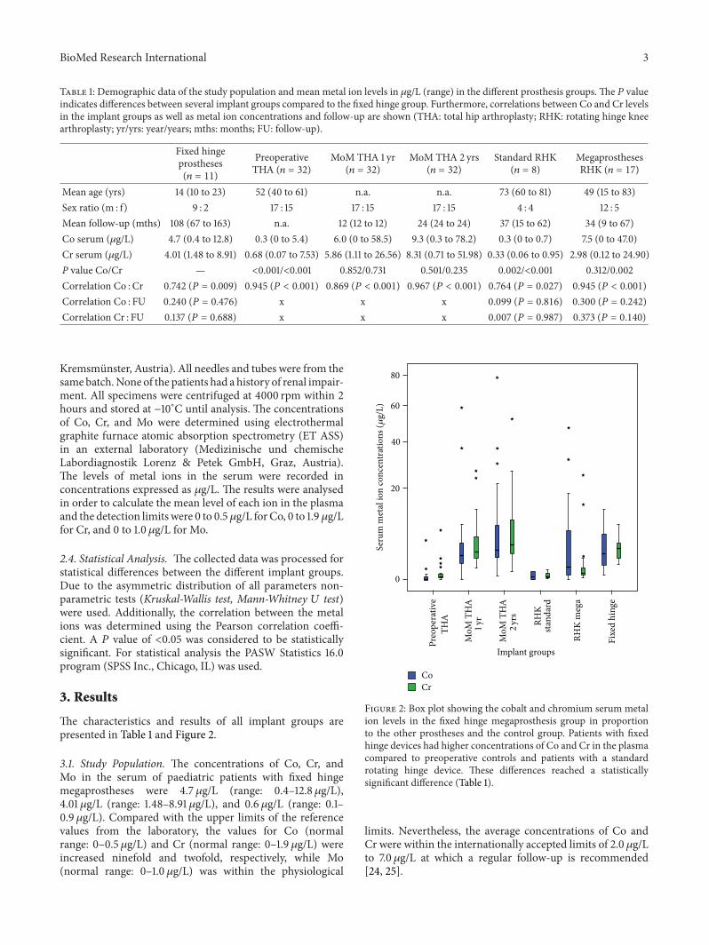
The characteristics and results of all implant groups arepresented in Table 1 and Figure 2.
3.1. Study Population. The concentrations of Co, Cr, andMo in the serum of paediatric patients with fixed hingemegaprostheses were 4.7𝜇g/L (range: 0.4–12.8 𝜇g/L),4.01 𝜇g/L (range: 1.48–8.91 𝜇g/L), and 0.6 𝜇g/L (range: 0.1–0.9 𝜇g/L). Compared with the upper limits of the referencevalues from the laboratory, the values for Co (normalrange: 0–0.5 𝜇g/L) and Cr (normal range: 0–1.9 𝜇g/L) wereincreased ninefold and twofold, respectively, while Mo(normal range: 0–1.0 𝜇g/L) was within the physiological
Seru
m m
etal
ion
conc
entr
atio
ns (𝜇
g/L)
80
60
40
20
0
Implant groups
Preo
pera
tive
THA
MoM
TH
A1
yr
MoM
TH
A2
yrs
RHK
stand
ard
RHK
meg
a
Fixe
d hi
nge
CoCr
Figure 2: Box plot showing the cobalt and chromium serum metalion levels in the fixed hinge megaprosthesis group in proportionto the other prostheses and the control group. Patients with fixedhinge devices had higher concentrations of Co and Cr in the plasmacompared to preoperative controls and patients with a standardrotating hinge device. These differences reached a statisticallysignificant difference (Table 1).
limits. Nevertheless, the average concentrations of Co andCr were within the internationally accepted limits of 2.0 𝜇g/Lto 7.0 𝜇g/L at which a regular follow-up is recommended[24, 25].

4 BioMed Research International
Statistical analysis showed that there was no correlationbetween implant length, follow-up, and serum metal ionconcentrations neither for Co nor for Cr (Table 1).
3.2. Fixed Hinge Megaprostheses versus Control Groups. Theserum concentrations of Co and Cr in the paediatric patientsfollowing fixed hinge total knee arthroplasty were signifi-cantly higher compared to the preoperativeTHAcontrols (CoandCr:𝑃 < 0.001,Mann-Whitney 𝑈 test) and the groupwiththe standard rotating hinge device (Co and Cr: 𝑃 = 0.002and 𝑃 < 0.001, Mann-Whitney 𝑈 test, Table 1). On the otherhand, the serum metal ion levels of patients following MoMTHA were higher at one and two years of follow-up likenedto the fixed hinge group, although these differences were notstatistically significant (Table 1).
Interestingly, the serum concentrations of Cowere higherin the patient group with the rotating hinge megaprosthesis,while the Cr values in this group were lower compared tothe paediatric patients with the fixed hinge prosthesis. Thedifference between theCo concentrationswas not statisticallysignificant (𝑃 = 0.312) while the Cr levels were significantlyhigher in the fixed hinge group (𝑃 = 0.002, Mann-Whitney𝑈 test, Table 1).
There was a positively significant correlation betweenserum Co and Cr concentrations in all implant groups underinvestigation (Pearson correlation coefficient, Table 1).
4. Discussion
The current study revealed increased serum levels for Coand Cr in paediatric patients following fixed hinge total kneearthroplasty using megaprostheses in comparison to severalother implant groups as well as the preoperative controls(Table 1). As expected, referring to previous published data,the values for Mo were within the limits [4, 18, 26–29]. Onthe other hand, patients with rotating hinge megaprosthesesand patients with MoM THA revealed higher serum metalion concentrations compared to the fixed hinge megapros-theses group although these differences were not statisticallysignificant.
Nevertheless, the hypothesis of this study was not sup-ported by the current results. However, continued long-termfollow-up is strictly recommended because especially youngpatients might suffer from possible late effects of chronic,high systemic metal ion exposure with unknown pathologiceffects.
Regarding the official guidelines of the EFORT andAAOSsocieties for MoM THA, the concentrations of Co and Crwere within the accepted limits of 2.0 𝜇g/L to 7.0 𝜇g/L atwhich a regular follow-up is recommended [24, 25], althoughthere are no standards defined for other orthopaedic devicesthan the hip. Nonetheless, the authors believe that theseguidelines should also be accepted for other orthopaedicdevices, especially with metal-on-metal articulation, to per-form continued follow-ups withmetal ion determination dueto the potential harmful effects of high Co and Cr levels.Further, the systemic toxicity of high Co and Cr levels isalways the same and is independent of the type of device.
One limitation of the study is the absence of preoperativeconcentrations of Co and Cr in the serum of patients withfixed hinge and rotating hinge knee prostheses. Another lim-itation was that only a small number of patients were enrolledin the study but it was not possible to include further patientsbecause the device under investigation has been pulled fromthe market several years ago. Furthermore, it should benoticed that there are differences between the follow-ups ofthe different implant groups. Patients with fixed hinge had thelongest follow-up but we do not think that this difference isa major confounding factor because all patients have passedthe running-in phase of the implant and the metal ion levelsmust have stabilized. In addition, statistical analysis did notshow any correlation between time of follow-up and serummetal ion concentrations in the different groups.
On the other hand, it should be noted as significantbenefit that this is the first study evaluating the increaseof serum metal ion levels in paediatric patients followingfixed hinge total knee arthroplasty using megaprostheses.Further, these results were compared to the concentrationsof different other implant groups. All samples of each groupwere evaluated at the same laboratory using the same studyprotocol.
An explanation for the increased concentrations of Coand Cr might be corrosion of the implants, which is knownto be proportional to the surface area of the components,and abrasive wear of the soft tissues. Furthermore, metalions might also be released from the conical junctions of themodular parts from the implants due to fretting.
Our data also shows that the implant’s size (mean resec-tion length: 20 cm)might also play an important role, becausepatientswithmegaprostheses (fixed hinge and rotating hinge)showed higher concentrations of Co and Cr than patientswith a standard rotating hinge device, although there was nocorrelation between serum metal ion levels and implant size[18].
Another reason for increments of Co and Cr in the fixedhinge group might also be the abrasive wear of the polyethy-lene bushes at the side of the hinge axle and the direct metal-on-metal contact (Figures 3(a) and 3(b)). Nevertheless, we areunable to provide a safe range of Co and Cr concentrations inthe serum because of the wide variation of levels encounteredand, therefore, it can be stated that there is a long-termion release following fixed hinge total knee arthroplasty asobserved after rotating hinge total knee arthroplasty [18].
Local soft tissue reactions, delayed hypersensitivity reac-tions, or development of soft tissue formations like pseudo-tumours as results of metal ion debris seems to be unlikelyfollowing total knee arthroplasty using megaprostheses nei-ther with fixed hinge nor with rotating hinge articulation.Nevertheless, there are two cases of soft tissue masses pos-terior to the implant following MoM TDR and one case ofa wear debris induced pseudotumour seven years followingTKA reported in the literature [30–32].
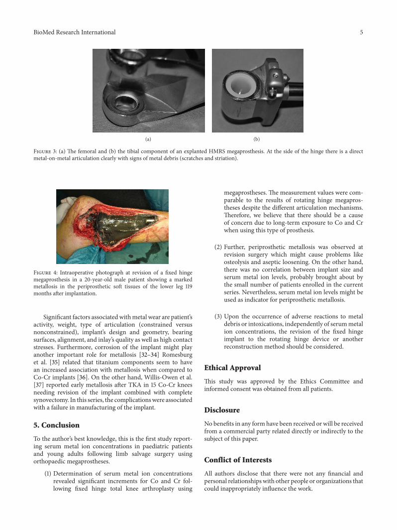
Another observation we made at revision of fixed hingemegaprostheses was periprosthetic metallosis, whereasmetal debris can be found in the joint fluid as well as thesurrounding soft tissues including the synovial layer andjoint capsule (Figure 4).
BioMed Research International 5
(a) (b)
Figure 3: (a) The femoral and (b) the tibial component of an explanted HMRS megaprosthesis. At the side of the hinge there is a directmetal-on-metal articulation clearly with signs of metal debris (scratches and striation).
Figure 4: Intraoperative photograph at revision of a fixed hingemegaprosthesis in a 20-year-old male patient showing a markedmetallosis in the periprosthetic soft tissues of the lower leg 119months after implantation.
Significant factors associatedwithmetal wear are patient’sactivity, weight, type of articulation (constrained versusnonconstrained), implant’s design and geometry, bearingsurfaces, alignment, and inlay’s quality as well as high contactstresses. Furthermore, corrosion of the implant might playanother important role for metallosis [32–34] Romesburget al. [35] related that titanium components seem to havean increased association with metallosis when compared toCo-Cr implants [36]. On the other hand, Willis-Owen et al.[37] reported early metallosis after TKA in 15 Co-Cr kneesneeding revision of the implant combined with completesynovectomy. In this series, the complicationswere associatedwith a failure in manufacturing of the implant.
5. Conclusion
To the author’s best knowledge, this is the first study report-ing serum metal ion concentrations in paediatric patientsand young adults following limb salvage surgery usingorthopaedic megaprostheses.
(1) Determination of serum metal ion concentrationsrevealed significant increments for Co and Cr fol-lowing fixed hinge total knee arthroplasty using
megaprostheses. The measurement values were com-parable to the results of rotating hinge megapros-theses despite the different articulation mechanisms.Therefore, we believe that there should be a causeof concern due to long-term exposure to Co and Crwhen using this type of prosthesis.
(2) Further, periprosthetic metallosis was observed atrevision surgery which might cause problems likeosteolysis and aseptic loosening. On the other hand,there was no correlation between implant size andserum metal ion levels, probably brought about bythe small number of patients enrolled in the currentseries. Nevertheless, serum metal ion levels might beused as indicator for periprosthetic metallosis.
(3) Upon the occurrence of adverse reactions to metaldebris or intoxications, independently of serummetalion concentrations, the revision of the fixed hingeimplant to the rotating hinge device or anotherreconstruction method should be considered.
Ethical Approval
This study was approved by the Ethics Committee andinformed consent was obtained from all patients.
Disclosure
No benefits in any formhave been received or will be receivedfrom a commercial party related directly or indirectly to thesubject of this paper.
Conflict of Interests
All authors disclose that there were not any financial andpersonal relationshipswith other people or organizations thatcould inappropriately influence the work.

6 BioMed Research International
References
[1] J. M. Smolders, A. Hol, W. J. Rijnberg, and J. L. van Susante,“Metal ion levels and functional results after either resurfacinghip arthroplasty or conventional metal-on-metal hip arthro-plasty,” Acta orthopaedica, vol. 82, no. 5, pp. 559–566, 2011.
[2] J. Daniel, H. Ziaee, C. Pradhan, P. B. Pynsent, and D. J. W.McMinn, “Blood and urine metal ion levels in young and activepatients after Birmingham hip resurfacing arthroplasty: four-year results of a prospective longitudinal study,” Journal of Boneand Joint Surgery B, vol. 89, no. 2, pp. 169–173, 2007.
[3] R. de Haan, P. A. Campbell, E. P. Su, and K. A. de Smet,“Revision ofmetal-on-metal resurfacing arthroplasty of the hip:the influence ofmalpositioning of the components,”The Journalof Bone & Joint Surgery, vol. 90, no. 9, pp. 1158–1163, 2008.
[4] D. J. Langton, S. S. Jameson, T. J. Joyce, N. J. Hallab, S. Natu,and A. V. F. Nargol, “Early failure of metal-on-metal bearingsin hip resurfacing and large-diameter total hip replacement: aconsequence of excess wear,” Journal of Bone and Joint SurgeryB, vol. 92, no. 1, pp. 38–46, 2010.
[5] D. J. Langton, T. J. Joyce, S. S. Jameson et al., “Adverse reactionto metal debris following hip resurfacing: the influence ofcomponent type, orientation and volumetric wear,” Journal ofBone and Joint Surgery—Series B, vol. 93, no. 2, pp. 164–171, 2011.
[6] M. Oldenburg, R.Wegner, and X. Baur, “Severe cobalt intoxica-tion due to prosthesis wear in repeated total hip arthroplasty,”Journal of Arthroplasty, vol. 24, no. 5, pp. e825–e820, 2009.
[7] U. E. Pazzaglia, P. Apostoli, T. Congiu, S. Catalani,M.Marchese,and G. Zarattini, “Cobalt, chromium and molybdenum ionskinetics in the human body: data gained from a total hipreplacement with massive third body wear of the head andneuropathy by cobalt intoxication,”Archives of Orthopaedic andTrauma Surgery, vol. 131, no. 9, pp. 1299–1308, 2011.
[8] P. Sauve, J. Mountney, T. Khan, J. de Beer, B. Higgins, and M.Grover, “Metal ion levels after metal-on-metal Ring total hipreplacement: a 30-year follow-up study,” Journal of Bone andJoint Surgery B, vol. 89, no. 5, pp. 586–590, 2007.
[9] W. Steens, J. F. Loehr, G. von Foerster, and A. Katzer, “Chroniccobalt poisoning in endoprosthetic replacement,” Orthopade,vol. 35, no. 8, pp. 860–864, 2006.
[10] C. P. Case, “Chromosomal changes after surgery for jointreplacement,” Journal of Bone and Joint Surgery B, vol. 83, no.8, pp. 1093–1095, 2001.
[11] R. de Haan, C. Pattyn, H. S. Gill, D. W. Murray, P. A. Camp-bell, and K. de Smet, “Correlation between inclination of theacetabular component and metal ion levels in metal-on-metalhip resurfacing replacement,” Journal of Bone and Joint SurgeryB, vol. 90, no. 10, pp. 1291–1297, 2008.
[12] R. M. deSouza, N. R. Parsons, T. Oni, P. Dalton, M. Costa, andS. Krikler, “Metal ion levels following resurfacing arthroplastyof the hip: serial results over a ten-year period,” Journal of Boneand Joint Surgery B, vol. 92, no. 12, pp. 1642–1647, 2010.
[13] D. Ladon, A. Doherty, R. Newson, J. Turner, M. Bhamra, and C.P. Case, “Changes in metal levels and chromosome aberrationsin the peripheral blood of patients after metal-on-metal hiparthroplasty,” Journal of Arthroplasty, vol. 19, no. 8, pp. 78–83,2004.
[14] A.Matthies, R. Underwood, P. Cann et al., “Retrieval analysis of240 metal-on-metal hip components, comparing modular totalhip replacement with hip resurfacing,” The Journal of Bone &Joint Surgery, vol. 93, no. 3, pp. 307–314, 2011.
[15] A. J. Hart, T. Hester, K. Sinclair et al., “The association betweenmetal ions from hip resurfacing and reduced T-cell counts,”TheJournal of Bone& Joint Surgery, vol. 88, no. 4, pp. 449–454, 2006.
[16] W.Maurer-Ertl, J. Friesenbichler, B. Liegl-Atzwanger, G. Kuerzl,R. Windhager, and A. Leithner, “Noninflammatory pseudotu-mor simulating venous thrombosis after metal-on-metal hipresurfacing,” Orthopedics, vol. 34, no. 10, pp. e678–e681, 2011.
[17] L. Savarino, D. Granchi, G. Ciapetti et al., “Ion release in stablehip arthroplasties using metal-on-metal articulating surfaces: acomparison between short-and medium-term results,” Journalof Biomedical Materials Research A, vol. 66, no. 3, pp. 450–456,2003.
[18] J. Friesenbichler,W.Maurer-Ertl, P. Sadoghi, T. Lovse, R.Wind-hager, and A. Leithner, “Serum metal ion levels after rotating-hinge knee arthroplasty: comparison between a standard deviceand a megaprosthesis,” International Orthopaedics, vol. 36, no.3, pp. 539–544, 2012.
[19] S. Garrett, N. Jacobs, P. Yates, A. Smith, and D. Wood, “Dif-ferences in metal ion release following cobalt-chromium andoxidized zirconium total knee arthroplasty,” Acta OrthopaedicaBelgica, vol. 76, no. 4, pp. 513–520, 2010.
[20] A. Zeh, C. Becker,M. Planert, P. Lattke, andD.Wohlrab, “Time-dependent release of cobalt and chromium ions into the serumfollowing implantation of the metal-on-metal Maverick typeartificial lumbar disc (Medtronic Sofamor Danek),” Archives ofOrthopaedic and Trauma Surgery, vol. 129, no. 6, pp. 741–746,2009.
[21] A. Zeh, M. Planert, G. Siegert, P. Lattke, A. Held, and W.Hein, “Release of cobalt and chromium ions into the serumfollowing implantation of the metal-on-metal maverick-typeartificial lumbar disc (Medtronic Sofamor Danek),” Spine, vol.32, no. 3, pp. 348–352, 2007.
[22] P. Bisseling, D. J. Zeilstra, A. M. Hol, and J. L. C. van Susante,“Metal ion levels in patients with a lumbarmetal-on-metal totaldisc replacement: shouldwe be concerned?” Journal of Bone andJoint Surgery B, vol. 93, no. 7, pp. 949–954, 2011.
[23] W. Maurer-Ertl, J. Friesenbichler, P. Sadoghi, M. Pechmann,M. Trennheuser, and A. Leithner, “Metal ion levels inlarge-diameter total hip and resurfacing hip arthroplasty—preliminary results of a prospective five year study after twoyears of follow-up,” BMC Musculoskeletal Disorders, vol. 13,article 56, 2012.
[24] EFORTGroup. Consensus statement, “Current Evidence on theManagement of Metal-on-Metal Bearings,” 2012.
[25] The American Academy of Orthopaedic Surgeons, Cur-rent Concerns with Metal-on-Metal Hip Arthroplasty, 2012,http://www.aaos.org/about/papers/advistmt/1035.asp.
[26] A.Grubl,M.Marker,W. Brodner et al., “Long-term follow-up ofmetal-on-metal total hip replacement,” Journal of OrthopaedicResearch, vol. 25, no. 7, pp. 841–848, 2007.
[27] T. Imanishi,M.Hasegawa, andA. Sudo, “Serummetal ion levelsafter second-generation metal-on-metal total hip arthroplasty,”Archives of Orthopaedic and Trauma Surgery, vol. 130, no. 12, pp.1447–1450, 2010.
[28] D. J. Langton, S. S. Jameson, T. J. Joyce, J. Webb, and A. V. F.Nargol, “The effect of component size and orientation on theconcentrations of metal ions after resurfacing arthroplasty ofthe hip,” Journal of Bone and Joint Surgery B, vol. 90, no. 9, pp.1143–1151, 2008.
[29] D. J. Langton, A. P. Sprowson, T. J. Joyce et al., “Blood metal ionconcentrations after hip resurfacing arthroplasty: a comparative

BioMed Research International 7
study of articular surface replacement and Birmingham hipresurfacing arthroplasties,”The Journal of Bone & Joint SurgeryB, vol. 91, no. 10, pp. 1287–1295, 2009.
[30] M. R. Berry, B. G. Peterson, and D. H. Alander, “A granulo-matous mass surrounding a maverick total disc replacementcausing iliac vein occlusion and spinal stenosis: a case report,”Journal of Bone and Joint Surgery—Series A, vol. 92, no. 5, pp.1242–1245, 2010.
[31] D. A. Cavanaugh, P. D. Nunley, E. J. Kerr, D. J. Werner, and A.Jawahar, “Delayed hyper-reactivity to metal ions after cervicaldisc arthroplasty,” Spine, vol. 34, no. 7, pp. E262–E265, 2009.
[32] A. F. Mavrogenis, G. N. Nomikos, V. I. Sakellariou, G. I.Karaliotas, P. Kontovazenitis, and P. J. Papagelopoulos, “Weardebris pseudotumor following total knee arthroplasty: a casereport,” Journal of Medical Case Reports, vol. 3, article 9304,2009.
[33] G. Ottaviani, M. A. Catagni, and L. Matturri, “Massive metal-losis due to metal-on-metal impingement in substitutive long-stemmed knee prosthesis,” Histopathology, vol. 46, no. 2, pp.237–238, 2005.
[34] V. Sanchis-Alfonso, “Severe metallosis after unicompartmen-tal knee arthroplasty,” Knee Surgery, Sports Traumatology,Arthroscopy, vol. 15, no. 4, pp. 361–364, 2007.
[35] J. W. Romesburg, P. L. Wasserman, and C. H. Schoppe,“Metallosis and Metal-induced synovitis following total kneearthroplasty: review of radiographic and CT findings,” Journalof Radiology Case Reports, vol. 4, no. 9, pp. 7–17, 2010.
[36] B. N. Weissman, R. D. Scott, G. W. Brick, and J. M. Corson,“Radiographic detection of metal-induced synovitis as a com-plication of arthroplasty of the knee,”The Journal of Bone& JointSurgery A, vol. 73, no. 7, pp. 1002–1007, 1991.
[37] C. A. Willis-Owen, G. C. Keene, and R. D. Oakeshott, “Earlymetallosis-related failure after total knee replacement: a reportof 15 cases,” Journal of Bone and Joint Surgery—Series B, vol. 93,no. 2, pp. 205–209, 2011.

Submit your manuscripts athttp://www.hindawi.com
ScientificaHindawi Publishing Corporationhttp://www.hindawi.com Volume 2014
CorrosionInternational Journal of
Hindawi Publishing Corporationhttp://www.hindawi.com Volume 2014
Polymer ScienceInternational Journal of
Hindawi Publishing Corporationhttp://www.hindawi.com Volume 2014
Hindawi Publishing Corporationhttp://www.hindawi.com Volume 2014
CeramicsJournal of
Hindawi Publishing Corporationhttp://www.hindawi.com Volume 2014
CompositesJournal of
NanoparticlesJournal of
Hindawi Publishing Corporationhttp://www.hindawi.com Volume 2014
Hindawi Publishing Corporationhttp://www.hindawi.com Volume 2014
International Journal of
Biomaterials
Hindawi Publishing Corporationhttp://www.hindawi.com Volume 2014
NanoscienceJournal of
TextilesHindawi Publishing Corporation http://www.hindawi.com Volume 2014
Journal of
NanotechnologyHindawi Publishing Corporationhttp://www.hindawi.com Volume 2014
Journal of
CrystallographyJournal of
Hindawi Publishing Corporationhttp://www.hindawi.com Volume 2014
The Scientific World JournalHindawi Publishing Corporation http://www.hindawi.com Volume 2014
Hindawi Publishing Corporationhttp://www.hindawi.com Volume 2014
CoatingsJournal of
Advances in
Materials Science and EngineeringHindawi Publishing Corporationhttp://www.hindawi.com Volume 2014
Smart Materials Research
Hindawi Publishing Corporationhttp://www.hindawi.com Volume 2014
Hindawi Publishing Corporationhttp://www.hindawi.com Volume 2014
MetallurgyJournal of
Hindawi Publishing Corporationhttp://www.hindawi.com Volume 2014
BioMed Research International
MaterialsJournal of
Hindawi Publishing Corporationhttp://www.hindawi.com Volume 2014
Nano
materials
Hindawi Publishing Corporationhttp://www.hindawi.com Volume 2014
Journal ofNanomaterials