clinical ultrasound transducer degradation effects on the
TRANSCRIPT
Clinical Ultrasound Transducer Degradation Effects on the Accuracy
of Spectral Doppler Velocity Measurements
Nicole R. Bloms, B.S., R.T.(R)
NCCAAPM 2012 Spring Meeting
Young Investigator Presentation

Overview
• Will be a poster at the 54th Annual AAPM Meeting
• Today:
• Brief ultrasound review
• Overview of equipment
• Summarize progress of study
• Preliminary results
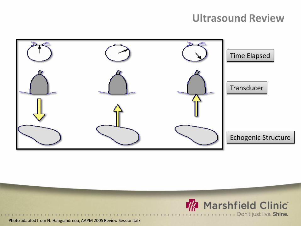
Ultrasound Review
Time Elapsed
Transducer
Echogenic Structure
Photo adapted from N. Hangiandreou, AAPM 2005 Review Session talk
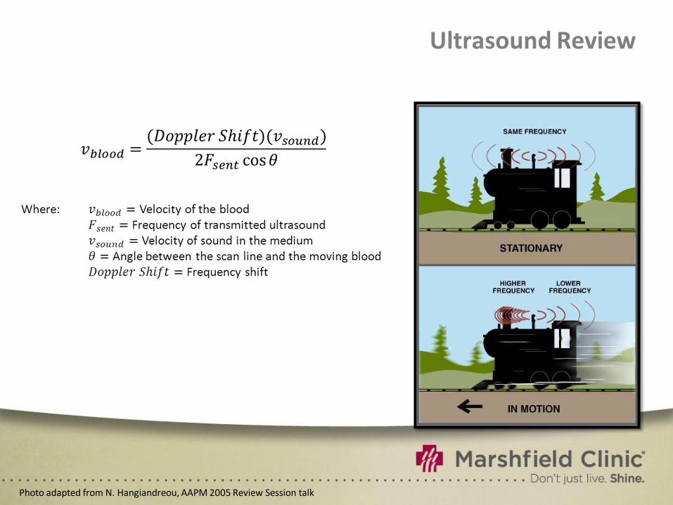
Ultrasound Review
Photo adapted from N. Hangiandreou, AAPM 2005 Review Session talk
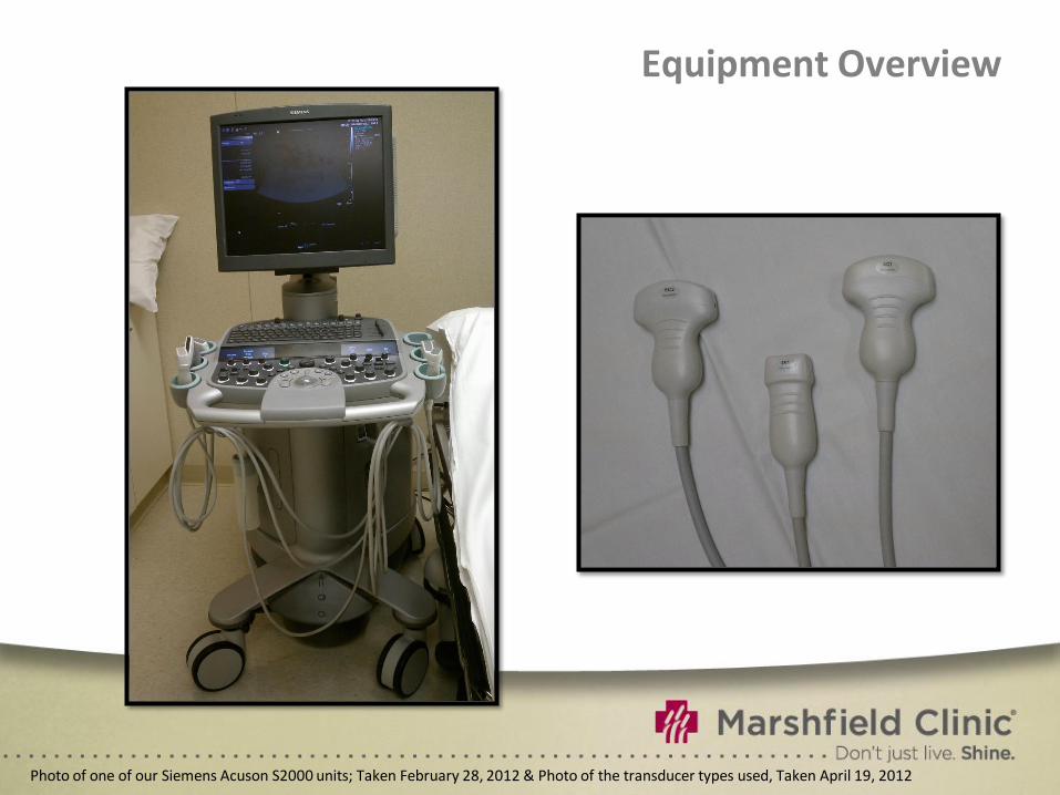
Equipment Overview
Photo of one of our Siemens Acuson S2000 units; Taken February 28, 2012 & Photo of the transducer types used, Taken April 19, 2012
Equipment Overview
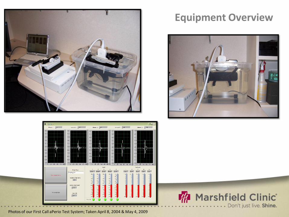
Photos of our First Call aPerio Test System; Taken April 8, 2004 & May 4, 2009
Equipment Overview
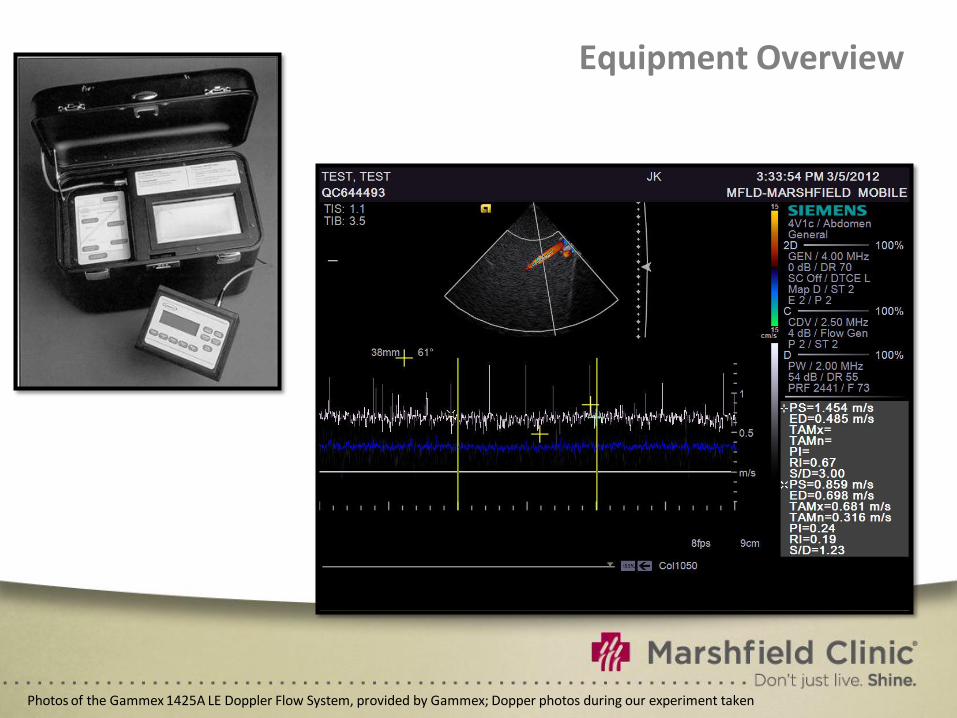
Photos of the Gammex 1425A LE Doppler Flow System, provided by Gammex; Dopper photos during our experiment taken
Equipment Overview
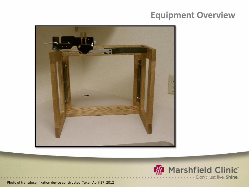
Photo of transducer fixation device constructed, Taken April 17, 2012
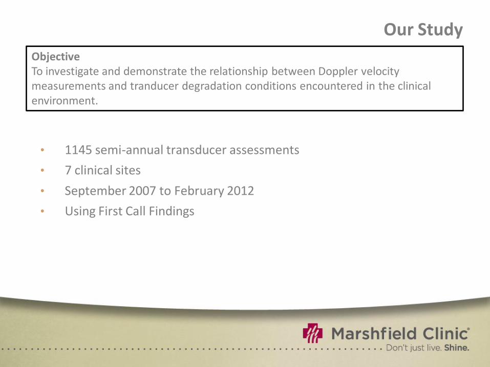
Our Study
• 1145 semi-annual transducer assessments
• 7 clinical sites
• September 2007 to February 2012
• Using First Call Findings
Objective To investigate and demonstrate the relationship between Doppler velocity measurements and tranducer degradation conditions encountered in the clinical environment.
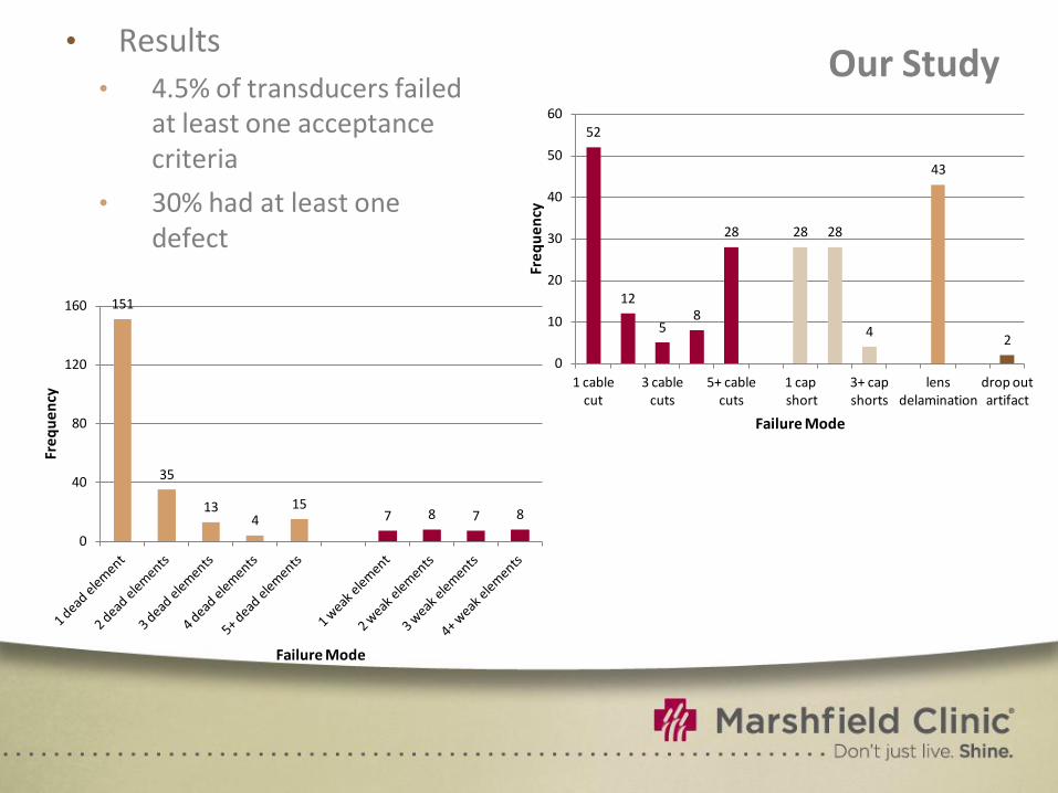
Our Study
151
35
13 4
15 7 8 7 8
0
40
80
120
160
Fre
qu
en
cy
Failure Mode
52
12
5 8
28 28 28
4
43
2
0
10
20
30
40
50
60
1 cable cut
3 cable cuts
5+ cable cuts
1 cap short
3+ cap shorts
lens delamination
drop out artifact
Fre
qu
en
cy
Failure Mode
• Results
• 4.5% of transducers failed at least one acceptance criteria
• 30% had at least one defect

Our Study
• We confirmed this hypothesis with simulated transducer degradation
Hypothesis Only the most severe defects and degradation conditions result in noticeable deviation in the Doppler velocity measurements.
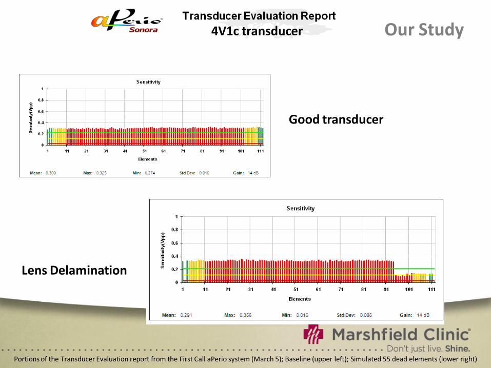
Our Study
Portions of the Transducer Evaluation report from the First Call aPerio system (March 5); Baseline (upper left); Simulated 55 dead elements (lower right)
Lens Delamination
4V1c transducer
Good transducer
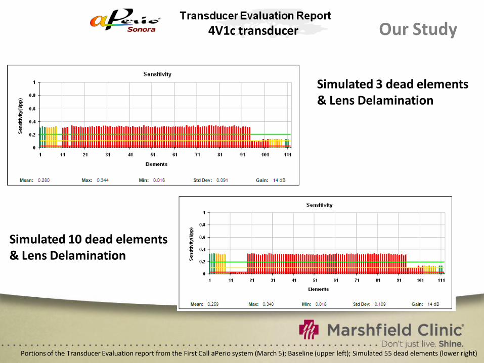
Our Study
Portions of the Transducer Evaluation report from the First Call aPerio system (March 5); Baseline (upper left); Simulated 55 dead elements (lower right)
Simulated 3 dead elements & Lens Delamination
Simulated 10 dead elements & Lens Delamination
4V1c transducer
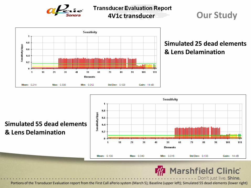
Our Study
Portions of the Transducer Evaluation report from the First Call aPerio system (March 5); Baseline (upper left); Simulated 55 dead elements (lower right)
Simulated 25 dead elements & Lens Delamination
Simulated 55 dead elements & Lens Delamination
4V1c transducer
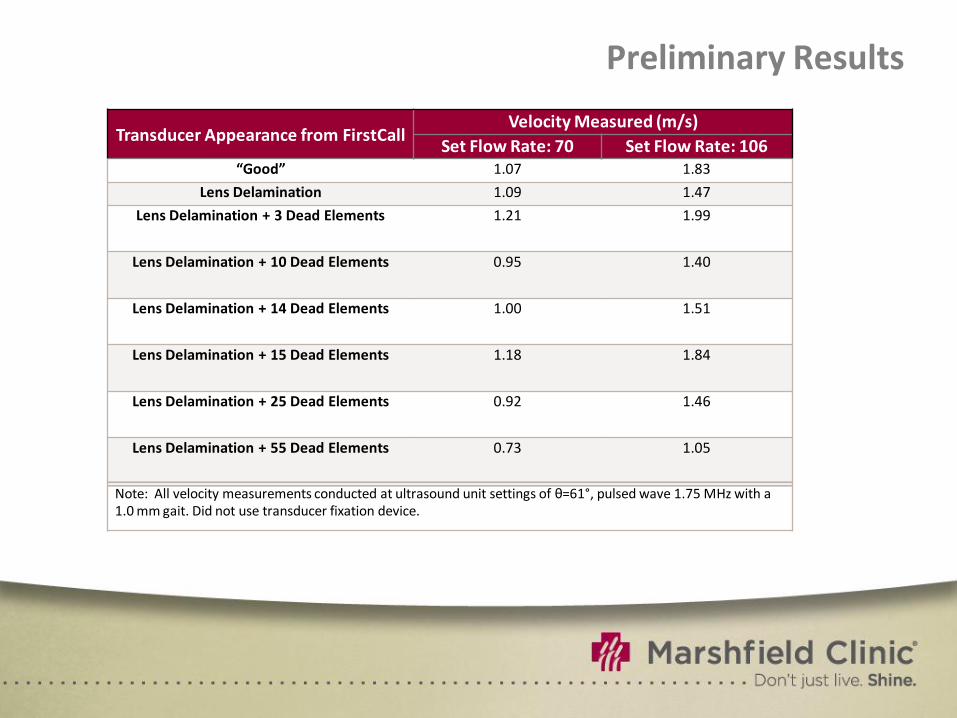
Preliminary Results
Transducer Appearance from FirstCall Velocity Measured (m/s)
Set Flow Rate: 70 Set Flow Rate: 106
“Good” 1.07 1.83
Lens Delamination 1.09 1.47
Lens Delamination + 3 Dead Elements 1.21 1.99
Lens Delamination + 10 Dead Elements 0.95 1.40
Lens Delamination + 14 Dead Elements 1.00 1.51
Lens Delamination + 15 Dead Elements 1.18 1.84
Lens Delamination + 25 Dead Elements 0.92 1.46
Lens Delamination + 55 Dead Elements 0.73 1.05
Note: All velocity measurements conducted at ultrasound unit settings of θ=61°, pulsed wave 1.75 MHz with a 1.0 mm gait. Did not use transducer fixation device.
Future Investigation
• Perform doppler flow measurements using tranducer fixation device
• In essence, repeating our ‘preliminary results’
• Streamline First Call testing procedure
• Possible diagram of back of transducer connection
Questions?