clinicalisolatesofmycobacteriumtuberculosisdifferin...
TRANSCRIPT
Hindawi Publishing CorporationClinical and Developmental ImmunologyVolume 2012, Article ID 152546, 11 pagesdoi:10.1155/2012/152546
Research Article
Clinical Isolates of Mycobacterium tuberculosis Differ inTheir Ability to Induce Respiratory Burst and Apoptosis inNeutrophils as a Possible Mechanism of Immune Escape
Marıa M. Romero,1 Luciana Balboa,1 Juan I. Basile,1 Beatriz Lopez,2 Viviana Ritacco,2
Silvia S. de la Barrera,1 Marıa C. Sasiain,1 Lucıa Barrera,2 and Mercedes Aleman1
1 IMEX-CONICET-ANM, Academia Nacional de Medicina, 1425 Buenos Aires, Argentina2 Servicio de Micobacterias, Hospital Malbran, Pacheco de Melo 3081, 1425 Buenos Aires, Argentina
Correspondence should be addressed to Mercedes Aleman, [email protected]
Received 4 April 2012; Accepted 29 April 2012
Academic Editor: Eyad Elkord
Copyright © 2012 Marıa M. Romero et al. This is an open access article distributed under the Creative Commons AttributionLicense, which permits unrestricted use, distribution, and reproduction in any medium, provided the original work is properlycited.
Tuberculosis pathogenesis was earlier thought to be mainly related to the host but now it appears to be clear that bacterial factorsare also involved. Genetic variability of Mycobacterium tuberculosis (Mtb) could be slight but it may lead to sharp phenotypicdifferences. We have previously reported that nonopsonized Mtb H37Rv induce apoptosis of polymorphonuclear neutrophils(PMNs) by a mechanism that involves the p38 pathway. Here we evaluated the capability to induce PMN apoptosis of two prevalentMtb lineages in Argentina, the Latin America and Mediterranean (LAM), and Haarlem, using the H37Rv as a reference strain.Results showed that LAM strains strongly induced apoptosis of PMN which correlated with the induction of reactive oxygenspecies (ROS) production and p38 activation. Interestingly, the highly prosperous multidrug-resistant M strain, belonging to theHaarlem lineage, lacked the ability to activate and to induce PMN apoptosis as a consequence of (1) a weak ROS production and(2) the contribution of antiapoptotic mechanisms mediated at least by ERK. Although with less skill, M is able to enter the PMNso that phenotypic differences could lead PMN to be a reservoir allowing some pathogens to prevail and persist over other strainsin the community.
1. Introduction
Polymorphonuclear neutrophils (PMNs) are key compo-nents of the first line of defense against bacterial and fungalpathogens. In tuberculosis (TB), the influx of PMN to thelung is one of the first events in the pathogenesis of thedisease. Bacterial products elicit the upregulation of the β2integrin, CD11b, as well as the release of pronflammatorycytokines by PMN contributing to the recruitment of leuko-cytes at the site of infection and amplifying the immuneresponse [1, 2]. Microbicidal mechanisms of PMNs includethe release of proteolytic enzymes and antimicrobial peptidesas well as the rapid production of reactive oxygen species(ROS) essential for bacterial killing which also enhanceinflammatory reactions [3].
We have previously demonstrated that nonopsonizedvirulent Mycobacterium tuberculosis (Mtb) strain H37Rv
induces PMN activation and accelerates their apoptosis invitro at a low Mtb : PMN ratio [4]. Moreover, in infected indi-viduals circulating PMN become activated [5] and arerecruited to the lungs in the early infection, where theyunderlie apoptosis [6]. Accelerated apoptosis has beenobserved in PMN after mycobacterial internalization, pos-sibly dependent on oxidative processes [4, 7], enhanced byhigh concentrations of TNF-alpha [8], and mediated viaTLR2- and mitogen-activated protein kinase (MAPK) p38-dependent pathways [9]. Along these lines, it has been dem-onstrated that PMN response to cytokines and other releasedpro-inflammatory agents as well as activation of PMNinvolves the MAPK pathway [10]. In the context of mycobac-terial infection, the mode of PMN cell death could influencedisease outcome: if a PMN cannot kill an ingested organ-ism, then necrosis could offer an inflammatory reactionwhereas, if PMN became apoptotic, then PMN-dendritic cell
2 Clinical and Developmental Immunology
Spoligotyping
H37Rv
LAM
T
LAM
Strain Lineage
H
M
Haarlem
Ra LAM
Haarlem
IS6110 RFLP
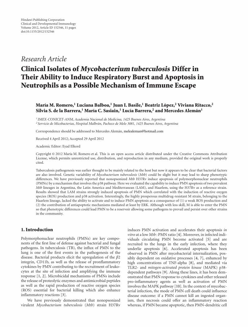
Figure 1: Spoligotyping and IS6110-RFLP pattern profiles of Mtb strains used as antigens in this study: reference virulent strain H37Rv,local-drug-sensitive (LAM, 10406) and drug-resistant (Ra, 11608) LAM strains, drug-sensitive Haarlem strain (H, 12425), and MDRHaarlem strain, M (6548).
cross-presentation and increased lymphocyte proliferationoccurs in response to Mtb, directly influencing the develop-ment of an acquired immune response [11].
In Argentina a total of 11,464 new cases of TB werereported in 2006, with an incidence of 29.1 per 100,000inhabitants. As in other South American countries [2, 12]the vast majority of Mtb strains circulating in Argentinabelong to the Latin American and Mediterranean (LAM)and the Haarlem lineages [2, 13, 14]. Within the Haarlemlineage, the multidrug-resistant (MDR) M strain is highlyprosperous in Argentina and is able to build up furtherdrug resistance without impairing its ability to spread [15].The genetic variability of Mtb strains might lead to sharpphenotypic differences which could be responsible for adifferential modulation of host immune response impactingin the onset and progression of the disease [16]. In thiscontext, differential immune responses among resistant Mtbstrains have been reported [17, 18].
Therefore, in this work we evaluated whether clinicalisolates of Mtb differ in their capability to induce PMNapoptosis, in order to evaluate apoptosis as a possible mech-anism that leads to pathogen survival inside PMN allowingsome strains to prevail and persist over other strains in thecommunity.
2. Methods
2.1. Isolate Selection and Preparation of Bacterial Strains. Mtbisolates representative of prevalent LAM and Haarlem lin-eages were obtained from sputum-culture-positive patientswith TB in Argentina. The isolates had been previouslysubmitted to drug susceptibility testing and genotypingby IS6110 DNA fingerprinting and spoligotyping usingstandardized protocols. The following strains were evaluated:a drug-susceptible strain (LAM, 10406) and a drug-resistantstrain (Ra, 11608) from the LAM lineage and a drug-susceptible Haarlem strain (H, 12425) and an MDR strainfrom the Haarlem lineage (isolate 6548 as representative ofthe outbreak strain M). Spoligotyping and IS6110 restriction
fragment length polymorphism pattern profiles of Mtbstrains used as antigens are depicted in Figure 1. Thestrains belonged to the collection kept at the ReferenceLaboratory for Mycobacteria at the Instituto Nacional deEnfermedades Infecciosas ANLIS “Carlos G. Malbran” inBuenos Aires, Argentina. Laboratory reference strain H37Rvfrom T family was kindly provided by I. N. de Kantor(Former Head of TB Laboratory, INPPAZ PAHO/WHO)(Figure 1). All strains were grown in Middlebrook 7H9 broth(Difco Laboratories, Detroit, MI, USA) at 37◦C in 5% CO2
until log phase. Mycobacteria were harvested, washed threetimes, and suspended in phosphate-buffered saline (PBS)free of pyrogen. Bacteria were killed by gamma irradiationor heat-killed and suspended in PBS at an OD600 nm of 1(∼108 bacteria/mL) and stored at −20◦C until their use.LPS from Escherichia coli 0111:B4 was purchased from SigmaChemicals Co. (St. Louis, USA).
2.2. Antibodies and Reagents. Oxidase inhibitor, diphenyleneiodonium DPI, was provided by Cayman Chemical (Michi-gan, USA), the specific inhibitor of p38, SB203580, and thespecific inhibitor of ERK, PD98059, were purchased fromCalbiochem-Behring (La Jolla, CA, USA). DMSO (SigmaCo.) was added to cultures at 0.1% (vol/vol) as a solventcontrol.
Mouse antibodies (Abs) against Caspase-3 (BD Bios-ciences Pharmingen, California, USA) phospho (Thr202/Tyr204)-ERK1/2, and phosphor (Thr180/Tyr182)-p38 werepurchased from Santa Cruz Biotechnology (Santa Cruz, CA,USA). Mouse antihuman CD11b, CD66b and CD16 werepurchased from eBioscience (San Diego, CA, USA).
2.3. PMN Purification and Culture. PMNs were isolated fromheparinized venous blood from healthy donors by Ficoll-Hypaque gradient centrifugation [19] followed by sedimen-tation in 3% dextran (Sigma Chem. Co, St. Louis Mo,USA). The PMN-rich supernatant was then collected andresidual red blood cells were removed by hypotonic lysis.The cells were washed immediately and resuspended at

Clinical and Developmental Immunology 3
3 × 10⁶ cells/mL in RPMI-1640 medium (Gibco, NY, USA)supplemented with 1% heat-inactivated Fetal Calf Serum(FCS) (Gibco) and 50 μg/mL gentamycin (complete media,CM). The viability was consistently >95% as determinedby trypan blue dye exclusion. The purity of the final PMNpreparation was up to 95% as assessed by morphologicalexamination by staining with Wright-Giemsa and by FACSlight scatter patterns. Cultures were performed by incubating1 mL of a PMN suspension (3× 10⁶ cell) in Falcon 2063 tubesstimulated with Mtb strains, at different Mtb : PMN ratios.
2.4. Surface Cell Staining. Cell surface expression of CD11band FcRIIIb (CD16) in recently isolated or 3 and 18-h-cultured PMN was evaluated by direct immunofluo-rescence using saturating concentrations of monoclonalmouse antihuman CD11b-PE- and -CD16-FITC-conjugatedantibodies. Briefly, 5 × 10⁵ cells were incubated with theantibody for 20 min on ice. Cells were washed, fixed in500 μL of 1% paraformaldehyde. Using a FACScan (Becton-Dickinson Immunocytometry Systems, San Jose, CA), 10,000events were collected in linear mode for forward scatter(FSC) and side scatter (SSC), and log amplification for FL-1 and FL-2. Analysis was performed using the CellQuestsoftware (Becton-Dickinson) and isotype-matched controlswere used to determine autofluorescence and nonspecificstaining. Results were expressed as percentages of positivecells and as mean fluorescence intensity (MFI).
2.5. Intracellular Cell Staining. The activated cytoplasmicprotein caspase-3 was measured in PMN by using a Fixand Perm kit (Caltag, Burlingame, CA, USA). Briefly, 3 ×10⁶ PMNs were incubated with different Mtb strains at 1 : 2Mtb : PMN ratio for 5 h and thereafter cells were washedand resuspended in 100 μL solution A (fixation) for 15 minat room temperature. After washing with PBS containing1% Na azide and 5% FCS, cells were resuspended insolution B (permeabilization) and mouse antihuman FITC-conjugated caspase-3 antibody specific for active caspase-3(BD Pharmingen). After 20-min incubation on ice in thedark, cells were washed, resuspended in isoflow, and analyzedin the same manner as mentioned above.
Phosphorylated form of p38 and ERK cytoplasmicproteins were measured in permeabilized cells as describedabove. PMNs were incubated with Mtb strains at a 1 : 2Mtb : PMN ratio for 1 h. Thereafter, cells were washed withPBS and resuspended in 100 μL solution A (fixation) for15 min at room temperature. After washing with PBS con-taining 1% Na azide and 5% FCS, cells were suspendedin solution B (permeabilization) and the mouse anti-human p-p38 IgM anti-p-p38-FITC- or anti-human p-ERK-conjugated antibody (Santa Cruz Biotechnology, Santa Cruz,CA., USA). After 20-min incubation on ice in the dark, cellswere washed, resuspended in isoflow and acquired in a flowcytometry and analyzed as previously described.
2.6. Detection of Apoptosis Using Annexin-FITC by FACS.The percentage of apoptotic PMN was assessed based onthe Annexin V-FITC (Sigma) protein-binding assay. Briefly,5 μL of Annexin V-FITC (10 μg/mL) and 5 μL of propidium
iodide (PI) (250 mg/mL) (Sigma) were added to 1.2 ×10⁶ cells in 500 μL of binding buffer and incubated for15 min at room temperature in the dark, as previouslydescribed [4]. Cells were washed, resuspended in bindingbuffer acquired in a flow cytometry, as previously described.AV−/PI− cell population was regarded as alive, AV+/PI− wasconsidered as an early apoptotic population, and the AV+/PI+
population represented late stage apoptotic or necrotic cells.Alternatively, to modify cellular p38 and ERK activity,SB20358 (20 μM) or PD98059 (50 μM) inhibitors was addedto 1 mL PMN suspension before culture, at 37◦C for 30 min.When indicated, 10 mM H2O2 were added 15 min after Mtbtreatment.
2.7. Assay for Oxidative Burst Using Dihydrorhodamine 123(DHR). Intracellular ROS levels were measured by Dihy-drorhodamine assays 123 (DHR). DHR was dissolved inDMSO at a concentration of 20 μg/mL and stored in aliquotsat −70◦C until use. Briefly, 5 × 10⁵ PMNs were incubatedwith 100 μl DHR (5 μg/mL) for 15 min at 37◦C. Afterward,gamma-irradiated Mtb strains (1 × 10⁶ bacilli/mL) wereadded to the culture at 1 : 2, 5 : 1, 20 : 1, and 50 : 1 Mtb : PMNratios for additional 90 min. When indicated, heated-killedMtb (ØMtb) were employed.
2.8. Phagocytosis Assay. As an indirect measure of phago-cytosis ROS production induced by DHR-labeled Mtb wasperformed. Briefly, Mtb were incubated with 5 μg/mL DHRfor 30 min at 37◦C followed by an extensive washing toremove free DHR and suspended in RPMI 1640 plus10% FCS. Thereafter, 5 × 10⁵ PMNs were incubated withDHRMtb strains at a 50 : 1 ratio for 90 min at 37◦C, washedand centrifuged at 100 g evaluated immediately in a flowcytometer as previously described.
Direct measure of phagocytosis, was performed by themethod described by Busetto et al. [20]. Briefly, 3 × 10⁶/mLPMNs were suspended in media 10% FSC and were incu-bated at 37◦C in a shaking water bath with nonopsonizedFITC-labeled Mtb strains. PMN-Mtb mixtures (ratio 10 : 1)were incubated for 2 hr at 37◦C. To stop phagocytosis,aliquots of the incubation mixtures were withdrawn intothe tubes used for cytometric analysis containing an equalvolume of 250 μg/mL Trypan Blue (TB) dissolved in ice-cold 0.1 M citrate buffer, pH 4.0. After 1 min incubationin ice, the samples were analyzed by flow cytometry. Eachsample was collected for 30 seconds at the slowest flow rateto minimize the coincidental appearance of free bacteria andPMN in the laser beam. The data were then analyzed byusing CellQuest software from Becton Dickinson. The meannumber of ingested bacteria per neutrophil was assessed (i.e.,both attached and internalized particles).
2.9. Statistics. The statistical analysis of the data was per-formed using one-way ANOVA followed by Tukey’s multiplecomparison tests to compare more than two groups followedby Wilcoxon test to compare two groups. The significancewas considered as P < 0.05. The graphical representation ofthe values is given by mean ± SEM.
4 Clinical and Developmental Immunology
H37Rv LAM Ra H M0
30
60
90A
v+(%
)
∗
##
∗∗
∗∗∗∗
—
(a)
H37Rv LAM Ra H M0
10
20
30#
#
∗∗
—
MFI
cas
pase
3
∗∗∗∗∗∗
∗
(b)
oh LAM Ra H M0
25
50
75
100
#
∗
∗ ∗∗
∗
— H37Rv
CD
16 (
%)
(c)
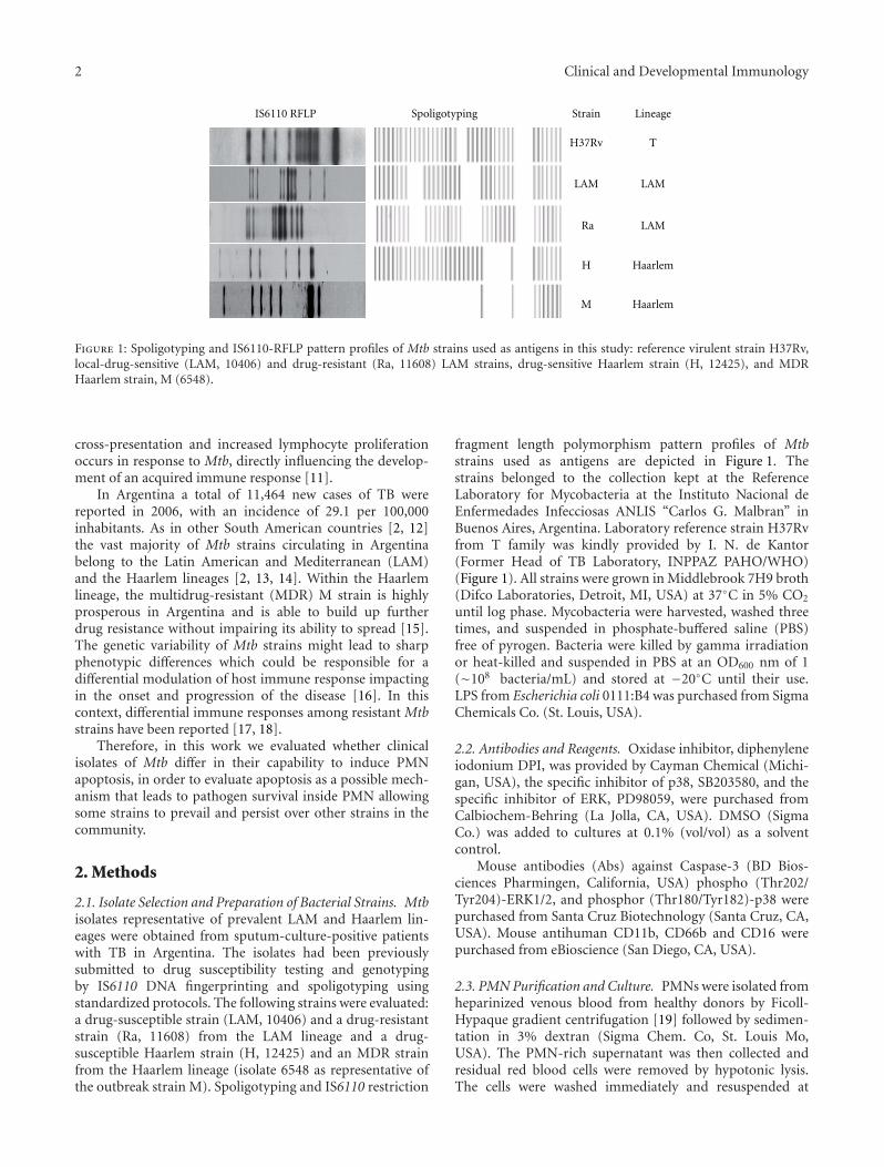
Figure 2: PMN apoptosis induced by different Mtb strains. PMNs (3 × 10⁶/mL) were cultured in media alone (control, —) or stimulatedwith Mtb strains (H37Rv, LAM, Ra, H, and M) at 1 : 2 Mtb : PMN ratio and parameters of apoptosis were evaluated by flow cytometricanalysis. (a) Expression of Annexin V-FITC (AV-FITC) on PMN cultured for 18 h. Results are expressed as media ± SEM of percentage ofpositive cells (n = 20). Control (—) versus LAM, Ra, and H: ∗∗P < 0.001; H37Rv: ∗P < 0.01; LAM and Ra versus other strains: #P < 0.001.(b) Intracellular expression of activated cytoplasmic protein caspase-3 in PMN after 5 h culture with or without Mtb stimulation. Resultsare expressed as media ± SEM of MFI. Control (—) versus LAM and Ra: ∗∗∗P < 0.001; H37Rv: ∗∗P < 0.01; H ∗P < 0.05; LAM and Raversus other strains: #P < 0.001. (c) The percentage of fresh (0 h) and 16 h cultured cells expressing FcRIIIb (CD16) was measured. 0 h versuscontrol: #P < 0.0002; control vesrus Mtb strains: ∗P < 0.0001.
3. Results
3.1. PMN Apoptosis Induced by Different Mtb Strains. Wehave previously described that nonopsonized strain MtbH37Rv is able to induce PMN apoptosis at low Mtb : PMNratio [4]. In order to evaluate whether Mtb strains differ intheir capability to induce apoptosis, PMNs were incubated at1 : 2 Mtb : PMN ratio for 18 h and, thereafter, apoptosis wereevaluated by measuring the percentage of cells expressingFITC-conjugated Annexin V (AV+). Flow cytometric analysisshows that all strains, except M, induced significant increaseof AV+ PMNs respect to spontaneous apoptosis. Further-more, clinical isolates belonging to LAM lineage (LAM andRa) were the highest inducers of apoptosis (Figure 2(a)).LPS, that exerts an antiapoptotic effect on cultured PMN[21], was used as a control in the apoptosis assay (data notshown). Most apoptotic signaling pathways are originatedfrom death receptor linkage or stress stimuli converging
on activation of caspases, key executors of apoptosis. Toassess the involvement of Caspase-3 (casp-3) in Mtb-inducedPMN apoptosis, expression of the activated form of Casp-3was evaluated in PMN incubated with different Mtb strains(Mtb : PMN 1 : 2) for 5 h. As it is shown in Figure 2(b), allstrains, except M, induced Casp-3 activation being LAMand Ra the highest inducers. These results are consistentwith those obtained in the apoptosis assay. In addition, nodifferences were observed in the loss of FcγRIIIb receptor(CD16) expression among strains. As it is known, theloss of CD16 expression on PMN surface correlates withspontaneous apoptosis in culture [22] so that our resultsshow differences in Mtb-induced apoptosis whereas CD16shedding was independent of the Mtb strain (Figure 2(c)).
3.2. CD11b, CD66b, and p-p38 Expression Induced by MtbStrains. CD11b and CD66b are proteins found in the
Clinical and Developmental Immunology 5
H37Rv Ra H M0
300
600
900
1200
1500
1800
— LAM
##
∗∗∗∗ ∗∗
∗∗
∗
MFI
CD
11b
(a)
H37Rv LAM Ra H M0
50
100
150
200
250
—
∗∗
∗∗ ∗∗
∗∗
##
∗
MFI
CD
66b
(b)
H37Rv LAM Ra H M0
5
10
15
20
25
#
—
∗∗
∗
#∗
MFI
p-p
38
(c)
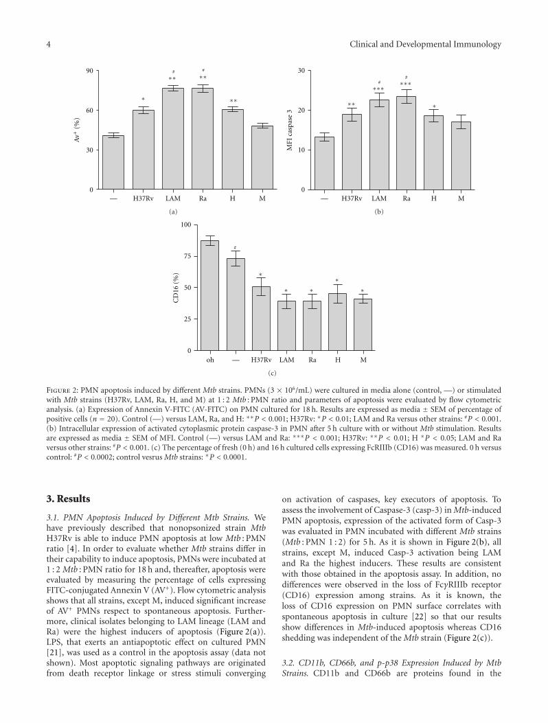
Figure 3: CD11b, CD66b, and p-p38 MAPK expression in PMN induced by Mtb strains. PMNs (3 ×10⁶/mL) were cultured in mediaalone (control, —) or stimulated with Mtb strains at 1 : 2 Mtb : PMN ratio for 3 h. (a) and (b) Surface expression of activation markers wasevaluated by flow cytometry and results are expressed as media ± SEM of MFI (n = 10). Statistical differences: CD11b and CD66b: control(—) versus H37Rv, LAM, Ra and H: ∗∗P < 0.001; control versus M: ∗P < 0.05; LAM and Ra versus other strains: #P < 0.01; (c) PMNs (3× 10⁶/mL) were cultured in media alone (control, —) or stimulated with Mtb strains at 1 : 2 Mtb : PMN ratio for 1 h. Intracellular activatedform of p38 (p-p38) was measured by flow cytometry. Results are expressed as media ± SEM (n = 10). Statistical differences: control versusH37Rv, LAM, Ra, and H: ∗P < 0.001; LAM and Ra versus other strains: #P < 0.001.
membrane of the various PMN granules and appear on thecell surface after exocytosis and release of granular contentsupon activation [23]. Indeed, the CD11b receptor is dra-matically upregulated at the surface of activated PMN whichextravasate through the vessels and migrate to the site ofinfection, thereby enhancing the effectors’ functions of thesecells. As shown in Figure 3, all Mtb strains enhanced CD11band CD66b expression in 3 h culture PMN. Particularly,we found that susceptible and drugs-resistant LAM strainsshowed the highest activation level, while M showed thelowest one (Figures 3(a) and 3(b)). As previously demon-strated, activation- and apoptosis-induced H37Rv involvesthe activation of mitogen-activated protein kinase p38 [4, 9].In this work, flow cytometric analysis of the activated formof p38 (p-p38) showed that susceptible and drugs resistantLAM induced the highest expression of p-p38 whereas, withM, p-p38 was not detectable. These results suggest a differentparticipation of p38 in apoptosis induced by Mtb strains(Figure 3(c)). From now on, we will refer to both LAM andRa as “LAM,” because no differences were found betweensusceptible or drugs-resistant LAM clinical isolates.
3.3. Role of p-p38, ERK, and ROS in Differential Mtb-InducedPMN Apoptosis. To evaluate if Mtb strains differ in themechanisms underlying the apoptotic process, PMNs werecultured with Mtb in presence of different inhibitors for 18 hand apoptosis was evaluated by AV expression. According toour previous results obtained with H37Rv [9], SB203580, aspecific p38 inhibitor, only inhibited the apoptosis inducedby LAM and H (Figure 4(a)). Considering that ROS areinvolved in the regulation of MAP-kinase-dependent apop-totic pathway [24], we used an oxidase inhibitor (DPI)to assess the participation of ROS in apoptosis and weobserved that generation of ROS was essential for triggeringapoptosis induced by all strains (Figure 4(a)). Strikingly, Mwas able to induce apoptosis when PMNs were incubatedwith a specific ERK inhibitor, PD98059, suggesting that thisstrain could be triggering anti-apoptotic mechanisms, whichinvolve ERK (Figure 4(a)). In order to evaluate the gradeof participation of anti-apoptotic signals, H2O2 was addedto the culture before challenging with different Mtb strainsand apoptosis was measured by AV. Interestingly, apoptosisinduced by all strains was enhanced in the presence of H2O2.

6 Clinical and Developmental Immunology
0
10
20
30
4050
60
70
80
SB—
H37
Rv
SB PD
DP
I
LAM SB PD
DP
I H SB PD
DP
I M PD
DP
I
Av+
(%)
∗
###
∗∗
∗∗∗
δ
δ δ
ϕ
(a)
100
101
102
103
104
100 101 102 103 104104
H37Rv
44%
AV
100
101
102
103
104
100 101 102 103
AV
100
101
102
103
104
100 101 102 103 104
AV
100
101
102
103
104
100 101 102 103 104
AV
100
101
102
103
104
100 101 102 103 104
AV
100
101
102
103
104
100 101 102 103 104104
AV
100
101
102
103
104
100 101 102 103
AV
100
101
102
103
104
100 101 102 103 104
AV
100
101
102
103
104
100 101 102 103 104
AV
100
101
102
103
104
100 101 102 103 104
AV
Pi
AV-F
ITC
(+H
2O
2)
Pi
AV-F
ITC
(—)
LAM H M
67% 67% 57% 42%
49% 81% 76% 65% 52%
(b)
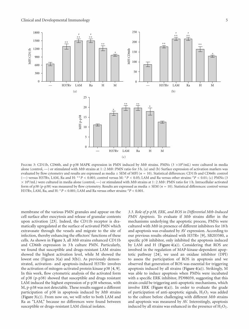
Figure 4: Role of MAP kinases and reactive oxygen species in PMN apoptosis induced by Mtb strains. (a) PMN (3× 10⁶/mL) were cultured inmedia alone (control, —) or stimulated with Mtb strains at 1 : 2 Mtb : PMN ratio for 18 h, in the presence or absence of specific inhibitors forp38 and ERK (SB203580 and PD98059, resp.), and an oxidase inhibitor DPI. Thereafter, Annexin V-FITC (AV-FITC) binding was measuredby flow cytometry. Results are expressed as media± SEM (n = 12). Statistical differences: Control versus LAM: ∗∗∗P < 0.001, control versusH: ∗∗P < 0.01, control versus H37Rv: ∗P < 0.05; H37Rv, LAM and H versus SB: #P < 0.01; H37RV, LAM and H versus DPI: σP < 0.001; Mversus PD: ϕP < 0.01. (b) PMN were treated with 10 mM H2O2 15 min before Mtb challenge and thereafter PMN were cultured as describedin (a). After 18 h-culture, Annexin V-FITC (AV-FITC) positive PMN was measured by flow cytometry. A representative experiment out offour is displayed.
However this effect was lower for the Haarlem strains, inparticular M, which effect was not enough to reach highlevels of apoptosis (Figure 4(b)), supporting the idea thatanti-apoptotic mechanisms could be involved.
3.4. Role of p-p38, ERK, and ROS in CD11b UpRegulation byMtb Strains. Previously we have shown that H37Rv-inducedapoptosis depends on PMN activation as Mtb induced alsoCD11b expression [4]. CD11b is a receptor that serves innonopsonic recognition of microbes which may confer anadvantage in the alveolar space, where serum opsonins arelimited. We further evaluated whether the activation ofp38 is involved in the upregulation of CD11b expressionand, as observed in Table 1, SB203580 abolished CD11bupregulation by all strains. Besides, PD inhibited M-inducedC11b enhancement suggesting that ERK was also requiredfor M. This data is in accordance with that observed to
apoptosis. However, CD11b expression was not abrogatedby DPI suggesting that ROS did not participate in CD11bupregulation.
3.5. ROS Production Induced by Mtb Strains in PMN.Considering that apoptosis in human PMN may be related toROS production [25] we wondered if M, that fails in inducePMN apoptosis, would exhibit any deficiency to induce ROS.Therefore, we evaluated the conversion of nonfluorescentdihydrorhodamine123 (DHR) to rhodamine123 as a mea-sure of H2O2 production. DHR freely enters the cell; itbinds to cellular and mitochondrial membranes and emits abright fluorescent signal mainly localized inside the cell [26].As it is shown in Figure 5(a), all Mtb strains induced ROSproduction in a dose-dependent manner. At low Mtb : PMNratio (1 : 2), LAM, H, and H37Rv strains were able to inducesignificant amount of ROS whereas M did not (Figure 5(b)).
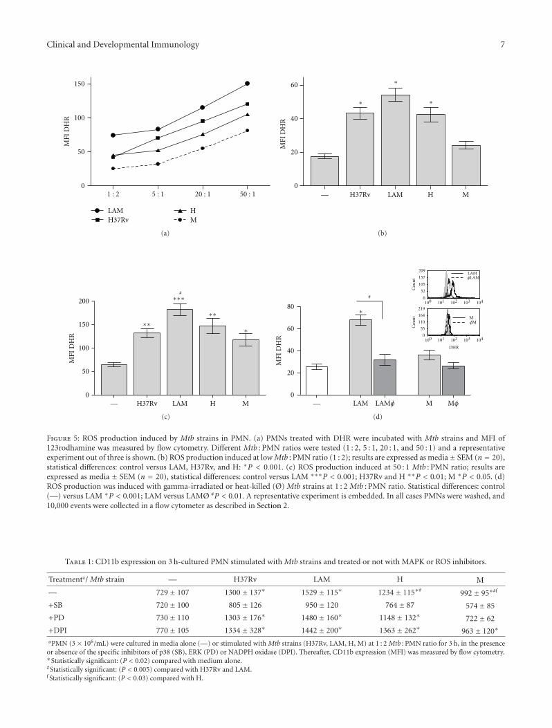
Clinical and Developmental Immunology 7
0
50
100
150
LAMH37Rv
HM
MFI
DH
R
1 : 2 5 : 1 20 : 1 50 : 1
(a)
H37Rv LAM H M0
20
40
60
MFI
DH
R
—
∗
∗
∗
(b)
H37Rv LAM H M0
50
100
150
200
MFI
DH
R
—
∗
#
∗∗∗∗
∗∗∗
(c)
LAM M0
20
40
60
80
MFI
DH
R
—
∗
#
Cou
nt
Cou
nt
0
52
105
157
209
0
55
110
164
219100 101 102 103 104
100 101 102 103 104
DHR
φLAMLAM
φMM
LAMφ Mφ
(d)
Figure 5: ROS production induced by Mtb strains in PMN. (a) PMNs treated with DHR were incubated with Mtb strains and MFI of123rodhamine was measured by flow cytometry. Different Mtb : PMN ratios were tested (1 : 2, 5 : 1, 20 : 1, and 50 : 1) and a representativeexperiment out of three is shown. (b) ROS production induced at low Mtb : PMN ratio (1 : 2); results are expressed as media± SEM (n = 20),statistical differences: control versus LAM, H37Rv, and H: ∗P < 0.001. (c) ROS production induced at 50 : 1 Mtb : PMN ratio; results areexpressed as media ± SEM (n = 20), statistical differences: control versus LAM ∗∗∗P < 0.001; H37Rv and H ∗∗P < 0.01; M ∗P < 0.05. (d)ROS production was induced with gamma-irradiated or heat-killed (Ø) Mtb strains at 1 : 2 Mtb : PMN ratio. Statistical differences: control(—) versus LAM ∗P < 0.001; LAM versus LAMØ #P < 0.01. A representative experiment is embedded. In all cases PMNs were washed, and10,000 events were collected in a flow cytometer as described in Section 2.
Table 1: CD11b expression on 3 h-cultured PMN stimulated with Mtb strains and treated or not with MAPK or ROS inhibitors.
Treatmenta/ Mtb strain — H37Rv LAM H M
— 729± 107 1300± 137∗ 1529± 115∗ 1234± 115∗# 992± 95∗#f
+SB 720± 100 805± 126 950± 120 764± 87 574± 85
+PD 730± 110 1303± 176∗ 1480± 160∗ 1148± 132∗ 722± 62
+DPI 770± 105 1334± 328∗ 1442± 200∗ 1363± 262∗ 963± 120∗
aPMN (3× 10⁶/mL) were cultured in media alone (—) or stimulated with Mtb strains (H37Rv, LAM, H, M) at 1 : 2 Mtb : PMN ratio for 3 h, in the presenceor absence of the specific inhibitors of p38 (SB), ERK (PD) or NADPH oxidase (DPI). Thereafter, CD11b expression (MFI) was measured by flow cytometry.∗Statistically significant: (P < 0.02) compared with medium alone.#Statistically significant: (P < 0.005) compared with H37Rv and LAM.f Statistically significant: (P < 0.03) compared with H.
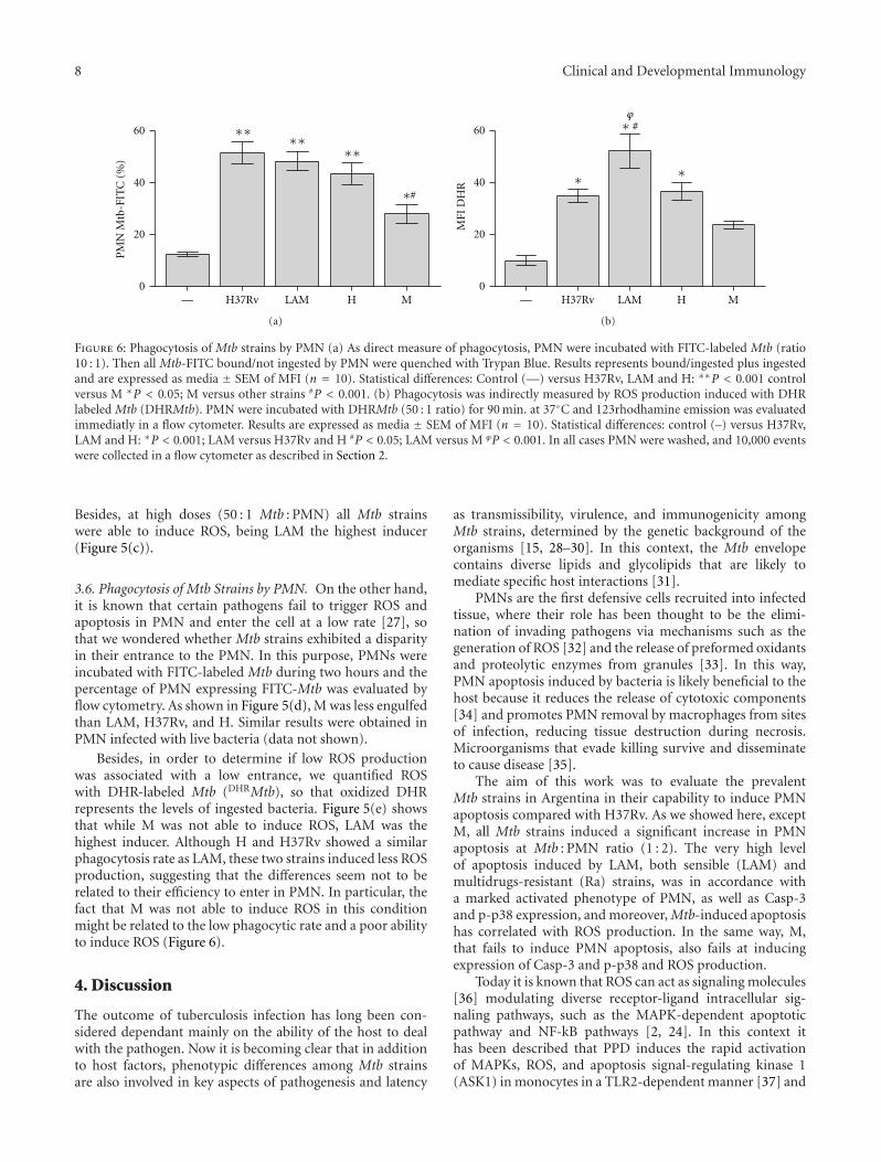
8 Clinical and Developmental Immunology
H37Rv LAM H M0
20
40
60
—
PM
N M
tb-F
ITC
(%
)∗∗ ∗∗
∗∗
#∗
(a)
H37Rv LAM H M0
20
40
60
MFI
DH
R
—
#∗
∗ ∗
ϕ
(b)
Figure 6: Phagocytosis of Mtb strains by PMN (a) As direct measure of phagocytosis, PMN were incubated with FITC-labeled Mtb (ratio10 : 1). Then all Mtb-FITC bound/not ingested by PMN were quenched with Trypan Blue. Results represents bound/ingested plus ingestedand are expressed as media ± SEM of MFI (n = 10). Statistical differences: Control (—) versus H37Rv, LAM and H: ∗∗P < 0.001 controlversus M ∗P < 0.05; M versus other strains #P < 0.001. (b) Phagocytosis was indirectly measured by ROS production induced with DHRlabeled Mtb (DHRMtb). PMN were incubated with DHRMtb (50 : 1 ratio) for 90 min. at 37◦C and 123rhodhamine emission was evaluatedimmediatly in a flow cytometer. Results are expressed as media ± SEM of MFI (n = 10). Statistical differences: control (–) versus H37Rv,LAM and H: ∗P < 0.001; LAM versus H37Rv and H #P < 0.05; LAM versus M ϕP < 0.001. In all cases PMN were washed, and 10,000 eventswere collected in a flow cytometer as described in Section 2.
Besides, at high doses (50 : 1 Mtb : PMN) all Mtb strainswere able to induce ROS, being LAM the highest inducer(Figure 5(c)).
3.6. Phagocytosis of Mtb Strains by PMN. On the other hand,it is known that certain pathogens fail to trigger ROS andapoptosis in PMN and enter the cell at a low rate [27], sothat we wondered whether Mtb strains exhibited a disparityin their entrance to the PMN. In this purpose, PMNs wereincubated with FITC-labeled Mtb during two hours and thepercentage of PMN expressing FITC-Mtb was evaluated byflow cytometry. As shown in Figure 5(d), M was less engulfedthan LAM, H37Rv, and H. Similar results were obtained inPMN infected with live bacteria (data not shown).
Besides, in order to determine if low ROS productionwas associated with a low entrance, we quantified ROSwith DHR-labeled Mtb (DHRMtb), so that oxidized DHRrepresents the levels of ingested bacteria. Figure 5(e) showsthat while M was not able to induce ROS, LAM was thehighest inducer. Although H and H37Rv showed a similarphagocytosis rate as LAM, these two strains induced less ROSproduction, suggesting that the differences seem not to berelated to their efficiency to enter in PMN. In particular, thefact that M was not able to induce ROS in this conditionmight be related to the low phagocytic rate and a poor abilityto induce ROS (Figure 6).
4. Discussion
The outcome of tuberculosis infection has long been con-sidered dependant mainly on the ability of the host to dealwith the pathogen. Now it is becoming clear that in additionto host factors, phenotypic differences among Mtb strainsare also involved in key aspects of pathogenesis and latency
as transmissibility, virulence, and immunogenicity amongMtb strains, determined by the genetic background of theorganisms [15, 28–30]. In this context, the Mtb envelopecontains diverse lipids and glycolipids that are likely tomediate specific host interactions [31].
PMNs are the first defensive cells recruited into infectedtissue, where their role has been thought to be the elimi-nation of invading pathogens via mechanisms such as thegeneration of ROS [32] and the release of preformed oxidantsand proteolytic enzymes from granules [33]. In this way,PMN apoptosis induced by bacteria is likely beneficial to thehost because it reduces the release of cytotoxic components[34] and promotes PMN removal by macrophages from sitesof infection, reducing tissue destruction during necrosis.Microorganisms that evade killing survive and disseminateto cause disease [35].
The aim of this work was to evaluate the prevalentMtb strains in Argentina in their capability to induce PMNapoptosis compared with H37Rv. As we showed here, exceptM, all Mtb strains induced a significant increase in PMNapoptosis at Mtb : PMN ratio (1 : 2). The very high levelof apoptosis induced by LAM, both sensible (LAM) andmultidrugs-resistant (Ra) strains, was in accordance witha marked activated phenotype of PMN, as well as Casp-3and p-p38 expression, and moreover, Mtb-induced apoptosishas correlated with ROS production. In the same way, M,that fails to induce PMN apoptosis, also fails at inducingexpression of Casp-3 and p-p38 and ROS production.
Today it is known that ROS can act as signaling molecules[36] modulating diverse receptor-ligand intracellular sig-naling pathways, such as the MAPK-dependent apoptoticpathway and NF-kB pathways [2, 24]. In this context ithas been described that PPD induces the rapid activationof MAPKs, ROS, and apoptosis signal-regulating kinase 1(ASK1) in monocytes in a TLR2-dependent manner [37] and

Clinical and Developmental Immunology 9
ASK1 could also be activated by oxidant stress, involving p38pathway. A strong evidence for the involvement of ROS inPMN apoptosis is the fact that phagocytes from patients withchronic granulomatous disease that are unable to produceROS due to a defect in NADPH oxidase are predisposedto recurring bacterial and fungal infections and developgranulomas linked to altered PMN apoptosis [38]. Patientswith chronic granulomatous disease are also susceptibleto TB and complications of vaccination with the bacillusCalmette-Guerin [39].
Here we show that both ROS and p38 are involved in theapoptosis triggered at low Mtb : PMN rate. Remarkably, Mwas able to trigger apoptosis when ERK was abrogated sug-gesting that M activates anti-apoptotic mechanisms whichcould also impact on final apoptosis outcome in PMN. Thisresult was supported by the fact that addition of H2O2
allowed a partial induction of apoptosis by M. The involve-ment of ERK in signaling triggered by M was also evidentas both p38 and ERK were involved in CD11b expressioninduced by M, whereas other strains only activated p-p38(Table 1). Although p-p38 induced by M was not detectable,it could be enough to activate PMN but not to overcomeantiapoptotic mechanisms triggered by this strain.
It has been described that PMN uptake of the obligatedintracellular pathogen Anaplasma phagocytophilum occurs ata slow rate compared with other bacteria [27], and evenmore, PMN infection with this pathogen fails to triggerROS and delays PMN spontaneous apoptosis [2, 25]. In thiscontext, albeit M was ingested at a lower rate, no differenceswere observed among LAM, H37Rv, and H (Figure 5(d)),suggesting that differences in ROS production were notrelated to the efficiency in bacterial entry. Consistently, albeitM was able to induced ROS at high Mtb : PMN ratio, it wasnot able to induce ROS when DHR came from Mtb that hasbeen ingested (MtbPMN), because of the sum of the lowphagocytic rate and a poor ability to induce ROS. Therefore,we can assume that LAM is more efficient in entering PMNand also has a major capability to induce ROS than otherstrains. Moreover, it is possible that Mtb strains would notbe qualitatively different in their capacity to induce ROS butmerely show a quantitatively different dose-response curvethat is finally reflected in apoptosis outcome.
Pathogens have developed strategies to counteractNADPH oxidase function and suppress generation of phago-somal ROS. The nuo G gene encodes the NuoG subunitof the type I NADH (nicotinamide adenine dinucleotide)-dehydrogenase of Mtb. Deletion of this gene leads to elevatedROS levels and apoptosis following infection of primarymacrophages [40]. Similarly, deletion of the secA2 gene,which encodes a protein required for the secretion of super-oxide dismutase A, results in enhanced ROS production andapoptosis. Superoxide dismutase A breaks down superoxideleading to diminished ROS generation, which is a mechanismthat Mtb uses to prevent apoptosis [41]. Nevertheless, ourdata obtained with heat-killed bacteria suggest that themechanism exerted by M might not involve those enzymes,but structural differences between clinical isolates leads todifferent ROS/apoptosis rate.
LAM and Haarlem are the two main families circulatingin Latin American countries [2, 13, 14]. Among them, twoclinical isolates differently interact with PMN in the host. Inparticular, M strain of the Haarlem family can be regarded asa highly successful genotype in Argentina because it has beenable to prevail and persist over other MDR Mtb strains inthe community [42, 43] being able to build up further drugresistance without impairing its ability to spread [15]. In thiscontext, it has been described that, when killing is absent, notonly could the disease progress locally, but the infected PMNcould also traffic organisms to distal sites, in particular ifbacilli reach the systemic circulation. Indeed, a granulocytic“Trojan horse” has been proposed by several authors in thecontext of mycobacterial infection [44]. Therefore, thoughless skillfully, M is able to enter the PMN and in turn generatefewer ROS. As a result, PMNs have less capacity to kill M andgreater capacity to prolong its life, becoming a reservoir forthis strain or another strain that uses this mechanism.
5. Conclusion
In conclusion, here we show that, independently of theability to entering PMN, certain Mtb strains (as LAM) inducehigh PMN apoptosis by triggering signaling mechanismsthat involves ROS generation via p38 activation, leading toenhanced effectors’ functions whereas others fail in activatingPMN to kill pathogen as well as inducing apoptosis as aconsequence of no. (1) a slight ROS production and (2)contribution of anti-apoptotic mechanism mediated at leastby ERK, making PMN a “Trojan horse,” which could be abeneficial mechanism allowing some strains to prevail andpersist over other strains in the community.
Abbreviations
Mtb: Mycobacterium tuberculosisPMN: polymorphonuclear neutrophilsROS: Reactive Oxygen SpeciesTB: tuberculosisDHR: dihydrorhodamineMAPK: mitogen-activated protein kinase.
Acknowledgments
This work was supported by Grants from the AgenciaNacional de Promocion Cientıfica y Tecnologica, ANPCyT(PAE-PICT 2007–2329 and PAE-PICT 2007–2328), ConsejoNacional de Investigaciones Cientıficas y Tecnicas, CON-ICET (PIP 112-200801-01476), and Fundacion Alberto JRoemmers. The authors thank to the National Commissionof Atomic Energy (CONEA) for Mtb irradiation and theyspecially thank Dr. Marta Finiazs for helpful comments andcritical reading of the paper.
References
[1] M. Faurschou and N. Borregaard, “Neutrophil granules andsecretory vesicles in inflammation,” Microbes and Infection,vol. 5, no. 14, pp. 1317–1327, 2003.

10 Clinical and Developmental Immunology
[2] L. Aristimuno, R. Armengol, A. Cebollada et al., “Molecularcharacterisation of Mycobacterium tuberculosis isolates in thefirst national survey of anti-tuberculosis drug resistance fromVenezuela,” BMC Microbiology, vol. 6, article 90, 2006.
[3] C. Nathan, “Neutrophils and immunity: challenges and op-portunities,” Nature Reviews Immunology, vol. 6, no. 3, pp.173–182, 2006.
[4] M. Aleman, A. Garcıa, M. A. Saab et al., “Mycobacterium tuber-culosis-induced activation accelerates apoptosis in peripheralblood neutrophils from patients with active tuberculosis,”American Journal of Respiratory Cell and Molecular Biology, vol.27, no. 5, pp. 583–592, 2002.
[5] M. Aleman, M. Beigier-Bompadre, C. Borghetti et al., “Activa-tion of peripheral blood neutrophils from patients with activeadvanced tuberculosis,” Clinical Immunology, vol. 100, no. 1,pp. 87–95, 2001.
[6] B. M. Babior, “Oxidants from phagocytes: agents of defenseand destruction,” Blood, vol. 64, no. 5, pp. 959–966, 1984.
[7] N. Perskvist, M. Long, O. Stendahl, and L. Zheng, “Mycobac-terium tuberculosis promotes apoptosis in human neutrophilsby activating caspase-3 and altering expression of Bax/Bcl-xLvia an oxygen-dependent pathway,” Journal of Immunology,vol. 168, no. 12, pp. 6358–6365, 2002.
[8] M. Aleman, S. S. de la Barrera, P. L. Schierloh et al., “In tuber-culous pleural effusions, activated neutrophils undergo apop-tosis and acquire a dendritic cell-like phenotype,” Journal ofInfectious Diseases, vol. 192, no. 3, pp. 399–409, 2005.
[9] M. Aleman, P. Schierloh, S. S. de la Barrera et al., “Mycobac-terium tuberculosis triggers apoptosis in peripheral neutrophilsinvolving Toll-like receptor 2 and p38 mitogen protein kinasein tuberculosis patients,” Infection and Immunity, vol. 72, no.9, pp. 5150–5158, 2004.
[10] Y. L. Zu, J. Qi, A. Gilchrist et al., “p38 mitogen-activated pro-tein kinase activation is required for human neutrophil func-tion triggered by TNF-α or FMLP stimulation,” Journal ofImmunology, vol. 160, no. 4, pp. 1982–1989, 1998.
[11] M. Aleman, S. de la Barrera, P. Schierloh et al., “Spontaneousor Mycobacterium tuberculosis-induced apoptotic neutrophilsexert opposite effects on the dendritic cell-mediated immuneresponse,” European Journal of Immunology, vol. 37, no. 6, pp.1524–1537, 2007.
[12] L. C. O. Lazzarini, S. M. Spindola, H. Bang et al., “RDRioMycobacterium tuberculosis infection is associated with ahigher frequency of cavitary pulmonary disease,” Journal ofClinical Microbiology, vol. 46, no. 7, pp. 2175–2183, 2008.
[13] N. Candia, B. Lopez, T. Zozio et al., “First insight into My-cobacterium tuberculosis genetic diversity in Paraguay,” BMCMicrobiology, vol. 7, article 75, 2007.
[14] L. C. O. Lazzarini, R. C. Huard, N. L. Boechat et al., “Discoveryof a novel Mycobacterium tuberculosis lineage that is a majorcause of tuberculosis in Rio de Janeiro, Brazil,” Journal ofClinical Microbiology, vol. 45, no. 12, pp. 3891–3902, 2007.
[15] B. G. Lopez, C. Latini, M. Ambroggi et al., “Two M. tuberculo-sis lineages are overrepresented among new cases of MDR andXDR TB in Argentina,” International Journal of Tuberculosisand Lung Disease, vol. 12, supplement 2, p. S173, 2008.
[16] C. M. Sassetti, D. H. Boyd, and E. J. Rubin, “Comprehensiveidentification of conditionally essential genes in mycobacte-ria,” Proceedings of the National Academy of Sciences of theUnited States of America, vol. 98, no. 22, pp. 12712–12717,2001.
[17] J. I. Basile, L. J. Geffner, M. M. Romero et al., “Outbreaksof Mycobacterium tuberculosis MDR strains induce high IL-17 T-cell response in patients with MDR tuberculosis that is
closely associated with high antigen load,” Journal of InfectiousDiseases, vol. 204, no. 7, pp. 1054–1064, 2011.
[18] L. Geffner, N. Yokobori, J. Basile et al., “Patients with mul-tidrug-resistant tuberculosis display impaired Th1 responsesand enhanced regulatory T-cell levels in response to anoutbreak of multidrug-resistant Mycobacterium tuberculosis Mand Ra strains,” Infection and Immunity, vol. 77, no. 11, pp.5025–5034, 2009.
[19] A. Boyum, “Isolation of mononuclear cells and granulocytesfrom human blood,” Scandinavian Journal of Clinical and Lab-oratory Investigation, Supplement, vol. 97, pp. 77–89, 1968.
[20] S. Busetto, E. Trevisan, P. Patriarca, and R. Menegazzi, “Asingle-step, sensitive flow cytofluorometric assay for the simul-taneous assessment of membrane-bound and ingested can-dida albicans in phagocytosing neutrophils,” Cytometry PartA, vol. 58, no. 2, pp. 201–206, 2004.
[21] F. Colotta, F. Re, N. Polentarutti, S. Sozzani, and A. Mantovani,“Modulation of granulocyte survival and programmed celldeath by cytokines and bacterial products,” Blood, vol. 80, no.8, pp. 2012–2020, 1992.
[22] C. H. E. Homburg, M. de Haas, A. E. G. K. von dem Borne, A.J. Verhoeven, C. P. M. Reutelingsperger, and D. Roos, “Humanneutrophils lose their surface FcγRIII and acquire Annexin Vbinding sites during apoptosis in vitro,” Blood, vol. 85, no. 2,pp. 532–540, 1995.
[23] S. C. Stocks, M. A. Kerr, C. Haslett, and I. Dransfield, “CD66-dependent neutrophil activation: a possible mechanism forvascular selectin-mediated regulation of neutrophil adhesion,”Journal of Leukocyte Biology, vol. 58, no. 1, pp. 40–48, 1995.
[24] V. V. Sumbayev and I. M. Yasinska, “Regulation of MAPkinase-dependent apoptotic pathway: implication of reactiveoxygen and nitrogen species,” Archives of Biochemistry andBiophysics, vol. 436, no. 2, pp. 406–412, 2005.
[25] D. L. Borjesson, S. D. Kobayashi, A. R. Whitney, J. M. Voyich,C. M. Argue, and F. R. DeLeo, “Insights into pathogenimmune evasion mechanisms: Anaplasma phagocytophilumfails to induce an apoptosis differentiation program in humanneutrophils,” Journal of Immunology, vol. 174, no. 10, pp.6364–6372, 2005.
[26] A. Emmendorffer, M. Hecht, M. L. Lohmann-Matthes, and J.Roesler, “A fast and easy method to determine the productionof reactive oxygen intermediates by human and murine pha-gocytes using dihydrorhodamine 123,” Journal of Immunolog-ical Methods, vol. 131, no. 2, pp. 269–275, 1990.
[27] J. A. Carlyon, D. A. Latif, M. Pypaert, P. Lacy, and E. Fikrig,“Anaplasma phagocytophilum utilizes multiple host evasionmechanisms to thwart NADPH oxidase-mediated killingduring neutrophil infection,” Infection and Immunity, vol. 72,no. 8, pp. 4772–4783, 2004.
[28] J. Keane, H. G. Remold, and H. Kornfeld, “Virulent Mycobac-terium tuberculosis strains evade apoptosis of infected alveolarmacrophages,” Journal of Immunology, vol. 164, no. 4, pp.2016–2020, 2000.
[29] A. N. J. Malik and P. Godfrey-Faussett, “Effects of genetic vari-ability of Mycobacterium tuberculosis strains on the presenta-tion of disease,” The Lancet Infectious Diseases, vol. 5, no. 3, pp.174–183, 2005.
[30] R. F. Silver, Q. Li, and J. J. Ellner, “Expression of virulenceof Mycobacterium tuberculosis within human monocytes: vir-ulence correlates with intracellular growth and inductionof tumor necrosis factor alpha but not with evasion oflymphocyte-dependent monocyte effector functions,” Infec-tion and Immunity, vol. 66, no. 3, pp. 1190–1199, 1998.
Clinical and Developmental Immunology 11
[31] M. S. Glickman and W. R. Jacobs Jr., “Microbial pathogenesisof Mycobacterium tuberculosis: dawn of a discipline,” Cell, vol.104, no. 4, pp. 477–485, 2001.
[32] M. E. May and P. J. Spagnuolo, “Evidence for activation of arespiratory burst in the interaction of human neutrophils withMycobacterium tuberculosis,” Infection and Immunity, vol. 55,no. 9, pp. 2304–2307, 1987.
[33] K. Kasahara, I. Sato, K. Ogura, H. Takeuchi, K. Kobayashi, andM. Adachi, “Expression of chemokines and induction of rapidcell death in human blood neutrophils by Mycobacteriumtuberculosis,” Journal of Infectious Diseases, vol. 178, no. 1, pp.127–137, 1998.
[34] F. R. DeLeo, “Modulation of phagocyte apoptosis by bacterialpathogens,” Apoptosis, vol. 9, no. 4, pp. 399–413, 2004.
[35] S. D. Kobayashi, K. R. Braughton, A. R. Whitney et al., “Bacte-rial pathogens modulate an apoptosis differentiation programin human neutrophils,” Proceedings of the National Academy ofSciences of the United States of America, vol. 100, no. 19, pp.10948–10953, 2003.
[36] B. Zhang, J. Hirahashi, X. Cullere, and T. N. Mayadas, “Elu-cidation of molecular events leading to neutrophil apoptosisfollowing phagocytosis. Cross-talk between caspase 8, reactiveoxygen species, and MAPK/ERK activation,” Journal of Biolog-ical Chemistry, vol. 278, no. 31, pp. 28443–28454, 2003.
[37] C. S. Yang, D. M. Shin, H. M. Lee et al., “ASK1-p38 MAPK-p47phox activation is essential for inflammatory responsesduring tuberculosis via TLR2-ROS signalling,” Cellular Micro-biology, vol. 10, no. 3, pp. 741–754, 2008.
[38] M. B. Hampton, M. C. M. Vissers, J. I. Keenan, and C. C.Winterbourn, “Oxidant-mediated phosphatidylserine expo-sure and macrophage uptake of activated neutrophils: possibleimpairment in chronic granulomatous disease,” Journal ofLeukocyte Biology, vol. 71, no. 5, pp. 775–781, 2002.
[39] P. P. W. Lee, K. W. Chan, L. Jiang et al., “Susceptibility tomycobacterial infections in children with x-linked chronicgranulomatous disease: a review of 17 patients living in aregion endemic for tuberculosis,” Pediatric Infectious DiseaseJournal, vol. 27, no. 3, pp. 224–230, 2008.
[40] K. Velmurugan, B. Chen, J. L. Miller et al., “Mycobacteriumtuberculosis nuoG is a virulence gene that inhibits apoptosis ofinfected host cells.,” PLoS pathogens, vol. 3, no. 7, Article IDe110, 2007.
[41] R. Kahl, A. Kampkotter, W. Watjen, and Y. Chovolou, “Antiox-idant enzymes and apoptosis,” Drug Metabolism Reviews, vol.36, no. 3-4, pp. 747–762, 2004.
[42] D. J. Palmero, M. Ambroggi, A. Brea et al., “Treatment andfollow-up of HIV-negative multidrug-resistant tuberculosispatients in an infectious diseases reference hospital, BuenosAires, Argentina,” International Journal of Tuberculosis andLung Disease, vol. 8, no. 6, pp. 778–784, 2004.
[43] V. Ritacco, M. Di Lonardo, A. Reniero et al., “Nosocomialspread of human immunodeficiency virus-related multidrug-resistant tuberculosis in Buenos Aires,” Journal of InfectiousDiseases, vol. 176, no. 3, pp. 637–642, 1997.
[44] V. Abadie, E. Badell, P. Douillard et al., “Neutrophils rapidlymigrate via lymphatics after Mycobacterium bovis BCG intra-dermal vaccination and shuttle live bacilli to the draininglymph nodes,” Blood, vol. 106, no. 5, pp. 1843–1850, 2005.

Submit your manuscripts athttp://www.hindawi.com
Stem CellsInternational
Hindawi Publishing Corporationhttp://www.hindawi.com Volume 2014
Hindawi Publishing Corporationhttp://www.hindawi.com Volume 2014
MEDIATORSINFLAMMATION
of
Hindawi Publishing Corporationhttp://www.hindawi.com Volume 2014
Behavioural Neurology
EndocrinologyInternational Journal of
Hindawi Publishing Corporationhttp://www.hindawi.com Volume 2014
Hindawi Publishing Corporationhttp://www.hindawi.com Volume 2014
Disease Markers
Hindawi Publishing Corporationhttp://www.hindawi.com Volume 2014
BioMed Research International
OncologyJournal of
Hindawi Publishing Corporationhttp://www.hindawi.com Volume 2014
Hindawi Publishing Corporationhttp://www.hindawi.com Volume 2014
Oxidative Medicine and Cellular Longevity
Hindawi Publishing Corporationhttp://www.hindawi.com Volume 2014
PPAR Research
The Scientific World JournalHindawi Publishing Corporation http://www.hindawi.com Volume 2014
Immunology ResearchHindawi Publishing Corporationhttp://www.hindawi.com Volume 2014
Journal of
ObesityJournal of
Hindawi Publishing Corporationhttp://www.hindawi.com Volume 2014
Hindawi Publishing Corporationhttp://www.hindawi.com Volume 2014
Computational and Mathematical Methods in Medicine
OphthalmologyJournal of
Hindawi Publishing Corporationhttp://www.hindawi.com Volume 2014
Diabetes ResearchJournal of
Hindawi Publishing Corporationhttp://www.hindawi.com Volume 2014
Hindawi Publishing Corporationhttp://www.hindawi.com Volume 2014
Research and TreatmentAIDS
Hindawi Publishing Corporationhttp://www.hindawi.com Volume 2014
Gastroenterology Research and Practice
Hindawi Publishing Corporationhttp://www.hindawi.com Volume 2014
Parkinson’s Disease
Evidence-Based Complementary and Alternative Medicine
Volume 2014Hindawi Publishing Corporationhttp://www.hindawi.com