cloning and characterization of the growth hormone-dependent insulin-like growth factor binding...
TRANSCRIPT

Vol. 166, No. 2, 1990 January 30, 1990
BIOCHEMICAL AND BIOPHYSICAL RESEARCH COMMUNICATIONS Pages 892-897
CLONING AND CHARACTERIZATION OFTHE GROWTH HORMONE-DEPENDENT INSULIN-LIKE GROWTH FACTOR BINDING PROTEIN
(IGFBP-3) IN THE RAT
Anthony L. Albiston and Adrian C. Herington
Prince Henry’s Institute of Medical Research, Prhrce Henry’s Hospital Campus, Monash Medical Centm, Melbourne, Australia 3004
Received December 12, 1989
We report for the first time the complete ammo acid sequence for the growth hormone dependent insulin-like growth factor bmding protein (IGFBP3) in the rat. A human IGFBP-3 clone was generated using the polymemse chain reaction (PCR) and used to screen a rat liver cDNA library. cDNA clones of the rat IGFBP-3 were isolated and the full ammo acid sequence deduced. The sequence begins with a putative, 26 amino acid signal peptide followed by a 265 ammo acid binding protein. The amino acid sequence is over 80% homologous with the equivalent human IGFBP-3 form and shows complete conservation of 18 cysteine residues that are clustered at the amino and carboxy ends of the protein. IGFBP3 is the binding subunit of the major circulating IGFBP in the rat, and hence the availability of precise structural data and cDNA probes provides an important opportunity for a detailed study of the control of IGFBP-3 synthesis at the level of gene expression. o 1990 Academic
Press, mc.
The insulin-like growth factors -1 and -II (IGF I, -11) are circulating mitogens that stimulate
growth both in vivo and in vitro (1.2). In serum the IGFs circulate essentially completely
bound to a family of evolutionary te!ated, specific binding proteins (3,4). These binding
proteins prolong the half life of the IGFs and have been shown to regulate IGF action on cells
in culture ($6). In rat or human sera two major forms of binding protein am detected by
radioligand binding studies, a M,lSO,OOO (150 K) form and a smaller M, 35 K form (4.7).
The M, 150 K binding protein is acid-dissociable into an IGF binding subunit, termed
IGFBP-3, of M,-50 K and an acid labile non-binding subunit of M, 80,000-90,000 (8). The
biosynthesis of IGFBPS is regulated by growth hormone (GH) whereas the smaller form,
termed IGFBP-I. is not GH-dependent (3,4). The human forms of IGFBP-1 and -3 have
recently been cloned (9-l 1). A third class of binding protein, IGFBP-2, was originally
isolated from a fetal rat liver cell line (BRL9A) (12). The cDNA for the rat IGFBP-2 has
recently been isolated (13, 14). Although IGFBP-3 which is synthesized primarily by the
liver (7,15) is the predominant IGF binding protein in adult rat serum, it has not been fully
characterized.
0006-291X/90 $1.50 Copyright 0 1990 by Academic Press, Inc. All rights of reproduction in any form reserved. 892

Vol. 166, No. 2, 1990 BIOCHEMICAL AND BIOPHYSICAL RESEARCH COMMUNICATIONS
In this paper we report the isolation of a cDNA encoding the complete sequence of the rat
IGFBP-3. ‘Ihe deduced amino acid sequence of the mature protein is cysteine rich, 265
residues in length and over 80% homologous with the human equivalent (11). Genomic
Southern analysis suggests that this is a single copy gene and northern analysis shows a single
mRNA species in the liver, approximately 2.4 kb in size.
MATERIALS AND METHODS
Polvruerase chain (PCR)
Based upon the published cDNA sequence of the human IGFBP-3 (11) two oligonucleotide primers were constructed, primer A: 531-550 nt and primer B: the complement of 882-863 nt. cDNA was prepared by reverse transcription of 5~ of human liver poly A+ RNA using 20 pmol of primer B and 110 units of AMV reverse transcriptase (Pharmacia, Uppsala, Sweden) under the conditions recommended by the supplier. A cDNA aliquot corresponding to 0.5% of RNA was used for PCR amplification in 67 mM Tris pH 8.8 at 25-C 17 mM (NH4)2SO4,3mM MgCl2, 10 mM B-mercaptoethanol, 2 mg/ml gelatine, 0.5 M of each primer, 500 M dNTP and 2 U of Taq DNA polymerase (Perkin Elmer Cetus, Norwalk, U.S.A.). Thirty cycles (one cycle: 1 min at 92’C, 2 min at 55’C. 2 min at 72’C) were carried out in a 50@ volume using a thermal cycler (Bartlett, Melbourne. Australia). The 35 1 bp product was subcloned into a Gem 42 plasmid vector and its identity confirmed by dideoxy sequencing.
Isolation of a cDNA clone
A rat liver cDNA library in a&t 10 vector (kindly donated by Professor G. Schreiber, University of Melbourne) was screened for clones of IGFBP-3. The 351 bp PCR product was 32P-labelled by random priming (16), hybridization of duplicate filters was performed at 42-C in 50% fonnamide and washes were in 2 X SSC at 42-C. Among 5 X lo6 plaques screened 7 were found to be positive. The largest clone, 2.0 kb in size, was subcloned into the Gem 42 plasmid vector for further manipulation.
Relevant restriction fragments were subcloned into either Gem 42 or Ml3 vectors. Dideoxy chain termination was performed using SP6 and T7 promoter primers and Ml3 universal primers respectively (17). In regions that lacked convenient restriction sites specifically designed oligonucleotides were synthesized on a DNA synthesizer (Applied Biosystems, Foster City, U.S.A.).
Genomic Southern Analvsis
High molecular weight rat liver DNA was digested with restriction endonucleases according to the supplier’s recommended reaction conditions. The fragments were separated on a 0.7% agarose gel and transferred to a nylon membrane as described by Reed and Mann (18). The restriction fragment probe used was 930 bp in length, containing the entire coding region of the gene, and was 32 P-labelled by random priming. Hybridization was at 42-C in 50% formamide and the filter was given a final wash in 0.1 X SSC, 1% SDS at 5o’C.
Pol A(+) mRNA was purified from adult rat liver as previously described (19). Tenllg of ~01~ A f’) mRNA was glyoxylated and then separated by electmphoresis on a 1.2% agarose gel. The RNA was transferred to a nylon membrane and hybridized with the probe described for genomic Southern analysis. The filter was given a fmal wash in 0.1 X SSC. 0.1% SDS at 55°C.
893

Vol. 166, No. 2, 1990 BIOCHEMICAL AND BIOPHYSICAL RESEARCH COMMUNICATIONS
RESULTS AND DISCUSSION
A 351 bp polymerase chain reaction product of the human IGPBP-3 was used to screen a
rat liver cDNA library. Approximately 5 x lo6 clones were screened revealing seven
independent positive clones. The largest clone, of 2.0 kb, contained the entire coding region
except for the start codon, ATG. A second overlapping clone, 700 bp in length, contained the
ATG codon plus 36 bp of 5’ untranslated sequence. The total sequence obtained from these
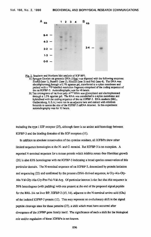
clones was 2058 bp (Pig. 1). Northern blot analysis of liver mRNA showed a single mRNA
species approximately 2.4 kb in size indicating mat we do not have a full length mRNA
sequence (Pig. 2B). The 3’ untranslated sequence obtained does not contain a polyadenylation
signal suggesting that the 3’ region is incomplete. An unusual feature of the 3’ untranslated
sequence is a 36 bp repetitive TC sequence beginning at position 1012. This repeating
sequence is also found in the 3’ untranslated sequence of the rat pmlactin receptor mRNA
(20), however the significance of this sequence is not known. Genomic Southern analysis
after hybridization to the coding region of the rat IGPBP-3 cDNA showed only 2 distinct
bands per lane for each of 4 restriction enzymes suggesting that this is a single copy gene
(Pig. 2 A).
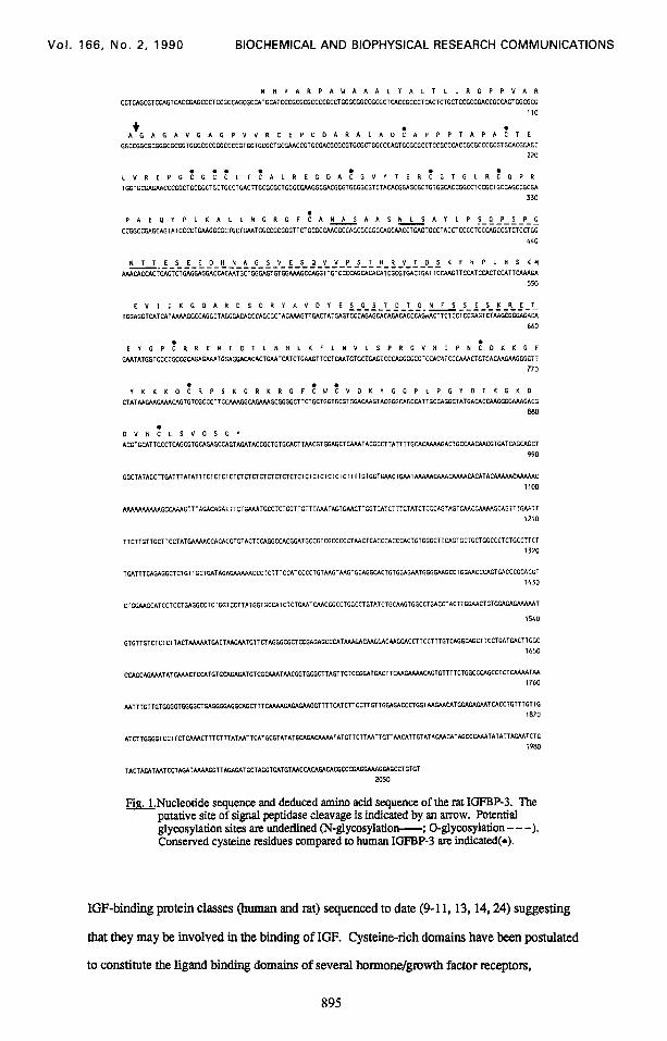
The sequence has an open reading frame of 29 1 amino acids beginning with a methionine
residue. The first 26 residues, containing a hydrophobic core, form a potential secretion
signal peptide (21). The mature peptide is 265 residues in length beginning at Gly 27. The
deduced amino acid sequence of rat IGPBP-3, residues 27-68 (Fig. 1). matches exactly the
NH2-terminal amino acid sequence reported by Zapf et al (22).
The protein has 4 potential N-linked glycosylation sites (NXT or NXS), and 2 short serine-
threonine rich domains which represent potential O-lied glycosylation sites (23). It is
known that rat IGPBP-3 is N-glycosylatcd (22) and this accounts for the discrepancy between
the predicted translated molecular weight of 28.8 K and the electmphoretically determined
molecular weight of -50 K (7).
The rat IGFBP-3 is 83% homologous with the equivalent human BP (11). An insertion of
two amino acids at residue 30 and one deletion at position 103 of the mature protein are
necessary to align the human sequence with the rat. The sequences have complete
conservation of the eighteen cysteine residues present, which am clustered at the N- and C-
termlni of the protein. The cysteine residues am highly conserved across all of the distinct
Vol. 166, No. 2, 1990 BIOCHEMICAL AND BIOPHYSICAL RESEARCH COMMUNICATIONS
Fia.Nucleotide sequence and deduced amino acid sequence of the rat IGFBP-3. The putative site of signal peptidase cleavage is indicated by an arrow. Potential glycosylation sites are underlined (N-glycosylatiow; 0-glycosylation - - -). Conserved cystelne residues compared to human IGFBP-3 are indicated(*).
IGF-binding protein classes (human and rat) sequenced to date (9-l 1,13,14,24) suggesting
that they may be involved in the binding of IGF. Cysteine-rich domains have been postulated
to constitute the ligand binding domains of several hormone/gmwth factor receptors,
895
Vol. 166, No. 2, 1990 BIOCHEMICAL AND BIOPHYSICAL RESEARCH COMMUNICATIONS
0.8 -
Fig. Southern and Northern blot analysis of IGF BP3. A) Sprague-Dawley rat genomic DNA (10~) was digested with the following enzymes:
Ecoe (lane l), BamHl (lane 2), Hind111 (lane 3) and PstI (lane 4). The DNA was electrbphorese probed with a h
through a 0.7% agarose gel, transferred to a nylon membrane and P-labelled restriction fragment comprised of the coding sequence of
the rat IGFEP-3. Autoradiography was for 48 hours. B) Ten micrograms of rat liver poly A(+) RNA was glyoxylated and electrophoresed
through a 1.2% agarose gel. The RNA was transferred to a nylon membrane and hybridized with the coding sequence of the rat IGFEP-3. RNA markers (ERL, Gaithersburg, U.S.A.) were run in an adjacent lane and stained with ethidium bromide to assess the size of the IGFEP3 mRNA detected. In this experiment autoradiography was for 12 hours.
including the type I IGF receptor (25), although there is no amino acid homology between
IGFBP3 and the binding domains of the IGF receptors (11).
In addition to absolute conservation of the cysteine residues, all IGFBPs show other
limited sequence homologies at the N- and C- tennini. Rat IGFBP-3 is no exception. A
reported N-terminal sequence for a mouse protein which inhibits serum-free fibroblast growth
(26) is also 83% homologous with rat IGFBP-3 indicating a broad species conservation of this
particular domain. The N-terminal sequence of rat IGFBP-3, determined by protein isolation
and sequencing (22) and conlirrned by the present cDNA-derived sequence, is Gly-Ala-Gly-
Ala-Val-Gly-Ala-Gly-Pro-Val-Val-Arg. Of particular interest is the fact that this sequence is
58% homologous (with padding) with one present at the end of the proposed signal peptide
for the BRL3A rat liver BP, IGFBP-2 (13,14), adjacent to the N-terminal amino acid (Glu)
of the isolated IGFBP-2 protein (12). This may represent an evolutionary shift in the signal
peptide cleavage sites for these proteins (27). a shift which must have occurred tier
divergence of the IGFBP gene family itself. The significance of such a shift for the biological
role and/or regulation of these IGFBPs is not known.
896
Vol. 166, No. 2, 1990 BIOCHEMICAL AND BIOPHYSICAL RESEARCH COMMUNICATIONS
In conclusion, we have isolated a cDNA encoding the rat growth hormone dependent
insulin-like growth factor binding protein, IGFBP-3, and report for the first time the complete
amino acid sequence of this important regulatory rat protein. The deduced amino acid
sequence is over 80% homologous to human IGFBP-3. The availability of the rat cDNA will
provide an opportunity to examine, in a very convenient model system, the nature and tissue
distribution of IGFBP-3 mRNA and its pharmacological/physiological regulation.
ACKNOWLEDGMENTS
These studies were supported in part by the National Health and Medical Research Council of Australia. We thank Mrs. Joni Law and MS Sue Panckrldge for help with the preparation of the manuscript.
1. 2.
Baxter, R.C. (1986) Adv. Clin. Chem. 25: 49-115. Froesch, E.R., S&mid, C., Schwander, J, and Zapf, J. (1985) Ann. Rev. Physiol. 47: 443-
468. 3. Ooi, G.T.. and Herington, A.C. (1988) J. Endocrinol. 118: 7-18. 4. Baxter, R.C. and Martin, J.L. (1989) Pmg. in Growth Factor Res. 1: 49-68. 5. Moses, A.C.. Nissley, S.P., and Cohen, K.L. (1976) Nature 263: 137-140. 6. Elgln, R.G., Busby, W.H., Jr. and Clemmons, D.R. (1987) Proc. Natl. Acad. Sci. USA
84: 3254-3258. 7.
;:
Baxter, R.L. and Martin, J.L. (1987) Biochem. Biophys. Res. Comm. 147: 408415. Baxter, R.C. (1988) J. Clin Endocrinol Metab 67: 265-272. Brinkman, A., G&fen, C., Kortleve, D.J., van Kessel, A.G. and Drop, S.L.S. (1988)
Embo J. 7: 2417-2423.
REFJXRENCES
lO.Lee, Y.L., Hmtz, R.L., James, P.M., Lee, P.D.K., Shively, J.E. and Powell, D.R. (1988) Mol. Endocrinol 2: 404411.
1 l.Wood, W.I., Cachianes, G., Henzel, W.J., Winslow, G.A., Spencer, S.A., Hellmiss, R., Martin, J.L. and Baxter, R.C. (1988) Mol. Endocrinol. 2: 1176-l 185.
12.Mottola C., MacDonald, R.G., Brackett, J.L., Mole, J.E., Anderson, J.K., and Czech, M.P. (1986) J. Biol. Chem. 261: 11180-11188.
13. Margot. J.B., Binkert. C., Mary, J-L., Landwehr, J., Heinrich, G. and Schwander, J. (1989) Mol. Endocrlnol. 3: 1053-1060.
14. Brown, AL., Chiamtti. L., Orlowski, C.C., Mehhnan, T.. Burgess, W.H.. Ackerman, E.J., Bnmi, C.B., and Rechler. M.M. (1989) J. Biol. Chem. 264: 5148-5154.
15.Scott. C.D., Martin, J.L. and Baxter, R.C. (1985) Endocrinology 116: 1094-1101. 16.Feinberg, A.P., and Vogelstein, B. (1983) Anal. Biochem. 132: 6-13. 17Zhang H., Scholl, R., Browse, J. and Somerville C. (1988) Nucleic Acid Res 16: 1220. 18.Reed. K.C. and Mann, D.A. (1985) Nucleic Acid Res. 13: 7207-7221. 19.Aviv, H., and Leder, P. (1972) hoc. Natl. Acad. Sci. USA. 69: 1408-1412. 20.Boutir1, J-M., Jolicoeur C., Okamura, H., Gagnon, J., Edery, M.. Shirota, M.. Banville, D.,
Dusanter-Fourt, I., Djiane, J. and Kelly, P.A. (1988) Cell 53: 69-77. 21.Watson, M.E.E. (1984) Nucleic Acids Res. 12:5145-5165. 22.Zapf, J., Born, W., Chang, J.-Y., James, P., Froesch, E.R., and Fischer, J.A. (1988)
Biochem. Biophys. Res. Comm. 156: 1187-1194. 23.Russell, D.W., Schneider, W.J., Yamamoto, T., Luskey, K.L., Brown, M.S. and Goldstein,
J.L. (1984) Cell 37: 577-585. 24.Binkei-t C., Landwehr, J., Mary, J.-L., Schwander, J. and Heinrich, G. (1989) Embo J. 8:
2497-2502. 25.Ullrich, A., Gray, A., Tam, A.W., Yang-Feng, T., Tsubokawa, M., Collins, L., Henzel, W..
Le Bon, T., Kathunia. S., Chen, E., Jacobs, S., Francke, U., Ramachandnm, J. and Fujita-Yamaguchl, Y. (1986) Embo J. 10: 2503-2512.
26.Blat, C., Bohlen, P., Villaudy, J., Chatelain, G., Golde, A. and Ha@, L. (1989) J. Biol. Chem. 264: 6021-6024.
27.Weisman, L.S., Krummel, B.M. and Wilson, A.C. (1986) J. Biol. Chem. 261: 2309-2313.
897