cloning, expression, and characterization of a dna binding domain of gpnu1, a phage λ dna packaging...
TRANSCRIPT
Cloning, Expression, and Characterization of a DNA Binding Domain of gpNu1, aPhageλ DNA Packaging Protein†
Qin Yang,‡ Tonny de Beer,§ Liping Woods,‡ Jeffrey D. Meyer,‡ Mark C. Manning,‡ Michael Overduin,§ andCarlos Enrique Catalano*,‡,|
Department of Pharmaceutical Sciences, Department of Pharmacology, and Molecular Biology Program,UniVersity of Colorado Health Sciences Center, DenVer, Colorado 80262
ReceiVed May 28, 1998; ReVised Manuscript ReceiVed September 16, 1998
ABSTRACT: Terminase is an enzyme from bacteriophageλ that is required for insertion of the viral genomeinto an empty pro-capsid. This enzyme is composed of the viral proteins gpNu1 (20.4 kDa) and gpA(73.3 kDa) in a holoenzyme complex. Current models for terminase assembly onto DNA suggest thatgpNu1 binds to three repeating elements within a region of theλ genome known ascosBwhich, in turn,stimulates the assembly of a gpA dimer at thecosNsubsite. This prenicking complex is the first of severalstable nucleoprotein intermediates required for DNA packaging. We have noted a hydrophobic regionwithin the primary amino acid sequence of the terminase gpNu1 subunit and hypothesized that this regionconstitutes a protein-protein interaction domain required for cooperative assembly atcosBand that isalso responsible for the observed aggregation behavior of the isolated protein. We therefore constructeda mutant of gpNu1 in which this hydrophobic “domain” has been deleted in order to test these hypotheses.The deletion mutant protein, gpNu1∆K, is fully soluble and, unlike full-length protein, shows no tendencytoward aggregation; However, the protein is a dimer under all experimental conditions examined asdetermined by gel permeation and sedimentation equilibrium analysis. The truncated protein is foldedwith evidence of secondary and tertiary structural elements by circular dichroism and NMR spectroscopy.While physical and biological assays demonstrate that gpNu1∆K does not interact with the terminasegpA subunit, the deletion mutant binds with specificity tocos-containing DNA. We have thus constructeda deletion mutant of the phageλ terminase gpNu1 subunit which constitutes a highly soluble DNA bindingdomain of the protein. We further propose that the hydrophobic amino acids found between Lys100 andPro141 define a self-association domain that is required for the assembly of stable nucleoprotein packagingcomplexes and that the C-terminal tail of the protein defines a distinct gpA-binding site that is responsiblefor terminase holoenzyme formation.
Bacteriophageλ consists of a linear, 48.5 kb1 double-stranded DNA genome tightly packaged within the viralcapsid and a complex structure known as the tail whosefunction is to “inject” the genome into anEscherichia coli(E. coli) cell to initiate the infectious process (1-3). Onceinside the bacterial cell, the genome circularizes through
complementary 12 base single-stranded “sticky” ends form-ing a complex sequence known as thecohesive endsite (cos)of the viral genome (1, 4). During the latter stages ofinfection, the circular genome is replicated by a rolling-circlemechanism that yields linear concatemers of the viralgenome, the preferred packaging substrate (4-6). Theterminase enzyme, a major component of the multiprotein“machine” responsible for DNA packaging in phageλ,excises a single genome from the concatemer and inserts itinto an empty pro-capsid (6-9). Similar packaging machinesare also required for virus assembly in all of the tailed,double-stranded DNA bacteriophages (10, 11) and theeukaryotic herpes virus groups (12).
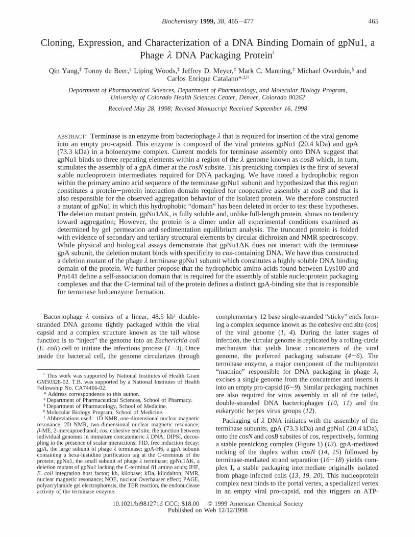
Packaging ofλ DNA initiates with the assembly of theterminase subunits, gpA (73.3 kDa) and gpNu1 (20.4 kDa),onto thecosNandcosBsubsites ofcos, respectively, forminga stable prenicking complex (Figure 1) (13). gpA-mediatednicking of the duplex withincosN (14, 15) followed byterminase-mediated strand separation (16-18) yields com-plex I , a stable packaging intermediate originally isolatedfrom phage-infected cells (13, 19, 20). This nucleoproteincomplex next binds to the portal vertex, a specialized vertexin an empty viral pro-capsid, and this triggers an ATP-
† This work was supported by National Institutes of Health GrantGM50328-02. T.B. was supported by a National Institutes of HealthFellowship No. CA74466-02.
* Address correspondence to this author.‡ Department of Pharmaceutical Sciences, School of Pharmacy.§ Department of Pharmacology, School of Medicine.| Molecular Biology Program, School of Medicine.1 Abbreviations used: 1D NMR, one-dimensional nuclear magnetic
resonance; 2D NMR, two-dimensional nuclear magnetic resonance;â-ME, 2-mercaptoethanol;cos, cohesive end site, the junction betweenindividual genomes in immature concatemericλ DNA; DIPSI, decou-pling in the presence of scalar interactions; FID, free induction decay;gpA, the large subunit of phageλ terminase; gpA-H6, a gpA subunitcontaining a hexa-histidine purification tag at the C-terminus of theprotein; gpNu1, the small subunit of phageλ terminase; gpNu1∆K, adeletion mutant of gpNu1 lacking the C-terminal 81 amino acids; IHF,E. coli integration host factor; kb, kilobase; kDa, kilodalton; NMR,nuclear magnetic resonance; NOE, nuclear Overhauser effect; PAGE,polyacrylamide gel electrophoresis; the TER reaction, the endonucleaseactivity of the terminase enzyme.
465Biochemistry1999,38, 465-477
10.1021/bi981271d CCC: $18.00 © 1999 American Chemical SocietyPublished on Web 12/12/1998
dependent translocation of the terminase subunits across theduplex and packaging of DNA within the capsid (Figure 1)(9, 21, 22). Though no direct evidence exists, it is likelythat a combination of the portal vertex proteins and the DNA-bound terminase subunits constitute the packaging apparatus.Upon encountering the next downstreamcosin the concate-mer, terminase again symmetrically nicks the duplex atcosNand strand separation simultaneously releases the DNA-filledcapsid and regenerates complexI (6, 8, 9). The addition ofa tail to the DNA-filled capsid yields a fully infectious virus,while the regenerated complexI again captures an emptypro-capsid to initiate a second round of genome packaging(23).
While the process of DNA packaging byλ terminase isreasonably well-characterized, the nature of the nucleoproteinintermediates involved in the packaging process are ill-defined. Terminase assembly models presume that gpNu1binds to the individual R-elements found withincosB, andthat gpNu1 assembly atcosB is required for efficientassembly of a gpA dimer atcosNand duplex nicking (6, 9,19) (Figure 1). These models are based on indirect evidenceand speculation, however, and there are no data availableon the protein composition of any of the packaging inter-
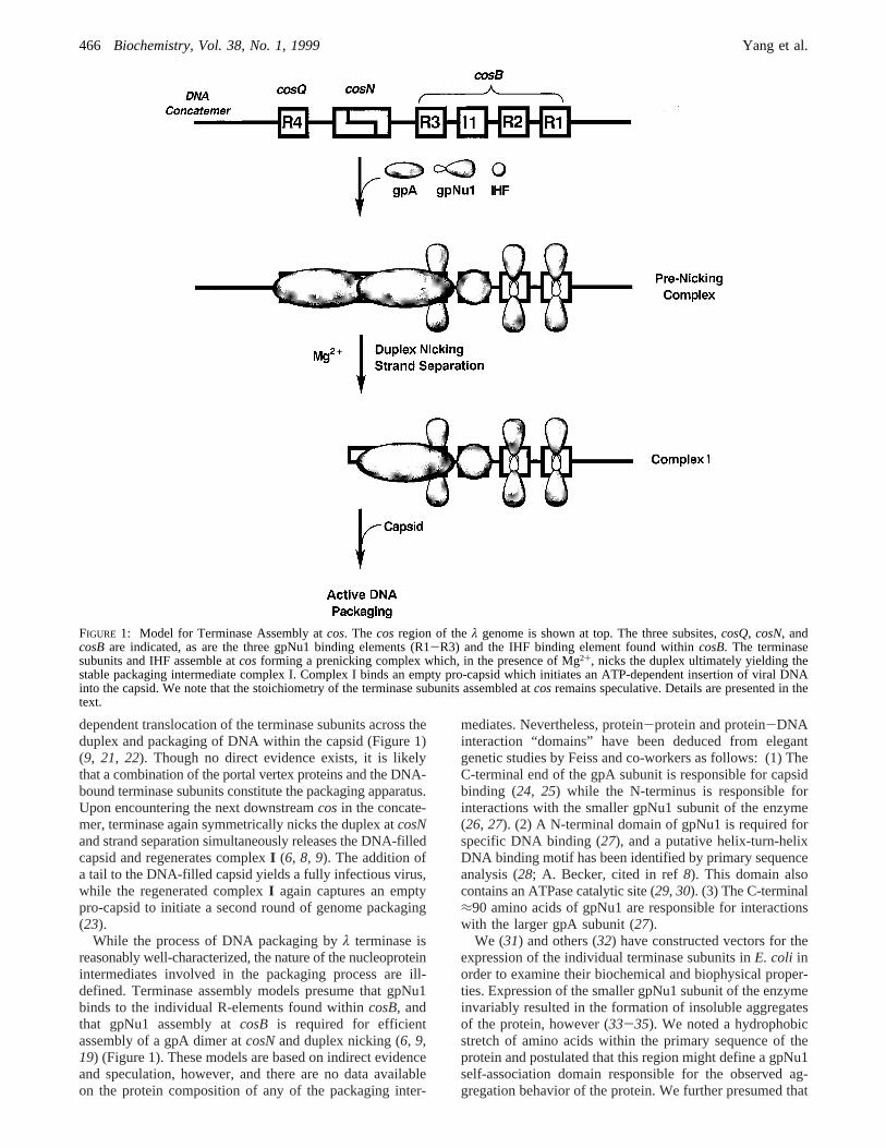
mediates. Nevertheless, protein-protein and protein-DNAinteraction “domains” have been deduced from elegantgenetic studies by Feiss and co-workers as follows: (1) TheC-terminal end of the gpA subunit is responsible for capsidbinding (24, 25) while the N-terminus is responsible forinteractions with the smaller gpNu1 subunit of the enzyme(26, 27). (2) A N-terminal domain of gpNu1 is required forspecific DNA binding (27), and a putative helix-turn-helixDNA binding motif has been identified by primary sequenceanalysis (28; A. Becker, cited in ref8). This domain alsocontains an ATPase catalytic site (29, 30). (3) The C-terminal≈90 amino acids of gpNu1 are responsible for interactionswith the larger gpA subunit (27).
We (31) and others (32) have constructed vectors for theexpression of the individual terminase subunits inE. coli inorder to examine their biochemical and biophysical proper-ties. Expression of the smaller gpNu1 subunit of the enzymeinvariably resulted in the formation of insoluble aggregatesof the protein, however (33-35). We noted a hydrophobicstretch of amino acids within the primary sequence of theprotein and postulated that this region might define a gpNu1self-association domain responsible for the observed ag-gregation behavior of the protein. We further presumed that
FIGURE 1: Model for Terminase Assembly atcos. The cos region of theλ genome is shown at top. The three subsites,cosQ, cosN, andcosBare indicated, as are the three gpNu1 binding elements (R1-R3) and the IHF binding element found withincosB. The terminasesubunits and IHF assemble atcosforming a prenicking complex which, in the presence of Mg2+, nicks the duplex ultimately yielding thestable packaging intermediate complex I. Complex I binds an empty pro-capsid which initiates an ATP-dependent insertion of viral DNAinto the capsid. We note that the stoichiometry of the terminase subunits assembled atcosremains speculative. Details are presented in thetext.
466 Biochemistry, Vol. 38, No. 1, 1999 Yang et al.
this region might mediate protein-protein interactionsrequired for the assembly of a stable gpNu1 nucleoproteincomplex atcosB. In order to examine these possibilities andto define functional domains within the gpNu1 polypeptide,we have constructed a deletion mutant of gpNu1 (amino acids1-100) which lacks the hydrophobic region of the protein.The construction, expression, and purification of this mutantprotein and the characterization of its structural and functionalfeatures are presented here.
EXPERIMENTAL PROCEDURES
Materials and Methods. Tryptone, yeast extract, and agarwere purchased from DIFCO. Restriction enzymes werepurchased from Promega. Mono-Q HR5/5 FPLC columns,DEAE-sepharose FF, and SP-sepharose FF chromatographyresins were purchased from Pharmacia.R32P-ATP waspurchased from ICN. Unlabeled nucleoside triphosphateswere purchased from Boehringer Mannheim Biochemicals.Ni-NTA agarose was purchased from Qiagen. The ECLWestern blotting kit was purchased from Amersham. Proteinmolecular weight standards for gel permeation chromatog-raphy were purchased from Pharmacia. All other materialswere of the highest quality commercially available.
Bacterial cultures were grown in shaker flasks utilizing aNew Brunswick Scientific series 25 incubator-shaker. Allprotein purifications utilized a Pharmacia FPLC systemwhich consisted of two P500 pumps, a GP250-plus controller,a V7 injector, and a Uvicord SII variable wavelengthdetector. UV-vis absorbance spectra were recorded on aHewlett-Packard HP8452A spectrophotometer. Fluorescencespectra were recorded at room temperature on a ShimadzuRF-1501 spectrofluorophotometer. A protein concentrationof 10 µg/mL in 10 mM potassium phosphate buffer, pH 7.4,was used, and a buffer blank was subtracted from thefluorescence spectrum. Circular dichroism (CD) spectra wererecorded on an Aviv model 62DS circular dichroism spec-tropolarimeter equipped with a Brinkmann Lauda RM6circulating water bath and a thermostated cell holder. Near-UV CD spectra utilized a protein concentration of 1.1 mg/mL in a 1 cm strain-free cuvette. Data were collectedbetween 250 and 350 nm at 0.5 nm intervals using abandwidth of 1.0 nm and a dwell time of 5 s. Far-UV CDspectra utilized a protein concentration of 110µg/mL in a 1mm strain-free cuvette. Data were collected from 180 to 250nm at 0.5 nm intervals using a bandwidth of 1.5 nm and adwell time of 5 s. MOLDI-TOF mass spectral analysis wasperformed at the University of Colorado Health SciencesCenter Macromolecular Resources Center. Prediction ofprotein secondary structures and hydrophobicity based uponprimary sequence data was performed by the methods ofChou and Fasman and Kyte and Doolittle, respectively, usingthe DNASIS program (Macintosh version 2.0). Calculationof protein secondary structures based upon the far-UV CDdata was performed using the SELCON program (36).Automated DNA sequence analysis was performed by theUniversity of Colorado Cancer Center MacromolecularResources Core facility. Both strands of the duplex wereexamined to ensure the expected DNA sequence.
Bacterial Strains, DNA Preparation, and Protein Purifica-tion. E. coli BL21(DE3) cells were a generous gift of D.Kroll (University of Colorado Health Sciences Center,
Denver, CO). All synthetic oligonucleotides used in this studywere purchased from Gibco/BRL and were used withoutfurther purification. Plasmids pSF1 and pAFP1, kindlyprovided by M. Feiss (University of Iowa, Iowa City, IA),were purified from theE. coli cell lines C600[pSF1] andJM107[pAFP1], respectively, using Qiagen DNA prepcolumns. Antiterminase antibodies were also a generous giftof Dr. Feiss, and Western blot analysis was performed usingthe ECL nonradioactive method according to the manufac-turer’s protocol (Amersham). Construction of a plasmidwhich expresses the terminase gpA subunit containing aC-terminal hexa-histidine sequence (gpA-H6) and purifica-tion of this protein to homogeneity were performed byWoods and Catalano (in preparation). Purification of gpAand full-length gpNu1 was performed as previously described(35). All of our purified proteins were homogeneous asdetermined by SDS-PAGE and densitometric analysis usinga molecular dynamics (MD) laser densitometer and theImageQuant data analysis package. Unless otherwise indi-cated, protein concentrations were determined spectrallyusing millimolar extinction coefficients (20, 35).
Construction of the gpNu1 Deletion Mutant OVerexpres-sion Plasmid.A truncatedNu1gene was amplified by PCRusing pSF1 as a DNA template. This plasmid contains thewild-typeNu1gene cloned into a pBR322 background (37).Primers were designed such thatEcoRI andBamHI restric-tion sequences were present at the 5′ and 3′ ends, respec-tively, of the PCR product. The primer sequences were asfollows: forward primer,5′-CCT CTC CCT TTC TCCGAATTC ATG GAA GTC AAC AAA AAG C- 3′; reverseprimer,5-CTT CCT GGA TCCTTA CTT CAG TTC CTGTGC GTC-3′′. TheEcoRI andBamHI restriction sequencesare indicated in italics while the f-MET (forward primer)and stop (reverse primer) codons are shown in bold type.Sequences complementary to theNu1 gene are underlined.The stop codon present in the reverse PCR primer yields,upon amplification, a truncated gpNu1 gene which expressesonly the first 100 amino acids of the protein. Amplificationof the truncated gene, isolation of the PCR product, andconstruction of the overexpression plasmid (pNu1∆K) wasperformed as described previously for the full-length protein(35). Colonies from BL21(DE3) cells transfected with thisplasmid efficiently expressed the C-terminal deleted gpNu1mutant protein, gpNu1∆K, as determined from whole celllysates analyzed by SDS-PAGE.
Expression and Purification of gpNu1∆K. Four liters of2X-YT media containing 50µg/mL ampicillin, 25 mMpotassium phosphate, pH 7.5, and 5 mM glucose wasinoculated with a 40 mL overnight culture of BL21(DE3)-[pNu1∆K] derived from an isolated colony. The culture wasmaintained at 37°C until an OD of 0.45 (600 nm) wasobtained at which point IPTG (1.2 mM) was added. The cellswere maintained at 30°C for an additional 2.5 h and thenharvested by centrifugation. Unless otherwise indicated, allsubsequent steps were performed at 0-4 °C. The cell pelletwas resuspended in ice cold buffer A (20 mM Tris, pH 8.0,2 mM EDTA, 7 mM â-ME, and 10% glycerol) containing100 mM NaCl, and the cells were disrupted by sonification.Insoluble cellular debris was removed by centrifugation(12000g, 30 min), and solid ammonium sulfate was addedto the clarified supernatant to 50% saturation. Insolubleprotein was removed by centrifugation (12000g, 30 min),
gpNu1 DNA Binding Domain Biochemistry, Vol. 38, No. 1, 1999467

and gpNu1∆K was then precipitated with the addition ofammonium sulfate to 75% followed by centrifugation. ThegpNu1∆K-containing pellet was taken into buffer A and,after dialysis against the same buffer, loaded onto a DEAE-sepharose column (200 mL) also equilibrated with bufferA. The column was developed with a salt gradient withgpNu1∆K eluting at 180 mM NaCl. Column fractions wereexamined by SDS-PAGE, and the appropriate fractions werepooled, dialyzed against buffer A, and loaded onto a SP-sepharose column equilibrated with the same buffer. Thecolumn was developed with a salt gradient with gpNu1∆Keluting at 200 mM NaCl. As before, column fractions wereexamined by SDS-PAGE; the appropriate fractions werepooled, dialyzed against buffer A, and loaded onto a MonoQHR5/5 column equilibrated with the same buffer. The columnwas developed with a salt gradient with gpNu1∆K elutingat 400 mM NaCl. Column fractions were examined by SDS-PAGE, and the appropriate fractions were pooled, dialyzedagainst 20 mM Tris buffer, pH 8.0, containing 1 mM EDTA,7 mM â-ME, and 20% glycerol, and stored at-80 °C.
NMR Spectra. NMR samples contained 0.5-1.0 mMgpNu1∆K in 10 mM sodium phosphate buffer, pH 7.2, and10% (v/v) 2H2O/1H2O. One-dimensional (1D)1H NMRexperiments were recorded at 7.5, 15, 25, and 35°C usinga Varian INOVA 600 MHz spectrometer equipped with ashielded triple resonance 5 mm probe. For each 1D spectrum,16 or 64 FIDs were acquired of 2048 data points each. Atwo-dimensional (2D)1H NOE spectrum (38) was recordedat 25 °C with a mixing time of 150 ms, collecting 512t1experiments of 2048 complex data points each and acquiring64 FIDs pert1 experiment. 2D1H DIPSI spectra (39-41)were recorded at 25°C with mixing times of 20 and 40 ms,collecting 860t1 experiments of 2048 complex data pointseach and acquiring 80 FIDs pert1 experiment. The waterresonance was suppressed by low-power saturation duringthe relaxation delay of 1.2 s. NMR spectra were processedusing the NMRPipe software package (42). In short, timedomain data were multiplied by phase-shifted squared-sine-bell functions, followed by zero-filling, Fourier transforma-tion, and base-line corrections.
Analysis of Subunit Association. Analytical gel filtrationanalysis utilized a Superose 12 HR10/30 column equilibratedwith 50 mM sodium phosphate buffer, pH 7.2, containing150 mM NaCl running at 0.4 mL/min. A molecular weightstandard curve was constructed by standard methods (43)using the following molecular weight standards: Bluedextran (FW> 2 000 000, RT) 19.8 min), bovine serumalbumin (67 000, 34.1 min), ovalbumin (43 000, 35.7 min),chymotrypsinogen A (25 000, 39.6 min), and ribonucleaseA (13 700, 41.1 min).
Sedimentation equilibrium experiments utilized a Beck-mann XL-A analytical ultracentrifuge equipped with a Ti-60 four-place rotor. Protein samples, at the concentrationsindicated in each individual experiment, were prepared byextensive dialysis against 10 mM sodium phosphate buffer,pH 7.2. The samples (100µL) were analyzed in six-channelcharcoal-epon centerpieces (12 mm) with 10µL of FC-43(Beckmann fluorocarbon-43) added to each sample as a basefluid. Absorbance optics were used to monitor the opticaldensity of each sample at the indicated wavelengths. Datawere collected at 0.001 cm radial increments and stored asthe average of five replicate measurements. Data sets were
collected until successive scans were superimposable toensure that equilibrium had been achieved. The data wereanalyzed with the program NONLIN (44) using only thosedata within the absorbance range between 0.1 and 1.6 OD.The reduced apparent molecular weight (σ) for gpNu1∆Kwas calculated from
whereMr andν are the molecular weight and partial specificvolume, respectively, of gpNu1∆K, F is the buffer density,ω is the radial velocity,R is the ideal gas constant, andT isthe temperature (45, 46). The buffer density was calculatedas described by Laue et al. (46), and a value ofν ) 0.7289was calculated from the amino acid composition of theprotein (46).
Interaction of the Terminase Subunits. C-terminal hexa-HIS-containing gpA (gpA-H6, 2µM) was preincubated witheither full-length gpNu1 (4µM) or the truncated gpNu1∆Kmutant (4µM) in 50 mM sodium phosphate, pH 8.0, 100mM NaCl buffer for 20 min on ice. Ni-NTA agarose resin(0.5 mL in the same buffer) was added to the protein mixture,and the incubation was continued for an additional 60 minwith mild shaking. The resin was then washed twice with300 µL of wash buffer (50 mM sodium phosphate, pH 8.0,20 mM imidazole, and 500 mM NaCl), and gpA-H6 wasfinally eluted from the column with elution buffer (50 mMsodium phosphate, pH 8.0, 250 mM imidazole, and 500 mMNaCl). Fractions (200µL) were collected and analyzed bySDS-PAGE.
ActiVity Assays.Gel mobility shift assays were conductedas described by Yang et al. (13) except that the radiolabeledDNA substrate (cos-containing or nonspecific) was addedat a concentration of 20 pM. The concentration of proteinand salt added to the binding reactions is indicated in eachindividual experiment. Thecos-cleavage assay was per-formed as described by Tomka and Catalano using pAFP1as a nuclease substrate (20). ATPase catalytic activity wasexamined as described previously (47) and utilized an ATPconcentration of 50µM and a DNA (Sca1-linearized pAFP1)concentration of 300µM (total nucleotide). The concentrationof protein used in these assays is indicated in each individualexperiment.
RESULTS
Construction, Expression, and Purification of gpNu1∆K.Purification of phageλ terminase has historically beenfrustrated by the insolubility of the overexpressed protein(20, 33), likely due to self-association of gpNu1, the smallterminase subunit (34, 35). The hydropathy plot for gpNu1shown in Figure 2A reveals a hydrophobic region locatedbetween amino acids≈100 and 140 of the protein primarysequence. We reasoned that this region of the protein might,via hydrophobic interactions, be responsible for the self-association behavior of gpNu1, resulting in the insolubilityof the isolated protein. We have therefore constructed adeletion mutant of gpNu1 which truncates the protein atlysine 100 in the primary sequence (gpNu1∆K). Importantly,limited proteolysis studies have demonstrated a relativelystable N-terminal digestion intermediate of gpNu1 with amolecular weight expected from a similar deletion of the
σ ) Mr*(1 - ν*F)*ω2/RT
468 Biochemistry, Vol. 38, No. 1, 1999 Yang et al.
C-terminus of the protein (Catalano, C. E., and Hanagan,A., unpublished).
Virtually all of the expressed gpNu1∆K protein waspresent in the soluble fraction of the crude cell lysate (Figure2B). This is in stark contrast to full-length gpNu1, whichpartitions exclusively into the insoluble lysis pellet (34, 35).Figure 2B shows gpNu1∆K at each stage of the purificationprocedure and demonstrates that the protein was purified tohomogeneity. Analysis of the gel data yielded an apparentmolecular weight of≈11 500, consistent with theMr )11 478 predicted from the gene sequence. Consistently,MOLDI-TOF mass spectral analysis of the purified proteinyielded a molecular weight of 11 478.9. Western blot analysis
shows a single immunoreactive band with no evidence forproteolysis (Figure 2C), suggesting that the truncated proteinis folded and stable in the crude cell lysate and throughoutthe purification. The protocol described here yields 6.5 mgof highly purified protein per liter of cell growth.
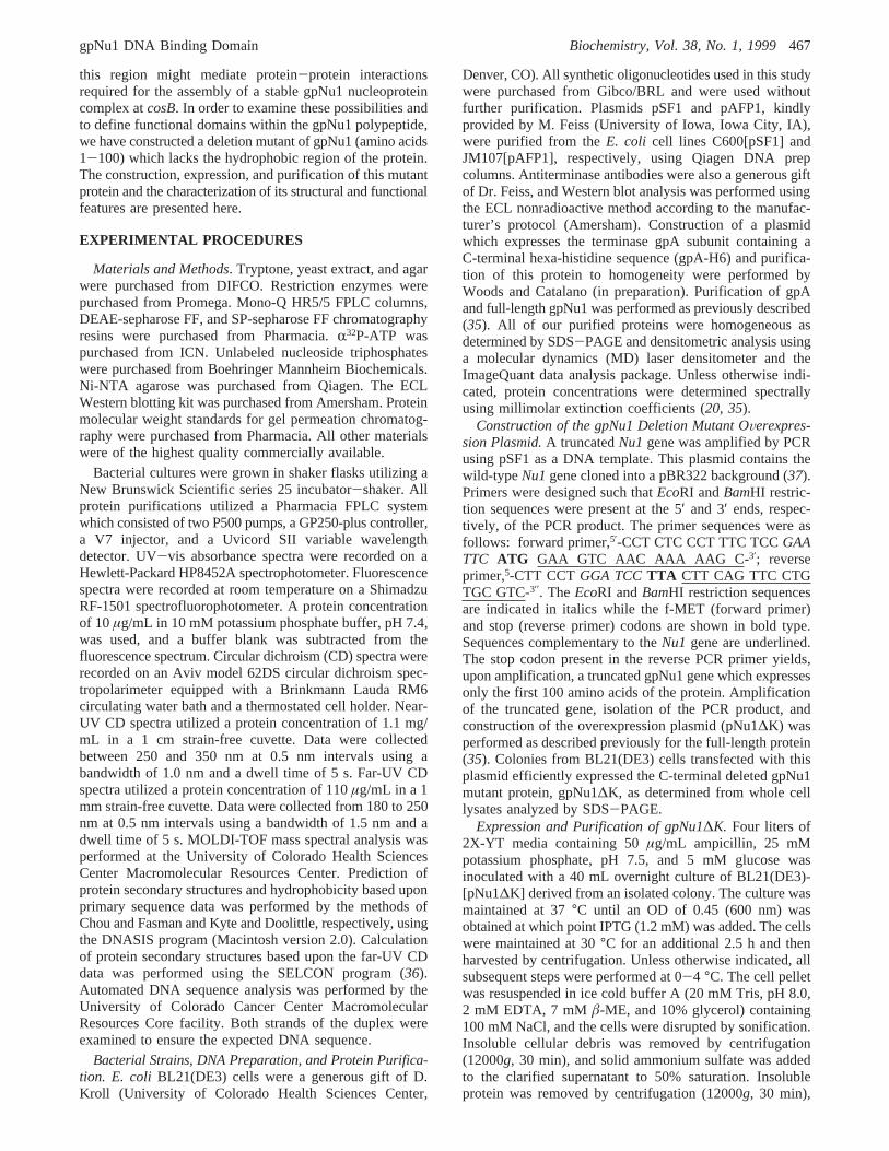
Preliminary Characterization of gpNu1∆K. Unlike full-length gpNu1, purified gpNu1∆K was fully soluble under avariety of buffer conditions and at elevated (>15 mg/mL)protein concentrations (see NMR experiments below). Theprotein possesses a UV spectrum (not shown) typical of aglobular protein that is devoid of contaminating DNA (A280:A260 ) 1.8) (48-50). An extinction coefficient (ε280) of 15.2mM-1‚cm-1 for gpNu1∆K was determined by the methodof Gill and von Hipple (51, 52). The fluorescence spectrumof the protein exhibits an emission maximum of 336 nm thatremains unchanged using excitation frequencies between 260and 285 nm (λex,max ) 273 nm) (Figure 3A); however,selective excitation of tryptophan residues (λex ) 295 nm)results in a red shift of the emission maximum to from 336to 342 nm. This suggests that, unlike most tyrosines inproteins, the three tyrosines in gpNu1∆K contribute
FIGURE 2: Cloning, Expression, and Purification of gpNu1∆K. (A)Kyte-Doolitle hydropathy plot of full-length gpNu1. The locationsof the hydrophobic domain (HD), the putative helix-turn-helix DNAbinding motif (HTH, Lys3 to Glu22), and the Walker A (ATPase)sequence (ATP, Val29 to Asp48) are indicated. The location ofthe C-terminal end in the gpNu1∆K deletion mutant protein(Lys100) is indicated with an arrow. (B) SDS-PAGE showing thepurification of gpNu1∆K: lane 1, preinduced crude cell lysate, lane2, post induced crude cell lysate; lane 3, lysis supernatant; lane 4,pooled DEAE sepharose fractions; lane 5, pooled SP-sepharosefractions; lane 6, pooled Mono-Q fractions. (C) Western blotanalysis of the gel shown in panel B.
FIGURE 3: Fluorescence and CD Spectra of Purified gpNu1∆K.(A) Fluorescence spectrum of gpNu1∆K. The full emissionspectrum is shown for excitation frequencies (λex) of 285 (solidline) and 295 nm (dashed line). (B) Far UV-CD spectrum ofgpNu1∆K. (C) Near UV-CD spectrum of gpNu1∆K. All spectrawere recorded as described in Experimental Procedures.
gpNu1 DNA Binding Domain Biochemistry, Vol. 38, No. 1, 1999469

significantly to the fluorescence spectrum when excited atshorter wavelengths (49, 53). Furthermore, this red-shiftedemission maximum is close to that of tryptophan free insolution (348 nm), suggesting that at least one of the twotryptophans in the protein is partially solvent exposed (49,53).
The far-UV circular dichroism (CD) spectrum of gpNu1∆Kshows strong negative maxima at 222 and 208 nm, suggest-ing that this protein possesses a significant amount ofR-helical structure (Figure 3B) (49, 54). Deconvolutionanalysis of the spectrum is consistent with a protein contain-ing primarily (50%)R-helical structure as well as 27% ofthe residues being in aâ-sheet conformation. These valuesagree well with the Chou and Fasman secondary structurepredictions for the protein (53%R-helix, 26%â-sheet). Thenear-UV CD spectrum of gpNu1∆K is shown in Figure 3C.Unlike the full-length protein, which exhibits little signal inthis region of the spectrum (35), the deletion mutant displaysa signal rich in vibronic fine structure consistent with theexistence of significant tertiary structure. The intense bandsobserved at 296 and 289 nm likely represent the two vibroniccomponents of the tryptophan1Lb band transition (54, 55).The intensity of these bands suggests that at least one of thetwo tryptophans in the protein has restricted mobility.Furthermore, these bands are significantly red-shifted fromtheir canonical positions of 292 and 285 nm, suggesting thatthere are specific interactions with nearby side chains. Thismay result from proximity to a nearby carboxylate residue(56) and/or through space electronic coupling to the lowestenergy states of a nearby aromatic group (55).2
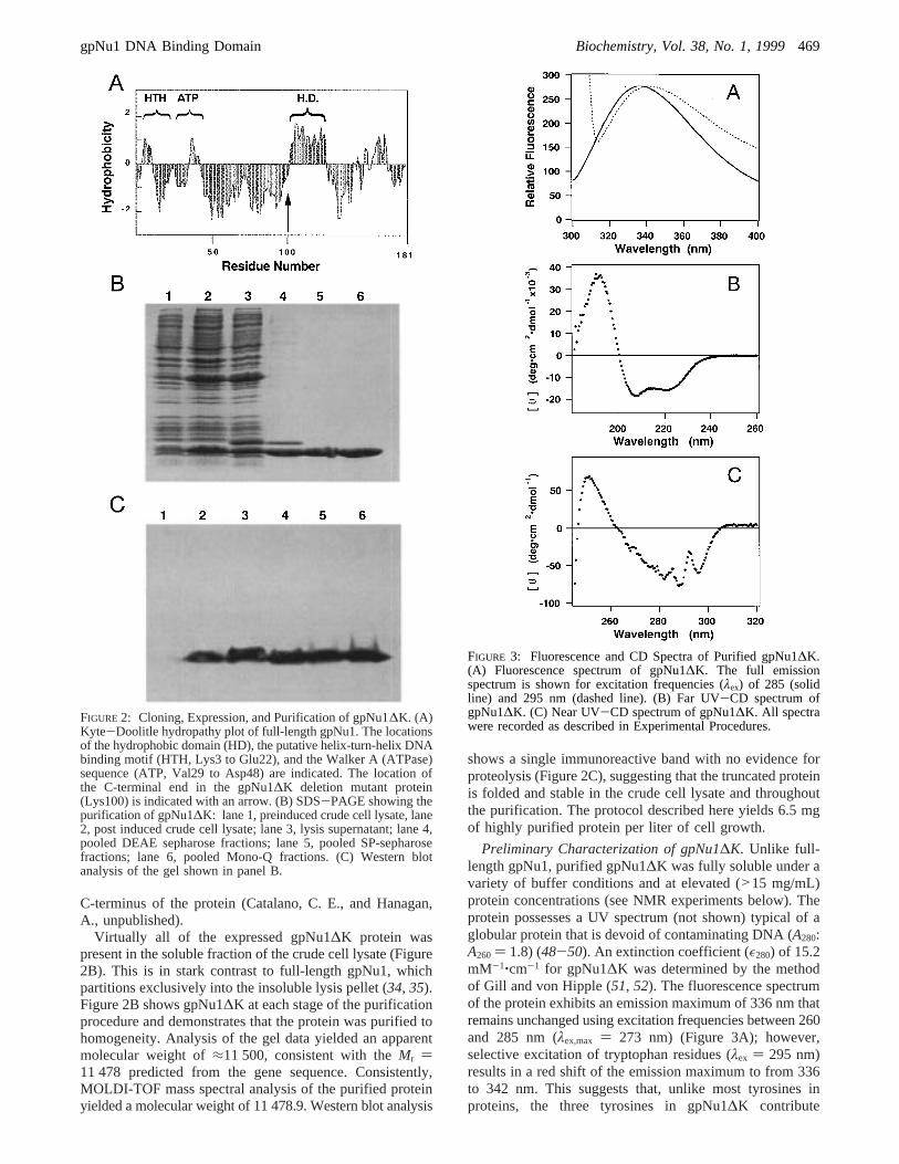
NMR Spectra of gpNu1∆K. The fold and structuralintegrity of gpNu1∆K were further investigated using NMRspectroscopy, and a typical 1D1H NMR spectrum isdisplayed in Figure 4A. This spectrum did not changesubsequent to prolonged storage (>100 days) at 4°C,demonstrating the structural stability of the protein. More-over, spectra collected between 7.5 and 35°C exhibited nosignificant differences, other than the expected temperature-related changes in resonance line widths and amide protonchemical shifts (not shown). The line widths of1H resonancesin NMR spectra are related to the molecular mass and theshape of the molecule. Analysis of the line widths in the 1D1H NMR spectrum of gpNu1∆K suggests that this proteinis at least a dimer at these protein concentrations. Further-more, the presence of only two tryptophan Hε1 resonancesin the spectrum (“Trp-Hε1s”, Figure 4A) suggest that thedimer is symmetric, or that neither of the two tryptophansin the protein resides within the subunit interface.
Amino acid HR chemical shifts have been correlated withthe participation of the respective amino acids in secondarystructural elements (57). It is difficult to resolveR-helicalcontent in a 1D spectrum due to spectral overlap; however,the NMR spectrum for gpNu1∆K possesses several reso-nances in the chemical shift region associated with aminoacids involved inâ-sheet structures (“HRs”, Figure 4A).These data are consistent with theR/â-fold predicted by CD
spectroscopy (Vide supra). The 1D spectrum further showsa broad dispersion of amide (HN), aromatic, HR, and methyl1H chemical shifts. Such a dispersion results from equivalentprotons that reside in different local environments, which isthe direct consequence of the folded nature of a protein. Forexample, the upfield shifts of several methyl protons(“resolved CH3 protons”, Figure 4A) reflects the shieldingof these protons from the solvent by nearby amino acids. Inaddition, the downfield shift of several amide protons withrespect to their random coil values (“downfield HNs”, Figure
2 Through space coupling to the lowest energy states of a nearbydisulfide may also result in red-shifted tryptophan bands; however, thereare no cysteine residues in gpNu1∆K. A final, though unlikely,possibility is that the red-shifted bands in the near-UV CD spectrumresult from a deprotonated phenolate, where the 0-0 band is know tooccur at 295 nm (55).
FIGURE 4: NMR Spectra of gpNu1∆K. (A) 1D 1H 600 MHz NMRspectrum of gpNu1∆K (1 mM) in 10 mM sodium phosphate buffer,pH 7.2, recorded at 35°C. Regions of resolved1H resonancesstemming from the two tryptophan indole protons (Trp-Hε1s), amideprotons (downfield HNs), aromatic protons,R protons (HR), andmethyl (CH3) protons are indicated in the figure. H2O points to theposition of the water resonance, whileφ indicates the resonancesof contaminating glycerol. (B) Fragment of the 2D1H 600 MHzNOE spectrum recorded at 25°C. WX indicates putative throughspace NOE correlations between the side chains of a tryptophanresidue and another aromatic residue, probably a tyrosine. Thenomenclature of tryptophan, phenylalanine, and tyrosine side chainshas been included in the figure for clarity. Tentative assignmentsof Trp and Tyr/Phe resonances were made on the basis of NOEand DIPSI (not shown) spectra.
470 Biochemistry, Vol. 38, No. 1, 1999 Yang et al.
4A) indicates their involvement in strong hydrogen bondsor their proximity to aromatic rings (58, 59).
The 2D 1H NOE spectrum of gpNu1∆K shows thepresence of numerous methyl-aromatic, HR-aromatic, andaromatic-aromatic1H-1H NOE cross-peaks (Figure 4B).1H NOEs are observed only for proton pairs positioned within5 Å of each other, and this 2D spectrum provides additionalevidence that the deletion mutant is folded in solution.Preliminary analysis of this NOE spectrum confirms andextends some of the observations made with the otherspectroscopic techniques. The Hε1 chemical shifts of the twotryptophans in gpNu1∆K (also depicted in the 1D spectrum,Figure 4A) and the NOE patterns involving these protonsare clearly different, indicating that the two tryptophansreside in different chemical environments. This suggests thatthe solvent-exposed Trp observed in the fluorescence spec-trum (Figure 3A) and the mobility-restricted Trp observedin the near-UV CD spectrum (Figure 3C) represent differentresidues. The NMR data thus confirm that one of thetryptophans in gpNu1∆K is burried and mobility-restrictedwithin the protein while the other is partially solvent-exposed,presumably closer to the protein surface. An attractivecandidate for the latter is Trp22, which lies within the helix-turn-helix DNA binding motif and must be located near thesurface in the DNA binding cleft. The second tryptophanresidue observed in the 2D spectrum shows1H-1H NOEsconsistent with proximity to an aromatic residue in theprotein, possibly a tyrosine (see Figure 4B). This tryptophanis likely the same residue observed in the near-UV CDspectrum of the protein and suggests that the red-shifted CDbands result from this aromatic-aromatic interaction. Thesedata suggest, but do not prove, that the second tryptophan,Trp49, represents the buried tryptophan observed in both theCD and 2D NMR spectra.
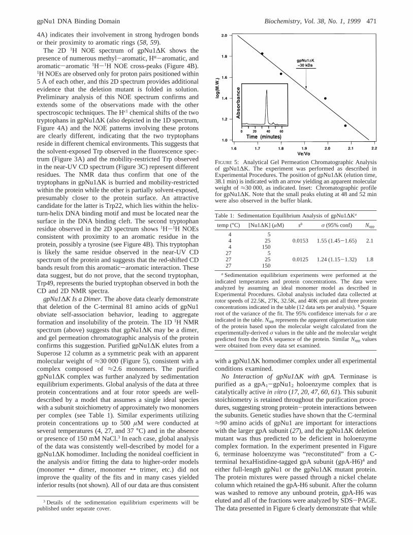
gpNu1∆K Is a Dimer.The above data clearly demonstratethat deletion of the C-terminal 81 amino acids of gpNu1obviate self-association behavior, leading to aggregateformation and insolubility of the protein. The 1D1H NMRspectrum (above) suggests that gpNu1∆K may be a dimer,and gel permeation chromatographic analysis of the proteinconfirms this suggestion. Purified gpNu1∆K elutes from aSuperose 12 column as a symmetric peak with an apparentmolecular weight of≈30 000 (Figure 5), consistent with acomplex composed of≈2.6 monomers. The purifiedgpNu1∆K complex was further analyzed by sedimentationequilibrium experiments. Global analysis of the data at threeprotein concentrations and at four rotor speeds are well-described by a model that assumes a single ideal specieswith a subunit stoichiometry of approximately two monomersper complex (see Table 1). Similar experiments utilizingprotein concentrations up to 500µM were conducted atseveral temperatures (4, 27, and 37°C) and in the absenceor presence of 150 mM NaCl.3 In each case, global analysisof the data was consistently well-described by model for agpNu1∆K homodimer. Including the nonideal coefficient inthe analysis and/or fitting the data to higher-order models(monomer T dimer, monomerT trimer, etc.) did notimprove the quality of the fits and in many cases yieldedinferior results (not shown). All of our data are thus consistent
with a gpNu1∆K homodimer complex under all experimentalconditions examined.
No Interaction of gpNu1∆K with gpA. Terminase ispurified as a gpA1-gpNu12 holoenzyme complex that iscatalytically activein Vitro (17, 20, 47, 60, 61). This subunitstoichiometry is retained throughout the purification proce-dures, suggesting strong protein-protein interactions betweenthe subunits. Genetic studies have shown that the C-terminal≈90 amino acids of gpNu1 are important for interactionswith the larger gpA subunit (27), and the gpNu1∆K deletionmutant was thus predicted to be deficient in holoenzymecomplex formation. In the experiment presented in Figure6, terminase holoenzyme was “reconstituted” from a C-terminal hexaHistidine-tagged gpA subunit (gpA-H6)4 andeither full-length gpNu1 or the gpNu1∆K mutant protein.The protein mixtures were passed through a nickel chelatecolumn which retained the gpA-H6 subunit. After the columnwas washed to remove any unbound protein, gpA-H6 waseluted and all of the fractions were analyzed by SDS-PAGE.The data presented in Figure 6 clearly demonstrate that while
3 Details of the sedimentation equilibrium experiments will bepublished under separate cover.
FIGURE 5: Analytical Gel Permeation Chromatographic Analysisof gpNu1∆K. The experiment was performed as described inExperimental Procedures. The position of gpNu1∆K (elution time,38.1 min) is indicated with an arrow yielding an apparent molecularweight of ≈30 000, as indicated. Inset: Chromatographic profilefor gpNu1∆K. Note that the small peaks eluting at 48 and 52 minwere also observed in the buffer blank.
Table 1: Sedimentation Equilibrium Analysis of gpNu1∆Ka
temp (°C) [Nu1∆K] (µM) sb σ (95% conf) Napp
4 54 25 0.0153 1.55 (1.45-1.65) 2.14 150
27 527 25 0.0125 1.24 (1.15-1.32) 1.827 150
a Sedimentation equilibrium experiments were performed at theindicated temperatures and protein concentrations. The data wereanalyzed by assuming an ideal monomer model as described inExperimental Procedures. Global analysis included data collected atrotor speeds of 22.5K, 27K, 32.5K, and 40K rpm and all three proteinconcentrations indicated in the table (12 data sets per analysis).b Squareroot of the variance of the fit. The 95% confidence intervals forσ areindicated in the table.Napprepresents the apparent oligomerization stateof the protein based upon the molecular weight calculated from theexperimentally-derivedσ values in the table and the molecular weightpredicted from the DNA sequence of the protein. SimilarNapp valueswere obtained from every data set examined.
gpNu1 DNA Binding Domain Biochemistry, Vol. 38, No. 1, 1999471
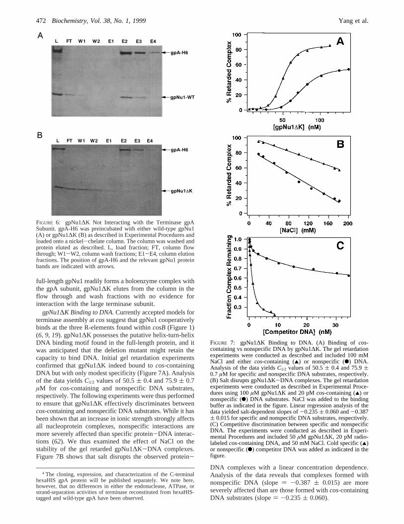
full-length gpNu1 readily forms a holoenzyme complex withthe gpA subunit, gpNu1∆K elutes from the column in theflow through and wash fractions with no evidence forinteraction with the large terminase subunit.
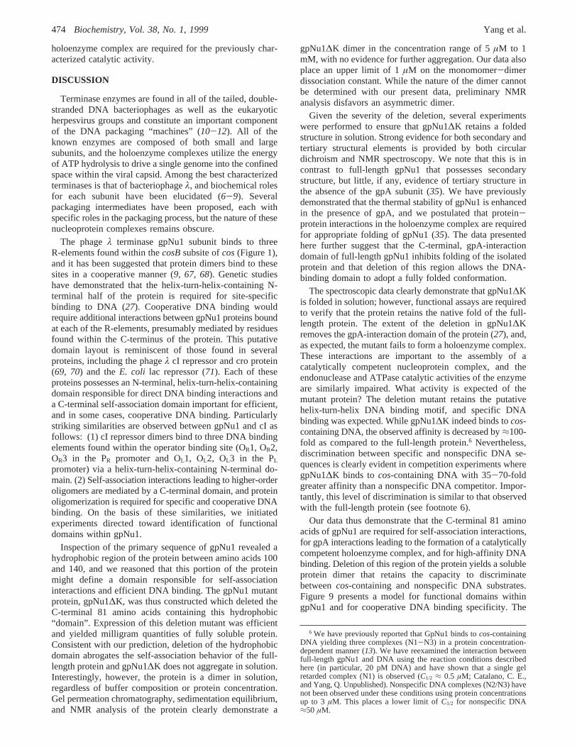
gpNu1∆K Binding to DNA.Currently accepted models forterminase assembly atcossuggest that gpNu1 cooperativelybinds at the three R-elements found withincosB(Figure 1)(6, 9, 19). gpNu1∆K possesses the putative helix-turn-helixDNA binding motif found in the full-length protein, and itwas anticipated that the deletion mutant might retain thecapacity to bind DNA. Initial gel retardation experimentsconfirmed that gpNu1∆K indeed bound tocos-containingDNA but with only modest specificity (Figure 7A). Analysisof the data yieldsC1/2 values of 50.5( 0.4 and 75.9( 0.7µM for cos-containing and nonspecific DNA substrates,respectively. The following experiments were thus performedto ensure that gpNu1∆K effectively discriminates betweencos-containing and nonspecific DNA substrates. While it hasbeen shown that an increase in ionic strength strongly affectsall nucleoprotein complexes, nonspecific interactions aremore severely affected than specific protein-DNA interac-tions (62). We thus examined the effect of NaCl on thestability of the gel retarded gpNu1∆K-DNA complexes.Figure 7B shows that salt disrupts the observed protein-
DNA complexes with a linear concentration dependence.Analysis of the data reveals that complexes formed withnonspecific DNA (slope) -0.387 ( 0.015) are moreseverely affected than are those formed withcos-containingDNA substrates (slope) -0.235( 0.060).
4 The cloning, expression, and characterization of the C-terminalhexaHIS gpA protein will be published separately. We note here,however, that no differences in either the endonuclease, ATPase, orstrand-separation activities of terminase reconstituted from hexaHIS-tagged and wild-type gpA have been observed.
FIGURE 6: gpNu1∆K Not Interacting with the Terminase gpASubunit. gpA-H6 was preincubated with either wild-type gpNu1(A) or gpNu1∆K (B) as described in Experimental Procedures andloaded onto a nickel-chelate column. The column was washed andprotein eluted as described. L, load fraction; FT, column flowthrough; W1-W2, column wash fractions; E1-E4, column elutionfractions. The position of gpA-H6 and the relevant gpNu1 proteinbands are indicated with arrows.
FIGURE 7: gpNu1∆K Binding to DNA. (A) Binding of cos-containing vs nonspecific DNA by gpNu1∆K. The gel retardationexperiments were conducted as described and included 100 mMNaCl and eithercos-containing (2) or nonspecific (b) DNA.Analysis of the data yieldsC1/2 values of 50.5( 0.4 and 75.9(0.7 µM for specific and nonspecific DNA substrates, respectively.(B) Salt disrupts gpNu1∆K-DNA complexes. The gel retardationexperiments were conducted as described in Experimental Proce-dures using 100µM gpNu1∆K and 20 pMcos-containing (2) ornonspecific (b) DNA substrates. NaCl was added to the bindingbuffer as indicated in the figure. Linear regression analysis of thedata yielded salt-dependent slopes of-0.235( 0.060 and-0.387( 0.015 for specific and nonspecific DNA substrates, respectively.(C) Competitive discrimination between specific and nonspecificDNA. The experiments were conducted as described in Experi-mental Procedures and included 50µM gpNu1∆K, 20 pM radio-labeledcos-containing DNA, and 50 mM NaCl. Cold specific (2)or nonspecific (b) competitor DNA was added as indicated in thefigure.
472 Biochemistry, Vol. 38, No. 1, 1999 Yang et al.

The experiment presented in Figure 7A suggests thatgpNu1∆K binds tocos-containing DNA with only modestspecificity. This is not entirely unexpected, however, asdiscrimination between specific and nonspecific DNA ispoorly demonstrated in gel retardation experiments performedin this manner (63), and DNA binding specificity is betterdemonstrated by direct competition between the substrates(64-66). In the experiment presented in Figure 7C, gpNu1∆Kwas added to a binding mixture containing a32P-radiolabeledcos-containing DNA substrate and increasing concentrationsof either specific (cos-containing) or nonspecific coldcompetitor DNA. The figure demonstrates that a specificDNA substrate effectively competes with complex formationand that≈1.5 nM cold competitor is required to decreasethe radiolabeled complex by 50%. Similar titrations with anonspecific DNA competitor demonstrate that significantlyhigher concentrations (≈50-100 nM, estimated from thecurve) are required to disrupt the radiolabeled complex to asimilar extent and that gpNu1∆K bindscos-containing DNAwith 35-70-fold greater affinity than nonspecific DNAsequences.
Catalytic ActiVity of gpNu1∆K. Binding of gpNu1 tocosBis critical to the assembly of gpA atcosN and thecos-cleavage reaction (Figure 1). We have shown that gpNu1∆Kdoes not interact with the terminase gpA subunit in solution(see Figure 6). The protein retains DNA binding activity,however, and it was feasible that a DNA-bound holoenzymecomplex might form and possess catalytic activity. Figure8A demonstrates that while neither the isolated gpA subunitnor the full-length gpNu1 subunit alone possess any detect-able nuclease activity under these experimental conditions,terminase enzyme reconstituted from the individual subunits(gpA1-gpNu12) is fully active. Reconstitution of terminaseholoenzyme from gpA and the gpNu1∆K deletion mutantdoes not, however, yield any detectable nuclease activity,even at significantly elevated protein concentrations. It isimportant to note that nocos-cleavage activity is detectedeven at 50µM gpNu1∆K, a concentration where strongbinding tocos-containing DNA is observed (see Figure 7).
We have previously characterized the ATPase activity ofterminase holoenzyme and have identified catalytic siteswithin each subunit of the enzyme (30, 47). The active siteP-loop motif is retained within gpNu1∆K, and it was possiblethat this mutant protein might possess ATPase activity. Wethus directly examined the catalytic activity of the isolatedgpNu1∆K subunit and terminase enzyme “reconstituted”from this protein. Figure 8B demonstrates that while theisolated gpA subunit possesses modest ATPase activity,neither the full-length gpNu1 subunit nor the C-terminaldeletion mutant exhibits any detectable catalytic activity
under these experimental conditions.5 Figure 8B and Table2 demonstrate that while reconstitution of terminase holoen-zyme from gpA and the full-length gpNu1 subunit signifi-cantly stimulates ATP hydrolysis, enzyme prepared fromgpNu1∆K does not exhibit activity beyond that observedfor gpA alone. Interestingly, these data suggest that theATPase activity of the gpNu1 subunit is essentially silent inthe isolated protein and that interactions with gpA in the
5 Early studies suggested that the isolated gpNu1 subunit possessedan intrinsic ATPase activity with a turnover number of 38 min-1 andthat, unlike terminase holoenzyme, it was not stimulated by DNA (34).More recent studies have similarly reported a gpNu1 ATPase activity,but with a much lower turnover number of 0.4-0.8 min-1 (61). OurgpNu1 preparations similarly possess an extremely weak ATPaseactivity, estimated to be≈0.03 min-1, that is not stimulated by DNAand that approaches wild-type levels only upon reconstitution into aholoenzyme complex. Given the ubiquitous nature of contaminatingATPases, the low ATPase activity of the isolated gpNu1 subunit, andthe lack of DNA stimulation in our gpNu1 preparations, we cannotattribute the observed ATPase activity directly to gpNu1 without furtherexperimentation.
FIGURE 8: Catalytic Activity of gpNu1∆K. (A) The cos-cleavageassay was performed as described in Experimental Procedures withthe following additions: lane 1, wild-type gpNu1 alone (2µM);lane 2, gpA alone (1µM); lane 3, terminase reconstituted fromgpA (1 µM) and wild-type gpNu1 (2µM); lanes 4-7, terminasereconstituted from gpA (1µM) and increasing concentrations ofgpNu1∆K (2, 20, 40, and 50µM, respectively); lane 8: gpNu1∆Kalone (50µM). The positions of the DNA product bands areindicated with arrows. (B) ATPase assay was performed asdescribed in Experimental Procedures with the following addi-tions: lane 1, no additions; lane 2, gpA alone (1µM); lane 3, full-length gpNu1 alone (2µM); lane 4, gpNu1∆K alone (2µM); lane5, gpNu1∆K alone (50µM); lane 6, terminase reconstituted fromgpA (1µM) and full-length gpNu1 (2µM); lanes 7 and 8, terminasereconstituted from gpA (1µM) and gpNu1∆K (2 µM and 50µM,respectively). The position of the ADP product band is indicatedwith an arrow. Quantitation of the ATP hydrolysis data is presentedin Table 2.
Table 2: ATPase Activity of the Terminase Subunitsa
additions ADP formed (µM)
none NDgpA 6gpNu1-FL NDgpNu1∆K NDgpNu1∆K (50 µM) NDgpA + gpNu1-FL 21gpA + gpNu1∆K 5gpA + gpNu1∆K (50 µM) 4
a The ATPase assays were performed as described in ExperimentalProcedures. The terminase gpA subunit was added, as indicated, to afinal concentration of 1µM. Unless otherwise stated, the gpNu1 subunit(full length or mutant) was added, as indicated, to a final concentrationof 2 µM. ND, no detectable ADP formation (<0.5 µM).
gpNu1 DNA Binding Domain Biochemistry, Vol. 38, No. 1, 1999473
holoenzyme complex are required for the previously char-acterized catalytic activity.
DISCUSSION
Terminase enzymes are found in all of the tailed, double-stranded DNA bacteriophages as well as the eukaryoticherpesvirus groups and constitute an important componentof the DNA packaging “machines” (10-12). All of theknown enzymes are composed of both small and largesubunits, and the holoenzyme complexes utilize the energyof ATP hydrolysis to drive a single genome into the confinedspace within the viral capsid. Among the best characterizedterminases is that of bacteriophageλ, and biochemical rolesfor each subunit have been elucidated (6-9). Severalpackaging intermediates have been proposed, each withspecific roles in the packaging process, but the nature of thesenucleoprotein complexes remains obscure.
The phageλ terminase gpNu1 subunit binds to threeR-elements found within thecosBsubsite ofcos(Figure 1),and it has been suggested that protein dimers bind to thesesites in a cooperative manner (9, 67, 68). Genetic studieshave demonstrated that the helix-turn-helix-containing N-terminal half of the protein is required for site-specificbinding to DNA (27). Cooperative DNA binding wouldrequire additional interactions between gpNu1 proteins boundat each of the R-elements, presumably mediated by residuesfound within the C-terminus of the protein. This putativedomain layout is reminiscent of those found in severalproteins, including the phageλ cI repressor and cro protein(69, 70) and theE. coli lac repressor (71). Each of theseproteins possesses an N-terminal, helix-turn-helix-containingdomain responsible for direct DNA binding interactions anda C-terminal self-association domain important for efficient,and in some cases, cooperative DNA binding. Particularlystriking similarities are observed between gpNu1 and cI asfollows: (1) cI repressor dimers bind to three DNA bindingelements found within the operator binding site (OR1, OR2,OR3 in the PR promoter and OL1, OL2, OL3 in the PL
promoter) via a helix-turn-helix-containing N-terminal do-main. (2) Self-association interactions leading to higher-orderoligomers are mediated by a C-terminal domain, and proteinoligomerization is required for specific and cooperative DNAbinding. On the basis of these similarities, we initiatedexperiments directed toward identification of functionaldomains within gpNu1.
Inspection of the primary sequence of gpNu1 revealed ahydrophobic region of the protein between amino acids 100and 140, and we reasoned that this portion of the proteinmight define a domain responsible for self-associationinteractions and efficient DNA binding. The gpNu1 mutantprotein, gpNu1∆K, was thus constructed which deleted theC-terminal 81 amino acids containing this hydrophobic“domain”. Expression of this deletion mutant was efficientand yielded milligram quantities of fully soluble protein.Consistent with our prediction, deletion of the hydrophobicdomain abrogates the self-association behavior of the full-length protein and gpNu1∆K does not aggregate in solution.Interestingly, however, the protein is a dimer in solution,regardless of buffer composition or protein concentration.Gel permeation chromatography, sedimentation equilibrium,and NMR analysis of the protein clearly demonstrate a
gpNu1∆K dimer in the concentration range of 5µM to 1mM, with no evidence for further aggregation. Our data alsoplace an upper limit of 1µM on the monomomer-dimerdissociation constant. While the nature of the dimer cannotbe determined with our present data, preliminary NMRanalysis disfavors an asymmetric dimer.
Given the severity of the deletion, several experimentswere performed to ensure that gpNu1∆K retains a foldedstructure in solution. Strong evidence for both secondary andtertiary structural elements is provided by both circulardichroism and NMR spectroscopy. We note that this is incontrast to full-length gpNu1 that possesses secondarystructure, but little, if any, evidence of tertiary structure inthe absence of the gpA subunit (35). We have previouslydemonstrated that the thermal stability of gpNu1 is enhancedin the presence of gpA, and we postulated that protein-protein interactions in the holoenzyme complex are requiredfor appropriate folding of gpNu1 (35). The data presentedhere further suggest that the C-terminal, gpA-interactiondomain of full-length gpNu1 inhibits folding of the isolatedprotein and that deletion of this region allows the DNA-binding domain to adopt a fully folded conformation.
The spectroscopic data clearly demonstrate that gpNu1∆Kis folded in solution; however, functional assays are requiredto verify that the protein retains the native fold of the full-length protein. The extent of the deletion in gpNu1∆Kremoves the gpA-interaction domain of the protein (27), and,as expected, the mutant fails to form a holoenzyme complex.These interactions are important to the assembly of acatalytically competent nucleoprotein complex, and theendonuclease and ATPase catalytic activities of the enzymeare similarly impaired. What activity is expected of themutant protein? The deletion mutant retains the putativehelix-turn-helix DNA binding motif, and specific DNAbinding was expected. While gpNu1∆K indeed binds tocos-containing DNA, the observed affinity is decreased by≈100-fold as compared to the full-length protein.6 Nevertheless,discrimination between specific and nonspecific DNA se-quences is clearly evident in competition experiments wheregpNu1∆K binds to cos-containing DNA with 35-70-foldgreater affinity than a nonspecific DNA competitor. Impor-tantly, this level of discrimination is similar to that observedwith the full-length protein (see footnote 6).
Our data thus demonstrate that the C-terminal 81 aminoacids of gpNu1 are required for self-association interactions,for gpA interactions leading to the formation of a catalyticallycompetent holoenzyme complex, and for high-affinity DNAbinding. Deletion of this region of the protein yields a solubleprotein dimer that retains the capacity to discriminatebetweencos-containing and nonspecific DNA substrates.Figure 9 presents a model for functional domains withingpNu1 and for cooperative DNA binding specificity. The
6 We have previously reported that GpNu1 binds tocos-containingDNA yielding three complexes (N1-N3) in a protein concentration-dependent manner (13). We have reexamined the interaction betweenfull-length gpNu1 and DNA using the reaction conditions describedhere (in particular, 20 pM DNA) and have shown that a single gelretarded complex (N1) is observed (C1/2 ≈ 0.5 µM; Catalano, C. E.,and Yang, Q. Unpublished). Nonspecific DNA complexes (N2/N3) havenot been observed under these conditions using protein concentrationsup to 3 µM. This places a lower limit ofC1/2 for nonspecific DNA≈50 µM.
474 Biochemistry, Vol. 38, No. 1, 1999 Yang et al.

N-terminal≈100 amino acids of the protein define a DNAbinding domain required for specific DNA binding interac-tions (Figure 9, upper panel). Efficient assembly of gpNu1at cosBrequires strong interactions between proteins boundat contiguous R-elements, however, and the hydrophobicregion of gpNu1 located between Lys100 and Pro141 in theprimary sequence defines a domain that enhances specificand cooperative DNA binding. At this point, it is instructiveto compare the nucleoprotein complexes formed by gpNu1and the phageλ cI protein. Assembly of cI at OR and OL
similarly involves protein dimers binding to contiguous DNArecognition elements (intrinsic binding) and in addition,protein-protein interactions (cooperative binding) in thecomplex (72). This complex has been described as a “delicateswitch” that must respond to cellular conditions and eithermaintain a lysogenic state or allow lytic replication. Thismodel requires reversible, facile assembly and disassemblyof the nucleoprotein complex. Conversely, gpNu1 is respon-sible, at least in part, for the assembly of an extremely stablenucleoprotein complex whose function is to protect the newlyformed “sticky end” of the viral genome from cellularnucleases. Premature disassembly of this intermediate wouldresult in degradation of the viral chromosome and abortedviral replication. It is thus imperative that gpNu1 remainstably bound atcosB until a viral pro-capsid triggersterminase movement. We suggest that while intrinsic DNA
binding energies are important in the specific recognition ofcosBby gpNu1, cooperative binding energies, presumablymediated by the C-terminal domain of the protein, are equallyimportant and play a major role in the assembly and stabilityof the resulting nucleoprotein complex (Figure 9, lowerpanel).
Finally, genetic studies that demonstrated a gpA-interactivedomain within gpNu1 localized this region to the penultimate≈90 amino acids of the protein. We suggest that thisconstitutes a third domain of gpNu1, separate and distinctfrom the self-association domain described here. We furthersuggest that this functional domain resides in the extremeC-terminus of the protein (Figure 9, upper panel). Thispredicts that deletion of the C-terminal≈40 amino acids ofgpNu1, a deletion that would retain the self-associationdomain of the protein, would exhibit aggregation behaviorand cooperative DNA binding, but lack gpA interactions andbe catalytically impaired. Construction of gpNu1∆P, atruncation mutant deleted at Pro141, and characterization ofits structural and functional properties is currently underwayin our laboratory. Characterization of these, and other, mutantproteins will yield significant insight into the domainstructure of gpNu1 and the role of these domains in theassembly of nucleoprotein complexes required for DNApackaging in phageλ.
FIGURE 9: (Upper Panel) Model for the Domain Structure of gpNu1. Putative domains involved in DNA binding, self-association, and gpAinteractions are indicated. HTH indicates the helix-turn-helix DNA binding motif (Lys3 to Glu22), and ATP represents the ATPase catalyticsite (Val29 to Asp48) found in gpNu1∆K. (Lower Panel) Model for the Assembly of gpNu1 atcosB. gpNu1 dimers are depicted as bilobedstructures that bind to the three R-elements found incosB. Assembly of a stable nucleoprotein complex requires occupancy of all threeR-elements. Details are given in the text.
gpNu1 DNA Binding Domain Biochemistry, Vol. 38, No. 1, 1999475
ACKNOWLEDGMENT
The authors are indebted to Drs. David Bain and MichaelFeiss for critical review of this manuscript. We furtheracknowledge the Howard Hughes Medical Institute (HHMI)for support of the University of Colorado Health SciencesCenter NMR facility.
REFERENCES
1. Sanger, F., Coulson, G. F., Hill, D. F., and Petersen, G. B.(1982)J. Mol. Biol. 162, 729-773.
2. Hershey, A. D., and Dove, W. (1971) inLambda II(Hendrix,R., Roberts, J., Stahl, F., and Weisberg, R., Eds.) pp 3-11,Cold Spring Harbor Laboratory, Cold Spring Harbor, NY.
3. Katsura, I. (1983) inLambda II(Hendrix, R. W., Roberts, J.W., Stahl, F. W., and Weisberg, R. A., Eds.) pp 331-346,Cold Spring Harbor Laboratory, Cold Spring Harbor, NY.
4. Furth, M. E., and Wickner, S. H. (1983) inLambda II(Hendrix, R. W., Roberts, J. W., Stahl, F. W., and Weisberg,R. A., Eds.) pp 145-155, Cold Spring Harbor Laboratory,Cold Spring Harbor, NY.
5. Feiss, M., and Becker, A. (1983) inLambda II (Hendrix, R.W., Roberts, J. W., Stahl, F. W., and Weisberg, R. A., Eds.)pp 305-330, Cold Spring Harbor Laboratory, Cold SpringHarbor, NY.
6. Murialdo, H. (1991)Annu. ReV. Biochem. 60, 125-153.7. Becker, A., and Murialdo, H. (1990)J. Bacteriol. 172, 2819-
2824.8. Feiss, M. (1986)Trends Genet. 2, 100-104.9. Catalano, C. E., Cue, D., and Feiss, M. (1995)Mol. Microbiol.
16, 1075-1086.10. Casjens, S. R. (1985) inVirus Structure and Assembly
(Casjens, S. R., Ed.) pp 1-28, Jones and Bartlett Publishers,Inc., Boston, MA.
11. Black, L. W. (1988) inThe Bacteriophages(Calendar, R., Ed.)pp 321-373, Plenum Publishing Corp., New York.
12. Roizman, B., and Sears, A. E. (1991) inFundamental Virology(Fields, B. N., Knipe, D. M., and Chanock, R. M., Eds.) pp863-865, Raven Press, New York.
13. Yang, Q., Hanagan, A., and Catalano, C. E. (1997)Biochem-istry 36, 2744-2752.
14. Rubinchik, S., Parris, W., and Gold, M. (1994)J. Biol. Chem.269, 13575-13585.
15. Woods, L., Terpening, C., and Catalano, C. E. (1997)Biochemistry 36, 5777-5785.
16. Rubinchik, S., Parris, W., and Gold, M. (1994)J. Biol. Chem.269, 13586-13593.
17. Yang, Q., and Catalano, C. E. (1997)Biochemistry 36, 10638-10645.
18. Higgins, R. R., Lucko, H. J., and Becker, A. (1988)Cell 54,765-775.
19. Becker, A., Marko, M., and Gold, M. (1977)Virology 78,291-305.
20. Tomka, M. A., and Catalano, C. E. (1993)J. Biol. Chem. 268,3056-3065.
21. Rubinchik, S., Parris, W., and Gold, M. (1995)J. Biol. Chem.270, 20059-20066.
22. Hwang, Y., and Feiss, M. (1995)Virology 211, 367-376.23. Feiss, M., Sippy, J., and Miller, G. (1985)J. Mol. Biol. 186,
759-771.24. Sippy, J., and Feiss, M. (1992)J. Bacteriol. 174, 850-856.25. Frackman, S., Siegele, D. A., and Feiss, M. (1984)J. Mol.
Biol. 180, 283-300.26. Wu, W.-F., Christiansen, S., and Feiss, M. (1988)Genetics
119, 477-484.27. Frackman, S., Siegele, D. A., and Feiss, M. (1985)J. Mol.
Biol. 183, 225-238.28. Kypr, J., and Mrazek, J. (1986)J. Mol. Biol. 191, 139-140.29. Becker, A., and Gold, M. (1988)J. Mol. Biol. 199, 219-222.30. Hwang, Y., Catalano, C. E., and Feiss, M. (1995)Biochemistry
35, 2796-2803.31. Hanagan, A., Meyer, J. D., Johnson, L., Manning, M. C., and
Catalano, C. E. (1998)Int. J. Biol. Macromol. 23, 37-48.
32. Chow, S., Daub, E., and Murialdo, H. (1987)Gene 60, 277-289.
33. Murialdo, H., Davidson, A., Chow, S., and Gold, M. (1987)Nucleic Acids Res. 15, 119-140.
34. Parris, W., Davidson, A., Keeler, C. L., and Gold, M. (1988)J. Biol. Chem. 263, 8413-8419.
35. Meyer, J. D., Hanagan, A., Manning, M. C., and Catalano, C.E. (1998)Int. J. Biol. Macromol. 23, 27-36.
36. Sreerama, N., and Woody, R. W. (1993)Anal. Biochem. 209,32-44.
37. Feiss, M., Siegele, D. A., Rudolph, C. F., and Frackman, M.(1982)Gene 17, 123-130.
38. Jeener, J., Meier, B. H., Bachmann, P., and Ernst, R. R. (1979)J. Chem. Phys. 71, 4546-4553.
39. Braunschweiler, L., and Ernst, R. R. (1983)J. Magn. Reson.53, 521-528.
40. Rucker, S. P., and Shaka, A. J. (1989)J. Mol. Phys. 68, 509-517.
41. Shaka, A. J., Lee, C. J., and Pines, A. (1988)J. Magn. Reson.77, 274-293.
42. Delaglio, F., Grzesiek, S., Vuister, G. W., Zhu, G., Pfeifer, J.,and Bax, A. (1995)J. Biomol. NMR 6, 277-293.
43. Preneta, A. Z. (1994) inProtein Purification Applications. APractical Approach(Harris, E. L. V., and Angal, S., Eds.) pp293-305, IRL Press, New York, NY.
44. Johnson, M. L., Correia, J. A., Yphantis, D. A., and Halvorson,H. R. (1981)Biophys. J. 36, 575-588.
45. Laue, T. M. (1995)Methods Enzymol 259, 427-453.46. Laue, T. M., Shah, B. D., Ridgeway, T. M., and Pelletier, S.
L. (1992) in Analytical Ultracentrifugation in Biochemistryand Polymer Science(Harding, S. E., Rowe, A. J., and Horton,J. C., Eds.) pp 90-125, The Royal Society of Chemistry,Cambridge, U.K.
47. Tomka, M. A., and Catalano, C. E. (1993)Biochemistry 32,11992-11997.
48. Dawson, R. M. C., Elliott, D. C., Elliot, W. H., and Jones, K.M. (1986)Data for Biochemical Research, Oxford UniversityPress, New York.
49. Schmid, F. X. (1990) inProtein Structure, a PracticalApproach(Creighton, T. E., Ed.) pp 251-285, IRL Press, NewYork.
50. Mach, H., Volkin, D. B., Burke, C. J., and Middaugh, C. R.(1995) inProtein Stability and Folding: Theory and Practice(Shirley, B. A., Ed.) pp 91-114, Humana Press, Inc., Totowa,NJ.
51. Gill, S. C., and von Hippel, P. H. (1989)Anal. Biochem. 182,319-326.
52. Gill, S. C., and von Hippel, P. H. (1990)Anal. Biochem. 189,283.
53. Lakowicz, J. R. (1983)Principles of Fluorescence Spectro-scopy, Plenum Press, New York.
54. Sears, D. W., and Beychok, S. (1973) inPhysical Principlesand Techniques of Protein Chemistry, Part C(Leach, S. J.,Ed.) pp 445-593, Academic Press, New York.
55. Woody, R. W., and Dunker, A. K. (1996) inCircularDichroism and the Conformational Analysis of Biomolecules(Fasman, G. D., Ed.) pp 109-157, Plenum Press, New York.
56. Goux, W. J., and Hooker, T. M. (1980)Biopolymers 19, 2192-2208.
57. Wishart, D. S., and Sykes, B. D. (1994)Methods Enzymol.239, 363-392.
58. Wagner, G., Pardi, A., and Wu¨thrich, K. (1983)J. Am. Chem.Soc. 105, 5948-5949.
59. Pardi, A., Wagner, G., and Wu¨thrich, K. (1983) Eur. J.Biochem. 137, 445-454.
60. Gold, M., and Becker, A. (1983)J. Biol. Chem. 258, 14619-14625.
61. Parris, W., Rubinchik, S., Yang, Y.-C., and Gold, M. (1994)J. Biol. Chem. 269, 13564-13574.
62. Record, T., Ha, J.-H., and Fisher, M. A. (1991)MethodsEnzymol. 208, 291-343.
63. Letovsky, J., and Dynan, W. S. (1989)Nucleic Acids Res. 17,2639-2653.
476 Biochemistry, Vol. 38, No. 1, 1999 Yang et al.
64. Singh, H., Sen, R., Baltimore, D., and Sharp, P. A. (1986)Nature 319, 154-158.
65. Carthew, R. W., Chodosh, L. A., and Sharp, P. A. (1985)Cell43, 439-448.
66. Carey, J. (1991)Methods Enzymol. 208, 103-118.67. Shinder, G., and Gold, M. (1988)J. Virol. 62, 387-392.68. Higgins, R. R., and Becker, A. (1995)J. Mol. Biol. 252, 31-
46.69. Pabo, C. O., Sauer, R. T., Sturtevant, J. M., and Ptashne, M.
(1979)Proc. Natl. Acad. Sci. U.S.A. 76, 1608-1612.
70. Gussin, G. N., Johnson, A. D., Pabo, C. O., and Sauer, R.(1983) inLambda II (Hendrix, R. W., Roberts, J. W., Stahl,F. W., and Weisberg, R. A., Eds.) pp 93-123, Cold SpringsHarbor Laboratory, Cold Springs Harbor, NY.
71. Khoury, A. M., Nick, H. S., and Lu, P. (1991)J. Mol. Biol.219, 623-634.
72. Ptashne, M. (1986)The Genetic Switch, Cell Press, Cambridge,MA.
BI981271D
gpNu1 DNA Binding Domain Biochemistry, Vol. 38, No. 1, 1999477