cloning of flagellar genes in chlamydomonas … of flagellar genes in chlamydomonas reinhardtii by...
TRANSCRIPT

Copyright 0 1993 by the Genetics Society of America
Cloning of Flagellar Genes in Chlamydomonas reinhardtii by DNA Insertional Mutagenesis
Lai-Wa Tam and Paul A. Lefebvre Department of Genetics and Cell Biology, University of Minnesota, St. Paul, Minnesota 55108
Manuscript received April 6, 1993 Accepted for publication June 12, 1993
ABSTRACT Chlamydomonas is a popular genetic model system for studying many cellular processes. In this
report, we describe a new approach to isolate Chlamydomonas genes using the cloned nitrate reductase gene (NZTl) as an insertional mutagen. A linearized plasmid containing the NIT1 gene was introduced into nit1 mutant cells by glass-bead transformation. Of 3000 Nit+ transformants examined, 74 showed motility defects of a wide range of phenotypes, suggesting that DNA transformation is an effective method for mutagenizing cells. For 13 of 15 such motility mutants backcrossed to nit- mutant strains, the motility phenotype cosegregated with the Nit+ phenotype, indicating that the motility defects of these 13 mutants may be caused by integration of the plasmid. Further genetic analysis indicated that three of these mutants contained alleles of previously identified loci: mbo2 (move backward only), pf13 (paralyzed Jagella) and vfll (variable flagellar number). Three other abnormal-flagellar-number mutants did not map to any previously described loci at which mutations produce similar phenotypes. Genomic sequences flanking the integrated plasmid in the mbo2 and vfll mutants were isolated and used as probes to obtain wild-type genomic clones, which complemented the motility defects upon transformation into cells. Our results demonstrate the potential of this new approach for cloning genes identified by mutation in Chlamydomonas.
T HE unicellular green alga, Chlamydomonas rein- hardt i i , has been widely used as a model system
for studying the genetic and molecular mechanisms of a number of cellular processes. Our focus is on the development and function of the flagellar apparatus. Each Chlamydomonas cell has two flagella that are attached to two basal bodies inside the cell. Common to all eukaryotic flagella and cilia is the flagellar axo- neme, which is composed of an array of nine doublet microtubules surrounding a pair of singlet microtu- bules and a number of accessory structures arranged regularly along the length of the organelle. Flagella are complex structures containing more than 200 different polypeptides (PIPERNO et al. 1981). So far, more than 270 mutations covering over 85 genetic loci affecting the function or assembly of this organelle have been identified in Chlamydomonas (DUTCHER and LUX 1989). However, due to the limited ap- proaches that have been available for cloning genes in Chlamydomonas, the protein products of many of these genes remain uncharacterized.
Recently, techniques for efficient transformation of Chlamydomonas using cloned Chlamydomonas genes such as nitrate reductase (FEKNANDEZ et al. 1989; KINDLE et al. 1989; KINDLE 1990), oxygen-evolving enhancer (MAYFIELD and KINDLE 1990), argininosuc- cinate lyase (DEBUCHY, PURTON and ROCHAIX 1989) and radial-spoke-protein 3 (DIENER et al. 1990) have become available. Chlamydomonas can be stably and
Genetics 135 375-384 (October, 1993)
efficiently transformed by simply agitating cells and DNA with glass beads (KINDLE 1990). The DNA is integrated in low copy number and can be maintained in the absence of selection for an indefinite number of generations (KINDLE et al. 1989; KINDLE 1990). Moreover, the integration of the transforming DNA into the nuclear genome occurs almost exclusively at nonhomologous sites (KINDLE et al. 1989). If such nonhomologous integration events are random, the transforming DNA may act as an insertional mutagen to disrupt functional genes. Such insertional muta- tions are potentially very useful because they may facilitate cloning of the genes that are disrupted and tagged by the exogenous DNA. Insertional mutagen- esis by DNA transformation has been successfully used for gene isolation in mouse (WOYCHIK et al. 1990; SINGH et al. 1991), Arabidopsis (FELDMAN et al. 1989; YANOFSKY et al. 1990), fungi (DIALLINAS and SCAZ- ZOCCHIO 1989; TILBURN, ROUSSEL and SCAZZOCCHIO 1990; KANC and METZENBERC 1993) and Dictyoste- lium (KUSPA and LOOMIS 1992). Several factors make it highly feasible to test this strategy for isolating genes in Chlamydomonas. First, thousands of transformants can easily be generated. Second, Chlamydomonas has a haploid genome, allowing the immediate phenotypic expression of nonlethal mutations. Third, genetic techniques including tetrad analysis are routine in Chlamydomonas, so that mutations created by DNA transformation can readily be analyzed.

376 L.-W. Tam and P. A. Lefebvre
Here we describe the use of this new approach to disrupt and clone genes required for flagellar assem- bly and function in C. reinhardtii. pMN24, a plasmid containing the wild-type structural gene for nitrate reductase ( N I T I ; FERNANDEZ et al. 1989; KINDLE et al. 1989), was transformed into a nitl mutant strain to generate motility mutants. Genetic analysis of these mutants indicates that DNA transformation is an ef- fective approach for mutagenesis and gene tagging. To demonstrate the procedure, we describe the clon- ing of two motility genes, MB02 (move backward only) and VFLl (variable flagellar number), using the pMN24 inserts as tags.
MATERIALS AND METHODS
Strains and cell cultures: Strain 5D, containing a nit l - 305 mutation in the nitrate reductase structural gene and a nu15 mutation which confers a cell-wall-less phenotype was used for transformation. Strains L5 (n i t l , apml-19 , mt+), L8 (n i t l , apml-19 , mt-) and B22 (nit2, acl7 , mt+) were used as parents in backcrosses to different motility mutants. (apml - 19, which confers resistance to amiprophos-methyl (APM), maps to linkage group XIX; JAMES et al. 1988.) For cotrans- formation experiments to test whether a genomic clone could complement a motility mutation, double mutant strains containing arg7 (derived from strains described in Lux and DUTCHER 1991) and the mutation of interest were constructed. Strains CC1388 ($1 , mt'), CC1686 (vJ3, mt'), CC1687 ($3, mt-), CC2530 (~$2, mt'), CC2531 ($2, mt-), CC2376 (mbol , mt'), CC2679 (mbol , mt+), CC2377 (mbo2, mt-) and CC2378 (mbo3, mt+) were provided by the Chlam- ydomonas Genetic Center, Duke University, Durham, North Carolina. (All of these strains also carry both nitl and nit2 mutations.) Strain B8 ( p f l 3 , n i t l , mt+) was mated to strain 7-4 (a paralyzed-flagella mutant) for construction of diploids (both strains were provided by R. A. SCHNELL). Unless otherwise stated, cells were grown either in minimal (M) medium I (SAGER and GRANICK 1953) or in TAP (GORMAN and LEVINE 1965) and supplemented with 0.005% arginine when necessary. Nit+ transformants were selected on M medium in which NH4NOs was replaced by 4 mM KNOs (MNOs medium) and Arg+ transformants were se- lected on M medium. Nit- cells were grown in SGII liquid medium (SAGER and GRANICK 1953; KINDLE 1990) and resuspended in SGII-N03 medium in which NHINOS was replaced with 4 mM KNOs for use in transformation exper- iments. For mating, cells were resuspended in M-N medium (KATES and JONES 1964) to induce gametogenesis.
Plasmid and phage DNA: pMN24, a plasmid containing the cloned wild-type nitrate reductase gene (FERNANDEZ et al. 1989; KINDLE et al. 1989), was digested to completion with EcoRI, cutting the plasmid twice to produce a 14.5-kb fragment that contained the entire NIT1 coding sequence plus the pUCll9 vector, and a 3-kb fragment (see Figure 2A). pARG7.8, which carries a 7.8-kb insert of the Chlam- ydomonas argininosuccinate lyase gene (DEBUCHY, PURTON and ROCHAIX 1989), was linearized with BamHI for use in cotransformation experiments. After digestion with restric- tion endonucleases, plasmid DNA was extracted twice with phenol/chloroform (1 : l), precipitated in ethanol and re- suspended in 10 mM Tris, 1 mM EDTA. Genomic clones were obtained from a library constructed with DNA from the wild-type strain 21gr mt+ (SCHNELL and LEFEBVRE 1993) in the X phage vector XFixII (Stratagene). These clones were
tested for complementation of the motility defects by co- transformation with pARG7.8 into motility mutants. Ge- nomic inserts containing the putative genes were then sub- cloned into the plasmid vector Bluescript KS+ (Stratagene) using restriction enzymes indicated in Figure 5 . CsCI-puri- fied plasmids were prepared by the alkaline-lysis method (SAMBROOK, FRITSCH and MANIATIS 1989). Bacteriophage DNA was prepared from 25-ml cultures by PEG precipita- tion as described (TAM and KIRK 199 1) and resuspended in 100 p1 of 10 mM Tris, 1 mM EDTA.
Transformation protocol: Cells were transformed using the glass-bead procedure of KINDLE (1990). Specifically, 1 pg of pMN24 DNA digested with EcoRI was agitated with 4 X lo7 nitl-305 cells in the presence of 5% PEG-8000 (Sigma) and 0.3 g of acid-washed glass beads (0.7-1.2 mm in diameter; Sigma), for 45 sec on a Fisher Vortex Genie I1 mixer. Cells were spread on 1 % MN03 agar plates to select for Nit* transformants. For cotransformation experiments, 1 pl of X DNA or 1 pg of plasmid DNA, along with 1 pg of pARG7.8 DNA digested with BamHI, was transformed into arg7 cells which had been treated with autolysin for 1 hr. Cells were plated on 1% M agar to select for Arg+ trans- formants. Autolysin was prepared using strains L5 and L8 as follows. Cells were grown on 1% M agar plates, resus- pended in M-N medium to a cell density of 1 X 10' cells/ml and incubated under bright lights for 3-4 hr to induce gametogenesis. Equal numbers of L5 and L8 gametes were then mixed together and placed under bright lights for 30 min. Cells were removed by centrifugation and the super- nate was aliquoted and stored at -80".
Motility assay: Transformant colonies were allowed to grow on plates for 7-10 days, then they were transferred by toothpick into liquid medium (0.2 ml) in 96-well micro- titer plates. After 2 days in liquid culture, the motility of each transformant line was examined using a stereo micro- scope (Zeiss DR-C) at 80 X magnification. Samples contain- ing putative motility mutants were fixed in 10% glutaral- dehyde and observed for flagellar-length defects using phase-contrast microscopy. Motility mutants were then sin- gle-colony isolated again for examination to ensure that the motility phenotype was stably maintained.
Genetic analysis: Techniques for mating and tetrad analysis were as described (JAMES et al. 1988). Meiotic prog- eny were scored for nitrate or arginine auxotrophy, motility phenotype and APM resistance. Dominance/recessiveness tests and complementation tests of mutations were per- formed in stable diploids which were constructed by mating complementing auxotrophic strains (arg7 X nitl nit2 or nitl x nit4) and selecting diploids on MNO3 medium. Dip- loid colonies were identified as prototrophs; diploid cells were larger than haploid and were always mating-type mi- nus. For each cross producing diploids, 16-24 diploid lines were recovered and analyzed.
DNA-blot analysis and library screening: Genomic DNA was isolated by an adaptation of the method of WEEKS, BEERMAN and GRIFFITH (1986) as described by SCHNELL and LEFEBVRE (1 993). Genomic-DNA blotting, plaque lifts and hybridization were performed using Nytran membrane, following protocols provided by the manufacturer (Schleicher & Schuell). After hybridization, filters were washed according to TAM and KIRK (1 991). "P-Labeled DNA probes were prepared by the random priming method (FEINBERG and VOGELSTEIN 1983). Approximately 2 X lo5 plaques from an amplified wild-type C. reinhardtii genomic library were screened with the cloned DNA fragment from each mutant.
Isolation of genomic sequence flanking the integrated plasmid A 1.2-kb genomic DNA fragment flanking the

Gene Cloning by DNA Transformation 377
integrated plasmid in the mbo2 mutant (strain 2G12) was cloned by plasmid rescue. Five micrograms of mutant DNA were digested with Sal1 for 5 hr at 37 O and the reaction was stopped by heating at 65" for 15 min. Ligation of DNA was performed with 2 units of T4 DNA ligase (BRL) in a total volume of 450 pl to favor circularization of DNA fragments. After phenol/chloroform extraction and precipitation of DNA in 70% ethanol, 1 pg of the DNA was transformed into frozen competent DH5a cells by electroporation using the BTX transfector 100 (Biotechnologies and Experimen- tal Research, Inc.) following protocols in the Bio-Rad Gene Pulser Application Guide. The transformed cells were then plated on LB agar plates containing 60-80 pg/ml ampicillin to select for resistant colonies.
A 4.4-kb junction fragment containing the 5' portion of the integrated NIT1 gene and its flanking host genomic sequence in the vfll mutant (strain 5E8) was cloned by screening a partial library constructed with DNA from mutant 5E8. Twenty micrograms of genomic DNA from 5E8 were digested with PstI and fractionated on a 1% Sea- Plaque agarose gel (FMC Corporation). After electropho- resis, 1-mm slices were cut out from the gel around the 4.4- kb region and DNA was extracted from the gel slices by melting at 65", followed by three phenol extractions and three ether extractions. The fraction containing the junction fragment was identified using DNA-blot hybridization: 0.1 volume of each recovered DNA fraction was electropho- resed on an agarose gel, transferred to Nytran filters and hybridized with a labeled probe prepared with a 0.9-kb XhoI fragment isolated from pMN24 (Figure 2A). The DNA fraction containing the junction fragment was ligated (using T4 DNA ligase) to 0.1 p g of Pstl-digested Bluescript DNA which had been dephosphorylated with calf intestinal alka- line phosphatase (Boehringer Mannheim) under conditions described by MANIATIS, FRITSCH and SAMBROOK (1982). The ligation mixture was transformed into DH5a cells as described above. Ampicillin-resistant colonies were trans- ferred to nitrocellulose membranes (SAMBROOK, FRITSCH and MANIATIS 1989) and hybridized with the "P-labeled 0.9-kb XhoI fragment from pMN24 under conditions similar to those used for plaque lifts.
RESULTS
Isolation of motility mutants: To generate motility mutants, a plasmid (pMN24) containing the nitrate reductase structural gene (NZTI) was digested with EcoRI and transformed into nitl mutant cells. Nit+ transformants were selected by growth on M N 0 9 medium in which nitrate is the sole source of nitrogen. T o minimize multiple insertion events, the ratio of plasmid DNA to cell number in transformation ex- periments was maintained at a relatively low level (see MATERIALS AND METHODS); several hundreds of trans- formants were typically obtained per microgram of plasmid DNA. Each transformed cell line was grown in liquid culture and scored for motility defects first by observation with a stereo microscope, then by phase-contrast microscopy. Of 3000 transformants screened, 74 independent cell lines showing abnormal flagellar motility were recovered. These motility mu- tants belonged to a wide range of phenotypic classes (Table 1). In addition to the many mutants with phenotypes similar to those of previously described
TABLE 1
Classes of motility mutants
Motility pllenotype
1. Par;llyzed flagella 2. Aberrant motility 5. Move b;lckward only 4. FIageIlell;t-less 5 . Abnorn1;d flagellar nunher 6. Short/stunlpy Ilagella 7. Long flagella 8. Unequal-length llagella 9. Monster dividers
No. of strains
I2 29
I 1 1 7
1 3 2
x
12C10, 4F1 2E4, J C I O , 4G1 2GI2
5E8, 8E4. 17114. I2AIO 1 C6 28D 1 1 l H 3 , 121)9
I
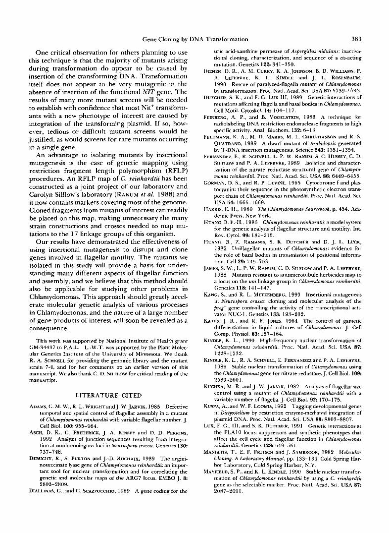
t ,:."..*,p><L>.$.. .1.,'., ., ' ,,(.,,",::' .. *I. " . . . FIGURE 1 .-Flagellar defects of unequal-length flagella mutants
visualized by differential interference contrast microscopy. (A) A wild-type cell showing normal flagella, (B) cells of strain 1 H 3 showing stumpy-flagella or (C) a pair of unequal-length flagella ( 3 0 0 0 ~ magnification).
strains (HUANG 1986), there was an interesting class of mutants which displayed an unusual new pheno- type: unequal-length flagella. While most of the cells in a population were flagella-less or had stumpy fla- gella, a small fraction of the cells had two flagella of different lengths (Figure 1). These cells with unequal- length flagella usually spun very slowly at the bottom of liquid cultures.
To determine whether these motility mutants con- tained DNA sequences derived from the pMN24 plas- mid integrated into the genome, DNA from 18 of these mutants was analyzed on blots using pMN24 DNA as the labeled hybridization probe. Under these conditions, three major PvuII fragments (Figure 2B, fragments A, B and C) from the untransformed strain 5D hybridized with the probe. The transformants contained these three fragments, which represented the endogenous nit1 gene, plus additional fragments which were derived from the integrated NIT1 plasmid (Figure 2B, 10 representative examples are shown). Some of these new fragments may represent the junc- tions between the integrated plasmid and host ge- nomic sequence. One of the extra fragments (D in Figure 2, A and B) was derived exclusively from the vector DNA and was present in most cases. The pattern of fragments was simple in most mutants analyzed, suggesting that these mutants contained only one or a few copies of the integrated plasmid.

378 L.-W. Tam and P. A. Lefebvre - - 1 k b A .
C Y C : A 7 xx x E PE
pMN24 i
EP P
Genomic P B P C P A P P I I I I 1
reglon xx x
B.
- B (4.7 kb)
- calc' (3.7 kb)
- D (3.0 kb)
FIGURE 2.--Genomic-DNA analysis of 10 different transform- ants with motility defects. (A) A restriction map of pMN24 showing PuulI (P), Xhol (X) and EcoRI (E) sites. pMN24 is shown linearized at an EcoRl site within the pUCll9 vector (open rectangle). The NIT1 coding region is indicated by the arrow above the map (D. ZHANG and P. A. LEFEBVRE, unpublished data). Restriction-endo- nuclease sites of the corresponding genomic region are illustrated on the lower map. Fragment D was derived exclusively from the vector and represented most of the vector sequence. (B) DNA (3 pg) from the untransformed strain (5D) and from 10 different motility mutants, plus 0.6 ng (1 copy equivalent) of pMN24 DNA were digested with PmII and probed with SzP-labeled pMN24 DNA. In strain 5D, three major bands were identified with the probe. In the transformants. extra fragments in addition to these three bands were detected.
Linkage of the motility mutations to the inte- grated NIT1 plasmid: To determine whether the motility phenotypes of the transformants were due to single mutations caused by the insertion of the NITl plasmid, 14 of the motility mutants were backcrossed to strains carrying the nit1 mutation. Also included in this analysis was a paralyzed-flagella mutant (strain 7- 4) generated by insertion of the cloned NIT2 gene (R. A. SCHNELL and P. A. LEFEBVRE, unpublished results); the mutant 7-4 was backcrossed to a nit2 mutant (strain B22). Progeny from these crosses were scored for their motility phenotype and for the ability to grow on MNOs medium.
In all 15 cases the motility phenotype segregated as a single Mendelian trait, as indicated by the equal ratio of normal to abnormal swimmers among the progeny (Table 2). If the motility mutations were
TABLE 2
Cosegregation tests for the motility and Nit phenotypes
Parental progeny Recombinant progeny Mutant strain Mor' Nit- Mot- Nit' Mot- Nit- Mot' Nit'
1 H3 52 60 0 0 12D9 56 64 0 0 5E8 207 192 0 0 8E4 61 51 0 0 17D4 125 131 0 0 12AlO 145 102 0 0 2G12 80 80 0 0 12c10 46 37 0 0 7-4 65 66 0 0 4F1 68 68 0 0 4G 1 42 36 0 0 3C10 37 46 0 0 1 C6 70 88 0 15 2E4 19 19 25 25 28D11 42 13 18 7
Abbreviations: Mot+ = normal motility: Mot- = abnormal motil- ity; Nit+ = growth on nitrate: Nit- = no growth on nitrate. The motility phenotypes of these mutants are listed on Table 1. Viability of the meiotic progeny for most of the crosses was lower than 20%. Similarly poor meiotic viability was also observed in crosses using the strain 5D (from which these mutants were derived) to other wild-type strains. Meiotic viability generally improved in subsequent backcrosses, except in cases involving mutants with abnormal fla- gellar number. The results presented were derived from all surviv- ing progeny from both incomplete and complete tetrads from one or more backcrosses and do not represent random-progeny data.
caused by insertion of the NITl transgene, then in backcrosses all of the progeny that displayed abnormal motility should also be able to grow on MNOs me- dium. Cosegregation of the motility and the Nit+ phenotypes was indeed observed for 13 of 15 mutants analyzed (Table 2). To determine whether the inte- grated plasmid DNA also cosegregated with the mo- tility and Nit+ phenotypes, PvuII-digested genomic DNA of six to eight progeny from backcrosses of six different mutants (2G12, 5E8, 8E4, 17D4, 7-4, 4F1) was analyzed on blots hybridized with "P-labeled pUCl19 or pMN24 DNA. For all six mutants, the integrated plasmid DNA was detected only in progeny that were Nit+ and showed abnormal motility (two examples are shown in Figure 3). Therefore, the results from both genetic and DNA analyses are con- sistent with the conclusion that insertion of the plas- mid DNA into the nuclear genome disrupted a gene that was involved in flagellar motility in 13 of 15 mutants analyzed.
Among the 13 mutants which showed cosegregation of the Nit+ and the defective motility phenotypes, the Nit+ phenotype also behaved as a single Mendelian trait in all but one case (Table 1). In mutant 1C6, which appeared to have more than 10 copies of the plasmid (Figure 2B), the number of Nit+ to Nit- progeny in the F1 generation was 63 to 30, far from the 1: 1 ratio indicative of a single Mendelian trait.

Gene Cloning by DNA Transformation 379
2G12 X L5 - + + - - + + - + e - + + - - +
FIGURE 3.-Genomic-DNA blots of progeny from 2G12 and 8E4 crossed to L5 showing cosegregation of the inte- grated plasmid with the phenotypic mark- ers. Each lane contains 2 pg of PvulI- digested genomic DNA hybridized with ’*P-labeled probes derived from pUCl19. pUCl19 has two PvulI sites flanking the polylinker region (Figure 2A). Fragment D is the major fragment derived from the integrated plasmid vector whereas the faint bands are restriction fragments con- taining the remaining pUCl19 sequence - - (3’0 kb) in junction with NIT1 or other genomic sequences. For abbreviations, see Table 2.
0.
Moreover, some of the Nit+ progeny were normal swimmers. I t was possible, however, to obtain FI Nit+ progeny with the motility defect, which in subsequent backcrosses segregated as expected for a single inser- tion event (two Nit+ to two Nit- progeny in 20 te- trads). Therefore, in this mutant at least two separate integration events of the pMN24 plasmid must have occurred, only one of which was linked to the muta- tion giving rise to the motility phenotype.
Three mutants contain alleles of previously iden- tified loci: Among the 13 mutants that appeared to be tagged with the NIT1 plasmid, one of them (2G12) was unable to swim forward, but it could move back- ward slowly. Mutants with a very similar “move-back- ward-only’’ phenotype have been previously described, and they define three distinct MBO loci (NAKAMURA 1979; SEGAL et al. 1984). In these mutants, the ciliary (forward) beat pattern is defective while the flagellar (backward) beat pattern is at least partly functional.
Two classes of mutations that have defective control of flagellar number have been identified previously. Mutations in three different VFL loci (KUCHKA and JARVIK 1982; WRIGHT, CHOJNACKI and JARVIK 1983; ADAMS, WRIGHT and JARVIK 1985) produce a “vari- able number offlagella” phenotype. Uniflagellar mu- tations, all of which mapped to a single locus closely linked to apml on linkage group XIX, produce cells with a single flagellum (HUANG et al. 1982). Both vJ and uniflagellar mutants are found in our collection (Table 1) and four of them have been analyzed genet- ically. Mutants 5E8 and 8E4 possessed 0-5 flagella with more than 10% of cells showing three or more flagella. Mutants 12A10 and 17D4 were similar to uniflagellar mutants; most cells in each population had 0 , 1 or 2 flagella with fewer than 2% of cells having three or more flagella.
T o test whether the mutations produced in this study represent new alleles of previously identified
TABLE 3
Pairwise crosses for tests of allelism
Parents No. of progeny
Mot- Mot+
1.2G12 X mbol mbo2 mbo2
2.5E8 x v f l v ! 2 $3
vf2 $3
3. BE4 x V I 1
17D4 12A10
4. 17D4 x v f l $2 vf3 12A10
5. 12A10 x v f l V f 2 $3
69 23 78 0 70 24
83 0 53 22 43 15
25 6 116 32 70 15 87 38 54 30
46 12 82 10 39 12 55 24
103 41 42 17 85 35
loci, we performed pairwise crosses of the putative mbo mutant 2G12 to the three previously described mbo mutants; we also crossed the four mutants with abnormal flagellar number to the three genetically characterized vfl mutants. In addition, we crossed the four abnormal-flagellar-number mutants in our col- lection inter se to determine if they are allelic. Nonal- lelism of two mutations is indicated by the appearance of wild-type recombinant progeny in a cross, whereas the absence of recombinant progeny indicates that the two mutations are either allelic or tightly linked. The results of these allelism tests are shown in Table 3. No recombinants were observed in a cross of 2G12 to mbo2, suggesting that these mutations are either allelic

380 L.-W. Tam and P. A. Lefebvre
or closely linked. Mutant 5E8 produced no wild-type recombinants in a cross to vfll. On the other hand, our results indicate that the mutations in strains 8E4, 17D4, 12A 10 were nonallelic to any of the three vfl mutations and to each other. In addition, they were unlinked to apml (data not shown), which is 6.5 cM from unil (JAMES et al. 1988). Therefore these three mutants contained lesions at three separate loci other than the VFL and UNZ loci that have been identified before. Strain 7-4, which had paralyzed flagella, was found to be closely linked to nitZ. Since pfZ3 is less than 0.5 cM from nit1 (HARRIS 1989), we suspected that mutant 7-4 might be a pf13 allele.
To determine whether 2G12, 5E8 and 7-4 were alleles of mbo2, vfll and pfZ3, respectively, we per- formed complementation tests in stable vegetative diploids. Mutants 2G12, 5E8 and 7-4 were first crossed to wild-type cells to generate heterozygous diploids to determine whether the mutations are re- cessive. Then 16-24 diploid lines from each cross were analyzed and they all showed wild-type motility and flagellar number, indicating that each mutation is recessive. Next we constructed stable diploids be- tween 2G12 and an mbo2 mutant, 5E8 and a vfll mutant, and 7-4 and a pf13 mutant. In all cases, the diploids displayed the abnormal motility characteristic of the haploid parents, suggesting that each pair of mutations tested is allelic. We have designated these new alleles as mbo2-3 (2G12), vf l l -2 (5E8) and pf l3 -3
Cloning of the MBo2 gene: T o test whether the insertion of pMN24 plasmid DNA will allow us to clone the affected genes in these mutants, we first cloned the MB02 gene, starting with the mutant 2G12. Analysis of genomic DNA digested with a variety of restriction enzymes indicated that only a single copy of the plasmid was present in the genome of 2G12 and that the pUC 1 19 vector sequence was intact and juxtaposed to an integration junction (as illustrated in Figure 4A). This configuration of the integrated plas- mid allowed us to clone the sequence flanking one side of the integration site by plasmid rescue. DNA of 2G12 was digested with SaZI, which cut in the poly- linker site of pUCll9, to release the junction frag- ment containing pUCl19 and a 1.2-kb genomic se- quence adjacent to the site of insertion (Figure 4A). After circularization and transformation into Esche- richia coli, ampicillin-resistant clones containing the rescued plasmid sequence were selected. The re- covered plasmid, p2G12S1, was used as a hybridiza- tion probe for analysis of genomic DNA. It had no homology to NIT1 DNA, as expected, but it contained a repetitive sequence (data not shown). A 0.6-kb A M - PvuII fragment (probe b in Figure 4A) was isolated from p2G12S1 and shown to hybridize to a single genomic fragment. The probe recognized restriction
(7-4).
H
2012 " , T I + """"" I
SSm Xb SaXb S S S I S Sa
B. Proboa: a b C d e
kb
9.1 - 6.6 - 4 . 4 -
2.1 - - FIGURE 4 4 A ) Top: Restriction map of the wild-type genomic
region which was disrupted in the mutant 2G12. Abbreviations: H HindII1; S , Sstl; Sa, Sall; Xb, XbaI. Five different fragments (shaded boxes) that were used as probes in (B) are indicated above the map. Bottom: A proposed map of the corresponding genomic region in 2G12. The junction fragment that was cloned by plasmid rescue is the fragment bordered by the two Sal1 sites marked with asterisks. The extent of deletion determined by genomic-DNA analysis as shown in (B) is represented by the combined span of the integrated plasmid and the dotted line. (B) Genomic-DNA analysis of the region around the integration junction in the mutant 2G12. Hindllldigested DNA from a wild-type strain and from mutant 2G 12 was hybridized with labeled probes corresponding to the five different fragments indicated in (A). Sequences corresponding to probes c, d and e were not detected in the mutant DNA. Digestion with other restriction endonucleases gave similar results (not shown).
fragments of different sizes in wild-type DNA and DNA from mutant 2G12, confirming that p2G12S1 was derived from the site of insertion (Figure 4B). A genomic library containing DNA from wild-type cells was screened with labeled probe b; nine different overlapping clones spanning a 30-kb region were ob- tained.
The presence of the complete MB02 gene in one of these genomic clones was demonstrated by transfor- mation as follows. Previous studies have shown that when a plasmid containing a selectable marker such as NIT1 and a second plasmid with an unselected gene were transformed together into cells, 10-70% of the Nit+ transformants also expressed the gene carried by the second plasmid (DIENER et al. 1990; KINDLE 1990). We constructed an arg7, mbo2-3 double mutant and performed cotransformation experiments in which genomic clones to be tested were transformed into cells along with the plasmid pARG7.8 (containing the

Gene Cloning by DNA Transformation 38 1
Probe b - 1 hb
I
li H H <
SSSXbSaXbSS SaS S S.S. S S SSSSSS. Phenolypic
Rescue A.
12012.4
12012.3 ,r.r...l-"<
12612.2 . ..j
12012.1 +
0. p20101
p20102
p20103
+ 8. ' Sa H H 1
n F. +
FIGURE 5.--Complernentation of the mbo2-3 mutation by trans- formation. Upper line represents t h e restriction map of the putative MBU2 gene region (see Figure 4A for abbreviations). Probe b, which was used to screen a wild-type genomic library, is indicated above the map. In (A), four different genomic clones covering this region were cotransformed (with pARG7.8 as the selected marker) into arg7, mbo2-3 recipient cells. Only X2G12-1 was able to restore wild-type motility to mbo mutant cells, as seen in 5% of the Arg' transformants. In (B). three subclones of X2G12-1 were tested for the ability to complement the mbo mutation.
wild-type argininosuccinate lyase gene) as selectable marker. Arg+ transformant colonies were selected, grown in liquid medium, and their motility was scored to test for the expression of the wild-type M B 0 2 gene in the transformants. Among the four overlapping X clones tested, only cotransformation with X2G 12-1 gave rise to swimming cells (Figure 5A). Eleven of 196 Arg+ transformants tested showed restoration of flagellar function. Analysis of DNA from five of these swimming transformants using DNA fragments from X2G12-1 as probes showed that they all contained DNA derived from the X clone (data not shown). Backcrosses of four motile transformants to an mb02- 3 strain indicated that the normal motility phenotype segregated as a single Mendelian trait (2:2 segregation in 20-24 tetrads for each cross). By crossing one of these Mbo+ transformants to the original mbo2-1 mu- tant isolated by SEGAL et d . (1 984), we found that the wild-type and the Mbo phenotypes segregated 2:2 among the tetrad progeny (six tetrads), indicating that the putative M B 0 2 transgene was able to restore nor- mal motility to the mbo2-1 mutant. Therefore, we conclude that a functional M B 0 2 gene must be con- tained within clone X2G 12- 1.
T o further define the extent of the functional M B 0 2 gene, a 13-kb fragment (p2G1 Ol), a 7.5-kb (p2G102) fragment and a 6.4-kb fragment (p2G103) from X2G12-1 were subcloned into the vector Blue- script and tested for phenotypic rescue of mbo2-3 in cotransformation experiments (Figure 5B). We found that both p2G 10 1 and p2G103 were able to correct the Mbo defect in 15-35% of Arg+ transformants but p2G102 could not. Thus the functional M B 0 2 gene has been localized to a 6.4-kb genomic region.
Probe - - 1 kb
N NN N
s s s s Phenolyplc rescue
h5EB-10 - + h5EB-13 I
A5E8-19 - - FIGURE B.--Complenlentation of the uJI-2 mutation by trans-
formation. Ssfl (S) and Not1 (N) sites of the wild-type genomic region corresponding to the region of plasmid integration in the mutant are shown. The 2-kb Psfl-Xhol fragment used to isolate the genomic clones is illustrated above the map. Three different lambda clones were tested by cotransformation into uJI-2, arg7 cells. Only X5E8-IO could restore wild-type motility to uJl-2 cells, as seen in about 5-15% of the Arg' transformants.
Cloning of the VFLl gene: We have also cloned the wild-type copy of the VFLl gene starting with mutant 5E8. Analysis of the mutant DNA with a number of restriction endonucleases and hybridization probes indicated that one copy of the pMN24 plasmid had integrated into the genome, but the pUCl l9 vector sequence was completely lost in this mutant. The genomic sequence flanking the site of insertion was therefore cloned as follows. A 4.4-kb PstI fragment was predicted by restriction mapping to contain the 5' portion of the integrated N I T l gene and several kilobases of DNA adjacent to the insertion. A partial library of genomic fragments was prepared (MATE- RIALS AND METHODS) and screened with a 0.9-kb XhoI fragment derived from the 5' portion of the N I T l gene (Figure 2A). A 4.4-kb clone containing the junc- tion fragment was isolated and shown to contain a 2- kb PstI-XhoI fragment (Figure 6) comprising only the genomic sequence adjacent to the site of insertion. This fragment was used as a hybridization probe to screen the genomic library to identify three overlap- ping clones. When tested for the ability to comple- ment the vfll-2 mutation upon transformation, only genomic clone X5E8-10 was able to complement the variable-flagellar-number phenotype (Figure 6).
Host DNA deletion accompanied DNA integra- tion: Host sequence rearrangements have been com- monly observed in association with nonhomologous integration of exogenous DNA into the genome of mouse or fungi (ROTH and WILSON 1988; ASCH et aZ. 1992). To investigate whether rearrangement of host genomic sequences occurred upon integration of pMN24 in strain 2G12, we used five different frag- ments (Figure 4A) from the wild-type genomic clones as hybridization probes for analysis of DNA from a wild-type strain and from 2G 12 (Figure 4B). Probes a and b, which are on the left of the integration junc- tion, hybridized to genomic fragments in both strains, although the band hybridizing to probe b in 2G12 was larger than that in wild type. In contrast, probes c, d and e, which are on the right side of the integra-

382 L.-W. Tam and P. A. Lefebvre
tion junction (Figure 4A), failed to hybridize to any sequence in the mutant strain (Figure 4B), suggesting that a large region to the right of the integration junction had been deleted. We have mapped the end- point of this deletion and determined the total size of the deletion to be 23 kb. The deletion covered the entire span of the MB02 gene as established by the phenotypic rescue experiments described above.
Similar studies of the region around the integration junctions in mutant 5E8 indicated that a 3-4-kb re- gion adjacent to the integrated junction was either rearranged or deleted in the mutant (data not shown). Preliminary studies of the genomic DNA around the site of plasmid insertion in a third mutant, 8E4, indi- cated that at least 12 kb of genomic DNA adjacent to an integration junction was deleted in the mutant (data not shown). Therefore, host DNA re- arrangement may be a common phenomenon associ- ated with nonhomologous DNA integration in Chlam- ydomonas.
DISCUSSION
In the present study, by introducing a plasmid con- taining the nitrate reductase gene into the nuclear genome of Chlamydomonas, 74 motility mutants were recovered. Motility mutants were also observed in transformants which resulted from integration of the cloned NIT2 gene (R. A. SCHNELL, personal commu- nication), and one of these mutants is an allele of Pf13. With a similar approach, a mutant with defective splicing of chloroplast RNA transcripts was obtained by insertion of the pARG7.8 plasmid (M. GOLD- SCHMIDT-CLERMONT, personal communication).
Several of our observations suggest that DNA trans- formation is a feasible and effective approach for tagging and cloning genes in Chlamydomonas. First, a very high percentage (2.5%) of the Nit+ transform- ants showed abnormal motility. Backcrosses of 15 of these mutants suggest that each was due to a single nuclear mutation. Second, the 74 motility mutants displayed a wide spectrum of phenotypes, represent- ing virtually all classes of motility mutants that have been described before (HUANG 1986), as well as a new class of mutants that produce unequal-length flagella. Recombination and diploid complementation tests of six mutants showed that three of them contained lesions at previously identified loci: MB02, VFLl and PF13, and the others, which displayed abnormal flagellar number, did not map to any of the previously described VFL or UNI loci. We conclude that the transforming DNA can integrate into many different sites in the genome, and that disruption of the nuclear genome by integration of exogenous DNA may be an efficient way to produce random mutations. Third, the tight linkage of the motility phenotype to the Nit+ phenotype in 13 of the 15 mutants analyzed is con-
sistent with the conclusion that most of the motility mutants obtained in this study were generated by the integration of the plasmid DNA. As a demonstration of the usefulness of the plasmid DNA as both a mu- tagen and a tag for cloning, we cloned two motility genes by different approaches. Since the majority of transformants contain only a few copies of the inte- grated plasmids, subsequent recovery of the sequences flanking the sites of integration should be straight forward. Moreover, as shown by the cloning of MB02, those mutants which retain a complete copy of the vector DNA will allow cloning of the flanking se- quence by simple plasmid rescue.
Studies of the genomic map around the sites of integration in three motility mutants revealed exten- sive deletion of the host sequence in two of the mu- tants and some sequence rearrangement or deletion in the third mutant. This observation raises the pos- sibility that integration of exogenous DNA into the Chlamydomonas nuclear genome may often be ac- companied by host DNA rearrangement. These se- quence rearrangements may provide an explanation for the unexpectedly high frequency of motility mu- tants observed among the Nit+ transformants. That is, deletions associated with integration events would be expected to inactivate a larger region of the ge- nome than simple integration. In mouse, the sequence rearrangements and deletions which accompany plas- mid insertion has complicated the isolation of genes in putative insertional mutants (MEISLER 1992). How- ever, with the ease of genetic analysis and the ability to easily complement mutant phenotypes by transfor- mation using cloned DNA in Chlamydomonas, dele- tions should not be a serious problem. Genes of inter- est should be readily isolated using clones with large DNA inserts for phenotypic rescue experiments. Our experience using genomic clones from a lambda li- brary (average size 18 kb; R. A. SCHNELL, personal communication) suggests that a few micrograms of a genomic DNA clone in X phage vector can be used directly for transformation without subcloning into plasmids. Alternatively, it should be possible to use chromosome walking to isolate the affected genes from mutants with large deletions. The ease and effi- ciency of cotransformation experiments makes it pos- sible to quickly test each set of overlapping clones for the ability to complement mutant phenotypes.
Not all genes of interest will be clonable using this procedure. Genes in which null mutants are lethal could not be obtained by this method because the insertion of plasmid DNA and the accompanying se- quence rearrangements will likely produce the null phenotype. There may also be genes which map close enough to essential genes that they will be difficult to clone because of the effect of sequence deletion on the neighboring genes.
Gene Cloning by DNA Transformation 383
One critical observation for others planning to use this technique is that the majority of mutants arising during transformation do appear to be caused by insertion of the transforming DNA. Transformation itself does not appear to be very mutagenic in the absence of insertion of the functional NIT gene. The results of many more mutant screens will be needed to establish with confidence that most Nit+ transform- ants with a new phenotype of interest are caused by integration of the transforming plasmid. If so, how- ever, tedious or difficult mutant screens would be justified, as would screens for rare mutants occurring in a single gene.
An advantage to isolating mutants by insertional mutagenesis is the ease of genetic mapping using restriction fragment length polymorphism (RFLP) procedures. An RFLP map of C. reinhardtii has been constructed as a joint project of our laboratory and Carolyn Silflow’s laboratory (RANUM et al. 1988) and it now contains markers covering most of the genome. Cloned fragments from mutants of interest can readily be placed on this map, making unnecessary the many strain constructions and crosses needed to map mu- tations to the 17 linkage groups of this organism.
Our results have demonstrated the effectiveness of using insertional mutagenesis to disrupt and clone genes involved in flagellar motility. The mutants we isolated in this study will provide a basis for under- standing many different aspects of flagellar function and assembly, and we believe that this method should also be applicable for studying other problems in Chlamydomonas. This approach should greatly accel- erate molecular genetic analysis of various processes in Chlamydomonas, and the nature of a large number of gene products of interest will soon be revealed as a consequence.
This work was supported by National Institute of Health grant GM-34437 to P.A.L. L.-W.T. was supported by the Plant Molec- ular Genetics Institute of the University of Minnesota. We thank R. A. SCHNELL for providing the genomic library and the mutant strain 7-4, and for her comments on an earlier version of this manuscript. We also thank C. D. SILFLOW for critical reading of the manuscript.
LITERATURE CITED
ADAMS, c. M. W., R. L. WRIGHTandJ. W. JARVIK, 1985 Defective temporal and spatial control of flagellar assembly in a mutant of Chlamydomonas reinhardtii with variable flagellar number. J. Cell Biol. 100 955-964.
ASCH, D. K., G. FREDERICK, J. A. KINSEY and D. D. PERKINS, 1992 Analysis of junction sequences resulting from integra- tion at nonhomologous loci in Neurospora crassa. Genetics 130:
DEBUCHY, R., S. PURTON and J.-D. ROCHAIX, 1989 The argini- nosuccinate lyase gene of Chlamydomonas reinhardtii: an impor- tant tool for nuclear transformation and for correlating the genetic and molecular maps of the ARC7 locus. EMBO J. 8:
DIALLINAS, G., and C. SCAZZOCCHIO, 1989 A gene coding for the
737-748.
2803-2809.
uric acid-xanthine permease of Aspergillus nidulans: inactiva- tional cloning, characterization, and sequence of a cis-acting mutation. Genetics 122: 341-350.
DIENER, D. R., A. M. CURRY, K. A. JOHNSON, B. D. WILLIAMS, P. A. LEFEBVRE, K. L. KINDLE and J. L. ROSENBAUM, 1990 Rescue of paralyzed-flagella mutant of Chlamydomonas by transformation. Proc. Natl. Acad. Sci. USA 87: 5739-5743.
DUTCHER, S. K., and F. G. Lux 111, 1989 Genetic interactions of mutations affecting flagella and basal bodies in Chlamydomonas. Cell Motil. Cytoskel. 14: 104-1 17.
FEINBERG, A. P., and B. VOGELSTEIN, 1983 A technique for radiolabeling DNA restriction endonuclease fragments to high specific activity. Anal. Biochem. 132: 6-13.
FELDMANN, K. A., M. D. MARKS, M. L. CHRISTIANSON and R. S. QUATRANO, 1989 A dwarf mutant of Arabidopsis generated by T-DNA insertion mutagenesis. Science 243: 1351-1354.
FERNANDEZ, E., R. SCHNELL, L. P. W. RANUM, S. C. HUSSEY, C. D. SILFLOW and P. A. LEFEBVRE, 1989 Isolation and character- ization of the nitrate reductase structural gene of Chlamydo- monas reinhardtii. Proc. Natl. Acad. Sci. USA 8 6 6449-6453.
GORMAN, D. S., and R. P. LEVINE, 1965 Cytochrome f and plas- tocyanin: their sequence in the photosynthetic electron trans- port chain of Chlamydomonas reinhardtii. Proc. Natl. Acad. Sci.
HARRIS, E. H., 1989 The Chlamydomonas Sourcebook, p. 454. Aca- demic Press, New York.
HUANG, B. P.-H., 1986 Chlamydomonas reinhardtii: a model system for the genetic analysis of flagellar structure and motility. Int. Rev. Cytol. 99: 181-215.
HUANG, B., Z. RAMANIS, S. K. DUTCHER and D. J. L. LUCK, 1982 Uniflagellar mutants of Chlamydomonas: evidence for the role of basal bodies in transmission of positional informa- tion. Cell 29: 745-753.
JAMES, S. W., L. P. W. RANUM, C. D. SILFLOW and P. A. LEFEBVRE, 1988 Mutants resistant to antimicrotubule herbicides map to a locus on the uni linkage group in Chlamydomonas reinhardtii. Genetics 118: 141-147.
KANG, S., and R. L. METZENBERG, 1993 Insertional mutagenesis in Neurospora crassa: cloning and molecular analysis of the p e g + gene controlling the activity of the transcriptional acti- vator NUC-1. Genetics 133: 193-202.
KATES, J. R., and R. F. JONES, 1964 The control of gametic differentiation in liquid cultures of Chlamydomonas. J. Cell Comp. Physiol. 63: 157-164.
KINDLE, K. L., 1990 High-frequency nuclear transformation of Chlamydomonas reinhardtii. Proc. Natl. Acad. Sci. USA 87:
KINDLE, K. L., R. A. SCHNELL, E. FERNANDEZ and P. A. LEFEBVRE, 1989 Stable nuclear transformation of Chlamydomonas using the Chlamydomonas gene for nitrate reductase. J. Cell Biol. 109
KUCHKA, M. R, and J. W. JARVIK, 1982 Analysis of flagellar size control using a mutant of Chlamydomonas reinhardtii with a variable number of flagella. J. Cell Biol. 92: 170-1 75.
KUSPA, A., and W. F. LOOMIS, 1992 Tagging developmental genes in Dictyostelium by restriction enzyme-mediated integration of plasmid DNA. Proc. Natl. Acad. Sci. USA 8 9 8803-8807.
LUX, F. G., 111, and S. K. DUTCHER, 1991 Genetic interactions at the FLAlO locus: suppressors and synthetic phenotypes that affect the cell cycle and flagellar function in Chlamydomonas reinhardtii. Genetics 128: 549-561.
MANIATIS, T. , E. F. FRITSCH and J. SAMBROOK, 1982 Molecular Cloning. A Laboratory Manual , pp. 133-134. Cold Spring Har- bor Laboratory, Cold Spring Harbor, N.Y.
MAYFIELD, S. P., and K. L. KINDLE, 1990 Stable nuclear transfor- mation of Chlamydomonas reinhardtii by using a C. reinhardtii gene as the selectable marker. Proc. Natl. Acad. Sci. USA 87:
USA 5 4 1665-1669.
1228-1232.
2589-2601.
2087-2091.

384 L.-W. Tam and P. A. Lefebvre
MEISLER, M. H., 1992 Insertional mutation of ‘classical’ and novel genes in transgenic mice. Trends Genet. 8: 341-344.
NAKAMURA, S., 1979 A backward swimming mutant of Chlamydo- monas reinhardtii. Exp. Cell Res. 123: 441-444.
PIPERNO, G., B. HUANG, 2. RAMANIS and D. J. L. LUCK, 198 1 Radial spokes of Chlamydomonas flagella: polypeptide composition and phosphorylation of stalk components. J. Cell Biol. 88: 73-79.
RANUM, L. P. W., M. D. THOMPSON, J. S. SCHLOSS, P. A. LEFEBVRE and C. D. SILFLOW, 1988 Mapping flagellar genes in Chlam- ydomonas using restriction fragment length polymorphism. Ge- netics 120: 109-122.
ROTH, D., and J. WILSON, 1988 Illegitimate recombination in mammalian cells, pp. 621-654 in Genetic Recombination, edited by R. KUCHERLAPATI and G. R. SMITH. American Society for Microbiology, Washington, D.C.
SAGER, R., and S. GRANICK, 1953 Nutritional studies in Chlamydo- monas reinhardtii. Ann. N.Y. Acad. Sci. 56: 831-838.
SAMBROOK, J., E. F. FRITSCH and T . MANIATIS, 1989 Molecular Cloning: A Laboratory Manual , Ed. 2. Cold Spring Harbor Laboratory, Cold Spring Harbor, N.Y.
SCHNELL, R. A,, and P. A. LEFEBVRE, 1993 Isolation of the Chlamydomonas regulatory gene NIT2 by transposon tagging. Genetics 134 737-747.
SEGAL, R. A,, B. HUANG, Z. RAMANIS and D. J. L. LUCK, 1984 Mutant strains of Chlamydomonas reinhardtii that move backward only. J. Cell Biol. 98: 2026-2034.
SINGH, G., D. M. SUPP, C. SCHREINER, J. MCNEISH, H.3. MERKER, N . G. COPELAND, N. A. JENKINS, S. S. POTTER and W. SCOTT, 199 1 legless insertional mutation: morphological, molecular, and genetic characterization. Genes Dev. 5: 2245-2255.
TAM, L.-W., and D. L. KIRK, 1991 Identification of cell-type- specific genes in Volvox carteri and characterization of their expression during the asexual life cycle. Dev. Biol. 1 4 5 51- 66.
TILBURN, J., F. ROUSSEL and C. SCAZZOCCHIO, 1990 Insertional inactivation and cloning of the wA gene of Aspergillus nidulans. Genetics 126: 8 1-90.
WEEKS, D. P., N. BEERMAN and 0. M. GRIFFITH, 1986 A small- scale five-hour procedure for isolating multiple samples of CsCI- purified DNA: application to isolations from mammalian, in- sect, higher plant, algal, yeast, and bacterial sources. Anal. Biochem. 152: 376-385.
WOYCHIK, R. L., MAAS, R. ZELLER, T. F. VOGT and P. LEDER, 1990 “Formins”: proteins deduced from the alternative tran- scripts of the limb deformity gene. Nature 346: 850-853.
WRIGHT, R. L., B. CHOJNACKI and J. W. JARVIK, 1983 Abnormal basal-body number, location and orientation in a striated fiber- defective mutant Chlamydomonas reinhardtii. J. Cell Biol. 96: 1697-1707.
YANOFSKY, M. F., H. MA, J. L. BOWMAN, G. N. DREWS, K. A. FELDMANN and E. M. MEYEROWITZ, 1990 The protein en- coded by the Arabidopsis homeotic gene agamous resembles transcription factors. Science 346: 35-38.
Communicating editor: M. T. FULLER