closed reduction, traction, and casting techniques - ota.org reduction, traction and casting... ·...
TRANSCRIPT
Closed Reduction, Traction, and Casting Techniques
Jason Tank, MD March 2014
Original Authors: Dan Horwitz, MD; March 2004; David Hak, MD;
Revised January 2006 & October 2008 New Author: Jason Tank, MD
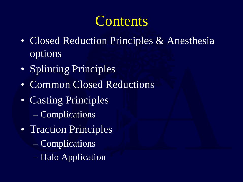
Contents • Closed Reduction Principles & Anesthesia
options • Splinting Principles • Common Closed Reductions • Casting Principles
– Complications • Traction Principles
– Complications – Halo Application
Closed Reduction Principles
• Identify need for closed reduction – Most displaced fractures should be reduced to
minimize soft tissue complications & injury • Includes injuries ultimately treated with surgery • Various resources for acceptable non-operative
fracture alignment parameters – Find & utilize a reliable source
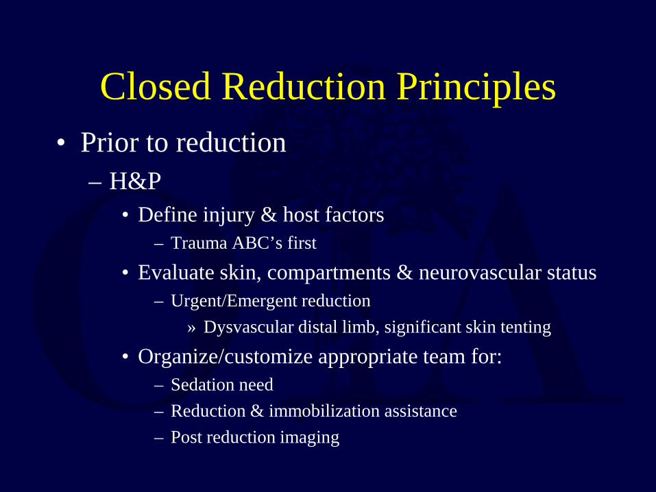
Closed Reduction Principles • Prior to reduction
– H&P • Define injury & host factors
– Trauma ABC’s first
• Evaluate skin, compartments & neurovascular status – Urgent/Emergent reduction
» Dysvascular distal limb, significant skin tenting
• Organize/customize appropriate team for: – Sedation need – Reduction & immobilization assistance – Post reduction imaging

Closed Reduction Principles
• Reduction maneuver specific for fracture location & pattern
• Goals: – Restore length, alignment & rotation
• Immobilize joint above & below • Quality post reduction radiographs
Anesthesia
• Adequate analgesia & muscle relaxation/fatigue are critical for success
• Determine goals of reduction & plan • Customize anesthesia for each patient &
injury combination
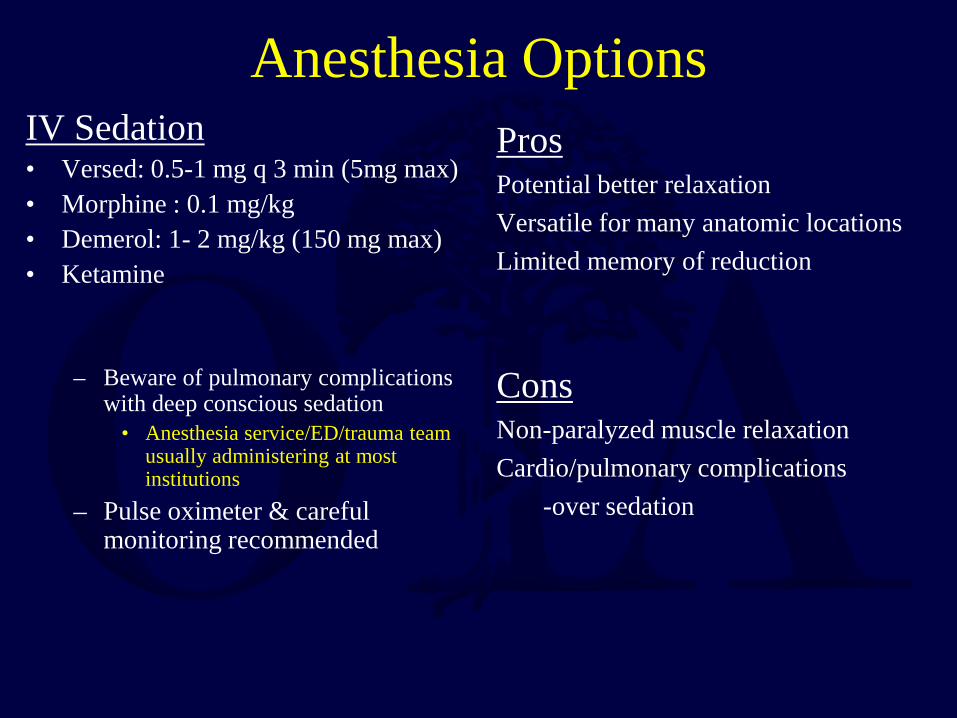
Anesthesia Options IV Sedation • Versed: 0.5-1 mg q 3 min (5mg max) • Morphine : 0.1 mg/kg • Demerol: 1- 2 mg/kg (150 mg max) • Ketamine
– Beware of pulmonary complications with deep conscious sedation
• Anesthesia service/ED/trauma team usually administering at most institutions
– Pulse oximeter & careful monitoring recommended
Pros Potential better relaxation Versatile for many anatomic locations Limited memory of reduction
Cons Non-paralyzed muscle relaxation Cardio/pulmonary complications -over sedation
Anesthesia Options
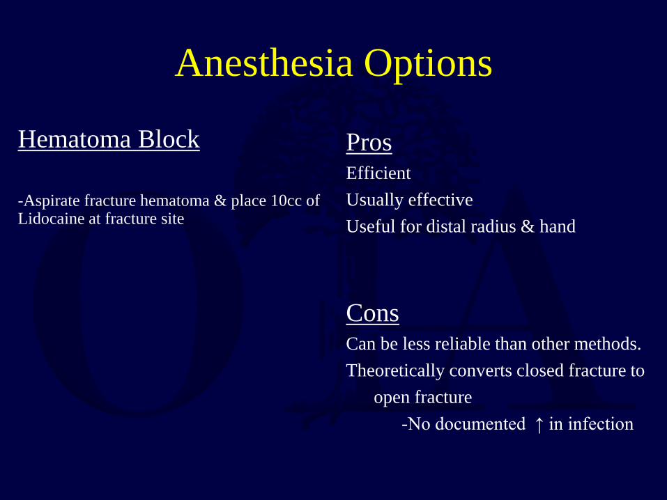
Hematoma Block -Aspirate fracture hematoma & place 10cc of Lidocaine at fracture site
Pros Efficient Usually effective Useful for distal radius & hand
Cons Can be less reliable than other methods. Theoretically converts closed fracture to open fracture -No documented ↑ in infection

Anesthesia Options
Intra-articular Block -Aspirate joint & place 10cc of Lidocaine (or equivalent local anesthesia) into joint
Pros Efficient Commonly effective Useful for certain ankle/knee injuries
Cons Can be less reliable than other methods Intra-articular violation Theoretically converts closed injury to open injury -No documented ↑ in infection
Anesthesia Options
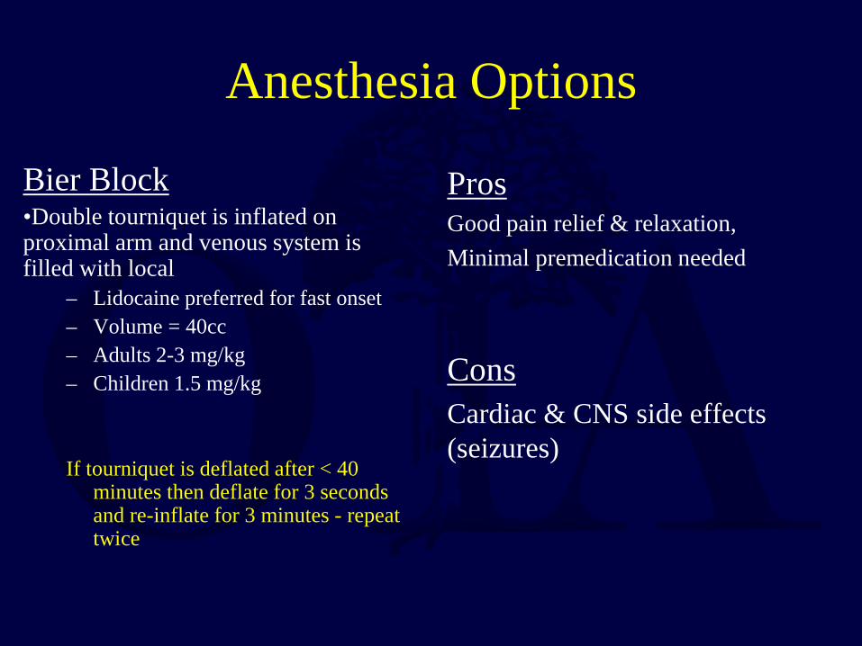
Bier Block •Double tourniquet is inflated on proximal arm and venous system is filled with local
– Lidocaine preferred for fast onset – Volume = 40cc – Adults 2-3 mg/kg – Children 1.5 mg/kg If tourniquet is deflated after < 40
minutes then deflate for 3 seconds and re-inflate for 3 minutes - repeat twice
Pros Good pain relief & relaxation, Minimal premedication needed
Cons Cardiac & CNS side effects (seizures)

Closed Reduction Principles • Prepare immobilization prior to reduction
– Splint pre-measured & ready for efficient application
– Sling or knee immobilizer in close proximity – Have extra supplies close – Assistant or assistive device (ex. Finger traps) available
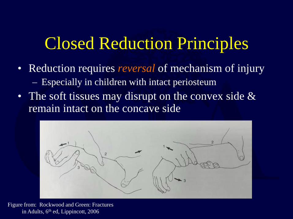
Closed Reduction Principles • Reduction requires reversal of mechanism of injury
– Especially in children with intact periosteum • The soft tissues may disrupt on the convex side &
remain intact on the concave side
Figure from: Rockwood and Green: Fractures in Adults, 6th ed, Lippincott, 2006

• Longitudinal traction alone may not allow the fragments to be disengaged & length re-established if there is an intact soft-tissue hinge – Especially in children with strong partially intact
periosteum
Closed Reduction Principles

Closed Reduction Principles Reproduce fracture mechanism
↓ Traction to disengage fracture fragments
↓ Re-align fracture
***Angulation beyond 90° is potentially required
Figure from: Rockwood and Green: Fractures in Adults, 6th ed, Lippincott, 2006

Splinting Principles
• Splint must be molded to resist deforming forces – “Straight casts lead to crooked bones”
– “Crooked casts lead to straight bones”

Splinting Principles
Three point contact (mold) is necessary to maintain closed reduction
Removal of any of the three forces results in loss of reduction
Figure from: Rockwood and Green: Fractures in Adults, 4th ed, Lippincott, 1996.
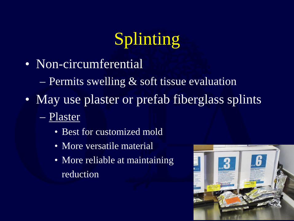
Splinting • Non-circumferential
– Permits swelling & soft tissue evaluation • May use plaster or prefab fiberglass splints
– Plaster • Best for customized mold • More versatile material • More reliable at maintaining reduction

Common Splinting Techniques
• Coaptation • Posterior long arm • Sugar-tong • Ulnar gutter • Volar/dorsal forearm • Volar/dorsal hand • Resting hand • Thumb spica
• Posterior long leg • Lateral long leg • Posterior slab (ankle)
+/- U splint +/- Foot plate +/-Side struts
• “Bulky” Jones
Splint Choice
• Considerations when customizing for each patient & injury – Overall patient condition
• Multi-trauma vs. isolated injury
– Soft tissue envelope – Reduction stability – Future treatment plan – Experience

Splint Padding
• 3-4 layers thick under ALL types of splints
• Padding Problems – Too thin skin pressure – Too thick less fracture
control (potential loss of reduction)
Unpadded fiber glass splint caused skin lesions
Common Closed Reductions
• Shoulder Dislocation • Humeral Shaft • Elbow Dislocation • Forearm Fracture • Distal Radius
• Hip Dislocation • Femur Fracture • Knee Dislocation • Tibia Fracture • Ankle Fracture • Talus Fracture • Calcaneus Fracture • Midfoot Fracture
Dislocation
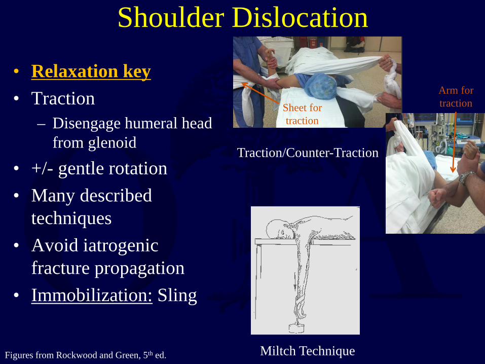
Shoulder Dislocation
• Relaxation key • Traction
– Disengage humeral head from glenoid
• +/- gentle rotation • Many described
techniques • Avoid iatrogenic
fracture propagation • Immobilization: Sling
Figures from Rockwood and Green, 5th ed. Miltch Technique
Traction/Counter-Traction
Sheet for traction
Arm for traction

Figure from Rockwood and Green, 4th ed.
Humeral Shaft • Gravity traction +/-
formal reduction maneuver
• Immobilization: Coaptation splint – Lateral splint extends
over the deltoid – Medial splint into axilla
& must be well padded (*ABD pad) to avoid skin breakdown
– Elbow unsupported permitting gravity traction

Elbow Dislocation • Traction, flexion & direct
manual palpation of olecranon – Reduce medial/lateral
displacement 1st – Address anterior/posterior
next – Supination/pronation may
assist reduction
• Cautious elbow range of motion after reduction – Can guide treatment plan
• Immobilization: Posterior long arm splint +/- sugar tong
Figure from Rockwood and Green, 5th ed.
Manual pressure over
olecranon
Multi-directional
traction

Forearm Fracture • Traction
– +/- need to significantly recreate the deformity
• Especially in pediatric pts
• Immobilization = Sugar tong splint with 3 point mold
• Pediatric – Splint Cast with nonop
mgnt
• Adult – Almost always surgical thus
temporizing until ORIF
-Splint around distal humerus to provide rotational control -Extra padding at the elbow
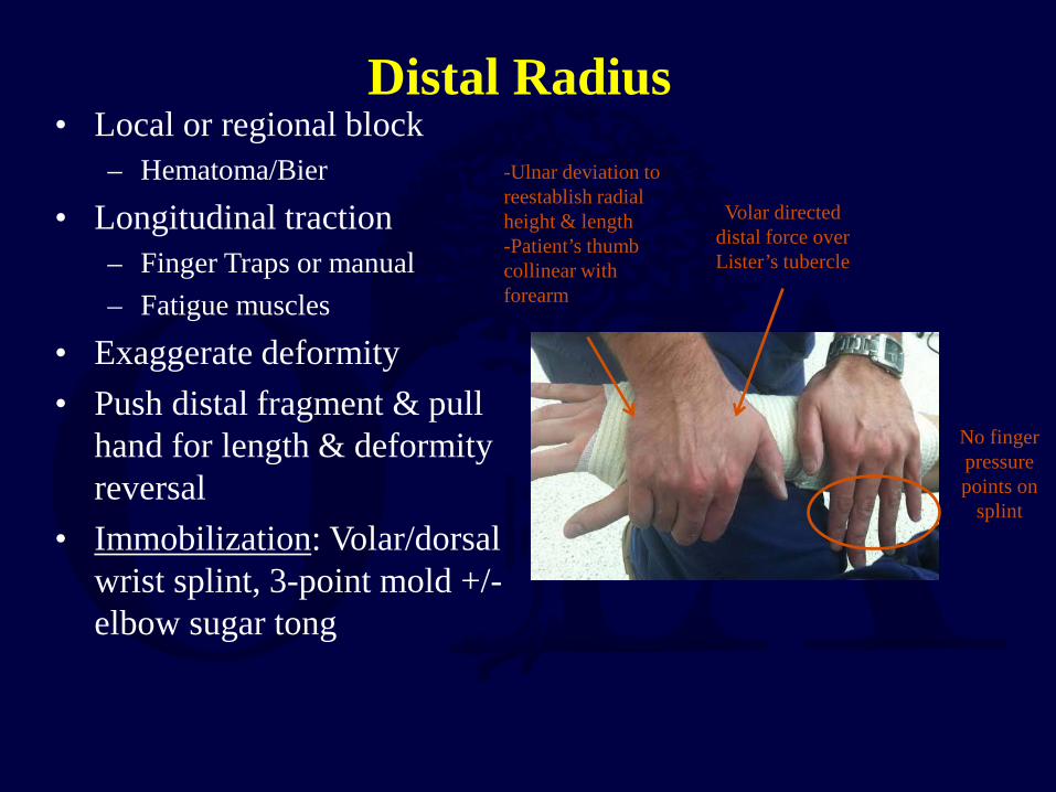
Distal Radius • Local or regional block
– Hematoma/Bier
• Longitudinal traction – Finger Traps or manual – Fatigue muscles
• Exaggerate deformity • Push distal fragment & pull
hand for length & deformity reversal
• Immobilization: Volar/dorsal wrist splint, 3-point mold +/- elbow sugar tong
Volar directed distal force over Lister’s tubercle
-Ulnar deviation to reestablish radial height & length -Patient’s thumb collinear with forearm
No finger pressure points on
splint

Hip Dislocation • IV Sedation (deep) with
Relaxation • Posterior: Flexion,
traction, adduction and internal rotation
• Anterior: Traction, abduction, lateralization, rotation
• Gentle & atraumatic • Reduction palpable &
permit significantly improved ROM
• Immobilization: Knee immobilizer vs. Abduction pillow
Figures from Rockwood and Green, 5th ed.
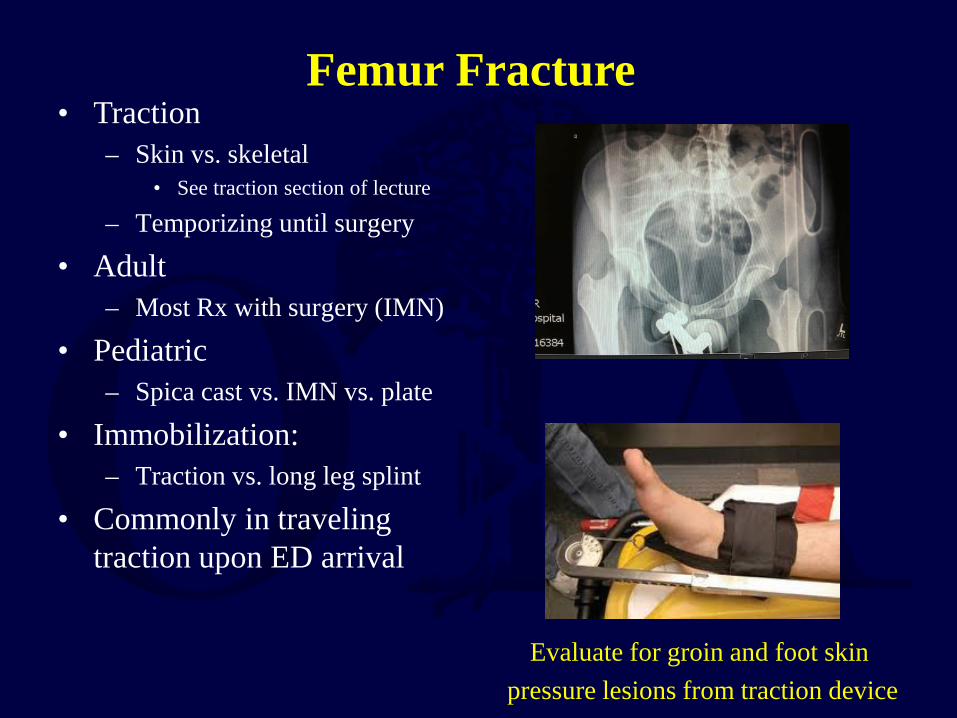
Femur Fracture • Traction
– Skin vs. skeletal • See traction section of lecture
– Temporizing until surgery
• Adult – Most Rx with surgery (IMN)
• Pediatric – Spica cast vs. IMN vs. plate
• Immobilization: – Traction vs. long leg splint
• Commonly in traveling traction upon ED arrival
Evaluate for groin and foot skin pressure lesions from traction device
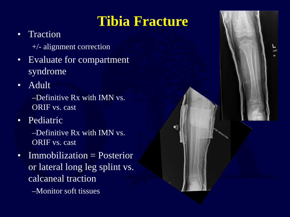
Tibia Fracture • Traction
+/- alignment correction
• Evaluate for compartment syndrome
• Adult –Definitive Rx with IMN vs. ORIF vs. cast
• Pediatric –Definitive Rx with IMN vs. ORIF vs. cast
• Immobilization = Posterior or lateral long leg splint vs. calcaneal traction –Monitor soft tissues

Knee Dislocation • Emergent Reduction
– Vascular injury common
• Traction with gentle flexion/extension after varus/valgus correction
• Check Pulse/ABI – Comprehensive NV exam
• Monitor compartments
• Immobilization = Knee Immobilizer +/- ExFix until surgical
reconstruction

Ankle Fracture • Traction with deformity
correction – Bend knee to relax
gastroc/soleus complex – Posterior & lateral dislocation
• +/- Quiggly Maneuver • Posterolateral to anterormedial
directed mold
– Medial • Traction reduction • Medial to lateral directed mold
– Customize mold to specific fracture/dislocation
• Immobilization: – U Splint
• +/-Posterior slab splint • +/- Foot plate • +/- Side struts
Quigley Maneuver:
Knee flexion & leg external
rotation, foot supination & adduction for
reduction
Posterolateral to anteromedial
mold for posterolateral ankle fractures
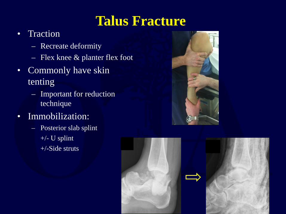
Talus Fracture • Traction
– Recreate deformity – Flex knee & planter flex foot
• Commonly have skin tenting – Important for reduction
technique
• Immobilization: – Posterior slab splint +/- U splint +/-Side struts

Calcaneus Fracture
• Traction & planterflexion if posterior significant skin pressure – Urgent operative indication
• Significant swelling common
• Immobilization: – Bulky Jones Splint
• Splint Cast if nonop mgnt after swelling decreases

Midfoot Fracture/dislocation • Traction & medial/lateral
with planter pressure • Commonly need pins to
hold reduction • ORIF frequently definitive
mgnt • Immobilization:
– Posterior slab splint +/- Foot plate +/-Side struts
Medial to lateral
reduction
Dorsal lateral to planter medial
reduction

Fracture Bracing
• Allows for early functional ROM and weight bearing
• Relies on intact soft tissues and muscle envelope to maintain reduction
• Most commonly used for humeral shaft & tibial shaft fractures
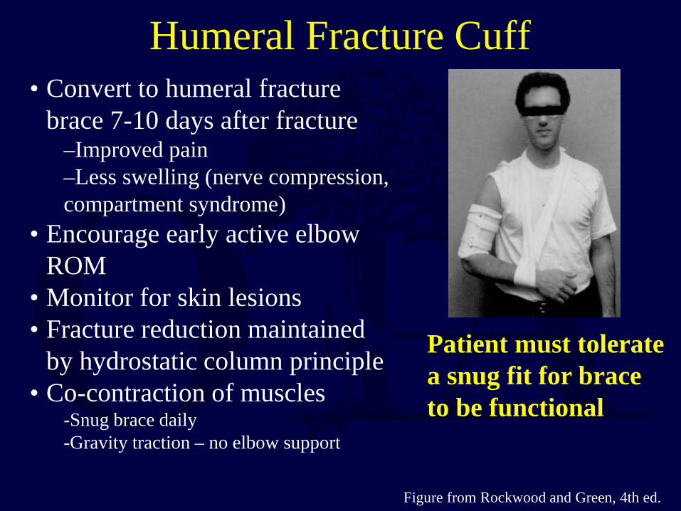
• Convert to humeral fracture brace 7-10 days after fracture
–Improved pain –Less swelling (nerve compression, compartment syndrome)
• Encourage early active elbow ROM
• Monitor for skin lesions • Fracture reduction maintained
by hydrostatic column principle • Co-contraction of muscles
-Snug brace daily -Gravity traction – no elbow support
Patient must tolerate a snug fit for brace to be functional
Figure from Rockwood and Green, 4th ed.
Humeral Fracture Cuff
Casting
• Goal of semi-rigid immobilization while avoiding pressure / skin complications
• Often a poor choice in the treatment of acute fractures due to swelling & other soft tissue pathology
• Good cast technique necessary to achieve predictable results
Casting Techniques
• Stockinette – May require two different diameters to avoid
over tight or loose, redundant material • Caution not to lift leg by stockinette
– Stretching the stockinette too tight around the heel may case high skin pressure

Casting Techniques • To avoid wrinkles in the
stockinette • Cut along the concave
surface and overlap to produce a smooth contour
• Applicable to ankle, elbow, posterior knee
Wrinkled stockinette
causing skin
pressure lesion to
antecubital fossa

Casting Techniques • Cast padding
– Roll distal to proximal – 50 % overlap – 2-3 layers minimum – Extra padding at boney
prominences • Fibular head, malleoli,
patella, and olecranon
Casting Material
• Plaster – Use cold water to maximize molding time &
limit exothermic heat reaction (can burn skin)
• Fiberglass – More difficult to mold but more durable &
resistant to breakdown – Generally 2 - 3 times stronger for any given
thickness
Width
• Casting materials are available in various widths – 4 - 6 inch for thigh – 3 - 4 inch for lower leg & upper arm – 2 - 3 inch for forearm

• Avoid molding with anything but the heels of the palm in order to avoid pressure points
• Mold applied to produce three point fixation
Cast Molding
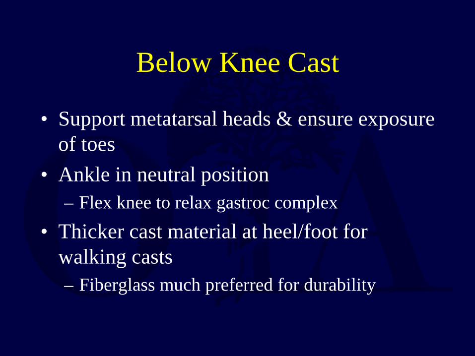
Below Knee Cast
• Support metatarsal heads & ensure exposure of toes
• Ankle in neutral position – Flex knee to relax gastroc complex
• Thicker cast material at heel/foot for walking casts – Fiberglass much preferred for durability
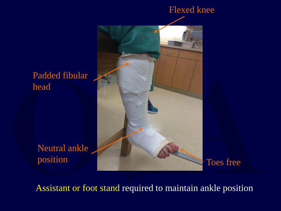
Padded fibular head
Flexed knee
Neutral ankle position Toes free
Assistant or foot stand required to maintain ankle position
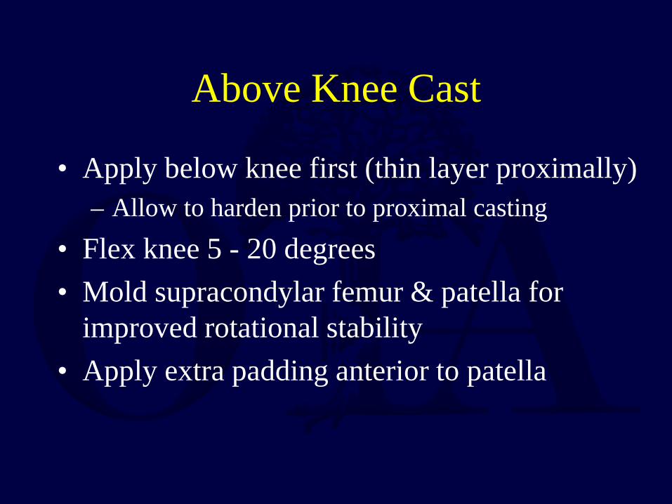
Above Knee Cast
• Apply below knee first (thin layer proximally) – Allow to harden prior to proximal casting
• Flex knee 5 - 20 degrees • Mold supracondylar femur & patella for
improved rotational stability • Apply extra padding anterior to patella
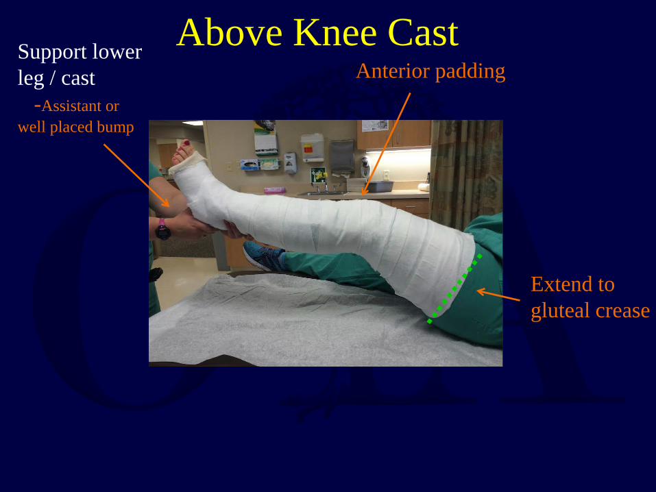
Anterior padding Support lower leg / cast -Assistant or well placed bump
Extend to gluteal crease
Above Knee Cast

Forearm Casts & Splints • MCP joints should be free for ROM if not
casting hand – Do not go past proximal palmar crease
• Thumb should be free to base of MC – Unobstructed opposition of thumb to little finger
Avoid digit impingement
Cast proximal to
palmar crease
permitting thumb
opposition
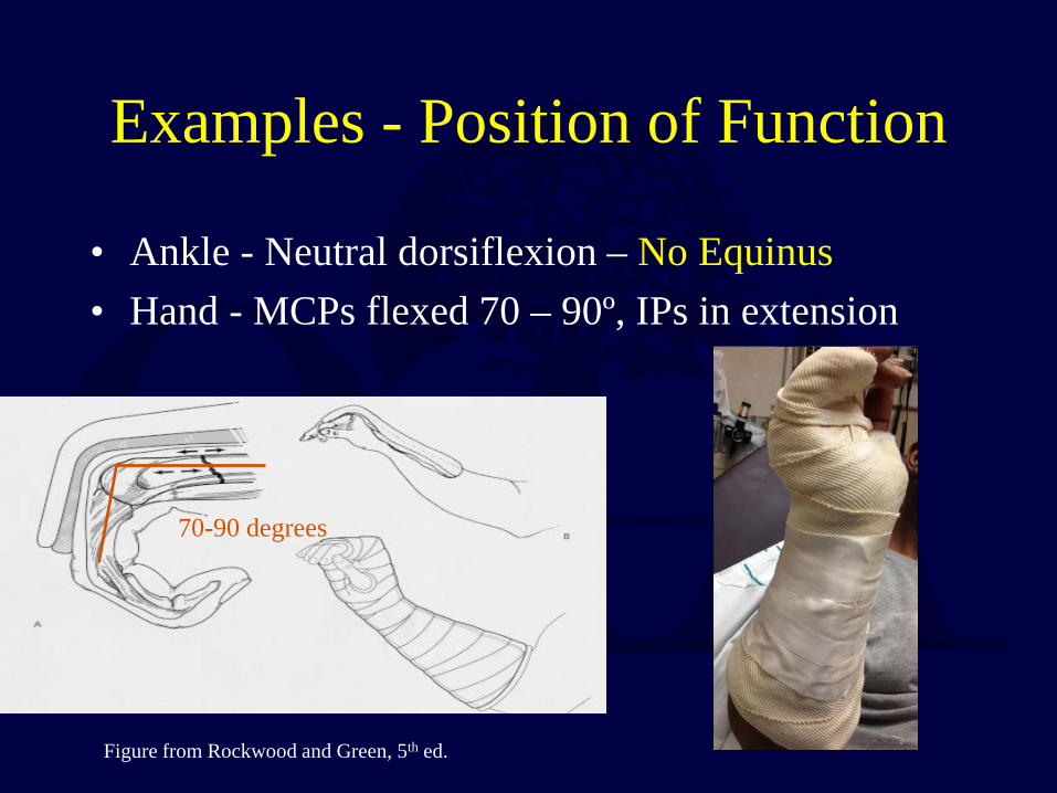
Examples - Position of Function
• Ankle - Neutral dorsiflexion – No Equinus • Hand - MCPs flexed 70 – 90º, IPs in extension
70-90 degrees
Figure from Rockwood and Green, 5th ed.

Cast Wedging
• Early follow-up x-rays are required to ensure acceptable reduction
• Cast may be “wedged” to correct reduction
• Deformity is drawn out on cast • Cast is cut circumferentially • Cast is wedged to correct
deformity & the over-wrapped
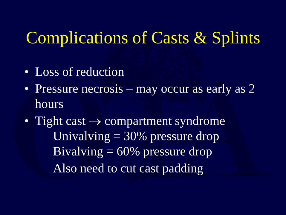
Complications of Casts & Splints
• Loss of reduction • Pressure necrosis – may occur as early as 2
hours • Tight cast → compartment syndrome
Univalving = 30% pressure drop Bivalving = 60% pressure drop
Also need to cut cast padding
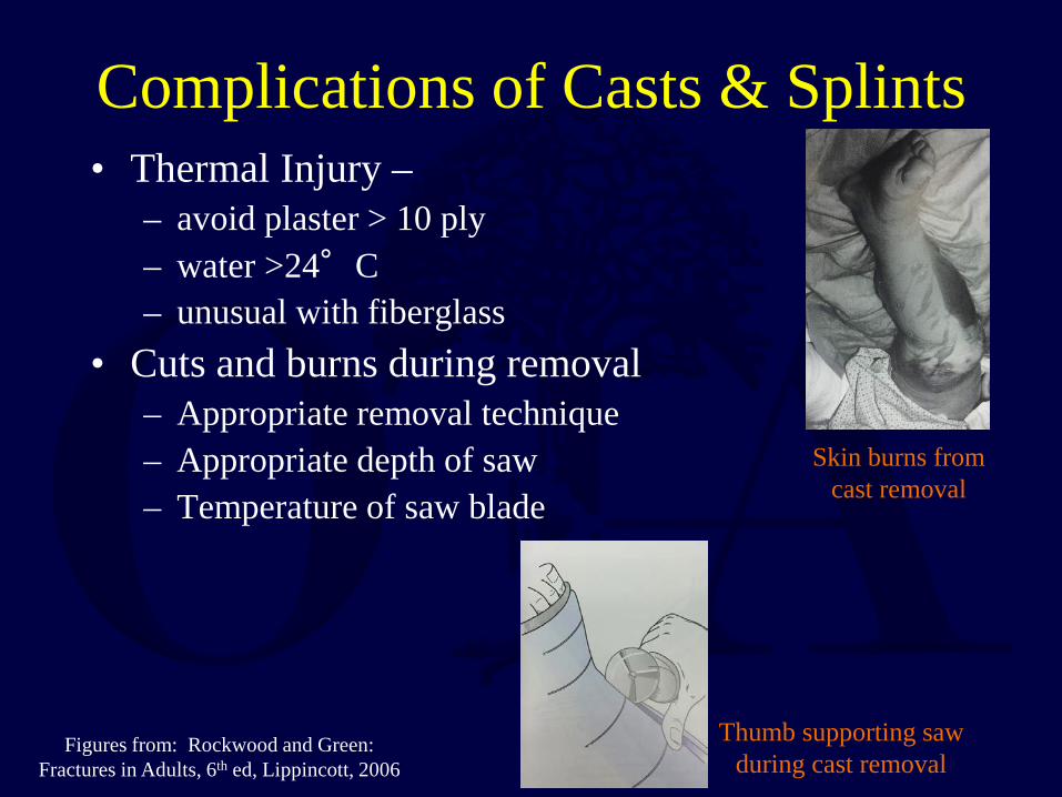
Complications of Casts & Splints • Thermal Injury –
– avoid plaster > 10 ply – water >24°C – unusual with fiberglass
• Cuts and burns during removal – Appropriate removal technique – Appropriate depth of saw – Temperature of saw blade
Figures from: Rockwood and Green: Fractures in Adults, 6th ed, Lippincott, 2006
Skin burns from cast removal
Thumb supporting saw during cast removal
Complications of Casts & Splints
• DVT/PE – Increased in lower extremity fracture – Prior history and family history – Birth control risk factor – Indications for prophylaxis controversial in patients
without risk factors • Joint stiffness
– Leave joints free when possible (ie. finger MCP for below elbow cast)
– Place joint in position of function • Limits long-term morbidity associated with stiffness

Traction
• Allows constant controlled force for initial stabilization of long bone fractures & aids reduction during operative procedure
• Skeletal vs. skin traction is case dependent

Skin (Bucks) Traction • Limited force can be applied
– Generally not to exceed 5 lbs • Commonly used in pediatric patients • Can cause soft tissue problems especially in
elderly or rheumatoid patients – Thin extremity skin
• Not as powerful when used during operative procedure for both length or rotational control

Skeletal Traction • More powerful than skin traction • May pull up to 20% of body weight for the
lower extremity • Requires anesthesia (local vs. sedation) for pin
insertion • Preferred method of temporizing:
– Femur fractures – Vertically unstable pelvic ring fractures – Acetabulum fractures
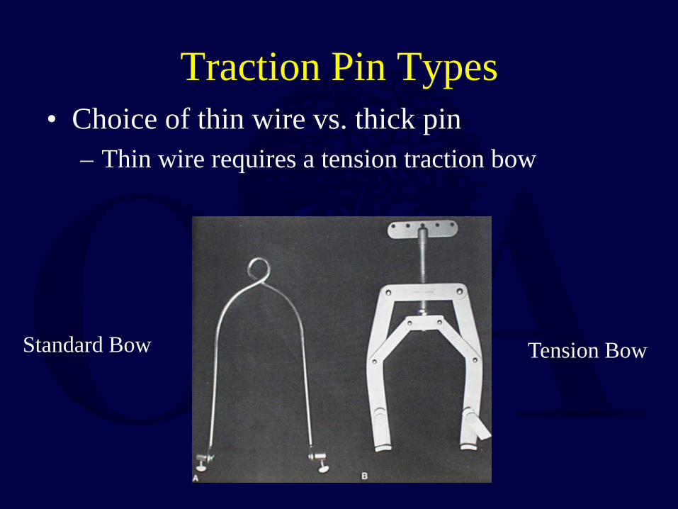
Traction Pin Types • Choice of thin wire vs. thick pin
– Thin wire requires a tension traction bow
Tension Bow Standard Bow

Traction Pin Types • Steinmann pin may be either smooth or threaded
– Smooth • Stronger but can slide if oblique
– Threaded pin • Weaker & can bend with higher weight application • Will not slide
• In general a 5 or 6 mm diameter pin is chosen for adults – Insertion may induce local bone thermal
necrosis
Bent non-tensioned thin wire

Traction Pin Placement • Sterile field with limb exposed • Local anesthesia + sedation • Insert pin from known area of neurovascular
structure – Distal femur: Medial → Lateral – Proximal Tibial: Lateral → Medial – Calcaneus: Medial → Lateral
• Place sterile dressing around pin site • Place protective caps over sharp pin ends

Distal Femoral Traction • Method of choice for acetabular/vertically
unstable pelvic ring & some femur fractures • If knee ligament injury suspected distal femur
instead of proximal tibial traction – Distraction through knee joint potential neurvascular
injury
Incline traction to prevent pretibial traction bow
pressure

Distal Femoral Traction • Place pin from medial to lateral at the
adductor tubercle - slightly proximal to epicondyle – Minimizes risk for vascular injury

Balanced Skeletal Traction
• Suspension of leg with longitudinal traction • Requires trapeze bar, traction cord, &
pulleys • Allows multiple adjustments for optimal
fracture alignment

• One of many options for setting up balanced suspension • In general the thigh support only requires 5-10 lbs of weight • Note the use of double pulleys at the foot to decrease the total
weight suspended off the bottom of the bed
Figure from: Rockwood and Green: Fractures in Adults, 4th ed, Lippincott, 1996.

Proximal Tibial Traction • Place pin 2 cm posterior and 1 cm distal to
tubercle • Place pin from lateral to medial
– Minimizes risk to peroneal nerve
Calcaneal Traction
• Most commonly used with a spanning ex fix for “travelling traction” or may be used with a Bohler-Braun frame
• Place pin medial to lateral 2 - 2.5 cm posterior and inferior to medial malleolus – Minimizes risk to posterior medial mal NV structures
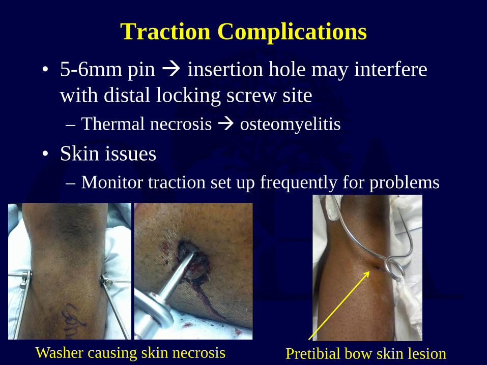
Traction Complications • 5-6mm pin insertion hole may interfere
with distal locking screw site – Thermal necrosis osteomyelitis
• Skin issues – Monitor traction set up frequently for problems
Washer causing skin necrosis Pretibial bow skin lesion
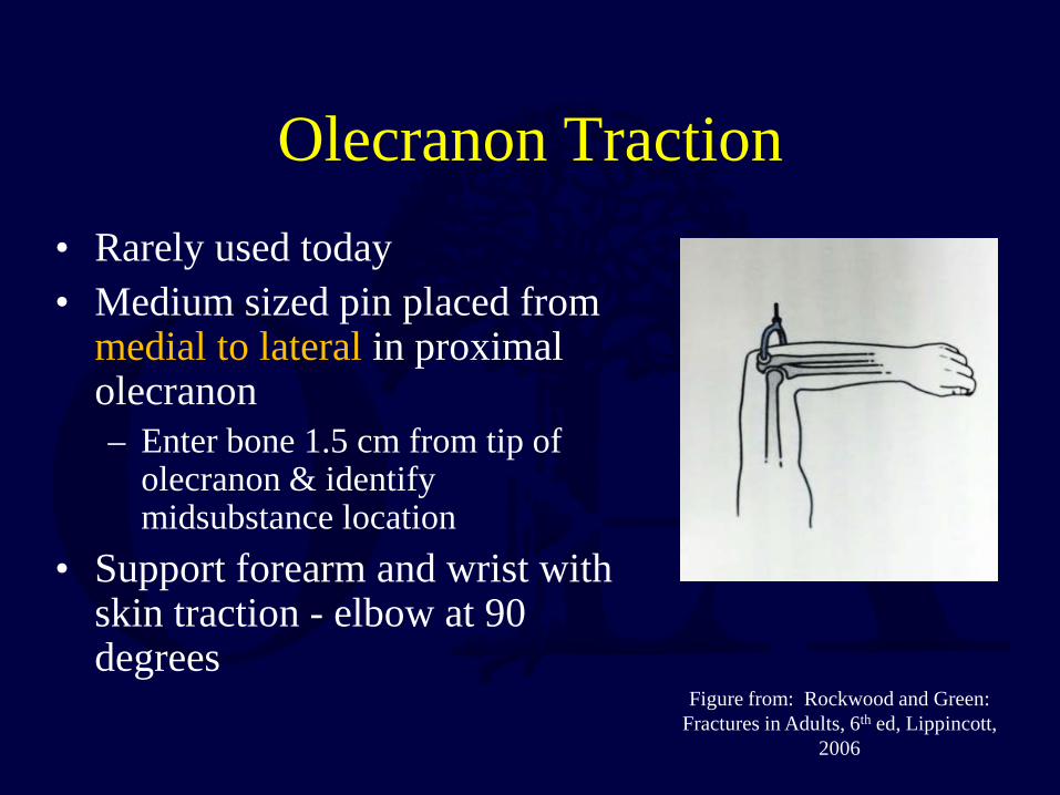
Olecranon Traction • Rarely used today • Medium sized pin placed from
medial to lateral in proximal olecranon – Enter bone 1.5 cm from tip of
olecranon & identify midsubstance location
• Support forearm and wrist with skin traction - elbow at 90 degrees
Figure from: Rockwood and Green: Fractures in Adults, 6th ed, Lippincott,
2006
Gardner Wells Tongs
• Used for C-spine reduction / traction • Pins are placed one finger breadth above
pinna & slightly posterior to external auditory meatus
• Apply traction beginning at 5 lbs. and increasing in 5 lb. increments with serial radiographs and clinical exam
Halo
• Indicated for certain cervical fractures as definitive treatment or supplementary protection to internal fixation
• Disadvantages – Pin problems – Respiratory compromise
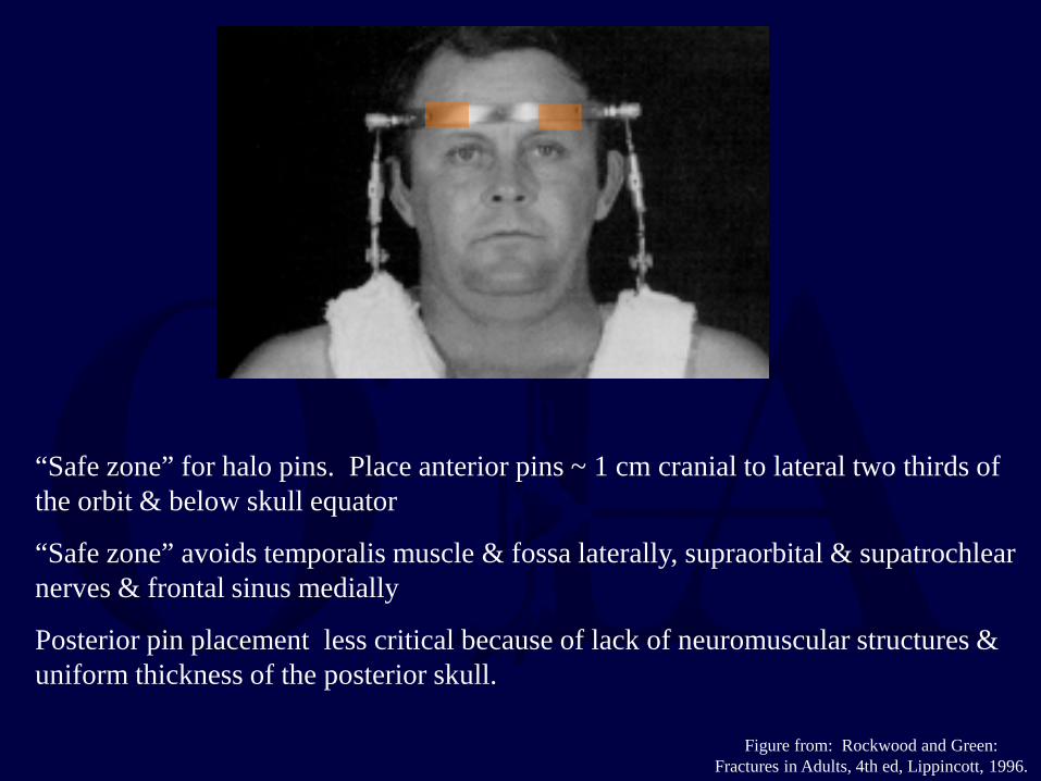
“Safe zone” for halo pins. Place anterior pins ~ 1 cm cranial to lateral two thirds of the orbit & below skull equator
“Safe zone” avoids temporalis muscle & fossa laterally, supraorbital & supatrochlear nerves & frontal sinus medially
Posterior pin placement less critical because of lack of neuromuscular structures & uniform thickness of the posterior skull.
Figure from: Rockwood and Green: Fractures in Adults, 4th ed, Lippincott, 1996.

Halo Application • Position patient maintaining spine
precautions • Fit Halo ring • Prep pin sites
– See previous slide for placement sites – Have patient gently close eyes for pin
placement to prevent eyelid dysfunction • Tighten pins to 6-8 ft-lbs. • Retighten if loose
– Pins only once at 24 hours
Figure from: Rockwood and Green: Fractures in Adults, 4th ed, Lippincott, 1996.

References • Freeland AE. Closed reduction of hand fractures. Clin Plast Surg.
2005 Oct;32(4):549-61. • Fernandez DL. Closed manipulation and casting of distal radius
fractures. Hand Clin. 2005 Aug;21(3):307-16. • Halanski M, Noonan KJ. Cast and splint immobilization:
complications. J Am Acad Orthop Surg. 2008 Jan;16(1):30-40. • Bebbington A, Lewis P, Savage R. Cast wedging for orthopaedic
surgeons. Injury. 2005;36:71-72. • Browner BD, Jupiter JB, Levine AM, Trafton PG, Krettek C. Skeletal
Trauma 4th ed. Philadelphia, PA: Saunders, 2009; 83-142. ISBN: 9781416048404
• Bucholz RW, Court-Brown CM, Heckman JD, Tornetta P, McQueen MM, Ricci WM. Rockwood and Green’s Fractures in Adults 7th ed. Philadelphia, PA: Lippincott Williams & Wilkins, 2010; 162-190. ISBN 9781605476773
References • Halanski MA, Halanski AD, Oza A, et al. Thermal injury
with contemporary cast-application techniques and methods to circumvent morbidity. J Bone Joint Surg Am. 2007 Nov;89(11):2369-77.
• Althausen PL, Hak DJ. Lower extremity traction pins: indications, technique, and complications. Am J Orthop. 2002 Jan;31(1):43-7.
• Alemdaroglu KB, Iltar S, Çimen O, et al.Risk Factors in Redisplacement of Distal Radial Fractures in Children. J Bone Joint Surg Am. 2008; 90: 1224 - 1230.
• Sarmiento A, Latta LL. Functional fracture bracing. J Am Acad Orthop Surg. 1999 Jan;7(1):66-75.

Classical References • Sarmiento A, Kinman PB, Galvin EG, Schmitt RH,
Phillips JG. Functional bracing of fractures of the shaft of the humerus. J Bone Joint Surg Am. 1977 Jul;59(5):596-601.
• Sarmiento A, Sobol PA, Sew Hoy AL, et al. Prefabricated Functional Braces for the Treatment of Fractures of the Tibial Diaphysis. JBone and Joint Surg. 1984. 66-A: 1328- 1339.
• Sarmiento A, Latta LL. 450 closed fractures of the distal third of the tibia treated with a functional brace. Clin Orthop Relat Res. 2004 Nov;(428):261-71.
• Sarmiento A. Fracture bracing. Clin Orthop Relat Res. 1974 Jul-Aug;(102):152-8.

Questions
Return to General/Principles
Index
E-mail OTA about
Questions/Comments
If you would like to volunteer as an author for the Resident Slide Project or recommend updates to any of the following slides, please send an e-mail to [email protected]