coix lacryma-jobi var. ma-yuen stapf sprout extract has...
TRANSCRIPT

RESEARCH ARTICLE Open Access
Coix lacryma-jobi var. ma-yuen Stapf sproutextract has anti-metastatic activity in coloncancer cells in vitroEun Suk Son1†, Young Ock Kim2†, Chun Geon Park2, Kyung Hun Park2, Sung Hwan Jeong1,Jeong-Woong Park1* and Se-Hee Kim3*
Abstract
Background: Coix lacryma-jobi var. ma-yuen (Rom.Caill.) Stapf has been used in China as an herbal medicine. Manystudies of this plant have reported anti-proliferative and apoptotic activities on human cancer cell lines. Therefore,this study of the anti-metastatic effect of Coix lacryma-jobi var. ma-yuen Stapf sprout extract (CLSE) in colorectal cancercells may provide a scientific basis for exploring anti-cancer effects of edible crops.
Methods: To evaluate the effect of CLSE on cell proliferation and signaling, we performed a Cell Counting Kit-8 (CCK-8)assay in HCT116 cells and used western blot analysis. Furthermore, scratch-wound healing, transwell migration,matrigel invasion, and adhesion assays were conducted to elucidate the anti-metastatic effects of CLSE underhypoxic conditions in colon cancer cells.
Results: First, CLSE decreased deferoxamine (DFO)-induced migration of colon cancer cells by 87%, and blocked coloncancer cell migration by 80% compared with hypoxia control cells. Second, CLSE treatment resulted in a 54% reductionin hypoxia-induced invasiveness of colon cancer cells, and 50% inhibition of adhesive potency through inactivation ofthe extracellular signal-regulated kinase (ERK) 1/2 and protein kinase b (AKT) pathways. Third, conditioned mediumcollected from CLSE-treated HCT116 cells suppressed tube formation of human umbilical vein endothelial cells(HUVECs) by 91%.
Conclusions: CLSE inhibited migration, invasion, and adhesion of colon cancer cells and tube formation byHUVECs via repression of the ERK1/2 and AKT pathways under hypoxic conditions. Therefore, CLSE may beused to treat patients with colon cancer.
Keywords: Coix, Colon cancer, Metastasis, Invasion, Hypoxia
BackgroundWorldwide, colon cancer is one of the most deadly can-cers because it is highly metastatic and invasive. An im-portant determinant of the prognosis of cancer patientsis the progression of tumor cell metastasis and invasion.The ability for metastasis and invasion enables cancer
cells to find new areas of the body to occupy when spaceand nutrients become limited in their current location.The metastatic cascade can be separated into three pro-cesses: invasion, intravasation, and extravasation. First,the process of invasion involves the dissociation oftumor cells from the primary tumor mass and subse-quent invasion into the surrounding tissue. Next, intra-vasation occurs when detached cells are transported viablood vessels to distant sites. Finally, tumor cells interactwith endothelial cells to form stronger bonds, andpenetrate the endothelium and basement membrane.Consequently, the new tumor cells can proliferate in sec-ondary sites. Therefore, the metastatic spread of tumortissue requires the growth of a vascular network [1].
* Correspondence: [email protected]; [email protected]†Equal contributors1Department of Internal Medicine, Gachon University Gil Medical Center, 21Namdong-daero 774 beon-gil, Namdong-gu, Incheon 405-760, Republic ofKorea3Gachon medical research institute, Gachon University Gil Medical Center, 21Namdong-daero 774 beon-gil, Namdong-gu, Incheon 405-760, Republic ofKoreaFull list of author information is available at the end of the article
© The Author(s). 2017 Open Access This article is distributed under the terms of the Creative Commons Attribution 4.0International License (http://creativecommons.org/licenses/by/4.0/), which permits unrestricted use, distribution, andreproduction in any medium, provided you give appropriate credit to the original author(s) and the source, provide a link tothe Creative Commons license, and indicate if changes were made. The Creative Commons Public Domain Dedication waiver(http://creativecommons.org/publicdomain/zero/1.0/) applies to the data made available in this article, unless otherwise stated.
Son et al. BMC Complementary and Alternative Medicine (2017) 17:486 DOI 10.1186/s12906-017-1990-y

The growth of new blood vessels (angiogenesis) isrequired for primary tumor growth as well as tumorinvasion and metastasis [1]. The vasculature that sup-plies oxygen and nutrients is important for cancer cellsurvival [2].Tumor hypoxia results from an imbalance between the
oxygen supply and demand due to uncontrolled tumorcell proliferation [3]. Because cancer cells rapidly prolif-erate, the tumor quickly exhausts the nutrient andoxygen supply from the normal vasculature, and be-comes hypoxic. This hypoxic condition upregulates theproduction of angiogenic factors from hypoxic tumorsites [4]. Therefore, hypoxia signaling can contribute totumor progression by promoting tumor cell migration,invasion, metastasis, and angiogenesis [5].Coix lacryma-jobi var. ma-yuen (Rom.Caill.) Stapf,
which is an important cereal crop for many indigenousgroups in upland areas, is characterized by having asimilar appearance and taste to rice, with a standingcrop comparable with corn. This plant is utilized as arice alternative, health-promoting staple crop, and as analternative livelihood and income source throughvalue-added products. An increase in the number ofhealth-conscious individuals has also contributed tothe popularity of Coix, with the market currentlygrowing due to increased acceptance of this product.Coix is largely consumed for household food securityas a rice alternative or used to make porridge, cham-porado, and other recipes.Previous studies have reported that Coix extract has
anti-proliferative and apoptotic activities on human lungcancer, histolytic lymphoma, and colon cancer cells, aswell as chemopreventive effects on lung cancer in vivo[6–9]. Although a few studies have reported that Coixhas anti-cancer effects in terms of regulating the prolif-eration and cell cycle of cancer cells, the effects of Coixlacryma-jobi var. ma-yuen Stapf sprout extract (CLSE)on cancer metastasis are unknown. Therefore, this studyaimed to explore the anti-cancer effects of CLSE in colo-rectal cancer cells.
MethodsReagentsCLSE was manufactured in the herbarium of the HerbalCrop Research Institute (Eumseong, Republic of Korea).Deferoxamine (DFO), Phorbol 12-myristate 13-acetate
(PMA), and SC79 were obtained from Sigma-Aldrich(St. Louis, MO, USA). CLSE and DFO were dissolved inwater. PMA and SC79 were dissolved in the solventdimethyl sulfoxide (DMSO).
CLSE preparationCoix cultivars were obtained from the National Instituteof Crop Science (Miryang, Republic of Korea). Coix were
germinated in a modified commercial soil bed (0.7–1.0mg/m3 soil bulk density, 450–650 mg/L available phos-phate, 800–1000 mg/kg nitrogen) (Punong Bed Soil,Gyeongju, Republic of Korea). The germinated Coix wasgrown at 22–23 °C with humidity of 60% in a 900–1000 lx environment. Between 15 and 22 d after germin-ation, young barley leaves about 8–13-cm long wereharvested and freeze-dried [10]. We used a water extrac-tion method because most traditional Oriental herbs aredecocted in boiling water. In addition, some componentsare more soluble in water than in organic solvents.Crushed plant materials (200 g each) were extractedthree times under reflux with distilled water. The waterextracts were combined and lyophilized. The yield was25% (wt/wt) of the dried Coix sprouts. Extracts werestored at −20 °C until usage. A voucher specimen (HPR-208) was deposited in the herbarium of Herbal CropResearch Institute (Eumseong, Republic of Korea).
Cell lines and cell culture conditionsHCT116 and CCD-18Co cells were obtained from theKorean Cell Line Bank (Seoul, Republic of Korea).Human umbilical vein endothelial cells (HUVECs) wereobtained from the Lonza (San Diego, CA, USA).HCT116 cells were cultured in McCoy’s medium (GibcoCell Culture, Carlsbad, CA, USA) supplemented with10% fetal bovine serum (FBS) (Gibco) and 1% penicillin-streptomycin (Gibco). CCD-18Co cells were cultured inMEM (Gibco) with 10% FBS (Gibco) and 1% penicillin-streptomycin (Gibco), and were used between passages 5and 6. HUVECs were grown in EBM-2 (Lonza) supple-mented with an EGM™-2 SingleQuots™ kit (Lonza), andused between passages 2 and 4 for experiments. Cellswere incubated at 37 °C in a humidified atmosphere with5% CO2. A hypoxia incubator (New Brunswick Scien-tific, Edison, NJ, USA) containing 1% O2, 5% CO2, and94% N2 was used to create hypoxic conditions.
Cell counting Kit-8 (CCK-8) assayCells were seeded into 96-well plates and exposed tovarious concentrations of CLSE for 24–72 h prior to theaddition of 10 μL CCK-8 solution (Dojindo MolecularTechnologies, Inc., Rockville, MD, USA) containing 2-(2-methoxy-4-nitrophenyl0–3-(4-nitrophenyl)-5-(2,4-dis-ulfophenyl)-2H–tetrazolium, monosodium salt (WST-8)to each well. After 1 h of incubation at 37 °C in a hu-midified atmosphere with 5% CO2, the absorbance wasdetermined at 450 nm.
Scratch-wound healing assayCells were seeded in 60-mm plates and then scratchedusing a pipette tip after 24-h incubation. After washingwith phosphate buffered saline (PBS), cells were incu-bated in medium containing CLSE and/or 100 μM DFO
Son et al. BMC Complementary and Alternative Medicine (2017) 17:486 Page 2 of 9

for 24 h. Images were then obtained at 0 and 24 h withan Olympus CFX41 microscope (Hamburg, Germany) at40× magnification. The area of cell migration was quan-tified using ImageJ software (https://imagej.nih.gov/ij/)and the percentage of wound closure was calculated asdescribed previously [11].
Transwell migration assayThe experiment was performed as described previously[11]. Briefly, cells in CLSE-containing serum-freemedium were seeded into the inner chamber with thelower surface coated with 0.2% gelatin. The cells wereincubated in normoxic or hypoxic conditions for 24 h.20% FBS-containing medium in the bottom chamberwas used as a chemoattractant. Cells on the upper mem-brane were wiped off using wet cotton swabs after fix-ation with methanol and crystal violet staining. Cells onthe lower surface were mounted using mounting solu-tion (Vectashield®; Vector Laboratories Burlingame, CA,USA). Stained cells were counted under a light micro-scope (DP72; Olympus, Hamburg, Germany) and imageswere taken at 200× magnification. All of the experimentswere independently repeated in triplicate.
Matrigel invasion assayThe matrigel invasion assay was performed as previouslydescribed [11]. Briefly, the lower surface were coatedwith 0.2% gelatin, and then the upper surfaces werecoated with Matrigel® (BD Biosciences, San Jose, CA,USA) at 37 °C for 2 h. Cells in CLSE-containing serum-free medium were plated into the inner chamber and in-cubated in normoxic or hypoxic conditions for 48 h with20% FBS in the bottom chamber as the chemoattractant.The cells were processed as described for the transwellmigration assay. All of the experiments were independ-ently repeated in triplicate.
Adhesion assayCells were treated with or without CLSE and/or DFOfor 24 h, suspended in serum-free McCoy’s medium, andthen seeded in a 96-well plate that had been pre-coatedwith Matrigel® (BD Biosciences). After incubation at 37 °Cfor 90 min, the cells were washed with PBS and treatedwith 0.5 mg/mL 3-(4,5-Dimethylthiazol-2-yl)-2,5-diphe-nyltetrazolium bromide (MTT). The absorbance of forma-zan crystals dissolved in 100 μL DMSO was determined at570 nm using a microplate reader. The experiments wereindependently performed in triplicate.
Western blot analysisCells were harvested with lysis buffer [50 mM Tris-HCl(pH 8.0), 0.5% sodium deoxycholate, 150 mM NaCl,0.1% sodium dodecyl sulfate (SDS), and 1% NP-40]. Anti-bodies to p65, phospho-p65, extracellular signal-regulated
kinase (ERK) 1/2, phospho-ERK1/2, protein kinase B(AKT), phospho-AKT, c-Jun N-terminal kinase (JNK),phospho-JNK, p38, phospho-p38, Signal transducer andactivator of transcription 3 (STAT3), and phospho-STAT3were all purchased from Cell Signaling Technology(Beverly, MA, USA). β-actin antibody was obtained fromSanta Cruz Biotechnology (Santa Cruz, CA, USA). Immu-noblot bands were quantified as a ratio of phosphorylatedprotein/total protein using ImageJ software.
Generation of conditioned mediumHCT116 cells were treated with or without CLSE (0.5,1 mg/mL) and/or DFO (100 μM) for 24 h in completemedium, and then the supernatant was saved. Thesupernatant, called conditioned medium, was filteredand stored at −70 °C.
Tube formation assayHUVECs were diluted in conditioned medium andseeded in a 48-well plate that had been pre-coated withMatrigel®. After a 12-h incubation period, images offormed tubules were taken at 40× magnification andquantified by determining the number of branchingpoints. The experiments were independently performedin triplicate.
Statistical analysisAll data were analyzed with GraphPad Prism® softwareusing a two-tailed Student t test. P values less than 0.05were significantly considered.
ResultsCLSE inhibits colon cancer cell migration under hypoxicconditionsTo examine effects on the viability of colon cancer cells,HCT116, and of normal cells, CCD-18Co, caused byCLSE treatment, we performed a CCK-8 assay. Asshown in Fig. 1, CLSE strongly decreased the viability ofHCT116 cells compared to CCD-18Co cells. Next, weperformed scratch-wound healing assays to determine ifCLSE had an effect on the migratory potency ofHCT116 cells. For this assay, we used DFO, a reagentused to simulate hypoxic conditions. As shown in Fig. 2aand b, DFO enhanced the migration of colon cancercells (p < 0.05). However, HCT116 cells co-treated withDFO and CLSE had 47–87% less healing ability thancells treated with DFO only (p < 0.05). Furthermore, toconfirm the inhibitory effect of CLSE on HCT116 cellmigration, we plated CLSE-treated HCT116 cells in aTranswell® chamber and incubated the cells for 24 hunder hypoxic conditions. The results showed that CLSEinhibited the migration of hypoxic HCT116 cells in theTranswell® chamber by 48–80% compared with the hyp-oxia control group (p < 0.01) (Fig. 2c, d). These results
Son et al. BMC Complementary and Alternative Medicine (2017) 17:486 Page 3 of 9

imply that CLSE has the potential to inhibit HCT116cell migration under hypoxic conditions.
CLSE inhibits invasion and adhesion of colon cancer cellsin hypoxiaIn cancer cell metastasis, invasion is as important as mi-gration. To investigate the effects of CLSE on colon can-cer cell invasion, we performed an invasion assay using aTranswell® coated with Matrigel®. As we showed, hypoxiapromoted the invasion of HCT116 cells (p < 0.01).However, CLSE-treated cells under hypoxic conditionsshowed a 21–54% reduction in invasion compared withthe hypoxia control group (p < 0.05) (Fig. 3a, b).Metastatic cancer cells have high adhesion potency to
migrate to secondary sites for new tumor growth. There-fore, cell adhesion assays are used to examine themetastatic potency of cancer cells. As shown in Fig. 3c,DFO-treated cells had increased adhesion comparedwith untreated cells (p < 0.05); however, cells co-treatedwith DFO and CLSE showed a 19–50% reduction in
adhesive ability compared with cells treated with DFOonly (p < 0.001, p < 0.01). Taken together, these resultssuggest that CLSE regulates the invasion and adhesionof HCT116 cells under hypoxic conditions.
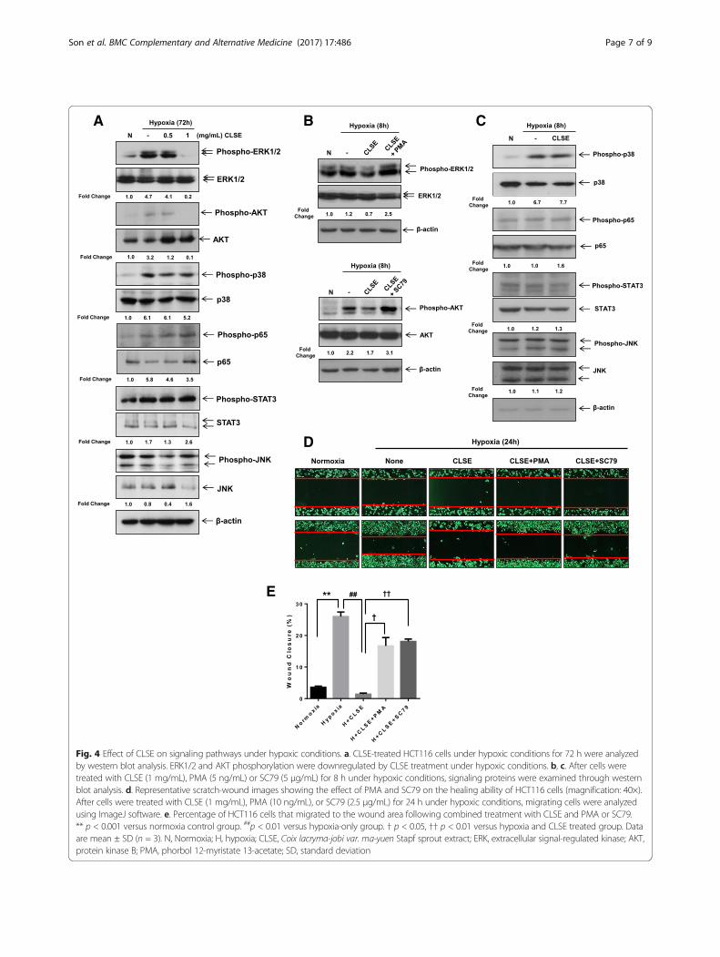
CLSE inactivates ERK1/2 and AKT activities in hypoxicconditionsTo identify the cellular signaling pathways regulated byCLSE during colon cancer cell metastasis under hypoxicconditions, we examined the expression of signalingmarkers using Western blot analysis. Among the varioussignaling markers investigated, hypoxia-induced activa-tion of ERK1/2 and AKT were downregulated by 1 mg/mL CLSE (Fig. 4a). To confirm whether the activation ofERK1/2 and AKT were repressed by CLSE in HCT116cells, we performed western blot and scratch-woundhealing assay at earlier stage in the presence of theERK1/2 and AKT activators, PMA and SC79, respect-ively. As shown in Fig. 4b, inhibition of the ERK1/2 andAKT pathways by CLSE was reversed in CLSE-treated
C L S E (m g /m L )
%o
fC
on
tro
l(C
ell
Via
bil
ity
)
0 .0 0 0 .2 5 0 .5 0 1 .0 0 2 .5 0 5 .0 0 1 0 .0 00
2 0
4 0
6 0
8 0
1 0 0
1 2 0 2 4 h
4 8 h
7 2 h
A HCT116
B CCD-18Co
C L S E (m g /m L )
%o
fC
on
tro
l(C
ell
Via
bil
i ty
)
0 .0 0 0 .5 0 2 .5 0 1 0 .0 00
2 0
4 0
6 0
8 0
1 0 0
1 2 0 2 4 h
4 8 h
7 2 h
Fig. 1 Cytotoxic effect of CLSE on HCT116 and CCD-18Co cells. a, b. A Cell Counting Kit (CCK)-8 assay was used to measure HCT116 and CCD-18Cocells proliferation for 24–72 h after treatment with various concentrations of CLSE. Data are mean ± SD. CLSE, Coix lacryma-jobi var. ma-yuen Straptsprout extract; SD, Standard Deviation
Son et al. BMC Complementary and Alternative Medicine (2017) 17:486 Page 4 of 9

HCT116 cells given PMA or SC79. However, activationof other signaling proteins (p38, p65, STAT3, JNK) wasnot affected by CLSE treatment at an earlier stage(Fig. 4c). In addition, in the scratch-wound healingassay, PMA and SC79 could compromise the inhibi-tory effect of CLSE on the migration of HCT116 cellsunder hypoxic conditions by 57–71% and 64–77%, re-spectively (p < 0.05, p < 0.01) (Fig. 4d, e). Therefore,
our results suggest that CLSE represses HCT116 cellmigration through inactivation of the ERK1/2 andAKT pathways under hypoxic conditions.
CLSE inhibits HUVEC tube formation under hypoxiaThe formation of new blood vessels (angiogenesis) iscrucial for invasive tumor growth and metastasis. Todemonstrate that CLSE affects angiogenesis, conditioned
B
*
CNormoxia
H+0.5 mg/mL CLSE H+1 mg/mL CLSE
HypoxiaD
None
0.5 mg/mL CLSE
1 mg/mL CLSE
None
001(O
FD
M)
0 h 24 hA
Wo
un
dC
los
ure
(%)
No n e
DF O
DF O
+ 0 .5m
g /mL
CL S
E
DF O
+ 1m
g /mL
CL S
E
0
2 0
4 0
6 0
Ce
llm
igra
tio
n(n
orm
ali
ze
dv
alu
es
)
No rm
o x ia
Hy p o x ia
H+ 0 .5
mg /m
LC
L SE
H+ 1
mg /m
LC
L SE
0
1
2
3
4
5
#
** ##
##
#
Fig. 2 Effect of CLSE on the migratory ability of HCT116 cells under hypoxic conditions. a. Representative scratch-wound images showing theeffect of CLSE on the healing ability of HCT116 cells (magnification: ×40). After with treatment DFO (100 μM) and CLSE (0.5, 1 mg/mL) for 24 h,the migratory ability was analyzed by scratch-wound healing assay. b. Percentage of HCT116 cells that migrated into the wound following CLSEtreatment relative to untreated control cells. * p < 0.05 versus untreated control group. #p < 0.05 versus DFO-only group. c. Representative imagesshowing the effect of CLSE on HCT116 cell migration through a Transwell® chamber membrane (magnification: ×200). After treatment with CLSE (0.5,1 mg/mL) for 24 h, the migration cells in transwells were stained by crystal violet solution. d. Percentage of HCT116 cells that migrated following CLSEtreatment relative to control cells (normoxia). ** p < 0.01 versus normoxia control group. ##p < 0.01 versus hypoxia-only group. Data are mean ± SD(n = 3). CLSE, Coix lacryma-jobi var. ma-yuen Strapt sprout extract; H, hypoxia; SD, Standard Deviation
Son et al. BMC Complementary and Alternative Medicine (2017) 17:486 Page 5 of 9

media, obtained from growing HCT116 cells in mediasupplemented with CLSE and/or DFO, was used to treatHUVEC cells. The media obtained from DFO-treatedHCT116 cells augmented tube formation by HUVECs(p < 0.01), whereas conditioned media, obtained fromCLSE and DFO-treated HCT116 cells, decreased tubeformation by 55–91% (p < 0.001). These results suggestthat CLSE may mediate its anti-angiogenic effect byregulating the secretion of angiogenic factors by coloncancer cells (Fig. 5).
DiscussionThe hallmarks of cancer are self-sufficiency of growthsignals, insensitivity to anti-growth signals, limitless rep-licative potential, evading cell death (apoptosis), tissueinvasion, metastasis, and sustained angiogenesis [2].
To investigate the anti-cancer effects of CLSE, we in-vestigated the physiological effect of CLSE on cancerusing human colon cancer cells.In this study, we showed that CLSE had an anti-
metastatic effect in colon cancer cells. Specifically, CLSEinhibited colon cancer cell migration, invasion, woundhealing, and adhesion. In addition, conditioned mediacollected from CLSE-treated HCT116 cells had an in-hibitory effect on HUVEC tube formation. On the otherhand, in the case of the cervical cancer cell line HeLa,CLSE promoted apoptosis and arrested the cell cycle (insubmission). Taken together, these results suggest thatthe mechanism on the anti-cancer effect of CLSE maybe organ-specific.Recently, many studies have tried to identify the active
components of Coix and determine their mechanism of
C
A Normoxia
H+0.5 mg/mL CLSE H+1 mg/mL CLSE
Hypoxia
#####
B
%o
fC
on
tro
l(A
dh
es
i on
)
No n e
DF O
DF O
+ 0 .5m
g /mL
CL S
E
DF O
+ 1m
g /mL
CL S
E
0
5 0
1 0 0
1 5 0
Ce
llin
va
sio
n(n
orm
ali
ze
dv
alu
es
)
No rm
o x ia
Hy p o x ia
H+ 0 .5
mg /m
LC
L SE
H+ 1
mg /m
LC
L SE
0 .0
0 .5
1 .0
1 .5
2 .0
#
*
#
**
Fig. 3 Effect of CLSE on HCT116 cell invasion and migration under hypoxic conditions. a. Representative images showing the effect of CLSE onHCT116 cell invasion through a Matrigel®-coated Transwell® chamber membrane (magnification: 200×). After treatment with CLSE (0.5, 1 mg/mL)for 48 h under hypoxic conditions, cells that had invaded the transwells were analyzed through crystal violet staining. b. Number of HCT116 cellsthat invaded following CLSE treatment. The values are normalized to the number of invaded control cells (normoxia). ** p < 0.01 versus normoxiacontrol group. #p < 0.05 versus hypoxia-only group. c. After treatment with DFO (100 μM) and/or CLSE (0.5, 1 mg/mL) for 24 h, cells were seededinto Matrigel-coated wells and then analyzed using an MTT assay. Number of HCT116 cells that adhered to the Matrigel®-coated plate followingCLSE treatment. The values are normalized to the number of adherent control cells (untreated). * p < 0.05 versus untreated control group. ##p < 0.01,###p < 0.001 versus DFO-only groups. Data are mean ± SD (n = 3). CLSE, Coix lacryma-jobi var. ma-yuen Stapf sprout extract; H, hypoxia; SD, standard deviation
Son et al. BMC Complementary and Alternative Medicine (2017) 17:486 Page 6 of 9
A B
D
E
C
Fig. 4 Effect of CLSE on signaling pathways under hypoxic conditions. a. CLSE-treated HCT116 cells under hypoxic conditions for 72 h were analyzedby western blot analysis. ERK1/2 and AKT phosphorylation were downregulated by CLSE treatment under hypoxic conditions. b, c. After cells weretreated with CLSE (1 mg/mL), PMA (5 ng/mL) or SC79 (5 μg/mL) for 8 h under hypoxic conditions, signaling proteins were examined through westernblot analysis. d. Representative scratch-wound images showing the effect of PMA and SC79 on the healing ability of HCT116 cells (magnification: 40×).After cells were treated with CLSE (1 mg/mL), PMA (10 ng/mL), or SC79 (2.5 μg/mL) for 24 h under hypoxic conditions, migrating cells were analyzedusing ImageJ software. e. Percentage of HCT116 cells that migrated to the wound area following combined treatment with CLSE and PMA or SC79.** p < 0.001 versus normoxia control group. ##p < 0.01 versus hypoxia-only group. † p < 0.05, †† p < 0.01 versus hypoxia and CLSE treated group. Dataare mean ± SD (n = 3). N, Normoxia; H, hypoxia; CLSE, Coix lacryma-jobi var. ma-yuen Stapf sprout extract; ERK, extracellular signal-regulated kinase; AKT,protein kinase B; PMA, phorbol 12-myristate 13-acetate; SD, standard deviation
Son et al. BMC Complementary and Alternative Medicine (2017) 17:486 Page 7 of 9

action. Specifically, neutral lipid isolated from endo-sperm of Coix inhibits the growth of pancreatic cancercells [12], and the ethyl acetate fraction from ethanolicextraction of adlay testa has an inhibitory effect on theallergic response [13]. In addition, five compounds(coixspirolactam A, coixspirolactam B, coixspirolactamC, coixlactam, methyl dioxindole-3-acetate) isolatedfrom Coix bran exhibit anti-proliferative effect on lungand colon cancer cells [9]. Because CLSE is obtainedfrom a young sprout form Coix, we believe it to share asimilar chemical composition with the mature plant. Assuch, it is highly likely that CLSE also contains coixspir-olactams and methyl dioxindole-3-acetate, which maycontribute to the anti-metastatic effects of CLSE.Previous reports analyzed the proliferation of cancer
cells or the expression of cell cycle regulatory proteinsto demonstrate the anti-cancer effects of Coix undernormal conditions. However, our study focused on theeffects of the sprout extract of Coix on colon cancer cellmetastasis and HUVEC tube formation under hypoxicconditions. Hypoxia, a characteristic feature of locallyadvanced solid tumors, has emerged as a pivotal factorfor the tumor physiome because it can promote tumor
progression and increase tumor resistance to therapy[14]. Migration, invasion, and adhesion of cancer cellsresult from the loss of epithelial markers and the degrad-ation of basement membrane. Therefore, we expect thatCLSE may have anti-metastatic effects via regulation ofE-cadherin, vimentin, MMP-2, and MMP-9 in coloncancer cells, and plan to undertake these experiments inthe future.Phosphorylation-mediated activation of AKT and ERK
signaling drives tumor invasion [15]. The ERK pathwaycontrols cell migration, invasion, proliferation, and theinduction of transcriptional programs [16]. Since AKTcontributes to the development or progression of cancer,many consequences of hyperactive AKT signaling areconsidered hallmarks of cancer [17]. Our data showedthat downregulation of ERK1/2 and AKT phosphoryl-ation by CLSE was reversed in the presence of theiractivators, PMA and SC79 under hypoxic conditions(Fig. 4); therefore, it is likely that CLSE blocked the mi-gration of colon cancer cells via inactivation of ERK1/2and AKT under hypoxic conditions.It has been reported in some animal research that Coix
consumption might cause embryotoxicity and enhance
None DFO DFO+0.5 mg/mL CLSE DFO+1 mg/mL CLSEA
B**
######
Nu
mb
er
of
bra
nc
hin
gp
oin
ts
No n e
DF O
DF O
+ 0 .5m
g /mL
CL S
E
DF O
+ 1m
g /mL
CL S
E
0
2 0
4 0
6 0
Fig. 5 Effect of CLSE on HUVECs capillary tube formation. a. After HUVECs were seeded in matrigel-coated plates and incubated with conditionedmedia from HCT116 cells treated with DFO (100 μM) and CLSE (0.5, 1 mg/mL) for 24 h. Representative images showing HUVEC tube formation followingtreatment with conditioned media collected from CLSE-treated HCT116 cells (magnification: ×40). b. Number of branching points per unit area followingtreatment with conditioned media. ** p < 0.01 versus untreated control group. ###p < 0.001 versus DFO-only group. Data are mean ± SD (n = 3). CLSE, Coixlacryma-jobi var. ma-yuen Strapt sprout extract; HUVECs, human umbilical vein endothelial cells; SD, standard deviation
Son et al. BMC Complementary and Alternative Medicine (2017) 17:486 Page 8 of 9

uterine contractility during pregnancy [18]. Furthermore,some herbal medicines are reported to interfere with theefficacy and safety of conventional medicines [19].Therefore, more research on the stability and efficacy ofherbal supplements such as Coix is needed. In addition,our primary focus in this research was to explore theoverall anti-cancer effects of CLSE, and we have suc-ceeded in this endeavor. Further studies will not onlyreinforce this research, but will also contribute greatly tothe field of oncology through detailed chemical profilingof CLSE and mechanistic studies of each component, aswell as in vivo experiments using CLSE.
ConclusionsCLSE can inhibit colon cancer cells migration, invasion,adhesion, and HUVEC tube formation in hypoxic condi-tions through inhibition of the ERK1/2 and AKT signal-ing pathways. However, further study is necessary toreveal the possible mechanism of the anti-metastaticeffect of CLSE and its active compounds under hypoxicconditions.
AbbreviationsAKT: Protein kinase B; CCK-8: Cell Counting Kit-8; CLSE: Coix lacryma-jobi var.ma-yuen Stapf sprout extract; DFO: Deferoxamine; ERK: Extracellular signal-regulated kinase; FBS: Fetal bovine serum; HUVECs: Human umbilical veinendothelial cells; JNK: c-Jun N-terminal kinase; KLT: Kanglaite®; MTT: 3-(4,5-Dimethylthiazol-2-yl)-2,5-diphenyltetrazolium bromide; PBS: Phosphatebuffered saline; PMA: Phorbol 12-myristate 13-acetate; SD: Standarddeviation; SDS: Sodium dodecyl sulfate; STAT3: Signal transducer andactivator of transcription 3; WST-8: 2-(2-methoxy-4-nitrophenyl0–3-(4-nitrophenyl)-5-(2,4-disulfophenyl)-2H–tetrazolium, monosodium salt
AcknowledgementsNot applicable.
FundingThis work was supported by a grant (FRD2014–10-2) funded by Gachon UniversityGil Medical Center in 2015 to Jeong Woong Park and a grant (PJ0127852017)from the Rural Development Administration to Young Ock Kim.
Availability of data and materialsThe datasets used and/or analyzed during the current study are availablefrom the corresponding author upon reasonable request.
Authors’ contributionsESS and YOK equally contributed to the research; CGP and KHP providedCoix lacryma-jobi var. ma-yuen Stapf sprout extract for this research; SHJanalyzed the data; JWP and S-HK wrote the manuscript and supervised thiswork. All authors read and approved the final manuscript.
Authors’ informationEnglish in this manuscript has been checked by at least two professionaleditors, both of whom are native speakers of English. For certification,please refer to the following website: http://www.textcheck.com/certificate/YxkEfe
Ethics approval and consent to participateNot applicable.
Consent for publicationNot applicable.
Competing interestsThe authors declare that they have no competing interests.
Publisher’s NoteSpringer Nature remains neutral with regard to jurisdictional claims inpublished maps and institutional affiliations.
Author details1Department of Internal Medicine, Gachon University Gil Medical Center, 21Namdong-daero 774 beon-gil, Namdong-gu, Incheon 405-760, Republic ofKorea. 2Department of Herbal Crop Research, National Institute ofHorticultural and Herbal Science, RDA, Cheongju, Chungbuk, Republic ofKorea. 3Gachon medical research institute, Gachon University Gil MedicalCenter, 21 Namdong-daero 774 beon-gil, Namdong-gu, Incheon 405-760,Republic of Korea.
Received: 19 July 2017 Accepted: 29 October 2017
References1. Weidner N, Semple JP, Welch WR, Folkman J. Tumor angiogenesis and
metastasis–correlation in invasive breast carcinoma. N Engl J Med. 1991;324(1):1–8.
2. Hanahan D, Weinberg RA. The hallmarks of cancer. Cell. 2000;100(1):57–70.3. Vaupel P, Briest S, Hockel M. Hypoxia in breast cancer: pathogenesis,
characterization and biological/therapeutic implications. Wien MedWochenschr. 2002;152(13–14):334–42.
4. Semenza GL. Hypoxia-inducible factors: mediators of cancer progressionand targets for cancer therapy. Trends Pharmacol Sci. 2012;33(4):207–14.
5. Chaudary N, Hill RP. Hypoxia and metastasis. Clin Cancer Res. 2007;13(7):1947–9.
6. Liu Y, Zhang W, Wang XJ, Liu S. Antitumor effect of Kanglaite(R) injection inhuman pancreatic cancer xenografts. BMC Complement Altern Med. 2014;14:228.
7. Chang HC, Huang YC, Hung WC. Antiproliferative and chemopreventiveeffects of adlay seed on lung cancer in vitro and in vivo. J Agric FoodChem. 2003;51(12):3656–60.
8. Kuo CC, Shih MC, Kuo YH, Chiang W. Antagonism of free-radical-induceddamage of adlay seed and its antiproliferative effect in human histolyticlymphoma U937 monocytic cells. J Agric Food Chem. 2001;49(3):1564–70.
9. Lee MY, Lin HY, Cheng F, Chiang W, Kuo YH. Isolation and characterizationof new lactam compounds that inhibit lung and colon cancer cells fromadlay (Coix Lachryma-Jobi L. Var. ma-Yuen Stapf) bran. Food Chem Toxicol.2008;46(6):1933–9.
10. Numata M, Yamamoto A, Moribayashi A, Yamada H. Antitumor componentsisolated from the Chinese herbal medicine Coix Lachryma-Jobi. Planta Med.1994;60(4):356–9.
11. Kim JH, Hwang YJ, Han SH, Lee YE, Kim S, Kim YJ, Cho JH, Kwon KA, Kim JH,Kim SH. Dexamethasone inhibits hypoxia-induced epithelial-mesenchymaltransition in colon cancer. World J Gastroenterol. 2015;21(34):9887–99.
12. Bao Y, Yuan Y, Xia L, Jiang H, Wu W, Zhang X. Neutral lipid isolated fromendosperm of Job's tears inhibits the growth of pancreatic cancer cells viaapoptosis, G2/M arrest, and regulation of gene expression. J GastroenterolHepatol. 2005;20(7):1046–53.
13. Chen HJ, Chung CP, Chiang W, Lin YL. Anti-inflammatory effects andchemical study of a flavonoid-enriched fraction from adlay bran. FoodChem. 2011;126(4):1741–8.
14. Vaupel P, Mayer A. Hypoxia in cancer: significance and impact on clinicaloutcome. Cancer Metastasis Rev. 2007;26(2):225–39.
15. Huang C, Jacobson K, Schaller MD. MAP kinases and cell migration. J CellSci. 2004;117(Pt 20):4619–28.
16. Provenzano PP, Inman DR, Eliceiri KW, Keely PJ. Matrix density-inducedmechanoregulation of breast cell phenotype, signaling and gene expressionthrough a FAK-ERK linkage. Oncogene. 2009;28(49):4326–43.
17. Altomare DA, Testa JR. Perturbations of the AKT signaling pathway in humancancer. Oncogene. 2005;24(50):7455–64.
18. Tzeng HP, Chiang W, Ueng TH, Liu SH. The abortifacient effects from theseeds of Coix Lachryma-Jobi L. Var. ma-Yuen Stapf. J Toxicol Environ HealthA. 2005;68(17–18):1557–65.
19. Alsanad SM, Howard RL, Williamson EM. An assessment of the impactof herb-drug combinations used by cancer patients. BMC ComplementAltern Med. 2016;16(1):393.
Son et al. BMC Complementary and Alternative Medicine (2017) 17:486 Page 9 of 9
本文献由“学霸图书馆-文献云下载”收集自网络,仅供学习交流使用。
学霸图书馆(www.xuebalib.com)是一个“整合众多图书馆数据库资源,
提供一站式文献检索和下载服务”的24 小时在线不限IP
图书馆。
图书馆致力于便利、促进学习与科研,提供最强文献下载服务。
图书馆导航:
图书馆首页 文献云下载 图书馆入口 外文数据库大全 疑难文献辅助工具