colchicine binding site agent dj95 overcomes drug
TRANSCRIPT

MOL#114801
1
Colchicine binding site agent DJ95 overcomes drug resistance and exhibits antitumor efficacy
Authors: Kinsie E. Arnst1, Yuxi Wang2,3, Zi-Ning Lei4, Dong-Jin Hwang1, Gyanendra Kumar5,
Dejian Ma1, Deanna N. Parke6, Qiang Chen2, Jinliang Yang2, Stephen W. White5, Tiffany N.
Seagroves6, Zhe-Sheng Chen4, Duane D. Miller*1, and Wei Li*1
Affiliation: 1Department of Pharmaceutical Sciences, College of Pharmacy, the University of
Tennessee Health Science Center, Memphis, TN. 2State Key Laboratory of Biotherapy and
Cancer Center, Collaborative Innovation Center of Biotherapy, 3 Department of Respiratory
Medicine, West China Hospital, Sichuan University, Chengdu, China. 4Department of
Pharmaceutical Sciences, College of Pharmacy and Health Sciences, St. John’s University,
Queens, NY. 5 Department of Structural Biology, St. Jude Children’s Research Hospital,
Memphis, TN. 6Department of Pathology, the University of Tennessee Health Science Center,
Memphis, TN
This article has not been copyedited and formatted. The final version may differ from this version.Molecular Pharmacology Fast Forward. Published on May 1, 2019 as DOI: 10.1124/mol.118.114801
at ASPE
T Journals on M
arch 11, 2022m
olpharm.aspetjournals.org
Dow
nloaded from
This article has not been copyedited and formatted. The final version may differ from this version.Molecular Pharmacology Fast Forward. Published on May 1, 2019 as DOI: 10.1124/mol.118.114801
at ASPE
T Journals on M
arch 11, 2022m
olpharm.aspetjournals.org
Dow
nloaded from
This article has not been copyedited and formatted. The final version may differ from this version.Molecular Pharmacology Fast Forward. Published on May 1, 2019 as DOI: 10.1124/mol.118.114801
at ASPE
T Journals on M
arch 11, 2022m
olpharm.aspetjournals.org
Dow
nloaded from
This article has not been copyedited and formatted. The final version may differ from this version.Molecular Pharmacology Fast Forward. Published on May 1, 2019 as DOI: 10.1124/mol.118.114801
at ASPE
T Journals on M
arch 11, 2022m
olpharm.aspetjournals.org
Dow
nloaded from
This article has not been copyedited and formatted. The final version may differ from this version.Molecular Pharmacology Fast Forward. Published on May 1, 2019 as DOI: 10.1124/mol.118.114801
at ASPE
T Journals on M
arch 11, 2022m
olpharm.aspetjournals.org
Dow
nloaded from
This article has not been copyedited and formatted. The final version may differ from this version.Molecular Pharmacology Fast Forward. Published on May 1, 2019 as DOI: 10.1124/mol.118.114801
at ASPE
T Journals on M
arch 11, 2022m
olpharm.aspetjournals.org
Dow
nloaded from
This article has not been copyedited and formatted. The final version may differ from this version.Molecular Pharmacology Fast Forward. Published on May 1, 2019 as DOI: 10.1124/mol.118.114801
at ASPE
T Journals on M
arch 11, 2022m
olpharm.aspetjournals.org
Dow
nloaded from
This article has not been copyedited and formatted. The final version may differ from this version.Molecular Pharmacology Fast Forward. Published on May 1, 2019 as DOI: 10.1124/mol.118.114801
at ASPE
T Journals on M
arch 11, 2022m
olpharm.aspetjournals.org
Dow
nloaded from
This article has not been copyedited and formatted. The final version may differ from this version.Molecular Pharmacology Fast Forward. Published on May 1, 2019 as DOI: 10.1124/mol.118.114801
at ASPE
T Journals on M
arch 11, 2022m
olpharm.aspetjournals.org
Dow
nloaded from
This article has not been copyedited and formatted. The final version may differ from this version.Molecular Pharmacology Fast Forward. Published on May 1, 2019 as DOI: 10.1124/mol.118.114801
at ASPE
T Journals on M
arch 11, 2022m
olpharm.aspetjournals.org
Dow
nloaded from
This article has not been copyedited and formatted. The final version may differ from this version.Molecular Pharmacology Fast Forward. Published on May 1, 2019 as DOI: 10.1124/mol.118.114801
at ASPE
T Journals on M
arch 11, 2022m
olpharm.aspetjournals.org
Dow
nloaded from
This article has not been copyedited and formatted. The final version may differ from this version.Molecular Pharmacology Fast Forward. Published on May 1, 2019 as DOI: 10.1124/mol.118.114801
at ASPE
T Journals on M
arch 11, 2022m
olpharm.aspetjournals.org
Dow
nloaded from

MOL#114801
2
RUNNING TITLE PAGE
Tubulin inhibitor DJ95 overcomes drug resistance in cancer
Corresponding author:
Wei Li, Ph.D., Address: 881 Madison Avenue, room 561, Memphis, TN 38163. Phone: 901-
448-7532, Email: [email protected]
Duane D. Miller, Ph.D., Address: 881 Madison Avenue, room 564, Memphis, TN 38163. Phone:
901-448-6027, Email: [email protected]
Text pages: 30
Tables: 4
Figures: 8
References: 89
Abstract: 241
Introduction: 684
Discussion: 1152
List of nonstandard abbreviations
ABC, ATP-binding cassette;
ANOVA, analysis of variance;
AUC, area under the curve;
BCRP, breast cancer resistant protein
COSMIC, Catalogue of Somatic Mutations in Cancer;
DMSO, dimethyl sulfoxide;
This article has not been copyedited and formatted. The final version may differ from this version.Molecular Pharmacology Fast Forward. Published on May 1, 2019 as DOI: 10.1124/mol.118.114801
at ASPE
T Journals on M
arch 11, 2022m
olpharm.aspetjournals.org
Dow
nloaded from

MOL#114801
3
FBS, fetal bovine serum;
GPCR, G-coupled protein receptor;
HUVEC, human umbilical vein endothelial cells;
i.p., intraperitoneal;
MDR, multidrug resistance;
MDR1, multidrug resistant protein 1
MRP1; multidrug resistant-associated protein 1;
MTD, maximum tolerable dose;
NCI/DTP, National Cancer Institute Developmental Therapeutics Program;
PBS, phosphate buffered saline;
PEG, polyethylene glycol;
P-gp, P-glycoprotein;
RI, resistant index;
TGI Tumor growth inhibition, total growth inhibition;
T2R-TTL, Tubulin-RB3_SLD-TTL;
TTL, tubulin tyrosine ligase;
TxR, taxane-resistant;
VDA, vascular disrupting agent
This article has not been copyedited and formatted. The final version may differ from this version.Molecular Pharmacology Fast Forward. Published on May 1, 2019 as DOI: 10.1124/mol.118.114801
at ASPE
T Journals on M
arch 11, 2022m
olpharm.aspetjournals.org
Dow
nloaded from

MOL#114801
4
ABSTRACT
Interfering with microtubule dynamics is a well-established strategy for cancer treatment, but
many microtubule targeting agents are associated with drug resistance and adverse effects.
Substantial evidence points to ATP-binding cassette transporters as critical players in the
development of resistance. Herein, we demonstrate the efficacy of DJ95, a novel tubulin
inhibitor, in a variety of cancer cell lines, including malignant melanomas, drug selected resistant
cell lines, specific ABC transporter over-expressing cell lines, and in the NCI-60 cell line panel.
DJ95 treatment inhibited cancer cell migration, caused morphological changes to the microtubule
network foundation, and severely disrupted mitotic spindle formation of mitotic cells. The high-
resolution crystal structure of DJ95 in complex with tubulin protein and the detailed molecular
interactions confirmed its direct binding to the colchicine site. In vitro pharmacological
screening of DJ95 using SafetyScreen44 revealed no significant off-target interactions, and
pharmacokinetic analysis showed that DJ95 was maintained at therapeutically relevant plasma
concentrations for up to 24 hours in ICR mice. In an A375 xenograft model in nude mice, DJ95
inhibited tumor growth and disrupted tumor vasculature in xenograft tumors. These results
demonstrate that DJ95 is potent against a variety of cell lines, demonstrated greater potency to
ABC transporters over-expressing cell lines than existing tubulin inhibitors, directly targets the
colchicine binding domain, exhibits significant anti-tumor efficacy, and demonstrates vascular-
disrupting properties. Collectively, these data suggest that DJ95 has great potential as a cancer
therapeutic, particularly for multidrug resistance phenotypes, and warrants further development.
This article has not been copyedited and formatted. The final version may differ from this version.Molecular Pharmacology Fast Forward. Published on May 1, 2019 as DOI: 10.1124/mol.118.114801
at ASPE
T Journals on M
arch 11, 2022m
olpharm.aspetjournals.org
Dow
nloaded from
MOL#114801
5
INTRODUCTION
Microtubules are cytoskeletal structures that are essential for a variety of cellular events including
movement, intracellular transport, cell signaling, and mitosis (Jordan, 2002; Kavallaris, 2010;
Pasquier and Kavallaris, 2008; Perez, 2009b). Microtubules are composed of α- and β-tubulin
proteins, which readily undergo polymerization and depolymerization in a phenomenon known as
dynamic instability (Mitchison and Kirschner, 1984). They also form the highly dynamic mitotic
spindles that are responsible for the alignment and segregation of chromosomes in the cell during
mitosis (Kline-Smith and Walczak, 2004). Interference with microtubule dynamics consequently
disrupts mitotic progression and ultimately leads to apoptosis and cell death (Schmidt and
Bastians, 2007). Therefore, interfering with microtubule dynamics by targeting tubulin with small
molecules is a validated anticancer strategy, and many agents are already used clinically or are
undergoing development (Dorleans et al., 2009; Gigant et al., 2005; Perez, 2009b; Ravelli et al.,
2004).
Tubulin inhibitors can be broadly divided into two categories: microtubule stabilizing agents (e.g.
taxanes) or destabilizing agents (e.g. vinca alkaloids, colchicine binding site agents). Currently, all
approved tubulin inhibitors for cancer therapy target the taxane or vinca alkaloid binding site.
However, many tubulin inhibitors, including paclitaxel (Taxol®), are associated with multidrug
resistance (MDR) mechanisms such as overexpression of drug efflux pumps or the βIII tubulin
isoform (Kamath et al., 2005; Lu et al., 2012; Morris and Fornier, 2008; Orr et al., 2003). Drug
efflux mediated by ATP-binding cassette (ABC) transporters is the most commonly observed
mechanism responsible for inhibiting the intracellular accumulation of therapeutic agents in
resistant cell lines(Szakacs et al., 2006). Of the known human ABC transporters, resistant
melanoma cells have been shown to overexpress ABCB1 (MDR1, P-gp), ABCC1 (MRP1),
This article has not been copyedited and formatted. The final version may differ from this version.Molecular Pharmacology Fast Forward. Published on May 1, 2019 as DOI: 10.1124/mol.118.114801
at ASPE
T Journals on M
arch 11, 2022m
olpharm.aspetjournals.org
Dow
nloaded from

MOL#114801
6
ABCC2 (MRP2), and ABCB5 amongst others (Chen et al., 2009; Frank et al., 2005; Luo et al.,
2012; Schadendorf et al., 1995). ABCG2 (BCRP) is also well-characterized and its overexpression
causes resistance to a variety of anticancer drugs including paclitaxel and docetaxel(Vlaming et
al., 2009; Wu et al., 2011). Additionally, studies have demonstrated that alterations in β-tubulin
isotypes can lead to resistance to taxanes in melanoma and other cancers (Hari et al., 2003; Kamath
et al., 2005; Mhaidat et al., 2008; Ranganathan et al., 1998). The colchicine binding site is located
at the interface between the α- and β-tubulin monomers, and agents that bind to this site may have
important advantages over other tubulin inhibitors targeting the taxane or vina alkaloid binding
domains. Extensive research efforts have addressed the issue of MDR, and numerous studies have
demonstrated that colchicine binding agents can overcome ABCB1(also known as P-glycoprotein)
overexpression-, and βIII tubulin-mediated drug resistance (Arnst et al., 2017; Devambatla et al.,
2017; Dong et al., 2016; Gangjee et al., 2013; Gangjee et al., 2010; Li et al., 2017b; Stengel et al.,
2010; Wang et al., 2012; Wu et al., 2016). While colchicine is not employed as an anticancer agent
due to its toxic side effects, other colchicine binding inhibitors have demonstrated promising
potential and some are currently being investigated as anticancer candidates (Lu et al., 2012;
Stanton et al., 2011).
More recently, there has been increasing interest in the vascular-disrupting capabilities possessed
by some microtubule binding agents (Canela et al., 2017; Ji et al., 2015; Schwartz, 2009). It is
well-known that tumor progression is dependent on blood vessels to supply oxygen, essential
nutrients, and growth factors. Vascular disrupting agents act on the tumor endothelium and induce
destructive changes that decrease blood flow, induce vascular collapse, initiate hypoxia within the
tumor, and cause necrosis(Canela et al., 2017). There is accumulating evidence that microtubule
This article has not been copyedited and formatted. The final version may differ from this version.Molecular Pharmacology Fast Forward. Published on May 1, 2019 as DOI: 10.1124/mol.118.114801
at ASPE
T Journals on M
arch 11, 2022m
olpharm.aspetjournals.org
Dow
nloaded from

MOL#114801
7
binding agents can act selectively on tumor endothelial cells, and this supports the pursuit of
targeting tumor vasculature (Schwartz, 2009).
We previously reported a variety of compounds that inhibit tubulin polymerization through
interactions with the colchicine binding site. (Ahn et al., 2010; Banerjee et al., 2018; Hwang et al.,
2015; Lu et al., 2011; Wang et al., 2012). This led to the development of a novel class of indolyl-
imidazopyridines, and several of these compounds demonstrated superior potency in vitro and
strong tubulin depolymerizing effects (Arnst et al., 2017; Hwang et al., 2015). Herein, we describe
one of the most potent of the imidazopyridines, DJ95, and evaluate its potential as a small-
molecule chemotherapeutic agent.
DJ95 is highly active against our melanoma panel, as well as many cancer types in the NCI-60
panel. Additionally, it presents a low resistance index against ABC-transporter overexpressing cell
lines and outperformed other tubulin targeting agents such as paclitaxel, colchicine, and
vincristine. We also evaluated its effect on cancer cell migration, clonogenic potential, and
endothelial cell tube formation in vitro. Its depolymerization effects were demonstrated through
visualization of the microtubule fragmentation as well as the distortion of mitotic spindles. The
binding of DJ95 to the colchicine site was confidently confirmed through X-ray crystallographic
analyses. Finally, DJ95 inhibited tumor growth in vivo in a melanoma xenograft model and did not
reveal significant off-target effects in pharmacological screening.
This article has not been copyedited and formatted. The final version may differ from this version.Molecular Pharmacology Fast Forward. Published on May 1, 2019 as DOI: 10.1124/mol.118.114801
at ASPE
T Journals on M
arch 11, 2022m
olpharm.aspetjournals.org
Dow
nloaded from
MOL#114801
8
MATERIALS AND METHODS
Cell culture and reagents
Human melanoma cell lines, A375, RPMI-7951, WM-164, WM115 and SK-MEL-1 (American
Type Culture Collection or ATCC, Manassas, VA, USA) were cultured in Dulbecco’s modified
Eagle’s medium (DMEM) (Corning, Manassas, VA) supplemented with 10% (v/v) fetal bovine
serum (FBS) (Atlanta Biologicals, Lawrenceville, GA) and 1% antibiotic/antimycotic mixture
(Sigma-Aldrich, St. Louis MO). HUVEC cells were cultured in Endothelial Cell Growth
Medium containing growth supplement (Promocell, Heidelberg, Germany) and 1%
antibiotic/antimycotic mixture (Sigma-Aldrich, St. Louis MO). The human epidermoid
carcinoma cell line KB-3-1, its drug-selected ABCB1-overexpressing KB-C2 cell line
(maintained in medium with 2 µg/mL colchicine) and ABCC1-overexpressing KB-CV60 cell
line (maintained in medium with 1 mg/mL of cepharanthine and 60 ng/mL of vincristine) (Wang
et al., 2018) were kindly provided by Dr. Shinchi Akiyama at Kagoshima University, Japan. The
human non-small cell lung cancer cell line NCI-H460 and its mitoxantrone-selected ABCG2-
overexpressing NCI-H460/MX20 cells (maintained in medium with 20 nM of mitoxantrone)
(Robey et al., 2001), and the transfected cell lines HEK293/pcDNA 3.1, HEK293/ABCB1,
HEK293/ABCC1, and HEK293/ABCG2-R482 were kindly provided by Drs. Susan E. Bates at
Columbia University, NY, and Robert W. Robey (NIH, Bethesda, MD). These cell lines were
established by transfecting HEK293 cells with either the empty pcDNA3.1 vector or the vector
containing full length ABCB1, ABCC1, and wild type ABCG2, respectively(Patel et al., 2017).
These cell lines were cultured in DMEM, supplemental with penicillin/streptomycin (Corning,
Manassas, VA) and FBS or Hyclone bovine calf serum (GE Healthcare Life Science, Pittsburgh,
PA). All cell lines were authenticated by ATCC by short tandem repeat profiling. Cultures were
This article has not been copyedited and formatted. The final version may differ from this version.Molecular Pharmacology Fast Forward. Published on May 1, 2019 as DOI: 10.1124/mol.118.114801
at ASPE
T Journals on M
arch 11, 2022m
olpharm.aspetjournals.org
Dow
nloaded from

MOL#114801
9
maintained to 80-90% confluency at 37 °C in a humidified atmosphere containing 5% CO2.
Compounds were dissolved in dimethyl sulfoxide (DMSO) (Sigma-Aldrich, St. Louis, MO) to
make a stock solution of 20 mM. Compound solutions were freshly prepared by diluting stocks
with cell culture medium before use.
Cytotoxicity assays
The cytotoxic effect against melanoma cell lines was previously described (Arnst et al., 2017).
Briefly, A375, RPMI-7951, WM-164, WM115, or SK-MEL-1 cells were seeded in 96-well
plates at a density of 1,000–3,500 cells per well, depending on the growth rate of the cell line.
After overnight incubation, test compounds were added to the wells at 10 concentrations ranging
from 0.03 nM to 1 µM plus a media-only control for 72 h in four replicates. Following treatment,
the MTS reagent (Promega, Madison, WI) was added to the cells and incubated in dark at 37 °C
for at least 1 hour. Absorbance at 490 nm was measured using a plate reader (BioTek
Instruments Inc., Winooski, VT).
The cytotoxic effects of DJ95 to KB-3-1, KB-C2, KB-CV60, NCI-H460 and NCIH460/MX20
cell lines, and the transfected cell lines HEK293/pcDNA3.1, HEK293/ABCB1,
HEK293/ABCC1, and HEK293/ABCG2-R482, were determined using the MTT reagent
(Thermo Fisher Scientific Inc., Haverhill, MA) as previously described (Fan et al., 2018).
Known tubulin inhibitors with different mechanisms of actions, including paclitaxel
(microtubule-stabilizing agent, targeting the taxane-binding site in tubulin), colchicine
(microtubule-destabilizing agent, targeting the colchicine-binding site in tubulin), and vincristine
(microtubule-destabilizing agent, targeting the vinca alkaloid-binding site in tubulin) (Li et al.,
2017a; Perez, 2009a), were selected as positive controls for comparison with DJ59. Paclitaxel,
colchicine and vincristine, which are existing substrates of ABCB1 and ABCC1(Chen et al.,
This article has not been copyedited and formatted. The final version may differ from this version.Molecular Pharmacology Fast Forward. Published on May 1, 2019 as DOI: 10.1124/mol.118.114801
at ASPE
T Journals on M
arch 11, 2022m
olpharm.aspetjournals.org
Dow
nloaded from

MOL#114801
10
2006; Deeley and Cole, 2006; Hodges et al., 2011), also served as positive substrate controls for
the experiments involving ABCB1- or ABCC1-overexpressing cell lines in this study.
Mitoxantrone, a known substrate of ABCG2 (Homolya et al., 2011), was used as positive
substrate control in ABCG2-overexpressing cells. Cisplatin was used as negative control since it
is not a substrate of ABCB1, ABCC1 or ABCG2 (Fan et al., 2018).
IC50 values were calculated by nonlinear regression analysis using GraphPad Prism (GraphPad
Software, San Diego, CA). In addition, DJ95 was evaluated in one dose and five dose assays
against the NCI-60 cell line panel by the National Cancer Institute Developmental Therapeutics
Program (NCI/DTP).
ATPase assay
The vanadate-sensitive ATPase activity of ABCB1 and ABCG2 using crude membranes of
High-five insect cells was determined with the presence of DJ-95 (0 to 10,000 nM) or positive
substrate drug by PREDEASY ATPase Kits with modified protocols as previously described
(Ambudkar, 1998; Wang et al., 2017b). Paclitaxel and topotecan were selected as positive
substrate drugs for ABCB1 and ABCG2, respectively.
Colony forming assay
A375 cells were seeded in 6 well plates (500 cells/well) in replicates of four and incubated at 37
°C overnight. Cells were treated with the compound or media only control and incubated for 10
days. Cells were then fixed with methanol and stained with 0.5% crystal violet. Images were
taken, and colony area was quantified with ImageJ software (NIH, Bethesda, MD).
This article has not been copyedited and formatted. The final version may differ from this version.Molecular Pharmacology Fast Forward. Published on May 1, 2019 as DOI: 10.1124/mol.118.114801
at ASPE
T Journals on M
arch 11, 2022m
olpharm.aspetjournals.org
Dow
nloaded from

MOL#114801
11
Scratch migration assay
A375 and RPMI7951 cells were seeded in 24 well plates (200,000 cells/well) in replicates of
four and incubated overnight. A 200 µL pipette tip was used to scratch a straight line through the
cell monolayer to remove an area of cells, then washed several times to remove any debris and
uprooted cells. Media was replaced containing equivalent vehicle (DMSO) control or DJ95 at 10
nM or 25 nM concentrations. Images were obtained at the start of the experiment and after 24 hrs
with Evos Fl Imaging System (LifeTechnologies, Carlsbad, CA). The analysis was performed
with ImageJ software (NIH, Bethesda, MD).
Endothelial cell tube formation assay
Matrigel (Corning, Manassas, VA) was thawed on ice overnight then diluted with serum-free
media for a final concentration of 10 mg/mL. Matrigel was plated in 48 well plates and incubated
at 37 °C for 1 hour. Low passage number HUVEC cells (<5) in logarithmic growth phase were
trypsinized and suspended in endothelial cell growth media. Cells (7 x10⁴) were plated on the
matrigel plates in quadruplicate containing the desired drug concentrations. Images were
captured after 6 hr incubation with Evos Fl Imaging System (LifeTechnologies, Carlsbad, CA).
Analysis was performed with angiogenesis tool plug-in with ImageJ software (NIH, Bethesda,
MD).
Immunofluorescent staining
WM-164 cells were seeded 2.5×10⁵ - 5×10⁵ on glass coverslips in 6 well plates and incubated
overnight. Media was changed, and cells were treated with DJ95, paclitaxel, or media only
control for 18 hrs. Cells were then fixed with 4% paraformaldehyde, permeabilized in 0.1%
Triton X (Sigma-Aldrich, St. Louis MO) in Phosphate Buffered Saline (PBS) (Thermo Fisher
Scientific Inc., Haverhill, MA) and blocked with 3% Bovine serum albumin (Cell Signaling
This article has not been copyedited and formatted. The final version may differ from this version.Molecular Pharmacology Fast Forward. Published on May 1, 2019 as DOI: 10.1124/mol.118.114801
at ASPE
T Journals on M
arch 11, 2022m
olpharm.aspetjournals.org
Dow
nloaded from

MOL#114801
12
Technology, Danvers, MA) prior to staining. Microtubules and mitotic spindles were visualized
after incubating with anti-α-tubulin antibody (catalog# 62240) (Thermo Scientific, Rockford, IL)
and Alexa Fluor 647 goat anti-mouse IgG (catalog#A21235) (Molecular Probes, Eugene, OR).
The coverslips were mounted with Prolong Diamond Antifade mounting media containing DAPI
(Invitrogen, Eugene, OR) and images acquired with a Keyence BZ-X700 fluorescence
microscope and BZ-X analyzer software (Keyence, Osaka, Japan).
X-ray crystallography
Protein expression and purification
The stathmin-like domain of RB3 (RB3-SLD) from rat was transformed into and over-expressed
in E. coli. The protein was purified by anion-exchange chromatography (QFF; GE Healthcare,
eluted with a 0–200 mM NaCl linear gradient in 20 mM Tris-HCl and 1 mM EGTA (pH 8.0))
and gel filtration chromatography (Superdex 75; GE-Healthcare, 10 mM HEPES(pH 7.2), 150
mM NaCl and 2 mM DTT). The peak fractions from the gel filtration column were concentrated
to 10 mg/mL and stored at -80 °C (Charbaut et al., 2001; Dorleans et al., 2009; Wang et al.,
2016). The TTL protein from chicken was expressed and purified from an E. coli expression
system as described previously (Prota et al., 2013). Briefly, the protein was expressed in E. coli
using Lysogeny broth, purified through Ni- nitrilotriacetic acid affinity chromatography and gel
filtration chromatography (buffer: Bis-Tris Propane pH 6.5, 200 mM NaCl, 2.5 mM MgCl2, 5
mM βMe, 1% glycerol). The peak fractions were concentrated to 20 mg/mL and saved at -80 °C.
The sodium dodecyl sulfate polyacrylamide gel electrophoresis was performed to check the
purity of RB3 and TTL. Porcine brain tubulin (catalog # T-238P) (Cytoskeleton Inc., Denver,
CO) was supplied at 10 mg/mL in G-PEM (General tubulin buffer:80 mM PIPES pH 6.9, 2 mM
MgCl2, 0.5 mM EGTA and 1 mM GTP) as a frozen liquid and saved at -80 °C until use.
This article has not been copyedited and formatted. The final version may differ from this version.Molecular Pharmacology Fast Forward. Published on May 1, 2019 as DOI: 10.1124/mol.118.114801
at ASPE
T Journals on M
arch 11, 2022m
olpharm.aspetjournals.org
Dow
nloaded from

MOL#114801
13
Crystallization and crystal soaking
The previously published process of obtaining crystals of the tubulin-RB3_SLD-TTL (T2R-
TTL) complex was applied (Prota et al., 2013; Wang et al., 2017a). In brief, tubulin (10 mg/mL),
TTL (20 mg/mL) and RB3 (10 mg/mL) were mixed at the molar ratio of 2:1.3:1.2 (Tubulin:
RB3_SLD: TTL) and incubated on ice with 1 mM AMPPCP acid adenylate ester, 5 mM
tyrosinol and 10 mM DDT, and the mixture was concentrated to 20 mg/mL at 4 °C. The
crystallization of the T2R-TTL complex was carried out at 20 °C using the sitting-drop vapor
diffusion method by mixing an equal volume of protein complex and crystallization buffer
containing 6% PEG 4000, 5% glycerol, 0.1 M MES, 30 mM CaCl2, 30 mM MgCl2, pH 6.7.
Seeding was used to obtain the well diffracting crystals. Initial crystals were observed after two
days of incubation and reached a final length of 200-300 µm within 3-5 days. Morphologically
superior crystals were selected, cryoprotected with crystallization buffer containing 20%
glycerol, and flash frozen in liquid nitrogen.
X-ray data collection and structure determination
Diffraction data were collected at 100K on the beamlines BL19U1 at Shanghai Synchrotron
Radiation Facility (SSRF) in Shanghai, China. Data were indexed, integrated and scaled using
HKL2000 (Otwinowski and Minor, 1997). The structure of T2R-TTL-DJ95 was determined by
molecular replacement using the previously published T2R-TTL structure (PDB ID: 4I55) as a
search model. The rotation and translation function searches were performed by the program
PHASER (McCoy et al., 2007). The model was further built with Coot (Emsley and Cowtan,
2004) and refined using the phenix.refine module Phenix (Adams et al., 2002). The model
quality was checked with PROCHECK and shows a good stereochemistry according to the
Ramachandran plot.
This article has not been copyedited and formatted. The final version may differ from this version.Molecular Pharmacology Fast Forward. Published on May 1, 2019 as DOI: 10.1124/mol.118.114801
at ASPE
T Journals on M
arch 11, 2022m
olpharm.aspetjournals.org
Dow
nloaded from

MOL#114801
14
Surface plasmon resonance (SPR) for tubulin binding affinity analyses.
To evaluate the binding affinities of DJ95 with tubulin protein, we performed SPR analyses
using a Biacore T200 system (GE Healthcare Life Sciences). A Series S Sensor Chip CM5 (GE
Healthcare Life Sciences) was pre-conditioned with three consecutive 1-min injections of 70%
(w/w) BIA normalizing solution. Then 20 μg/mL tubulin (catalog # T-238P) (Cytoskeleton Inc.,
Denver, CO) was immobilized to the sensor chip surface to attain 17,000RU (1,000 RU
correspond to an angle change of ~0.1°). One of the four flow cells on the chip was left free as a
negative control. DJ95, colchicine or CA-4 (positive controls) was injected over the sensor chip
surface for association analysis, followed by dissociation analysis. We adjusted the concentration
gradients for each of the three compounds based on their different affinities to tubulin and
different solubility. The experiment data were obtained at 25°C with running a buffer PBS (10
mM phosphate, 2.7 mM KCl, 137 mM NaCl), and 0.01% (v/v) surfactant P20, pH7.4. The flow
rate was 30 µl/min. The analytes bound on the sensor chips were connected for 120 s and
dissociated for 120 s. Regeneration of the sensor chips was performed for 30 s by 10 mM
glycine-HCl buffer (pH = 1.5). The equilibrium dissociation constant (Kd) was calculated by a
steady state fitting mode with Biacore T200 Evaluation Software, version 2.
In vitro pharmacological profiling to assess potential off-target effects
Screening of potentially significant off-target effects to DJ95 binding and enzyme targets was
performed via SafetyScreen44 offered by Eurofins Cerep-Panlabs. DJ95 was tested at 100 nM.
Compound binding was calculated as a % inhibition of the binding of a radioactively labeled
ligand specific for each target. Compound enzyme inhibition effect was calculated as a %
inhibition of control enzyme activity. Results showing an inhibition (or stimulation for assays
run in basal conditions) higher than 50% are considered to represent significant effects of the test
This article has not been copyedited and formatted. The final version may differ from this version.Molecular Pharmacology Fast Forward. Published on May 1, 2019 as DOI: 10.1124/mol.118.114801
at ASPE
T Journals on M
arch 11, 2022m
olpharm.aspetjournals.org
Dow
nloaded from

MOL#114801
15
compounds. Results showing an inhibition (or stimulation) between 25% and 50% are indicative
of weak effects. Results showing an inhibition (or stimulation) lower than 25% are not
considered significant and mostly attributable to variability of the signal around the control level.
In each experiment, the respective reference compound was tested concurrently with DJ95, and
the data were compared with historical values determined at Eurofins. The experiment was
accepted in accordance with Eurofins validation Standard Operating Procedure.
LC-MS/MS plasma concentration analysis
LC-MS/MS parameters
The LC-MS/MS system comprised a Sciex (Framingham, MA) 5500 triple quadrupole mass
spectrometer, equipped with a Turboionspray™ ionization interface and Analyst software version
1.6.3. Chromatographic separation was carried out using a ZORBAX SB-C18 column of 150 x 4.6
mm i.d., and 3.5 µm particle size (Agilent Technologies, Santa Clara, CA) maintained at 35°C using
Shimadzu (Columbia, MD) Nexera XR HPLC system and SIL-20ACXR autosampler. The mobile
phase (A: Milli-Q water, B: methanol) was eluted at a flow rate of 0.5 mL/min. The gradient started
at 50% of mobile phase B and maintained for 0.5 min , then linearly rose to 100 % B over 1 min.
Subsequently, the eluent composition was maintained at 100% B from 1.5 to 6 min before it was
decreased to initial condition 50 % mobile phase B for re-equilibration in 0.1 min. The total run
time was 6.5 min plus a pre-equilibrate of 0.5 min. A switching valve directed the mobile phase to
the MS system between 4.5 and 6.1 min. The electrospray ion source was operated in a positive
ionization mode for all the experiments. The typical parameters were: capillary 5.5 kV; entrance
potential (EP) 10 V; channel electron multiplier (CEM) 1800 V; source temperature 600°C. Other
compound specific parameters are listed in Supplement Table 1.
This article has not been copyedited and formatted. The final version may differ from this version.Molecular Pharmacology Fast Forward. Published on May 1, 2019 as DOI: 10.1124/mol.118.114801
at ASPE
T Journals on M
arch 11, 2022m
olpharm.aspetjournals.org
Dow
nloaded from

MOL#114801
16
LC-MS/MS sample preparation
Protein precipitation was used to extract DJ95 from plasma. Plasma (50 μL) was added in 150 μL
precipitation solution, methanol including 26.5 nM ABI-231 as IS, vortexed for 15 seconds, and
then centrifuged for 15 minutes at 4°C (circa 12,000 rpm). Supernatant (120 μL) was then
transferred to a 96-well plate and 1 μL sample was injected into the LC-MS/MS system.
Calibration standards were prepared by mixing DJ95 stock solution in pooled human plasma,
resulting in matrix concentrations of 1, 10, 100, 200, 1000, 2000, 10,000 nM. Blank samples were
prepared using blank plasma. All samples were stored at -20°C prior to use. The lowest standard of
1 nM was not detected. The LLOQ was determined to be 10 nM.
In vivo mouse models and treatments
All protocols and methods, including methods of anesthesia, administration of drugs, blood
collection and endpoints requiring euthanasia, were approved by the University of Tennessee
Health Science Center (UTHSC) Animal Care and Use Committee (ACUC), consistent with the
Guide for the Care and Use of Laboratory Animals, 8th edition as published by the National
Academy of Sciences. All animals were maintained in a room with a 12 h light/dark cycle and
provided food and water ad libitum.
Pharmacokinetic studies
Thirty CD-1 ICR mice from Charles River Laboratories (Wilmington, MA) of approximately 6
weeks of age were used for the study. A mix of males and females were used, and at least one
animal of each sex was collected at each time point. For each time point cohort, animals (n=3 per
time point) were dosed with 15 mg/kg DJ95 via intraperitoneal injection and the drug vehicle
was PEG300 (Sigma-Aldrich, St. Louis MO) and PBS at a 1:1 ratio.
This article has not been copyedited and formatted. The final version may differ from this version.Molecular Pharmacology Fast Forward. Published on May 1, 2019 as DOI: 10.1124/mol.118.114801
at ASPE
T Journals on M
arch 11, 2022m
olpharm.aspetjournals.org
Dow
nloaded from

MOL#114801
17
Blood was collected using heparinized syringes from mice deeply anesthetized with isoflurane
during the terminal blood collection via cardiac venipuncture into lithium heparinized tubes. At
each time point, blood (~0.6 mL) was collected from a separate cohort of three mice at the
following time points: 0, 15, 30, 60, 90, 180, 260, 480, 720 and 1440 minutes. Samples were
centrifuged at 3,000 rpm for 10 min. Plasma was collected into 1.5mL centrifuge tubes and
frozen at -80°C until analysis by LC/MS. Pharmacokinetic parameters were determined by
noncompartmental analysis using Phoenix WinNonlin 8.1 (Certara, Princeton, NJ). These
parameters included area under the concentration-time profile curve (AUC), half-life (t1/2),
clearance, volume of distribution and maximum concentration (Cmax).
MTD and Xenograft study
Nude mice, 50:50 male:female, age 6–8 weeks old, were purchased from Evigo Laboratories
(Indianapolis, IN). A maximum tolerable dose (MTD) study was performed by subjecting nude
mice up to a dose of 30 mg/kg of DJ95 formulated in the vehicle (equal parts PEG300:PBS) by
i.p. injection for 5 consecutive days. Because mice began to show signs of toxicity beyond 30
mg/kg for the 5 day treatment, we scaled the dose back to 15mg/kg for the xenograft study to
ensure an adequate safety margin. Logarithmic growth phase A375 cells were prepared in
phenyl red-free, FBS-free media and mixed with thawed matrigel prior to injecting into mice.
Tumors were established by injecting 100 μL of matrigel/cell suspension containing 2.5×10⁶
cells subcutaneously in the hind flank of each mouse. After tumors were established, mice were
ranked on tumor size and randomized into control or treatment groups, with each group receiving
half males and half females. 100 µL of the drug treatment or vehicle control solution was
administered via i.p. injection 5 times a week for the duration of the studies.
This article has not been copyedited and formatted. The final version may differ from this version.Molecular Pharmacology Fast Forward. Published on May 1, 2019 as DOI: 10.1124/mol.118.114801
at ASPE
T Journals on M
arch 11, 2022m
olpharm.aspetjournals.org
Dow
nloaded from
MOL#114801
18
Tumor volume was measured three times a week with a caliper and calculated by using the
formula a×b2×0.5, where a and b represent the larger and smaller diameters, respectively. Tumor
growth inhibition (TGI) at the conclusion of the experiments was calculated as 100 – 100 ×((T −
T0)/(C − C0)), where T, T0, C and C0 are the mean tumor volume for the specific group on the
last day of treatment, mean tumor volume of the same group on the first day of treatment, mean
tumor volume for the vehicle control group on the last day of treatment and mean tumor volume
for the vehicle control group on the first day of treatment, respectively (Wang et al., 2014).
Animal activity was monitored, and body weights were recorded throughout the study to assess
potential acute toxicity. At the end of the experiment, mice were sacrificed, and the tumors were
dissected out, weighed, and fixed in in 10% neutral buffered formalin solution prior to pathology
staining analysis.
Histology and immunohistochemistry
The fixed tumor xenograft tissues were embedded in paraffin. Serial sections were obtained for
immunohistochemistry analysis. Staining was performed with rabbit anti-CD31 (catalog #
D8V9E) (Cell Signaling Technology Inc., Danvers, MA) following ABC-DAB methods.
Antigen retrieval was performed with H-3300 antigen unmasking solution (Vector Laboratories,
Burlingame, CA). Images were captured with a Keyence BZ-X fluorescent microscope (Keyence
Corporation, Itasca, IL) at 10x and 20x magnification. Five representative fields of view from
three tumors per group at 20x magnification were analyzed in ImageJ to calculate positive
stained area.
Statistical analysis
In all studies, it was presumed that the null hypothesis would be that the DJ95 treatment had no
effect on the biological phenotype measured compared to the control, therefore, significance was
This article has not been copyedited and formatted. The final version may differ from this version.Molecular Pharmacology Fast Forward. Published on May 1, 2019 as DOI: 10.1124/mol.118.114801
at ASPE
T Journals on M
arch 11, 2022m
olpharm.aspetjournals.org
Dow
nloaded from

MOL#114801
19
based on two-tailed statistics tests. The sample sizes for all in vitro and in vivo studies were pre-
determined before the studies were initiated. In vitro studies included multiple technical
replicates per assay and were repeated at least three times (biological replicates). IC50 values
were calculated by nonlinear regression and SD was determined from at least 3 independent
experiments. For in vivo studies, sample sizes were based on our prior experience from studies
testing similar drug compounds in xenograft animal models using the subcutaneous injection
method. For in vitro experiments comparing multiple doses of DJ95 multiple doses (Figures 2,
8), the statistical significance (p < 0.05) was calculated by one-way analysis of variance
(ANOVA) followed by Dunnett’s multiple comparison test. For the in vivo experiments (Figure
7), two cohorts were compared to each other, the control mice (vehicle, n=7) and the DJ95-
treated mice (15 mg/kg, n=6) using the unpaired Student’s t-test. Data for endpoint tumor
volume and tumor wet weight were shown as a scatter plot presenting the mean with one SD. All
data were analyzed using Prism Software 5.0 (GraphPad Software, Inc., San Diego, CA).
This article has not been copyedited and formatted. The final version may differ from this version.Molecular Pharmacology Fast Forward. Published on May 1, 2019 as DOI: 10.1124/mol.118.114801
at ASPE
T Journals on M
arch 11, 2022m
olpharm.aspetjournals.org
Dow
nloaded from

MOL#114801
20
RESULTS
DJ95 is potent against cancer cell lines and overcomes ABC transporter-mediated
resistance.
We previously reported that DJ95 is active against a variety of melanoma and prostate cancer
cell lines (Hwang et al., 2015). To further explore these initial findings, we tested DJ95 against a
panel of malignant melanoma cell lines representing genomic complexity and heterogeneity,
including A375, RPMI-7951, WM-164, WM115, and SK-MEL-1 (Supplement Table 2). We
discovered that DJ95 was highly potent in melanoma cell lines and had IC50 values of less than
100 nM (Table 1). It was the most potent against A375, with an average IC50 of 24.7 ± 4.9 nM
and was similar to colchicine (10.6 ± 1.8 nM). Paclitaxel was generally the most potent against
the parental melanoma cell lines.
Because we were particularly interested in the ability of DJ95 to overcome transporter-mediated
drug resistance, we tested DJ95 against genetically engineered HEK293 cells lines, that stably
transfected full length ABCB1, ABCC1, wild type ABCG2, or the pcDNA3.1 blank vector
(Supplement Figure 1). Cisplatin, which is not a substrate for any of these ABC transporters, was
used as negative control. Greater than 100-fold resistance was observed for the ABCB1
overexpressing HEK293 cells against paclitaxel, and colchicine and vincristine (Table 2). On the
other hand, HEK293/ABCB1 resistant cells showed virtually no resistance to DJ95. Slight
resistance (RI= 3.7) was revealed for the ABCC1 overexpressing HEK293 transfected cells
against DJ95 compared to the vector-only cells, but all other microtubule targeting drugs that
were tested (paclitaxel, colchicine, vincristine) demonstrated significantly increased resistance,
with IC50s greater than 10,000 nM against HEK293/ABCC1. Similarly, against the
HEK293/ABCG2-R482 cells, resistance was evident for all tubulin inhibitors, though less
This article has not been copyedited and formatted. The final version may differ from this version.Molecular Pharmacology Fast Forward. Published on May 1, 2019 as DOI: 10.1124/mol.118.114801
at ASPE
T Journals on M
arch 11, 2022m
olpharm.aspetjournals.org
Dow
nloaded from

MOL#114801
21
resistance was developed against the DJ95 compound (about 8.7-fold). This was still lower than
mitoxantrone, a known substrate of ABCG2, showing about 11.2-fold decrease in potency in the
ABCG2 over-expressing cell line.
We also wanted to assess the effects of DJ95 against drug-selected, resistant cell lines. From the
parental epidermoid carcinoma cell line KB-3-1, the colchicine resistant/ABCB1-overexpressing
(KB-C2) cell line and vincristine resistant/ABCC1-overexpressing (KB-CV60) cell line were
developed. The mitoxantrone-selected ABCG2-overexpressing cell line NCI-H460/MX20
generated from the parental non-small cell lung cancer cell line NCI-H460 was utilized to further
evaluate the effects against ABCG2 upregulation. Overexpression of ABCB1, ABCC1, or
ABCG2 for each of these cell lines is shown in Supplement Figure 1.
Compared to existing tubulin inhibitors paclitaxel, colchicine and vincristine, DJ95 exhibited
higher cytotoxic levels in drug-selected ABCB1-overexpressing KB-C2 cells and ABCC1-
overexpressing KB-CV60 cell lines, with IC50 values of 719.54 ± 181.48 nM and 35.90 ± 4.50
nM, respectively (Table 3). Although there was still some increase in resistance against the KB-
C2 (colchicine selected/ABCB1 over-expressing) cells compared to the parental KB-3-1 cells,
both KB-C2 and KB-CV60 were much less resistant to DJ95 than they were against the three
other tubulin inhibitors tested. Since no resistance to DJ95 was revealed from ABCB1
transfected cell line, the increase in resistance observed for DJ95 against the colchicine-selected
KB-C2 may be related to mechanisms affiliated with the colchicine binding site and not solely
ABCB1 overexpression.
In both NCI-H460 and its mitoxantrone-selected ABCG2 overexpressing cell line, DJ95
demonstrated better anti-cancer activities than paclitaxel, colchicine and vincristine, with IC50
values of 673.04 ± 111.71 nM and 5361.50 ± 645.45 nM, respectively, whereas IC50 values of
This article has not been copyedited and formatted. The final version may differ from this version.Molecular Pharmacology Fast Forward. Published on May 1, 2019 as DOI: 10.1124/mol.118.114801
at ASPE
T Journals on M
arch 11, 2022m
olpharm.aspetjournals.org
Dow
nloaded from

MOL#114801
22
the three other tubulin inhibitors were higher than 10,000 nM in both cell lines. However, low
resistance against DJ95 was observed in NCI-H460/MX20 cell line compared to its parental cell
line (about 8-fold resistance), though the resistance index was not as high as that of the substrate
control mitoxantrone (about 20-fold resistance). The resistance index is the ratio of IC50 value of
the drug resistant cell (or transfected cell overexpressing ABC transporter) over IC50 value of the
parental cell (or vector control for the transfected cell). The results from the drug-selected
resistant cells are consistent with those obtained from transfected ABCG2-overexpressing cell
line HEK293/ABCG2-R482 and its vector control, which indicated that DJ95 might be a weak
substrate of ABCG2.
To follow up on these results, the ABCB1 or ABCG2 ATPase activity with the presence of DJ95
were investigated to assess the interaction between DJ95 and these ABC transporters. DJ95
inhibited ABCB1-mediated ATP hydrolysis, while paclitaxel, which is a substrate of ABCB1,
stimulated the vanadate-sensitive ATPase activity of ABCB1 (Supplement Figure 2A). This
suggests that DJ95 may not be a substrate of ABCB1 and may be able to circumvent ABCB1-
medited MDR, which is consistent with the cytotoxicity results from HEK293/ABCB1 and
HEK293/pcDNA3.1 cells. On the contrary, DJ95 showed stimulating effects on ABCG2 ATPase
activity, similarly to the positive substrate drug, topotecan (Supplement Figure 2B). This is in
accordance with the resistance to DJ95 shown by ABCG2-overexpressing cells, which further
supports the hypothesis that DJ95 may be a substrate of ABCG2.
Finally, DJ95 was tested in the NCI-60 screening, assaying a diverse selection of cancer types.
DJ95 was evaluated in a single-dose assay (Supplement Figure 3A and S3B) as well as a 5-
concentration assay to see if it was sensitive in other kinds of cancer (Figure 1). DJ95 was highly
active and demonstrated a low GI50 for the majority of melanoma cell lines, and was especially
This article has not been copyedited and formatted. The final version may differ from this version.Molecular Pharmacology Fast Forward. Published on May 1, 2019 as DOI: 10.1124/mol.118.114801
at ASPE
T Journals on M
arch 11, 2022m
olpharm.aspetjournals.org
Dow
nloaded from

MOL#114801
23
effective against leukemia, colon, CNS, and prostate cancers. The average total growth inhibition
(TGI) and LC50 was greatest for colon cancer and melanoma. Additionally, we assessed the dose-
response curves of each individual melanoma cell line tested and determined that the cell lines
most sensitive to DJ95 treatment were LOX IMVI and SK-MEL-5 (Supplement Figure 3C,
Supplement Table 3). Together, these results indicate that DJ95 is effective against a multitude
of cancer cell lines representing genomic heterogeneity, drug resistant profiles, and cancer types.
DJ95 impedes cancer cell colony formation and migration.
After verifying the potency of DJ95, it was further evaluated for functional inhibition of cell
proliferation and migration in vitro. We first determined its ability to inhibit colony formation in
a concentration-dependent manner compared to untreated A375 control cells (Figure 2A). A
significant decrease was observed for all treated cells, and concentrations as low as 10 nM
caused a 26.78 ± 1.9% inhibition of colony area compared to control (P < 0.001) and higher
concentrations of 25 nM and 50 nM inhibited colony formation by 44.73 ± 1.9% and 65.01 ±
3.08%, respectively (P <0.0001) (Figure 2B). At 100 nM, DJ95 almost completely eliminated
colony formation and only 1.25 ± 0.07% of the area compared to the control remained. DJ95 was
then tested in both A375 and RPMI-7951 melanoma cell lines to determine its effect on
migration ability of the cells in a scratch assay (Figure 2C). After 24 hrs, the untreated cells
migrated into 80.73 ± 4.48% of the wound channel, nearly closing the gap. Treatment to the
A375 cells with 10 nM and 25 nM of DJ95 decreased the cell migration and led to cells only
occupying 58.23± 2.15% (P < 0.01) and 35.60 ± 1.35% (P < 0.0001) of the scratch area, which
was significantly less than the control (Figure 2D). A similar phenomenon was also observed in
the RPMI-7951 cell line, where the control cells reclaimed 77.43 ± 3.91% of the scratch area
This article has not been copyedited and formatted. The final version may differ from this version.Molecular Pharmacology Fast Forward. Published on May 1, 2019 as DOI: 10.1124/mol.118.114801
at ASPE
T Journals on M
arch 11, 2022m
olpharm.aspetjournals.org
Dow
nloaded from

MOL#114801
24
(Figure 2E). Treatments of 10 nM and 25 nM of DJ95 caused the cells to only migrate into 34.37
± 6.09% (P < 0.001) and 23.10 ± 2.92% (P < 0.0001) of the scratch area, respectively. There was
no significant difference in the scratch channel area for each group at the beginning of the
experiment (time 0) as determined by two-way ANOVA analysis factoring in both time and
treatment group (Supplement Figure 4). To this end, it can be interpreted that DJ95 inhibits cell
proliferation and migration at low concentrations for these melanoma cell lines.
DJ95 disrupts microtubule networks and mitotic spindle formation.
Previously, we demonstrated that DJ95 is able to potently inhibit polymerization of purified
tubulin protein in a cell-free assay (Hwang et al., 2015). Here, we visually present the effect
DJ95 has on the microtubule networks of WM-164 melanoma cells (Figure 3A). The stabilizing
agent paclitaxel, a potent enhancer of polymerization, was also used for comparison. After 18 hrs
of treatment at a 50 nM concentration with DJ95, cells showed dramatically disrupted
microtubule networks and an increase in soluble, cytoplasmic tubulin. Furthermore, at 200 nM,
there appears to be very little polymeric tubulin framework left intact. In contrast, paclitaxel
treated cells demonstrated highly condensed, polymeric tubulin. For cells treated at the higher
concentration of 200 nM paclitaxel, the fluorescent signal was amplified and the aggregation of
the filaments was more pronounced, consistent with increased stabilization of microtubules.
Microtubules also make up the mitotic spindle and utilize the dynamic properties for
chromosome segregation during mitosis (Kline-Smith and Walczak, 2004). Therefore, tubulin
targeting agents also interfere with the formation and organization of mitotic spindles (Rai et al.,
2012; Rohena et al., 2016; Sakchaisri et al., 2017; Weiderhold et al., 2006). We investigated the
effects of DJ95 in a concentration-dependent manner compared to untreated control cells (Figure
This article has not been copyedited and formatted. The final version may differ from this version.Molecular Pharmacology Fast Forward. Published on May 1, 2019 as DOI: 10.1124/mol.118.114801
at ASPE
T Journals on M
arch 11, 2022m
olpharm.aspetjournals.org
Dow
nloaded from

MOL#114801
25
3B). While control cells exhibited normal, bilateral spindle formation extruding from
centrosomes, DJ95 treatment led to aberrant spindle development as evidenced by multi-polar
and disorganized spindle morphology. These abnormalities were exacerbated with increasing
drug concentrations. This is further proof that DJ95 inhibits the progression of mitosis by
interfering with microtubule dynamics.
X-ray crystallography confirms that DJ95 occupies the colchicine binding site.
We determined the crystal structure of the T2R-TTL complex bound with compound DJ95 at 2.4
Å resolution (Figure 4A), and the coordinates and structure factors have been deposited in the
Protein Data Bank with the PDB ID: 6NNG. The refinement statistics and overall geometric
parameters confirm that the structure quality is excellent; this information together with the data
collection parameters are presented in Table 4. DJ95 binds at the interface of the α/β-tubulin
heterodimer and occupies the same locale as colchicine in the β-tubulin protomer (PDB ID:
4O2B) directly opposite the GTP that binds within the α-tubulin. Unlike colchicine that binds
both available α/β interfaces in the T2R-TTL complex, DJ95 only binds to the interface
proximal to the TTL subunit. DJ95 is slightly more extended than colchicine and it appears that
the single occupancy is not biologically relevant but rather the result of small conformational
changes in the assembly that cannot easily be accommodated by the packing within the crystal
lattice. The 3-methoxyphenyl group of DJ95 occupies a deep pocket that is lined with residues
Tyr200, Val236, Cys239, Leu240, Leu250, Leu253, Ala314, Ile316, Ala352 and Ile368 (Figure
4B). The central imidazopyridine ring and the distal indole ring are linearly connected and
flanked on one side by residues from the short α-helix β-H8, specifically Lys252, Leu253,
Asn256 and Met257, and residues Lys350 and Leu246 on the other side. The DJ95 binding
This article has not been copyedited and formatted. The final version may differ from this version.Molecular Pharmacology Fast Forward. Published on May 1, 2019 as DOI: 10.1124/mol.118.114801
at ASPE
T Journals on M
arch 11, 2022m
olpharm.aspetjournals.org
Dow
nloaded from

MOL#114801
26
interactions are almost exclusively van der Waals in nature but there are several specific
interactions (Figure 4B). The central imidazopyridine ring forms two hydrogen bonds; a
nitrogen atom with the main chain amide of Asp249, and an NH group with the main chain
carbonyl oxygen of Thr179 of α-tubulin across the interface. The 3-methoxyphenyl moiety has
three interactions; the central methoxy group interacts via a water molecule with the main chain
NH of Cys239, and the side chains of Leu253 and Cys239 make flanking pi-H interactions with
the phenyl ring.
Comparison with the colchicine complex reveals that the binding interactions are very similar,
with most of the residues described above in equivalent conformations (Figure 4C). There are
two noticeable differences. First, the equivalent 3-methoxyphenyl moiety of colchicine does not
penetrate the pocket as deeply (by some 2.5 Å) which precludes the water mediated hydrogen
bond to the central methoxy group. Second, in the unliganded (PDB ID: 4I55) and colchicine
liganded T2R-TTL complexes, the side chain of Lys350 is extended and interacts across the
α/β interface with Ser178 and Thr179, respectively, in the flexible α-T5 loop of α-tubulin. In
the DJ95 complex, however, steric interference with the distal indole ring and favorable van der
Waals interactions with the molecule cause Lys350 to swing around, which in turn allows
Thr179 to form the main chain hydrogen bond described above. Finally, in a similar fashion to
colchicine, the binding of DJ95 elicits substantial conformational changes at the α/β interface,
including within loops β-T7 of β-tubulin and α-Τ5 of α-tubulin (Figure 4D). These are
incompatible with the ‘straight’ structure of the tubulin filament (PDB ID: 1JFF) and therefore
consistent with the DJ95 acting as a destabilizer of filament formation. The electron density map
is demonstrated in Supplement Figure 5.
This article has not been copyedited and formatted. The final version may differ from this version.Molecular Pharmacology Fast Forward. Published on May 1, 2019 as DOI: 10.1124/mol.118.114801
at ASPE
T Journals on M
arch 11, 2022m
olpharm.aspetjournals.org
Dow
nloaded from

MOL#114801
27
DJ95 shows a lower binding affinity to tubulin than that of colchicine and CA-4.
To quantitatively determine the binding affinity of DJ95 with tubulin protein, we performed
surface plasmon resonance experiments using Biacore T200 system (GE Healthcare Life
Sciences). A CM5 sensor chip with glucan on the surface was used to immobilize tubulin. DJ95,
colchicine or CA-4 was injected over the sensor chip surface for binding detection. As
determined by a 1:1 kinetics fitting model, the equilibrium dissociation constant Kd of DJ95,
colchicine, and CA-4 was 59.4 μM, 5.7 μM and 7.7 μM, respectively (Figure 5). These data
reveal that DJ95 has approximately 10-fold lower binding affinity than colchicine and CA-4, and
further modifications will be required to increase the affinity to tubulin and avoid potential
binding to additional molecular targets.
DJ95 has negligible interactions with 44 physiologically important targets.
Prior to in vivo studies, we wanted to determine if DJ95 would exhibit a relatively safe profile
based on interactions with off-targets and predict if there would be any potentially detrimental
clinical effects. In vitro pharmacological profiling involves screening the compound of interest
against a wide range of targets such as receptors, ion channels, transporters and enzymes other
than the intended therapeutic target in order to identify specific interactions that may elicit
adverse drug-related side effects (Bowes et al., 2012). Here, DJ95 was evaluated in binding and
enzyme uptake assays performed by Eurofins Cerep Panlabs. DJ95 demonstrated minimal
specific binding inhibition or stimulation against 37 radioactive labeled ligand targets (Figure
6A). Additionally, DJ95 did not induce significant alterations in enzyme function for COX1,
COX2, PDE3A, PDE4D2, Lck kinase or ACHE (Figure 6B). Results showing an inhibition or
stimulation higher than 50% are considered significant for test compound, none of which were
observed at any of the targets studied in this screening. Specific binding assay targets and
This article has not been copyedited and formatted. The final version may differ from this version.Molecular Pharmacology Fast Forward. Published on May 1, 2019 as DOI: 10.1124/mol.118.114801
at ASPE
T Journals on M
arch 11, 2022m
olpharm.aspetjournals.org
Dow
nloaded from
MOL#114801
28
enzyme-based assays, along with the corresponding reference compound, are summarized in
Supplement Table 4.
DJ95 pharmacokinetics and antitumor activity in vivo.
Based on the preliminary in vitro data suggesting that DJ95 is potently active against melanoma
cell lines and minimally interferes with off-targets, we sought to test the activity of DJ95 in vivo.
We determined that concentrations up to 30 mg/kg administered by i.p. injection daily was well-
tolerated for at least 5 days in nude mice, but additional treatments caused a decrease in mouse
weight and decline in behavioral activity. Therefore, we scaled back the dose to half of that (15
mg/kg) for the pharmacokinetic and xenograft study. To determine if DJ95 could reach
therapeutically relevant biological concentrations at doses of 15 mg/kg, we collected blood
samples from 15 minutes to 24 hrs following i.p. injection of the drug and analyzed the plasma
concentrations by LC-MS methods. The Cmax for DJ95 was 13.65 µM and the detected
concentrations stayed above 13 µM for at least 1.5 hrs in mouse plasma (Figure 7A). While the
concentration of DJ95 gradually decline over the course of the 24 hrs when samples were
collected, there was still an average of 126.5 nM at 12 hrs and 8.1 nM at 24 hrs. This data, along
with the AUC (50,500 hr*nM) suggests acceptable exposure for DJ95 over the course of a day.
We also determined additional pharmacokinetic parameters including half -life (3.28 hrs),
volume of distribution to (3.51 L/kg), and clearance (0.744 L/hr/kg). We reasoned that these
results supported a dosing regimen of 5 treatments/week, allowing for 2 recovery days to avoid
accumulating toxicities. To test the anticancer efficacy of DJ95 in vivo, xenografts were
established by subcutaneous inoculation of A375 cells, and treatment began after viable tumors
developed. Groups were dosed by i.p. injection with either 15 mg/kg treatments of DJ95 or
This article has not been copyedited and formatted. The final version may differ from this version.Molecular Pharmacology Fast Forward. Published on May 1, 2019 as DOI: 10.1124/mol.118.114801
at ASPE
T Journals on M
arch 11, 2022m
olpharm.aspetjournals.org
Dow
nloaded from

MOL#114801
29
vehicle solution only. After two weeks and a total of 10 treatments, tumor growth for the DJ95
treated group was significantly inhibited compared to the control group, with a tumor growth
inhibition (TGI) of 61.4% (Figure 7B). A student’s T test gave an overall P value of 0.0081
based on final percent change and 0.0382 based on final tumor volume volumes (Figure 7C),
compared to the control group. Animal behavior and mouse body weights were measured and
recorded throughout the course of the experiment to assess for acute toxicities, and major
deviations were not observed (Figure 7D). At the end of the study, tumors were resected and
weighed, and there was a significant decrease in tumor weight for the DJ95 group (P = 0.0406)
(Figure 7E). This in vivo study demonstrates the antitumor efficacy of DJ95 in a melanoma
xenograft model and supports its continued investigation as an anticancer agent.
DJ95 shows vascular disrupting capabilities.
In recent years, there has been extensive research on the vascular disrupting properties of tubulin
targeting agents and their ability to selectively target tumor vasculature (Banerjee et al., 2016; Ji
et al., 2015; Martel-Frachet et al., 2015; Su et al., 2016; Wang et al., 2012). To see whether DJ95
would exhibit these characteristics, we tested the in vitro effect on the formation of capillary-like
networks of HUVEC cells plated on matrigel. The matrigel basement membrane allows
endothelial cells to form tubules with tight cell-to-cell and cell-to-membrane contacts (Benton et
al., 2014). After 6 hrs of incubation, it was clearly observed that both DJ95 and colchicine
disrupted the tube cell formation in a concentration-dependent manner (Figure 8A). At
concentrations of 100 nM and 200 nM, DJ95 disrupted networks by 35.71 ± 5.62% and 51.81 ±
3.25% (P < 0.0001) (Figure 8B). This was similar to the effect observed upon colchicine
treatment at the same concentrations, inhibiting HUVEC tube cell formation by 60.43 ± 9.04 %
This article has not been copyedited and formatted. The final version may differ from this version.Molecular Pharmacology Fast Forward. Published on May 1, 2019 as DOI: 10.1124/mol.118.114801
at ASPE
T Journals on M
arch 11, 2022m
olpharm.aspetjournals.org
Dow
nloaded from

MOL#114801
30
and 81.59% ± 8.01, respectively (P < 0.0001). Since this result was observed in a short
timeframe, we expect that the drug action on tube cell formation was not a result of
antiproliferative activity.
We also wanted to assess the effect on tumor vasculature from the DJ95 treated xenografts.
CD31 staining of tumor sections revealed the change in microvessels of the DJ95 treated group
compared to the control group (Figure 8C.) It is apparent that the control group has more
abundant and in-tact microvessels throughout the tumors, whereas DJ95 treatment induced vessel
fragmentation and decreased overall density and occupied area. Quantification of the positive
CD31-stained area revealed that there was a 50.47% decrease in total microvessel area,
representing a significant difference (P= 0.0038) (Figure 8D). Detailed images used for
quantification can be found in Supplement Figure 6. This finding corroborates the in vitro results
and suggests that the anti-vascular capacities portrayed by DJ95 may contribute to its anticancer
efficacy.
This article has not been copyedited and formatted. The final version may differ from this version.Molecular Pharmacology Fast Forward. Published on May 1, 2019 as DOI: 10.1124/mol.118.114801
at ASPE
T Journals on M
arch 11, 2022m
olpharm.aspetjournals.org
Dow
nloaded from

MOL#114801
31
DISCUSSION
Importance of overcoming drug resistance.
Melanoma is the most aggressive form of skin cancer and is one of the most rapidly increasing
cancers worldwide (Linos et al., 2009). Malignant melanoma is characterized by resistance to
chemotherapy and is incurable in most affected patients (Kalal et al., 2017; Wu and Singh,
2011). Despite recent advances in treating melanoma with targeted therapies and
immunotherapies, significant obstacles still exist for finding satisfactory treatments. Intrinsic and
acquired resistance are the major causes of treatment failure, and it is of utmost importance to
discover and develop agents that can overcome drug resistance, improve response rates, and
extend survival for melanoma patients.
We previously identified DJ95, a small-molecule synthesized from a series of indolyl-
imidazopyridines, that targets the colchicine binding site and showed promising activity in vitro
(Hwang et al., 2015). Genetic heterogeneity among cancer cell lines of the same cancer type has
emphasized the necessity to study multiple cell lines in a panel (Gillet et al., 2011). To expand
on its cytotoxic potential, DJ95 was tested against our malignant melanoma cell line panel and
demonstrated IC50 values ranging from 25-100 nM, confirming its strong activity against a
variety melanoma cell lines. We also tested DJ95 against the NCI-60 cell panel, to assess its
efficacy against other cancers. The NCI-60 cell panel includes numerous cell lines from nine
different tumor types which are extensively characterized. DJ95 had an exceptionally low GI50 in
the majority of cell lines for all cancer types. In addition to melanoma, DJ95 was particularly
active against colon cancers based on the TGI and LC50.
One of the most significant factors that limit the efficacy of chemotherapeutics is the
development of MDR. One of the major culprits responsible for contributing to MDR is the
This article has not been copyedited and formatted. The final version may differ from this version.Molecular Pharmacology Fast Forward. Published on May 1, 2019 as DOI: 10.1124/mol.118.114801
at ASPE
T Journals on M
arch 11, 2022m
olpharm.aspetjournals.org
Dow
nloaded from

MOL#114801
32
overexpression of ABC transporters such as ABCB1 (P-gp/MDR1), ABCC1 (MRP1), and
ABCG2 (BCRP) (Gottesman et al., 2002). Agents that target the taxane and vinca binding sites
of tubulin are particularly susceptible to resistance from the overexpression ABCB1 and are
effectively effluxed from the cell (Callaghan et al., 2014; Fojo and Menefee, 2007; Xia and
Smith, 2012; Yusuf et al., 2003). These agents are also substrates to several ABCC/MRP
members that decrease their intracellular concentrations (Dumontet and Jordan, 2010). We
discovered that DJ95 had a lower resistance index than the other tested tubulin inhibitors in both
the drug-selected and gene-transfected cell lines overexpressing ABCC1. While it did not show
resistance to the transfected ABCB1 overexpressing cells, it did show some resistance to KB-C2
drug-selected cell line overexpressing ABCB1. Since DJ95 was considered not as a substrate of
ABCB1 based on the data that it did not stimulate ABCB1 ATPase activity, the resistance
showed from KB-C2 might be contributed by the more complex MDR mechanisms in drug-
selected MDR cells in comparison with gene-transfected cells. As KB-C2 cells were established
by continuous selection with colchicine (2 μg/mL), they may have incurred additional drug-
resistance mechanisms besides solely ABCB1 upregulation. For example, mutations to the
colchicine binding domain on tubulin could account for the cross-resistance to DJ95 or other
agents targeting this site. Taken together, we infer that DJ95 may be able to circumvent ABCB1-
or ABCC1-mediated MDR but may have reduced efficacy in colchicine-selected drug resistant
cases.
Targeting mitotic machinery.
The main function of microtubule targeting agents is to inhibit mitosis through disruption of
microtubule dynamics (Mukhtar et al., 2014). While stabilizing agents such as paclitaxel
This article has not been copyedited and formatted. The final version may differ from this version.Molecular Pharmacology Fast Forward. Published on May 1, 2019 as DOI: 10.1124/mol.118.114801
at ASPE
T Journals on M
arch 11, 2022m
olpharm.aspetjournals.org
Dow
nloaded from

MOL#114801
33
promote polymerization by blocking the disassembly of GDP-bound tubulin and form stable
tubulin polymers, destabilizing agents inhibit this assembly of tubulin into microtubules and
promote depolymerization (Field et al., 2014). This phenomenon was observed in DJ95 treated
cells, which displayed fragmented microtubules and a dramatic decrease of visible tubulin
filaments. On the contrary, paclitaxel treated cells demonstrated rigid and condensed polymeric
microtubules. Furthermore, microtubules constitute the mitotic spindle in cells undergoing
mitosis. Disruption of the mitotic spindle through suppression of treadmilling and dynamic
instability are the primary means by which microtubule inhibitors thwart cellular functions and
induce apoptosis (Loong and Yeo, 2014). Typical features of mitotic spindle suppression and
aberrant formation such as multiple asters in mitotic cells were evident in the DJ95 treated cells,
whereas the control cells demonstrated polar spindle formation and centrally aligned
chromosomes.
Optimizing scaffolds through x-ray crystallography.
The crystal structure of the T2R-TTL complex bound with DJ95 provides further evidence to the
mechanism of action for the anti-cancer activity of this compound. It binds at the colchicine
binding pocket at the interface of α- and β-tubulin heterodimer. Colchicine targets the β subunit
of tubulin and keeps it from adopting a straight conformation, thus inhibiting microtubule
assembly. The binding of DJ95 also blocked the curve-to-straight conformational change of
tubulin by the steric clashes between DJ95 and surrounding secondary structure elements (Figure
4C). Therefore, DJ95 likely shares the same inhibition mechanism as that of colchicine. The
crystal structure not only provides the structural basis for the anti-cancer activity of DJ95, it also
presents opportunities for designing better analogues of this compound that may show improved
This article has not been copyedited and formatted. The final version may differ from this version.Molecular Pharmacology Fast Forward. Published on May 1, 2019 as DOI: 10.1124/mol.118.114801
at ASPE
T Journals on M
arch 11, 2022m
olpharm.aspetjournals.org
Dow
nloaded from

MOL#114801
34
potency. For example, replacing a carbon atom that faces residue Asn347 by a hydrogen bond
donor (NH) could pick up an additional hydrogen bond with the main chain C=O of Asn347.
Minimizing failure in drug development.
Reducing the attrition rate of potential pharmaceutical drug candidates is an important step in
drug discovery and development. Identification of off-target, adverse drug reactions to important
pharmacological targets such as GPCRs, drug transporters, ion channels, nuclear receptors,
kinases and non-kinase enzymes can aid in determining if a drug may elicit side effects that
would prevent its further advancement. Use of predictive safety panels that implement assays to
identify significant off-target interactions of drug candidates is supported by major
pharmaceutical companies such as AstraZeneca, GlaxoSmithKline, Novartis and Pfizer (Bowes
et al., 2012). DJ95 underwent this pharmacological profiling in Eurofins SafetyScreen44™
Panel. DJ95 did not show significant inhibition or activation to any of the 44 physiologically
important targets tested in the screening. This suggests that DJ95 minimally interacts with or
activates some of the key targets that potentially cause adverse drug reactions. We therefore
proceeded with in vivo studies and found that DJ95 effectively inhibited melanoma tumor growth
at a 15 mg/kg dose. While colchicine is known to cause significant toxicity, no toxic side effects
based on animal body weight and behavior were observed in the DJ95 treated groups.
DJ95 disrupts tumor vasculature
It is well-known that tumor growth and metastasis require a reliable system of blood vessels to
support the tumor microenvironment, and interfering with this process is an attractive strategy
for inhibiting tumor growth (Banerjee et al., 2016; Matter, 2001). This has prompted research in
This article has not been copyedited and formatted. The final version may differ from this version.Molecular Pharmacology Fast Forward. Published on May 1, 2019 as DOI: 10.1124/mol.118.114801
at ASPE
T Journals on M
arch 11, 2022m
olpharm.aspetjournals.org
Dow
nloaded from

MOL#114801
35
the development of anti-angiogenic and vascular disrupting agents, and many tubulin inhibitors
that target the colchicine binding site demonstrate this capability (Arnst et al., 2017; Banerjee et
al., 2016; Galmarini et al., 2018; Ji et al., 2015; Wang et al., 2012). Microtubule stabilizing
agents such as paclitaxel, peloruside A, and laulimalide also have reported vascular disrupting
properties, but they have other shortcomings, such development of resistance and dose-limiting
toxicities (Akiyama et al., 2012; Bocci et al., 2013; Chan et al., 2015; Kanakkanthara et al.,
2014). Here, we demonstrate the ability of DJ95 in suppressing capillary-like networks in vitro in
HUVEC cells in a concentration-dependent manner. We also discovered vascular disrupting
action evident in mouse xenografts after DJ95 treatment, where there was significant disturbance
of endothelial vasculature and an overall decrease in microvessel area. These in vitro and in vivo
data also support the notion of DJ95 as potential dual inhibitor that may exert its anticancer
effects through multiple mechanisms.
Conclusions
In summary, we report the preclinical evaluation of DJ95. DJ95 is effective against a variety of
melanoma cell lines and other cancer types, significantly reduces tubulin polymerization, binds
to the colchicine binding site on tubulin, and reduces melanoma tumor growth in vivo without
causing toxicity, and has vascular disrupting properties. DJ95 incurred less resistance in ABC-
transporter overexpressing cell lines and could prove to be an effective alternative treatment
when other tubulin targeting agents fail to show efficacy due to MDR. There are currently no
colchicine binding site agents approved for chemotherapy, and DJ95 shows great potential for
the further development as an anticancer agent.
This article has not been copyedited and formatted. The final version may differ from this version.Molecular Pharmacology Fast Forward. Published on May 1, 2019 as DOI: 10.1124/mol.118.114801
at ASPE
T Journals on M
arch 11, 2022m
olpharm.aspetjournals.org
Dow
nloaded from

MOL#114801
36
ACKNOWLEDGMENTS
The authors thank Dr. Benoît Gigant (Institute for Integrative Biology of the Cell (I2BC), CEA,
CNRS, Univ. Paris-Sud, Universite´ Paris-Saclay, France) and Dr. Michel O. Steinmetz (Paul
Scherrer Institute, Switzerland) for kindly providing the plasmids of RB3-SLD and TTL. We
thank Dr. Shinichi Akiyama (Kagoshima University, Japan) for kindly provided the KB-3-1,
KB-C2 and KB-CV60 cell lines. We are thankful to Drs. Susan E. Bates (Columbia University,
NY, USA) and Robert W. Robey (NIH, Bethesda, MD, USA) for generously providing the NCI-
H460, NCI-H460/MX20, HEK293/pcDNA 3.1 and the transfected cell lines HEK293/ABCB1,
HEK293/ABCC1 and HEK293/ABCG2-482R. We also thank Dr. Yi Xue at UTHSC for helping
with the IHC analyses and Dr. Pradeep Lukka for his help with the LCMS analysis of the plasma
samples.
Funding.
This work is supported by NIH/NCI grant [R01CA148706] to WL and DDM; NIH grants
[1S10OD010678-01] and [1S10RR026377-01] to WL. Other support from grants includes
[1S10OD016226-01A1]. Its contents are solely the responsibility of the authors and do not
necessarily represent the official views of the NIH. Additional support from the University of
Tennessee College of Pharmacy Drug Discovery Center. The X-ray work was partially
supported by National Natural Science Foundation of China (Grant No. [81703553]) to YW, QC,
and JY. No NIH funds were used for any experiments contributed by YW, QC and JY to this
work. All intellectual properties are owned by the University of Tennessee Health Science
Center. GK and SWW acknowledge the support of American Lebanese Syrian Associated
Charities (ALSAC).
This article has not been copyedited and formatted. The final version may differ from this version.Molecular Pharmacology Fast Forward. Published on May 1, 2019 as DOI: 10.1124/mol.118.114801
at ASPE
T Journals on M
arch 11, 2022m
olpharm.aspetjournals.org
Dow
nloaded from

MOL#114801
37
AUTHORSHIP CONTRIBUTIONS
Participated in research design: Arnst, Wang, Miller, Li
Conducted experiments: Arnst, Wang, Lei, Ma, Parke
Contributed to reagents or analytical tools: Arnst, Wang, Lei, Hwang, Seagroves, Q. Chen, Yang, ZS Chen, Li
Wrote or contributed to writing of manuscript: Arnst, Lei, Kumar, White, Li
Conflict of Interest Disclosure: Dr. Wei Li started in January 2019 to serve as a consultant to
Veru Inc., who licensed the patent portfolios covering DJ95 and its related analogs for
commercial development. Veru Inc. did not provide any financial support or have any influence
on research design, experiments, data collection, data analyses, or the writing of this manuscript.
This article has not been copyedited and formatted. The final version may differ from this version.Molecular Pharmacology Fast Forward. Published on May 1, 2019 as DOI: 10.1124/mol.118.114801
at ASPE
T Journals on M
arch 11, 2022m
olpharm.aspetjournals.org
Dow
nloaded from

MOL#114801
38
REFERENCES
Adams PD, Grosse-Kunstleve RW, Hung LW, Ioerger TR, McCoy AJ, Moriarty NW, Read RJ,
Sacchettini JC, Sauter NK and Terwilliger TC (2002) PHENIX: building new software
for automated crystallographic structure determination. Acta crystallographica Section D,
Biological crystallography 58(Pt 11): 1948-1954.
Ahn S, Duke CB, 3rd, Barrett CM, Hwang DJ, Li CM, Miller DD and Dalton JT (2010) I-387, a
novel antimitotic indole, displays a potent in vitro and in vivo antitumor activity with less
neurotoxicity. Mol Cancer Ther 9(11): 2859-2868.
Akiyama K, Ohga N, Hida Y, Kawamoto T, Sadamoto Y, Ishikawa S, Maishi N, Akino T,
Kondoh M, Matsuda A, Inoue N, Shindoh M and Hida K (2012) Tumor endothelial cells
acquire drug resistance by MDR1 up-regulation via VEGF signaling in tumor
microenvironment. The American journal of pathology 180(3): 1283-1293.
Ambudkar SV (1998) Drug-stimulatable ATPase activity in crude membranes of human MDR1-
transfected mammalian cells. Methods in enzymology 292: 504-514.
Arnst KE, Wang Y, Hwang D-J, Xue Y, Costello T, Hamilton D, Chen Q, Yang J, Park F,
Dalton JT, Miller DD and Li W (2017) A potent, metabolically stable tubulin inhibitor
targets the colchicine binding site and overcomes taxane resistance. Cancer research.
Banerjee S, Arnst KE, Wang Y, Kumar G, Deng S, Yang L, Li G-b, Yang J, White SW, Li W
and Miller DD (2018) Heterocyclic-Fused Pyrimidines as Novel Tubulin Polymerization
Inhibitors Targeting the Colchicine Binding Site: Structural Basis and Antitumor
Efficacy. Journal of medicinal chemistry 61(4): 1704-1718.
This article has not been copyedited and formatted. The final version may differ from this version.Molecular Pharmacology Fast Forward. Published on May 1, 2019 as DOI: 10.1124/mol.118.114801
at ASPE
T Journals on M
arch 11, 2022m
olpharm.aspetjournals.org
Dow
nloaded from

MOL#114801
39
Banerjee S, Hwang DJ, Li W and Miller DD (2016) Current Advances of Tubulin Inhibitors in
Nanoparticle Drug Delivery and Vascular Disruption/Angiogenesis. Molecules (Basel,
Switzerland) 21(11).
Benton G, Arnaoutova I, George J, Kleinman HK and Koblinski J (2014) Matrigel: from
discovery and ECM mimicry to assays and models for cancer research. Advanced drug
delivery reviews 79-80: 3-18.
Bocci G, Di Paolo A and Danesi R (2013) The pharmacological bases of the antiangiogenic
activity of paclitaxel. Angiogenesis 16(3): 481-492.
Bowes J, Brown AJ, Hamon J, Jarolimek W, Sridhar A, Waldron G and Whitebread S (2012)
Reducing safety-related drug attrition: the use of in vitro pharmacological profiling.
Nature reviews Drug discovery 11(12): 909-922.
Callaghan R, Luk F and Bebawy M (2014) Inhibition of the multidrug resistance P-glycoprotein:
time for a change of strategy? Drug metabolism and disposition: the biological fate of
chemicals 42(4): 623-631.
Canela MD, Noppen S, Bueno O, Prota AE, Bargsten K, Saez-Calvo G, Jimeno ML, Benkheil
M, Ribatti D, Velazquez S, Camarasa MJ, Diaz JF, Steinmetz MO, Priego EM, Perez-
Perez MJ and Liekens S (2017) Antivascular and antitumor properties of the tubulin-
binding chalcone TUB091. Oncotarget 8(9): 14325-14342.
Chan A, Singh AJ, Northcote PT and Miller JH (2015) Inhibition of human vascular endothelial
cell migration and capillary-like tube formation by the microtubule-stabilizing agent
peloruside A. Investigational new drugs 33(3): 564-574.
This article has not been copyedited and formatted. The final version may differ from this version.Molecular Pharmacology Fast Forward. Published on May 1, 2019 as DOI: 10.1124/mol.118.114801
at ASPE
T Journals on M
arch 11, 2022m
olpharm.aspetjournals.org
Dow
nloaded from

MOL#114801
40
Charbaut E, Curmi PA, Ozon S, Lachkar S, Redeker V and Sobel A (2001) Stathmin family
proteins display specific molecular and tubulin binding properties. The Journal of
biological chemistry 276(19): 16146-16154.
Chen KG, Valencia JC, Gillet JP, Hearing VJ and Gottesman MM (2009) Involvement of ABC
transporters in melanogenesis and the development of multidrug resistance of melanoma.
Pigment cell & melanoma research 22(6): 740-749.
Chen Q, Yang Y, Li L and Zhang J-T (2006) The amino terminus of the human multidrug
resistance transporter ABCC1 has a U-shaped folding with a gating function. Journal of
Biological Chemistry 281(41): 31152-31163.
Deeley RG and Cole SPC (2006) Substrate recognition and transport by multidrug resistance
protein 1 (ABCC1). FEBS Letters 580(4): 1103-1111.
Devambatla RKV, Li W, Zaware N, Choudhary S, Hamel E, Mooberry SL and Gangjee A
(2017) Design, synthesis, and structure-activity relationships of pyrimido[4,5-b]indole-4-
amines as microtubule depolymerizing agents that are effective against multidrug
resistant cells. Bioorganic & medicinal chemistry letters 27(15): 3423-3430.
Dong M, Liu F, Zhou H, Zhai S and Yan B (2016) Novel Natural Product- and Privileged
Scaffold-Based Tubulin Inhibitors Targeting the Colchicine Binding Site. Molecules
(Basel, Switzerland) 21(10).
Dorleans A, Gigant B, Ravelli RB, Mailliet P, Mikol V and Knossow M (2009) Variations in the
colchicine-binding domain provide insight into the structural switch of tubulin.
Proceedings of the National Academy of Sciences of the United States of America
106(33): 13775-13779.
This article has not been copyedited and formatted. The final version may differ from this version.Molecular Pharmacology Fast Forward. Published on May 1, 2019 as DOI: 10.1124/mol.118.114801
at ASPE
T Journals on M
arch 11, 2022m
olpharm.aspetjournals.org
Dow
nloaded from

MOL#114801
41
Dumontet C and Jordan MA (2010) Microtubule-binding agents: a dynamic field of cancer
therapeutics. Nature reviews Drug discovery 9(10): 790-803.
Emsley P and Cowtan K (2004) Coot: model-building tools for molecular graphics. Acta
crystallographica Section D, Biological crystallography 60(Pt 12 Pt 1): 2126-2132.
Fan Y-F, Zhang W, Zeng L, Lei Z-N, Cai C-Y, Gupta P, Yang D-H, Cui Q, Qin Z-D and Chen
Z-S (2018) Dacomitinib antagonizes multidrug resistance (MDR) in cancer cells by
inhibiting the efflux activity of ABCB1 and ABCG2 transporters. Cancer letters 421:
186-198.
Field JJ, Kanakkanthara A and Miller JH (2014) Microtubule-targeting agents are clinically
successful due to both mitotic and interphase impairment of microtubule function.
Bioorganic & medicinal chemistry 22(18): 5050-5059.
Fojo T and Menefee M (2007) Mechanisms of multidrug resistance: the potential role of
microtubule-stabilizing agents. Annals of oncology : official journal of the European
Society for Medical Oncology 18 Suppl 5: v3-8.
Frank NY, Margaryan A, Huang Y, Schatton T, Waaga-Gasser AM, Gasser M, Sayegh MH,
Sadee W and Frank MH (2005) ABCB5-mediated doxorubicin transport and
chemoresistance in human malignant melanoma. Cancer research 65(10): 4320-4333.
Galmarini CM, Martin M, Bouchet BP, Guillen-Navarro MJ, Martinez-Diez M, Martinez-Leal
JF, Akhmanova A and Aviles P (2018) Plocabulin, a novel tubulin-binding agent, inhibits
angiogenesis by modulation of microtubule dynamics in endothelial cells. BMC cancer
18(1): 164.
Gangjee A, Zaware N, Devambatla RK, Raghavan S, Westbrook CD, Dybdal-Hargreaves NF,
Hamel E and Mooberry SL (2013) Synthesis of N(4)-(substituted phenyl)-N(4)-
This article has not been copyedited and formatted. The final version may differ from this version.Molecular Pharmacology Fast Forward. Published on May 1, 2019 as DOI: 10.1124/mol.118.114801
at ASPE
T Journals on M
arch 11, 2022m
olpharm.aspetjournals.org
Dow
nloaded from

MOL#114801
42
alkyl/desalkyl-9H-pyrimido[4,5-b]indole-2,4-diamines and identification of new
microtubule disrupting compounds that are effective against multidrug resistant cells.
Bioorganic & medicinal chemistry 21(4): 891-902.
Gangjee A, Zhao Y, Lin L, Raghavan S, Roberts EG, Risinger AL, Hamel E and Mooberry SL
(2010) Synthesis and discovery of water-soluble microtubule targeting agents that bind to
the colchicine site on tubulin and circumvent Pgp mediated resistance. J Med Chem
53(22): 8116-8128.
Gigant B, Wang C, Ravelli RB, Roussi F, Steinmetz MO, Curmi PA, Sobel A and Knossow M
(2005) Structural basis for the regulation of tubulin by vinblastine. Nature 435(7041):
519-522.
Gillet JP, Calcagno AM, Varma S, Marino M, Green LJ, Vora MI, Patel C, Orina JN, Eliseeva
TA, Singal V, Padmanabhan R, Davidson B, Ganapathi R, Sood AK, Rueda BR,
Ambudkar SV and Gottesman MM (2011) Redefining the relevance of established cancer
cell lines to the study of mechanisms of clinical anti-cancer drug resistance. Proceedings
of the National Academy of Sciences of the United States of America 108(46): 18708-
18713.
Gottesman MM, Fojo T and Bates SE (2002) Multidrug resistance in cancer: role of ATP-
dependent transporters. Nature reviews Cancer 2(1): 48-58.
Hari M, Yang H, Zeng C, Canizales M and Cabral F (2003) Expression of class III beta-tubulin
reduces microtubule assembly and confers resistance to paclitaxel. Cell motility and the
cytoskeleton 56(1): 45-56.
This article has not been copyedited and formatted. The final version may differ from this version.Molecular Pharmacology Fast Forward. Published on May 1, 2019 as DOI: 10.1124/mol.118.114801
at ASPE
T Journals on M
arch 11, 2022m
olpharm.aspetjournals.org
Dow
nloaded from

MOL#114801
43
Hodges LM, Markova SM, Chinn LW, Gow JM, Kroetz DL, Klein TE and Altman RB (2011)
Very important pharmacogene summary: ABCB1 (MDR1, P-glycoprotein).
Pharmacogenetics and Genomics 21(3): 152-161.
Homolya L, Orban TI, Csanady L and Sarkadi B (2011) Mitoxantrone is expelled by the ABCG2
multidrug transporter directly from the plasma membrane. Biochim Biophys Acta
1808(1): 154-163.
Hwang DJ, Wang J, Li W and Miller DD (2015) Structural Optimization of Indole Derivatives
Acting at Colchicine Binding Site as Potential Anticancer Agents. ACS medicinal
chemistry letters 6(9): 993-997.
Ji YT, Liu YN and Liu ZP (2015) Tubulin colchicine binding site inhibitors as vascular
disrupting agents in clinical developments. Current medicinal chemistry 22(11): 1348-
1360.
Jordan MA (2002) Mechanism of action of antitumor drugs that interact with microtubules and
tubulin. Current medicinal chemistry Anti-cancer agents 2(1): 1-17.
Kalal BS, Upadhya D and Pai VR (2017) Chemotherapy Resistance Mechanisms in Advanced
Skin Cancer. Oncology reviews 11(1): 326.
Kamath K, Wilson L, Cabral F and Jordan MA (2005) BetaIII-tubulin induces paclitaxel
resistance in association with reduced effects on microtubule dynamic instability. The
Journal of biological chemistry 280(13): 12902-12907.
Kanakkanthara A, Eras J, Northcote PT, Cabral F and Miller JH (2014) Resistance to peloruside
A and laulimalide: functional significance of acquired betaI-tubulin mutations at sites
important for drug-tubulin binding. Curr Cancer Drug Targets 14(1): 79-90.
This article has not been copyedited and formatted. The final version may differ from this version.Molecular Pharmacology Fast Forward. Published on May 1, 2019 as DOI: 10.1124/mol.118.114801
at ASPE
T Journals on M
arch 11, 2022m
olpharm.aspetjournals.org
Dow
nloaded from

MOL#114801
44
Kavallaris M (2010) Microtubules and resistance to tubulin-binding agents. Nature reviews
Cancer 10(3): 194-204.
Kline-Smith SL and Walczak CE (2004) Mitotic spindle assembly and chromosome segregation:
refocusing on microtubule dynamics. Molecular cell 15(3): 317-327.
Li W, Sun H, Xu S, Zhu Z and Xu J (2017a) Tubulin inhibitors targeting the colchicine binding
site: a perspective of privileged structures. Future medicinal chemistry 9(15): 1765-1794.
Li W, Sun H, Xu S, Zhu Z and Xu J (2017b) Tubulin inhibitors targeting the colchicine binding
site: a perspective of privileged structures. Future medicinal chemistry 9(15): 1765-1794.
Linos E, Swetter SM, Cockburn MG, Colditz GA and Clarke CA (2009) Increasing burden of
melanoma in the United States. The Journal of investigative dermatology 129(7): 1666-
1674.
Loong HH and Yeo W (2014) Microtubule-targeting agents in oncology and therapeutic
potential in hepatocellular carcinoma. OncoTargets and therapy 7: 575-585.
Lu Y, Chen J, Xiao M, Li W and Miller DD (2012) An overview of tubulin inhibitors that
interact with the colchicine binding site. Pharmaceutical research 29(11): 2943-2971.
Lu Y, Li CM, Wang Z, Chen J, Mohler ML, Li W, Dalton JT and Miller DD (2011) Design,
synthesis, and SAR studies of 4-substituted methoxylbenzoyl-aryl-thiazoles analogues as
potent and orally bioavailable anticancer agents. J Med Chem 54(13): 4678-4693.
Luo Y, Ellis LZ, Dallaglio K, Takeda M, Robinson WA, Robinson SE, Liu W, Lewis KD,
McCarter MD, Gonzalez R, Norris DA, Roop DR, Spritz RA, Ahn NG and Fujita M
(2012) Side population cells from human melanoma tumors reveal diverse mechanisms
for chemoresistance. The Journal of investigative dermatology 132(10): 2440-2450.
This article has not been copyedited and formatted. The final version may differ from this version.Molecular Pharmacology Fast Forward. Published on May 1, 2019 as DOI: 10.1124/mol.118.114801
at ASPE
T Journals on M
arch 11, 2022m
olpharm.aspetjournals.org
Dow
nloaded from

MOL#114801
45
Martel-Frachet V, Keramidas M, Nurisso A, DeBonis S, Rome C, Coll JL, Boumendjel A,
Skoufias DA and Ronot X (2015) IPP51, a chalcone acting as a microtubule inhibitor
with in vivo antitumor activity against bladder carcinoma. Oncotarget 6(16): 14669-
14686.
Matter A (2001) Tumor angiogenesis as a therapeutic target. Drug Discovery Today 6(19): 1005-
1024.
McCoy AJ, Grosse-Kunstleve RW, Adams PD, Winn MD, Storoni LC and Read RJ (2007)
Phaser crystallographic software. Journal of applied crystallography 40(Pt 4): 658-674.
Mhaidat NM, Thorne RF, de Bock CE, Zhang XD and Hersey P (2008) Melanoma cell
sensitivity to Docetaxel-induced apoptosis is determined by class III β-tubulin levels.
FEBS Letters 582(2): 267-272.
Mitchison T and Kirschner M (1984) Dynamic instability of microtubule growth. Nature
312(5991): 237-242.
Morris PG and Fornier MN (2008) Microtubule active agents: beyond the taxane frontier.
Clinical cancer research : an official journal of the American Association for Cancer
Research 14(22): 7167-7172.
Mukhtar E, Adhami VM and Mukhtar H (2014) Targeting microtubules by natural agents for
cancer therapy. Molecular cancer therapeutics 13(2): 275-284.
Orr GA, Verdier-Pinard P, McDaid H and Horwitz SB (2003) Mechanisms of Taxol resistance
related to microtubules. Oncogene 22(47): 7280-7295.
Otwinowski Z and Minor W (1997) [20] Processing of X-ray diffraction data collected in
oscillation mode. Methods in enzymology 276: 307-326.
This article has not been copyedited and formatted. The final version may differ from this version.Molecular Pharmacology Fast Forward. Published on May 1, 2019 as DOI: 10.1124/mol.118.114801
at ASPE
T Journals on M
arch 11, 2022m
olpharm.aspetjournals.org
Dow
nloaded from

MOL#114801
46
Pasquier E and Kavallaris M (2008) Microtubules: a dynamic target in cancer therapy. IUBMB
life 60(3): 165-170.
Patel A, Li T-W, Anreddy N, Wang D-S, Sodani K, Gadhia S, Kathawala R, Yang D-H, Cheng
C and Chen Z-S (2017) Suppression of ABCG2 mediated MDR in vitro and in vivo by a
novel inhibitor of ABCG2 drug transport. Pharmacological research 121: 184-193.
Perez EA (2009a) Microtubule inhibitors: Differentiating tubulin-inhibiting agents based on
mechanisms of action, clinical activity, and resistance. Molecular cancer therapeutics:
1535-7163. MCT-1509-0366.
Perez EA (2009b) Microtubule inhibitors: Differentiating tubulin-inhibiting agents based on
mechanisms of action, clinical activity, and resistance. Mol Cancer Ther 8(8): 2086-
2095.
Prota AE, Bargsten K, Zurwerra D, Field JJ, Diaz JF, Altmann KH and Steinmetz MO (2013)
Molecular mechanism of action of microtubule-stabilizing anticancer agents. Science
(New York, NY) 339(6119): 587-590.
Rai A, Surolia A and Panda D (2012) An antitubulin agent BCFMT inhibits proliferation of
cancer cells and induces cell death by inhibiting microtubule dynamics. PloS one 7(8):
e44311.
Ranganathan S, Benetatos CA, Colarusso PJ, Dexter DW and Hudes GR (1998) Altered beta-
tubulin isotype expression in paclitaxel-resistant human prostate carcinoma cells. British
journal of cancer 77(4): 562-566.
Ravelli RB, Gigant B, Curmi PA, Jourdain I, Lachkar S, Sobel A and Knossow M (2004) Insight
into tubulin regulation from a complex with colchicine and a stathmin-like domain.
Nature 428(6979): 198-202.
This article has not been copyedited and formatted. The final version may differ from this version.Molecular Pharmacology Fast Forward. Published on May 1, 2019 as DOI: 10.1124/mol.118.114801
at ASPE
T Journals on M
arch 11, 2022m
olpharm.aspetjournals.org
Dow
nloaded from

MOL#114801
47
Robey RW, Honjo Y, van de Laar A, Miyake K, Regis JT, Litman T and Bates SE (2001) A
functional assay for detection of the mitoxantrone resistance protein, MXR (ABCG2).
Biochimica et Biophysica Acta (BBA)-Biomembranes 1512(2): 171-182.
Rohena CC, Telang NS, Da C, Risinger AL, Sikorski JA, Kellogg GE, Gupton JT and Mooberry
SL (2016) Biological Characterization of an Improved Pyrrole-Based Colchicine Site
Agent Identified through Structure-Based Design. Molecular pharmacology 89(2): 287-
296.
Sakchaisri K, Kim SO, Hwang J, Soung NK, Lee KH, Choi TW, Lee Y, Park CM,
Thimmegowda NR, Lee PY, Shwetha B, Srinivasrao G, Pham TT, Jang JH, Yum HW,
Surh YJ, Lee KS, Park H, Kim SJ, Kwon YT, Ahn JS and Kim BY (2017) Anticancer
activity of a novel small molecule tubulin inhibitor STK899704. PloS one 12(3):
e0173311.
Schadendorf D, Herfordt R and Czarnetzki BM (1995) P-glycoprotein expression in primary and
metastatic malignant melanoma. The British journal of dermatology 132(4): 551-555.
Schmidt M and Bastians H (2007) Mitotic drug targets and the development of novel anti-mitotic
anticancer drugs. Drug Resist Updat 10(4-5): 162-181.
Schwartz EL (2009) Antivascular actions of microtubule-binding drugs. Clinical cancer
research : an official journal of the American Association for Cancer Research 15(8):
2594-2601.
Stanton RA, Gernert KM, Nettles JH and Aneja R (2011) Drugs that target dynamic
microtubules: a new molecular perspective. Medicinal research reviews 31(3): 443-481.
This article has not been copyedited and formatted. The final version may differ from this version.Molecular Pharmacology Fast Forward. Published on May 1, 2019 as DOI: 10.1124/mol.118.114801
at ASPE
T Journals on M
arch 11, 2022m
olpharm.aspetjournals.org
Dow
nloaded from

MOL#114801
48
Stengel C, Newman SP, Leese MP, Potter BV, Reed MJ and Purohit A (2010) Class III beta-
tubulin expression and in vitro resistance to microtubule targeting agents. British journal
of cancer 102(2): 316-324.
Su M, Huang J, Liu S, Xiao Y, Qin X, Liu J, Pi C, Luo T, Li J, Chen X and Luo Z (2016) The
anti-angiogenic effect and novel mechanisms of action of Combretastatin A-4. Sci Rep 6:
28139.
Szakacs G, Paterson JK, Ludwig JA, Booth-Genthe C and Gottesman MM (2006) Targeting
multidrug resistance in cancer. Nature reviews Drug discovery 5(3): 219-234.
Vlaming ML, Lagas JS and Schinkel AH (2009) Physiological and pharmacological roles of
ABCG2 (BCRP): recent findings in Abcg2 knockout mice. Advanced drug delivery
reviews 61(1): 14-25.
Wang B, Ma L-Y, Wang J-Q, Lei Z-N, Gupta P, Zhao Y-D, Li Z-H, Liu Y, Zhang X-H and Li
Y-N (2018) Discovery of 5-cyano-6-phenylpyrimidin derivatives containing an acylurea
moiety as orally bioavailable reversal agents against P-glycoprotein-mediated mutidrug
resistance. Journal of medicinal chemistry 61(14): 5988-6001.
Wang J, Chen J, Miller DD and Li W (2014) Synergistic combination of novel tubulin inhibitor
ABI-274 and vemurafenib overcome vemurafenib acquired resistance in BRAFV600E
melanoma. Mol Cancer Ther 13(1): 16-26.
Wang Y, Yu Y, Li GB, Li SA, Wu C, Gigant B, Qin W, Chen H, Wu Y, Chen Q and Yang J
(2017a) Mechanism of microtubule stabilization by taccalonolide AJ. Nature
communications 8: 15787.
This article has not been copyedited and formatted. The final version may differ from this version.Molecular Pharmacology Fast Forward. Published on May 1, 2019 as DOI: 10.1124/mol.118.114801
at ASPE
T Journals on M
arch 11, 2022m
olpharm.aspetjournals.org
Dow
nloaded from
MOL#114801
49
Wang Y, Zhang H, Gigant B, Yu Y, Wu Y, Chen X, Lai Q, Yang Z, Chen Q and Yang J (2016)
Structures of a diverse set of colchicine binding site inhibitors in complex with tubulin
provide a rationale for drug discovery. The FEBS journal 283(1): 102-111.
Wang YJ, Zhang YK, Zhang GN, Al Rihani SB, Wei MN, Gupta P, Zhang XY, Shukla S,
Ambudkar SV, Kaddoumi A, Shi Z and Chen ZS (2017b) Regorafenib overcomes
chemotherapeutic multidrug resistance mediated by ABCB1 transporter in colorectal
cancer: In vitro and in vivo study. Cancer Lett 396: 145-154.
Wang Z, Chen J, Wang J, Ahn S, Li CM, Lu Y, Loveless VS, Dalton JT, Miller DD and Li W
(2012) Novel tubulin polymerization inhibitors overcome multidrug resistance and
reduce melanoma lung metastasis. Pharmaceutical research 29(11): 3040-3052.
Weiderhold KN, Randall-Hlubek DA, Polin LA, Hamel E and Mooberry SL (2006) CB694, a
novel antimitotic with antitumor activities. International journal of cancer 118(4): 1032-
1040.
Wu CP, Hsieh CH and Wu YS (2011) The emergence of drug transporter-mediated multidrug
resistance to cancer chemotherapy. Molecular pharmaceutics 8(6): 1996-2011.
Wu S and Singh RK (2011) Resistance to chemotherapy and molecularly targeted therapies:
rationale for combination therapy in malignant melanoma. Current molecular medicine
11(7): 553-563.
Wu X, Wang Q and Li W (2016) Recent Advances in Heterocyclic Tubulin Inhibitors Targeting
the Colchicine Binding Site. Anti-cancer agents in medicinal chemistry 16(10): 1325-
1338.
This article has not been copyedited and formatted. The final version may differ from this version.Molecular Pharmacology Fast Forward. Published on May 1, 2019 as DOI: 10.1124/mol.118.114801
at ASPE
T Journals on M
arch 11, 2022m
olpharm.aspetjournals.org
Dow
nloaded from

MOL#114801
50
Xia CQ and Smith PG (2012) Drug efflux transporters and multidrug resistance in acute
leukemia: therapeutic impact and novel approaches to mediation. Molecular
pharmacology 82(6): 1008-1021.
Yusuf RZ, Duan Z, Lamendola DE, Penson RT and Seiden MV (2003) Paclitaxel resistance:
molecular mechanisms and pharmacologic manipulation. Curr Cancer Drug Targets
3(1): 1-19.
This article has not been copyedited and formatted. The final version may differ from this version.Molecular Pharmacology Fast Forward. Published on May 1, 2019 as DOI: 10.1124/mol.118.114801
at ASPE
T Journals on M
arch 11, 2022m
olpharm.aspetjournals.org
Dow
nloaded from

MOL#114801
51
FIGURES LEGENDS
Figure 1. DJ95 is potent against NCI-60 cell lines.
NCI DTP 5-dose assay mean graphs. Positive values indicate net cell growth, 0 represents no net
growth, and negative values indicate cell lethality.
Figure 2. DJ95 inhibits cell proliferation and migration of melanoma.
A) Colony formation assay of A375 cells treated with DJ95 in 6 well plates (n=3). Cells were
treated with indicated concentrations of DJ95 and media only was used as the control. (B)
Quantification of colony area using ImageJ software. Graph is represented as area ± SD. (C)
Representative images of A375 (top) and RPMI-7951 cells (bottom) in a wound healing assay
(n=3). A scratch was created through a monolayer of confluent cells, which were then treated
with DJ95 or control. The migrating ability was calculated from images taken at the start of the
treatment and after 24 hr incubation with compound. The yellow outline represents the leading
edge of the area boundary as determined by ImageJ software. (D) Quantification of A375 and (E)
RPMI-7951 migration represented as a percentage of the initial scratch area ± SD. Statistical
significance was determined by one-way ANOVA followed by Dunnett’s multiple comparison
test, comparing each treatment group to the control group for the above experiments. (**p< 0.01,
***p< 0.001, ****p< 0.0001).
Figure 3. DJ95 interferes with microtubule networks and mitotic spindle organization.
(A) Microtubule networks of WM-164 cells after 18 hr treatment with paclitaxel or DJ95 at
indicated concentrations compared to untreated cells. The fragmentation and destabilization of
the microtubules is evident from DJ95 treatment, whereas paclitaxel increased polymerization
and stabilization. (B) The effect on mitotic spindle organization after 18 hr treatment with 100
and 200 nM concentrations of DJ95. Mitotic spindle suppression is evidenced by multiple asters
This article has not been copyedited and formatted. The final version may differ from this version.Molecular Pharmacology Fast Forward. Published on May 1, 2019 as DOI: 10.1124/mol.118.114801
at ASPE
T Journals on M
arch 11, 2022m
olpharm.aspetjournals.org
Dow
nloaded from
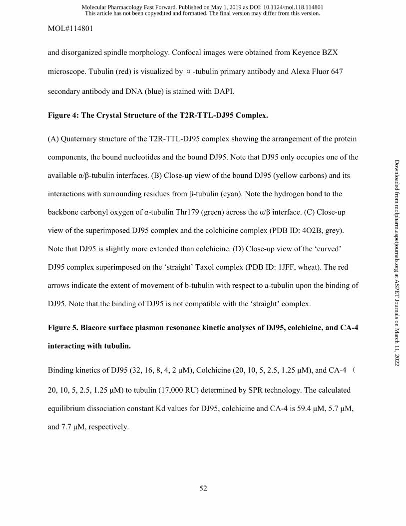
MOL#114801
52
and disorganized spindle morphology. Confocal images were obtained from Keyence BZX
microscope. Tubulin (red) is visualized by α-tubulin primary antibody and Alexa Fluor 647
secondary antibody and DNA (blue) is stained with DAPI.
Figure 4: The Crystal Structure of the T2R-TTL-DJ95 Complex.
(A) Quaternary structure of the T2R-TTL-DJ95 complex showing the arrangement of the protein
components, the bound nucleotides and the bound DJ95. Note that DJ95 only occupies one of the
available α/β-tubulin interfaces. (B) Close-up view of the bound DJ95 (yellow carbons) and its
interactions with surrounding residues from β-tubulin (cyan). Note the hydrogen bond to the
backbone carbonyl oxygen of α-tubulin Thr179 (green) across the α/β interface. (C) Close-up
view of the superimposed DJ95 complex and the colchicine complex (PDB ID: 4O2B, grey).
Note that DJ95 is slightly more extended than colchicine. (D) Close-up view of the ‘curved’
DJ95 complex superimposed on the ‘straight’ Taxol complex (PDB ID: 1JFF, wheat). The red
arrows indicate the extent of movement of b-tubulin with respect to a-tubulin upon the binding of
DJ95. Note that the binding of DJ95 is not compatible with the ‘straight’ complex.
Figure 5. Biacore surface plasmon resonance kinetic analyses of DJ95, colchicine, and CA-4
interacting with tubulin.
Binding kinetics of DJ95 (32, 16, 8, 4, 2 μM), Colchicine (20, 10, 5, 2.5, 1.25 μM), and CA-4 (
20, 10, 5, 2.5, 1.25 μM) to tubulin (17,000 RU) determined by SPR technology. The calculated
equilibrium dissociation constant Kd values for DJ95, colchicine and CA-4 is 59.4 μM, 5.7 μM,
and 7.7 μM, respectively.
This article has not been copyedited and formatted. The final version may differ from this version.Molecular Pharmacology Fast Forward. Published on May 1, 2019 as DOI: 10.1124/mol.118.114801
at ASPE
T Journals on M
arch 11, 2022m
olpharm.aspetjournals.org
Dow
nloaded from

MOL#114801
53
Figure 6. DJ95 in binding and enzyme and uptake assays.
(A) DJ95 control-specific binding as a % inhibition to the binding to a radioactively labeled
ligand specific for each target. (B) DJ95 enzyme based functional a % of control enzyme
activity. Results showing an inhibition or stimulation higher than 50% are considered to
represent significant effects of the test compounds. Graphs are represented as the mean of
duplicate assays ± SD. Screening was performed by Eurofins Cerep-Panlabs.
Figure 7. DJ95 inhibits melanoma tumor growth in vivo.
(A) Plasma concentrations at each timepoint following i.p. injection of DJ95 at 15 mg/kg
concentration shown as individual replicates connected by mean (n=3 per time point). (B) A375
melanoma xenograft model in nude mice. Mice were dosed by i.p. injection 5 times a week for 2
weeks with vehicle solution only or 15 mg/kg DJ95. Mean tumor volume is expressed as %
growth compared to tumor volume at the beginning of the experiment ± SEM (n=6 for treated
group,7 for control). (C) Individual tumor volumes at the end of the experiment shown as mean ±
SD. (D) Mouse weight change throughout the study shown as mean ± SD. (E) Resected tumor
weight at the end of the experiment expressed as mean ± SD. Statistical significance was
determined by student’s t test comparing treatment group to the vehicle control group (*p< 0.05,
**p< 0.01).
Figure 8. DJ95 targets tumor vascular and capillary network formation.
(A) Representative images of HUVEC capillary formation on matrigel. Images were taken after
6 hr incubation with indicated concentrations of DJ95 or colchicine. (B) Quantification of total
tube length using Image J analysis expressed as mean ± SD (n=4). Statistical analysis was
performed using one-way ANOVA comparing each group to the control. (C) Representative
This article has not been copyedited and formatted. The final version may differ from this version.Molecular Pharmacology Fast Forward. Published on May 1, 2019 as DOI: 10.1124/mol.118.114801
at ASPE
T Journals on M
arch 11, 2022m
olpharm.aspetjournals.org
Dow
nloaded from

MOL#114801
54
immunohistochemistry images of CD31 stained tumor sections taken at 10x. (D) Positive stained
area calculated for tumor sections at 20x magnification (5 images per tumor, 3 separate tumors
per group) represented as mean pixels ± SD. Statistical significance was determined by a
student’s t test without correction for multiple comparisons. (**p< 0.01, ***p< 0.001, ****p<
0.0001).
This article has not been copyedited and formatted. The final version may differ from this version.Molecular Pharmacology Fast Forward. Published on May 1, 2019 as DOI: 10.1124/mol.118.114801
at ASPE
T Journals on M
arch 11, 2022m
olpharm.aspetjournals.org
Dow
nloaded from

MOL#114801
55
TABLES
Table 1. Cytotoxic effects of DJ95 against malignant melanoma cancer cell lines.
Treatment IC50
a ± SD (nM) A375 RPMI-7951 WM-164 WM115 SK-MEL-1
DJ95 24.7 ± 4.9 58.9 ± 13.3 68.2 ± 12.4 99.8 ± 16.8 78.7 ± 16.5 Colchicine 10.6 ± 1.8 11.1 ± 0.7 10.1 ± 0.7 9.3 ± 1.0 11.5 ± 2.5 Paclitaxel 2.5 ± 1.0 3.4 ± 0.8 8.0 ± 1.0 3.3 ± 1.6 1.8 ± 0.8
a IC50 values represent the mean ± SD of three independent experiments performed in quadruplicate.
This article has not been copyedited and formatted. The final version may differ from this version.Molecular Pharmacology Fast Forward. Published on May 1, 2019 as DOI: 10.1124/mol.118.114801
at ASPE
T Journals on M
arch 11, 2022m
olpharm.aspetjournals.org
Dow
nloaded from

MOL#114801
56
Table 2. Cytotoxic effects of DJ95 against parental and gene transfected ABCB1-, ABCC1-
or ABCG2-overexpressing cancer cell lines.
Treatment IC50
b ± SD (nM)
HEK293/pcDNA3.1 HEK293/ABCB1 HEK293/ABCC1 HEK293/ABCG2-R482
DJ95 330.0 ± 44.1 314.7 ± 73.1 1213.9 ± 241.6 2869.2 ± 123.3
Paclitaxel 34.6 ± 6.3 3496.0 ± 657.8 >10000 >10000
Colchicine 112.1 ± 41.5 >10000 >10000 >10000
Vincristine 99.4 ± 23.1 >10000 >10000 >10000
Mitoxantrone 108.0 ± 27.5 — — 1209.7 ± 111.4
Cisplatin 2510.1 ± 362.6 2583.1 ± 474.5 2584.1 ± 151.8 2475.3 ± 127.4
b IC50 values represent the mean ± SD of three independent experiments performed in triplicate.
This article has not been copyedited and formatted. The final version may differ from this version.Molecular Pharmacology Fast Forward. Published on May 1, 2019 as DOI: 10.1124/mol.118.114801
at ASPE
T Journals on M
arch 11, 2022m
olpharm.aspetjournals.org
Dow
nloaded from

MOL#114801
57
Table 3. Cytotoxic effects of DJ95 against parental and drug-selected ABCB1-, ABCC1- or
ABCG2-overexpressing cancer cell lines.
Treatment
IC50b ± SD (nM)
KB-3-1 KB-C2 (ABCB1)
KB-CV60 (ABCC1) NCI-H460
NCI-H460/MX20
(ABCG2) DJ95 23.5 ± 2.2 719.5 ± 181.5 35.9 ± 4.5 673.0 ± 111.7 5361.5 ± 645.5
Paclitaxel 9.2 ± 2.1 1918.5 ± 106.3 120.0 ± 14.6 >10000 >10000 Colchicine 23.9 ± 4.9 7445.3 ± 446.3 91.7 ± 7.7 >10000 >10000 Vincristine 1.3 ± 0.2 748.8 ± 99.8 218.6 ± 1.9 >10000 >10000
Mitoxantrone — — — 118.7 ± 40.2 2456.5 ± 420.4 Cisplatin 2024.5 ± 463.7 2871.2 ± 115.3 1796.5 ± 198.8 2226.6 ± 179.5 2870.6 ± 281.7
b IC50 values represent the mean ± SD of three independent experiments performed in triplicate.
This article has not been copyedited and formatted. The final version may differ from this version.Molecular Pharmacology Fast Forward. Published on May 1, 2019 as DOI: 10.1124/mol.118.114801
at ASPE
T Journals on M
arch 11, 2022m
olpharm.aspetjournals.org
Dow
nloaded from
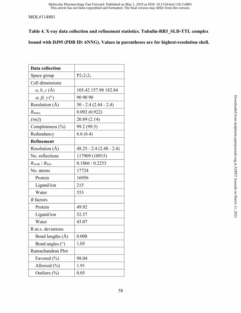
MOL#114801
58
Table 4. X-ray data collection and refinement statistics. Tubulin-RB3_SLD-TTL complex
bound with DJ95 (PDB ID: 6NNG). Values in parentheses are for highest-resolution shell.
Data collection Space group P212121 Cell dimensions a, b, c (Å) 105.42 157.98 182.84 α, β, γ (°) 90 90 90 Resolution (Å) 50 - 2.4 (2.44 - 2.4) Rmeas, 0.092 (0.922) I/σ(I) 20.89 (2.14) Completeness (%) 99.2 (99.5) Redundancy 6.6 (6.4) Refinement Resolution (Å) 48.25 - 2.4 (2.48 - 2.4) No. reflections 117909 (10915) Rwork / Rfree 0.1866 / 0.2253 No. atoms 17724 Protein 16956 Ligand/ion 215 Water 553 B factors Protein 49.92 Ligand/ion 52.57 Water 43.07 R.m.s. deviations Bond lengths (Å) 0.008 Bond angles (°) 1.05 Ramachandran Plot Favored (%) 98.04 Allowed (%) 1.91 Outliers (%) 0.05
This article has not been copyedited and formatted. The final version may differ from this version.Molecular Pharmacology Fast Forward. Published on May 1, 2019 as DOI: 10.1124/mol.118.114801
at ASPE
T Journals on M
arch 11, 2022m
olpharm.aspetjournals.org
Dow
nloaded from

MOL#114801
59
FIGURES
Figure 1.
This article has not been copyedited and formatted. The final version may differ from this version.Molecular Pharmacology Fast Forward. Published on May 1, 2019 as DOI: 10.1124/mol.118.114801
at ASPE
T Journals on M
arch 11, 2022m
olpharm.aspetjournals.org
Dow
nloaded from

MOL#114801
60
Figure 2.
This article has not been copyedited and formatted. The final version may differ from this version.Molecular Pharmacology Fast Forward. Published on May 1, 2019 as DOI: 10.1124/mol.118.114801
at ASPE
T Journals on M
arch 11, 2022m
olpharm.aspetjournals.org
Dow
nloaded from

MOL#114801
61
Figure 3.
This article has not been copyedited and formatted. The final version may differ from this version.Molecular Pharmacology Fast Forward. Published on May 1, 2019 as DOI: 10.1124/mol.118.114801
at ASPE
T Journals on M
arch 11, 2022m
olpharm.aspetjournals.org
Dow
nloaded from

MOL#114801
62
Figure 4.
This article has not been copyedited and formatted. The final version may differ from this version.Molecular Pharmacology Fast Forward. Published on May 1, 2019 as DOI: 10.1124/mol.118.114801
at ASPE
T Journals on M
arch 11, 2022m
olpharm.aspetjournals.org
Dow
nloaded from

MOL#114801
63
Figure 5.
This article has not been copyedited and formatted. The final version may differ from this version.Molecular Pharmacology Fast Forward. Published on May 1, 2019 as DOI: 10.1124/mol.118.114801
at ASPE
T Journals on M
arch 11, 2022m
olpharm.aspetjournals.org
Dow
nloaded from

MOL#114801
64
Figure 6.
This article has not been copyedited and formatted. The final version may differ from this version.Molecular Pharmacology Fast Forward. Published on May 1, 2019 as DOI: 10.1124/mol.118.114801
at ASPE
T Journals on M
arch 11, 2022m
olpharm.aspetjournals.org
Dow
nloaded from

MOL#114801
65
Figure 7.
This article has not been copyedited and formatted. The final version may differ from this version.Molecular Pharmacology Fast Forward. Published on May 1, 2019 as DOI: 10.1124/mol.118.114801
at ASPE
T Journals on M
arch 11, 2022m
olpharm.aspetjournals.org
Dow
nloaded from

MOL#114801
66
Figure 8.
This article has not been copyedited and formatted. The final version may differ from this version.Molecular Pharmacology Fast Forward. Published on May 1, 2019 as DOI: 10.1124/mol.118.114801
at ASPE
T Journals on M
arch 11, 2022m
olpharm.aspetjournals.org
Dow
nloaded from

S-1
Supporting information
Colchicine binding site agent DJ95 overcomes drug resistance and exhibits antitumor efficacy
Authors: Kinsie E. Arnst1, Yuxi Wang2,3, Zi-Ning Lei4, Dong-Jin Hwang1, Gyanendra Kumar5,
Dejian Ma1, Deanna N. Parke6, Qiang Chen2, Jinliang Yang2, Stephen W. White5, Tiffany N.
Seagroves6, Zhe-Sheng Chen4, Duane D. Miller*1, and Wei Li*1
Affiliation: 1Department of Pharmaceutical Sciences, College of Pharmacy, the University of
Tennessee Health Science Center, Memphis, TN. 2State Key Laboratory of Biotherapy and
Cancer Center, Collaborative Innovation Center of Biotherapy, 3 Department of Respiratory
Medicine, West China Hospital, Sichuan University, Chengdu, China. 4Department of
Pharmaceutical Sciences, College of Pharmacy and Health Sciences, St. John’s University,
Queens, NY. 5 Department of Structural Biology, St. Jude Children’s Research Hospital,
Memphis, TN. 6Department of Pathology, the University of Tennessee Health Science Center,
Memphis, TN.

S-2
Supplement Table 1. Multiple reaction-monitoring (MRM) parameters based compound
optimization.
Analyte Transition (m/z) TR(min) DP (V) CE (V) CXP (V)
DJ95 400.7/340.3 5.38 196 39 22
ABI-231 (IS) 378.3/210.2 5.10 186 37 12
TR Retention time; DP, declustering potential; CE, collision energy; CXP, collision cell exit
potential; IS internal standard
Supplement Table 2. Melanoma cell lines contain genomic mutations in the following genes
according to the Sanger COSMIC database: BRAF, CKD4, CTNNB1, CDKN2A, NRAS, PTEN,
TP53, and PPP6C.
Cell line site Reported gene mutations
A375 primary BRAF, CDKN2A, PPP6C
RPMI-7951 metastatic BRAF, CDKN2A, PTEN, TP53
WM-164 metastatic BRAF, CDKN2A, TP53,
WM-115 primary BRAF, NRAS, CDKN2A, PTEN,
SK-MEL-1 primary BRAF, CDK4, CTNNB1, PPP6C
http://cancer.sanger.ac.uk

S-3
Supplement Table 3. Cytotoxic activities of DJ95 against NCI60 human cancer cell lines. GI50,
TGI, and LC50 values were obtained from the NCI-DTP five-dose assay and the mean growth %
was obtained from the one-dose assay.
PANEL/CELL LINE GI50
(NM) TGI (µM) LC50
(µM) MEAN
GROWTH %
Leukemia
CCRF-CEM <10.0 12.3 >100.0 5.3
HL-60(TB) <10.0 <0.01 >100.0 -48.7
K-562 <10.0 2.3 >100.0 -14.4
MOLT-4 <10.0 7.2 >100.0 18.5
RPMI-8226 44.7 5.1 >100.0 -43.0
SR <10.0 12.0 >100.0 -4.9
NSCLC
A549/ATCC 11.2 1.8 7.1 -28.5
EKVX
5.9 40.7 30.9
HOP-62 <10.0 2.7 11.2 -19.6
HOP-92 309.0 5.0 70.8 -8.6
NCI-H226 1174.9 3.0 >100.0 19.3
NCI-H23 <10.0 4.2 81.3 26.1
NCI-H322M <10.0 11.7 44.7 45.0
NCI-460 <10.0 1.0 6.3 2.6
NCI-H522 <10.0
23.4 -59.5
Colon cancer
COLO 205 <10.0 <0.01 3.2 -71.5
HCC-2998 <10.0 1.4 5.1 -19.2
HCT-116 <10.0 1.1 5.5 4.4
HCT-15 <10.0 1.3 6.2 -7.0
HT29 <10.0 1.1 10.7 -37.8
KM12 <10.0 1.5 9.8 3.6
SW-620 <10.0 1.6 5.0 -15.6
CNS cancer
SF-268 <10.0 3.2 60.3 19.3
SF-295 <10.0 <0.01 3.6 -59.4
SF-539 <10.0 <0.01 12.9 -19.5
SNB-19 <10.0 15.8 93.3 34.8
SNB-75 <10.0 6.6 61.7 18.9
U251 <10.0 1.9 8.3 -16.3
Melanoma
LOX IMVI <10.0 1.5 4.5 -67.5
MALME-3M 1288.2 3.2 7.9 21.7
M14 <10.0
5.9 4.3
MDA-MB-435 <10.0 <0.01 >100 5.2
SK-MEL-2 <10.0 3.1 20.4 15.2
SK-MEL-28
2.9 7.9 42.1
SK-MEL-5 <10.0 0.6 3.1 -38.4
UACC-62 <10.0 2.6 8.1 27.4

S-4
Supplement Table 3 continued
Panel/Cell line GI50
(nM) TGI (µM) LC50
(µM) mean growth %
Ovarian cancer
IGROV1 15.1 3.8 61.7 29.5
OVCAR-3 <10.0
4.1 2.2
OVCAR-4 30.9 17.0 56.2 39.3
OVCAR-5 1288.2 12.0 44.7 41.5
OVCAR-8 <10.0 11.2 >100.0 12.4
NCI/ADR-RES <10.0 10.7 >100.0 12.7
SK-OV-3 <10.0 5.6 30.9 1.3
Renal cancer
786–0 <10.0 2.0 9.1 -56.0
ACHN <10.0 3.0 21.4 2.8
CAKI-1 <10.0 11.2 38.9 29.7
SN12C <10.0 10.0 45.7 30.9
TK-10 1122.0 7.9 49.0 10.7
UO-31 <10.0 3.0 35.5 13.6
Prostate Cancer
PC-3 <10.0 4.8 34.7 -0.9
DU-145 <10.0 11.5 38.0 18.2
Breast cancer
MCF7 56.2 2.9 49.0 2.1
MDA-MB-231 <10.0 1.9 30.2 -20.3
HS 578T <10.0 10.2 >100.0 31.1
BT-549 <10.0 2.7 10.5 18.4
T-47D 1258.9 4.4 53.7 30.5
MDA-MB-468 17.8 1.5 6.9 -36.7

S-5
Supplement Table 4. Assay and reference compound for safety screen data
ASSAY REFERENCE COMPOUND
specific
binding
assay
A2A (h) (agonist radioligand) NECA
alpha 1A (h) (antagonist radioligand) WB 4101
alpha 2A (h) (antagonist radioligand) yohimbine
beta 1 (h) (agonist radioligand) atenolol
beta 2 (h) (agonist radioligand) ICI 118551
BZD (central) (agonist radioligand) diazepam
CB1 (h) (agonist radioligand) CP 55940
CB2 (h) (agonist radioligand) WIN 55212-2
CCK1 (CCKA) (h) (agonist radioligand) CCK-8s
D1 (h) (antagonist radioligand) SCH 23390
D2S (h) (agonist radioligand) 7-OH-DPAT
ETA (h) (agonist radioligand) endothelin-1
NMDA (antagonist radioligand) CGS 19755
H1 (h) (antagonist radioligand) pyrilamine
H2 (h) (antagonist radioligand) cimetidine
MAO-A (antagonist radioligand) clorgyline
M1 (h) (antagonist radioligand) pirenzepine
M2 (h) (antagonist radioligand) methoctramine
M3 (h) (antagonist radioligand) 4-DAMP
alpha 4beta 2 (h) (agonist radioligand) nicotine
delta 2 (DOP) (h) (agonist radioligand) DPDPE
kappa (KOP) (agonist radioligand) U 50488
mu (MOP) (h) (agonist radioligand) DAMGO
5-HT1A (h) (agonist radioligand) 8-OH-DPAT
5-HT1B (antagonist radioligand) serotonin
5-HT2A (h) (agonist radioligand) (±)DOI
5-HT2B (h) (agonist radioligand) (±)DOI
5-HT3 (h) (antagonist radioligand) MDL 72222
GR (h) (agonist radioligand) dexamethasone
AR (h) (agonist radioligand) mibolerone
V1a (h) (agonist radioligand) [d(CH2)51,Tyr(Me)2]-AVP
Ca2+ channel (L, dihydropyridine site) (antagonist radioligand) nitrendipine
KV channel (antagonist radioligand) alpha -dendrotoxin
Na+ channel (site 2) (antagonist radioligand) veratridine
NE transporter (h) (antagonist radioligand) protriptyline
DA transporter (h) (antagonist radioligand) BTCP
5-HT transporter (h) (antagonist radioligand) imipramine
enzyme-
based
assay
COX1 (h) diclofenac
COX2 (h) NS 398
PDE3A (h) milrinone
PDE4D2 (h) rolipram
Lck kinase (h) staurosporine
acetylcholinesterase (h) neostigmine

S-6
Supplement Figure 1. Western blot showing the elevated protein concentrations for each of the
drug-selected and gene-transfected cell lines over-expressing ABCB1, ABCC1, and ABCG2.
Primary antibodies against ABCB1 (#P7965, Sigma-Aldrich, St. Louis, MO), ABCC1 (#72202,
Cell Signaling Technology, Danvers, MA), ABCG2 (#MAB4146, Sigma-Aldrich, St. Louis,
MO) and GAPDH (#MA5-15738, Thermo Fisher Scientific, Rockford, IL) were used. HRP-
linked secondary antibodies used were anti-mouse IgG (#7076) and anti-rabbit IgG (#7074) from
Cell Signaling Technology, Danvers, MA.
Supplement Figure 2. Effect of DJ95 on ABCB1 or ABCG2 vanadate-sensitive ATPase
activity. (A) ABCB1-mediated ATP hydrolysis in the presence of DJ95 or positive substrate
paclitaxel (0-10,000 nM). (B) ABCG2-mediated ATP hydrolysis in the presence of DJ95 or
positive substrate topotecan (0-10,000 nM). Data are plotted with mean and error bars depict SD
obtained from three independent experiments.

S-7
Supplement Figure 3. NCI-60 data for DJ95. (A) Sensitivity of NCI cancer cell lines to DJ95.
10 µM of DJ95 was used against cell lines to compare the growth percentage of each cell line
relative to the no-drug control, and relative to the time zero number of cells. The percentage
growth is represented by the horizontal bar, showing both growth inhibition (values between 0
and 100) and lethality (< 0). (B) Average growth percentage for each cancer type represented as
mean ± SEM. (C) Dose-response curves for DJ95 against melanoma cancer cell lines.

S-8
Supplement Figure 4. Scratch assay quantification. A375 and RPMI-7951 migration
represented as total area in scratch channel ± SD. Statistical significance was determined by two-
way ANOVA followed by Dunnett’s multiple comparison test, comparing each treatment group
to the control group at that specific time point. There was no significant difference for the area at
time zero for any group. (*p< 0.05, ****p< 0.0001).

S-9
Supplement Figure 5. Electron density maps for DJ95. (A) 2Fo-Fc map contoured at 1 sigma.
(B) Simulated annealing omit map contoured at 3 sigma.

S-10
Supplement Figure 6. Images obtained from immunohistochemistry CD31 stained tumor
sections at 20x magnification used for quantification of positive stained area for (A) control
group and (B) DJ95 treated group. Five images were obtained from 3 separate tumors per group.
Each row represents a separate tumor. Necrotic tissue area was avoided.