colloidal aggregation: from screening nuisance to
TRANSCRIPT
Nano Today 19 (2018) 188–200
Contents lists available at ScienceDirect
Nano Today
journa l h om epa ge: www.elsev ier .com/ locate /nanotoday
Review
Colloidal aggregation: From screening nuisance to formulation nuance
Ahil N. Ganesha,b, Eric N. Dondersa,b, Brian K. Shoichetc, Molly S. Shoicheta,b,d,∗
a Department of Chemical Engineering and Applied Chemistry, University of Toronto, ON, Canadab Institute of Biomaterials and Biomedical Engineering, University of Toronto, ON, Canadac Department of Pharmaceutical Chemistry, University of California – San Francisco, CA, United Statesd Department of Chemistry, University of Toronto, ON, Canada
a r t i c l e i n f o
Article history:Received 14 November 2017Received in revised form 30 January 2018Accepted 20 February 2018Available online 10 March 2018
Keywords:Colloidal aggregatesSelf-assemblyHigh-throughput screeningProtein adsorptionNanoparticles
a b s t r a c t
It is well known that small molecule colloidal aggregation is a leading cause of false positives in early drugdiscovery. Colloid-formers are diverse and well represented among corporate and academic screeningdecks, and even among approved drugs. Less appreciated is how colloid formation by drug-like com-pounds fits into the wider understanding of colloid physical chemistry. Here we introduce the impactthat colloidal aggregation has had on early drug discovery, and then turn to the physical and thermo-dynamic driving forces for small molecule colloidal aggregation, including the particulate nature of thecolloids, their critical aggregation concentration-governed formation, their mechanism of protein adsorp-tion and subsequent inhibition, and their sensitivity to detergent. We describe methods that have beenused extensively to both identify aggregate-formers and to study and control their physical chemistry.While colloidal aggregation is widely recognized as a problem in early drug discovery, we highlight theopportunities for exploiting this phenomenon in biological milieus and for drug formulation.
© 2018 Elsevier Ltd. All rights reserved.
Contents
Introduction . . . . . . . . . . . . . . . . . . . . . . . . . . . . . . . . . . . . . . . . . . . . . . . . . . . . . . . . . . . . . . . . . . . . . . . . . . . . . . . . . . . . . . . . . . . . . . . . . . . . . . . . . . . . . . . . . . . . . . . . . . . . . . . . . . . . . . . . . . . . . 189Properties of colloidal aggregates . . . . . . . . . . . . . . . . . . . . . . . . . . . . . . . . . . . . . . . . . . . . . . . . . . . . . . . . . . . . . . . . . . . . . . . . . . . . . . . . . . . . . . . . . . . . . . . . . . . . . . . . . . . . . . . . . . . . . . . 189
Colloidal particle formation . . . . . . . . . . . . . . . . . . . . . . . . . . . . . . . . . . . . . . . . . . . . . . . . . . . . . . . . . . . . . . . . . . . . . . . . . . . . . . . . . . . . . . . . . . . . . . . . . . . . . . . . . . . . . . . . . . . . . . . 189Aggregation thermodynamics . . . . . . . . . . . . . . . . . . . . . . . . . . . . . . . . . . . . . . . . . . . . . . . . . . . . . . . . . . . . . . . . . . . . . . . . . . . . . . . . . . . . . . . . . . . . . . . . . . . . . . . . . . . . . . . . . . . . . 189Factors that influence the critical aggregation concentration . . . . . . . . . . . . . . . . . . . . . . . . . . . . . . . . . . . . . . . . . . . . . . . . . . . . . . . . . . . . . . . . . . . . . . . . . . . . . . . . . . . . 192Aggregation of multiple compounds . . . . . . . . . . . . . . . . . . . . . . . . . . . . . . . . . . . . . . . . . . . . . . . . . . . . . . . . . . . . . . . . . . . . . . . . . . . . . . . . . . . . . . . . . . . . . . . . . . . . . . . . . . . . . . 192Macromolecule adsorption onto colloidal aggregates . . . . . . . . . . . . . . . . . . . . . . . . . . . . . . . . . . . . . . . . . . . . . . . . . . . . . . . . . . . . . . . . . . . . . . . . . . . . . . . . . . . . . . . . . . . . 192Detergent reversibility . . . . . . . . . . . . . . . . . . . . . . . . . . . . . . . . . . . . . . . . . . . . . . . . . . . . . . . . . . . . . . . . . . . . . . . . . . . . . . . . . . . . . . . . . . . . . . . . . . . . . . . . . . . . . . . . . . . . . . . . . . . . . 193
Identifying and characterizing colloidal aggregates . . . . . . . . . . . . . . . . . . . . . . . . . . . . . . . . . . . . . . . . . . . . . . . . . . . . . . . . . . . . . . . . . . . . . . . . . . . . . . . . . . . . . . . . . . . . . . . . . . . . 193Light scattering . . . . . . . . . . . . . . . . . . . . . . . . . . . . . . . . . . . . . . . . . . . . . . . . . . . . . . . . . . . . . . . . . . . . . . . . . . . . . . . . . . . . . . . . . . . . . . . . . . . . . . . . . . . . . . . . . . . . . . . . . . . . . . . . . . . . 193Electron microscopy . . . . . . . . . . . . . . . . . . . . . . . . . . . . . . . . . . . . . . . . . . . . . . . . . . . . . . . . . . . . . . . . . . . . . . . . . . . . . . . . . . . . . . . . . . . . . . . . . . . . . . . . . . . . . . . . . . . . . . . . . . . . . . . 193Fluorescence assays . . . . . . . . . . . . . . . . . . . . . . . . . . . . . . . . . . . . . . . . . . . . . . . . . . . . . . . . . . . . . . . . . . . . . . . . . . . . . . . . . . . . . . . . . . . . . . . . . . . . . . . . . . . . . . . . . . . . . . . . . . . . . . . . 193Nuclear magnetic resonance . . . . . . . . . . . . . . . . . . . . . . . . . . . . . . . . . . . . . . . . . . . . . . . . . . . . . . . . . . . . . . . . . . . . . . . . . . . . . . . . . . . . . . . . . . . . . . . . . . . . . . . . . . . . . . . . . . . . . . 194Other methods . . . . . . . . . . . . . . . . . . . . . . . . . . . . . . . . . . . . . . . . . . . . . . . . . . . . . . . . . . . . . . . . . . . . . . . . . . . . . . . . . . . . . . . . . . . . . . . . . . . . . . . . . . . . . . . . . . . . . . . . . . . . . . . . . . . . . 194
Implications of colloidal aggregates. . . . . . . . . . . . . . . . . . . . . . . . . . . . . . . . . . . . . . . . . . . . . . . . . . . . . . . . . . . . . . . . . . . . . . . . . . . . . . . . . . . . . . . . . . . . . . . . . . . . . . . . . . . . . . . . . . . . .195Interactions of colloidal aggregates with proteins . . . . . . . . . . . . . . . . . . . . . . . . . . . . . . . . . . . . . . . . . . . . . . . . . . . . . . . . . . . . . . . . . . . . . . . . . . . . . . . . . . . . . . . . . . . . . . . . 195Membrane transport of colloidal drug aggregates . . . . . . . . . . . . . . . . . . . . . . . . . . . . . . . . . . . . . . . . . . . . . . . . . . . . . . . . . . . . . . . . . . . . . . . . . . . . . . . . . . . . . . . . . . . . . . . . 195Colloidal drug aggregates in cell culture . . . . . . . . . . . . . . . . . . . . . . . . . . . . . . . . . . . . . . . . . . . . . . . . . . . . . . . . . . . . . . . . . . . . . . . . . . . . . . . . . . . . . . . . . . . . . . . . . . . . . . . . . . 196Persistence of colloidal drug aggregates in vivo . . . . . . . . . . . . . . . . . . . . . . . . . . . . . . . . . . . . . . . . . . . . . . . . . . . . . . . . . . . . . . . . . . . . . . . . . . . . . . . . . . . . . . . . . . . . . . . . . . 196
Stabilizing colloidal drug aggregates . . . . . . . . . . . . . . . . . . . . . . . . . . . . . . . . . . . . . . . . . . . . . . . . . . . . . . . . . . . . . . . . . . . . . . . . . . . . . . . . . . . . . . . . . . . . . . . . . . . . . . . . . . . . . . . . . . . . 197Mechanisms of colloid destabilization . . . . . . . . . . . . . . . . . . . . . . . . . . . . . . . . . . . . . . . . . . . . . . . . . . . . . . . . . . . . . . . . . . . . . . . . . . . . . . . . . . . . . . . . . . . . . . . . . . . . . . . . . . . . 197
∗ Corresponding author at: University of Toronto, 160 College Street, Room 514,Toronto, ON M5S 3E1, Canada.
E-mail address: [email protected] (M.S. Shoichet).
https://doi.org/10.1016/j.nantod.2018.02.0111748-0132/© 2018 Elsevier Ltd. All rights reserved.
A.N. Ganesh et al. / Nano Today 19 (2018) 188–200 189
Stabilizing colloids with polymeric excipients . . . . . . . . . . . . . . . . . . . . . . . . . . . . . . . . . . . . . . . . . . . . . . . . . . . . . . . . . . . . . . . . . . . . . . . . . . . . . . . . . . . . . . . . . . . . . . . . . . . . 197Co-aggregation to stabilize drug colloids. . . . . . . . . . . . . . . . . . . . . . . . . . . . . . . . . . . . . . . . . . . . . . . . . . . . . . . . . . . . . . . . . . . . . . . . . . . . . . . . . . . . . . . . . . . . . . . . . . . . . . . . . .198Exploiting protein adsorption to stabilize colloidal aggregates . . . . . . . . . . . . . . . . . . . . . . . . . . . . . . . . . . . . . . . . . . . . . . . . . . . . . . . . . . . . . . . . . . . . . . . . . . . . . . . . . . .198
Conclusions and outlook . . . . . . . . . . . . . . . . . . . . . . . . . . . . . . . . . . . . . . . . . . . . . . . . . . . . . . . . . . . . . . . . . . . . . . . . . . . . . . . . . . . . . . . . . . . . . . . . . . . . . . . . . . . . . . . . . . . . . . . . . . . . . . . . 198Acknowledgements . . . . . . . . . . . . . . . . . . . . . . . . . . . . . . . . . . . . . . . . . . . . . . . . . . . . . . . . . . . . . . . . . . . . . . . . . . . . . . . . . . . . . . . . . . . . . . . . . . . . . . . . . . . . . . . . . . . . . . . . . . . . . . . . . . . 199References . . . . . . . . . . . . . . . . . . . . . . . . . . . . . . . . . . . . . . . . . . . . . . . . . . . . . . . . . . . . . . . . . . . . . . . . . . . . . . . . . . . . . . . . . . . . . . . . . . . . . . . . . . . . . . . . . . . . . . . . . . . . . . . . . . . . . . . . . . . . . 199
Introduction
Drug discovery often begins with screening libraries of overone million molecules to find early compounds that may becomeleads to drug candidates [1,2]. While they remain the most widelyused strategy in pharmaceutical research to discover new disease-related targets, these high-throughput screening (HTS) campaignsare dominated by false-positive “hits” [3–5]. Often, far more timeand resources are spent distinguishing between true and false pos-itives, and prioritizing well-behaved hits for progression, than wasspent developing and executing the HTS in the first place.
Among the most common mechanisms for false-positive hits inHTS is the colloidal aggregation of small molecules, first discov-ered 15 years ago [3] and now widely accepted [6]. Subsequentmechanistic work demonstrated that aggregation occurs via phaseseparation and particle formation when the small molecules arepresent above a compound-specific critical aggregation concentra-tion (CAC) [7]. The resulting colloidal aggregates non-specificallybind proteins to their surface causing local unfolding events, which,in the case of enzymes, result in loss of catalytic activity [7,8].Compound aggregation alone explained the flat structure-activityrelationships and high sensitivity to assay conditions that hadbeen a common feature of the HTS false positives [2,5]. A thirdwidespread feature of these pathological hits, their steep Hill coef-ficients in concentration-response curves, was explained by theaggregates having binding affinities for their target proteins thatwere substantially higher than the concentration of the targets inthe assays [5].
The formation of colloidal particles in biochemical buffers andtheir interactions with biological molecules have had many impli-cations for drug discovery, formulation and activity. While manyof these properties have rendered colloidal aggregates to be con-sidered as nuisance artifacts, they can also be exploited to turncolloidal aggregation into an advantage. The aim of this review is toinform the reader of these unique properties of colloidal aggregates,their implications in biochemical assay and drug development, andcurrent efforts to exploit these properties.
Properties of colloidal aggregates
Many organic small molecules spontaneously self-assemble inaqueous media into nano-sized colloidal aggregates without chem-ical manipulation. These molecules cover a range of chemicalproperties and structures, and include compounds from screen-ing libraries, for which the phenomenon was first characterized,dyes, and even clinically approved drugs [3,9,10] (Table 1 ). Inthe following sections, we highlight properties that make colloidalaggregates unique as nanostructures and quite different from otherself-assembled drug nanoparticles, which have been reviewed else-where [11–13].
Colloidal particle formation
The formation of stable, amorphous, nano-sized particles isa characteristic property of colloidal aggregation [3]. These col-loids have diameters typically between 50 and 1000 nm and form
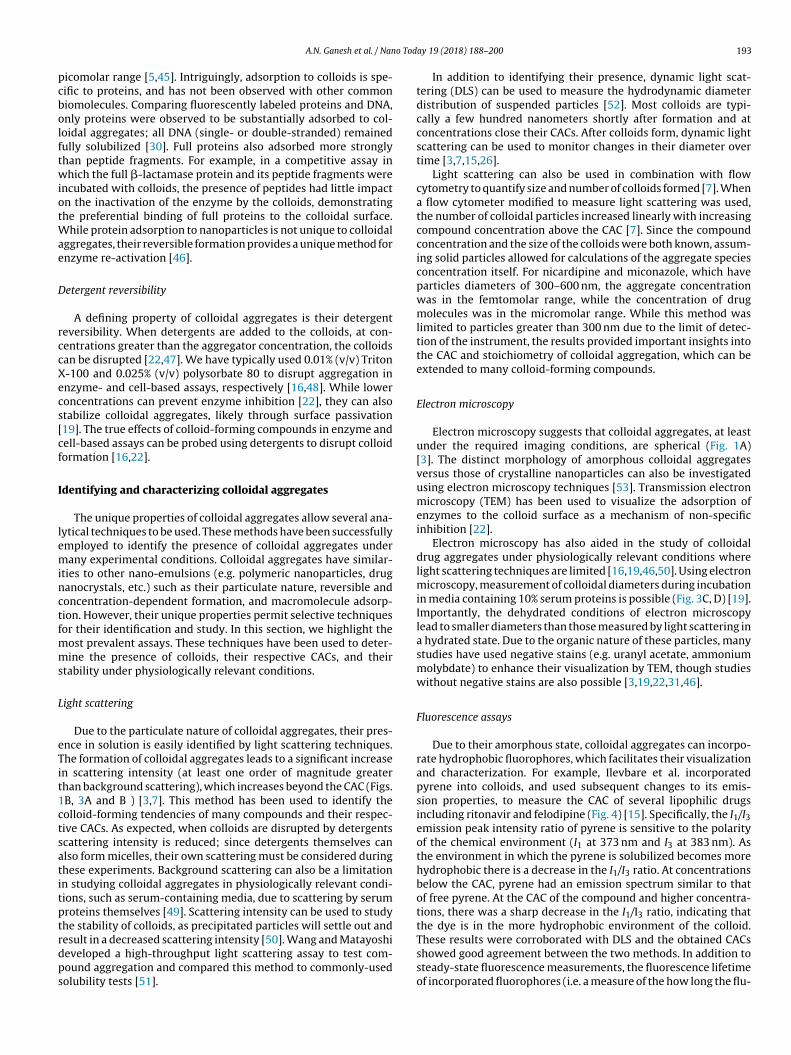
through spontaneous phase separation on addition to aqueousmedia from, often, an organic stock solution such as DMSO (Fig. 1A).Aggregation is concentration dependent; at low concentrations, thecompound is fully solubilized, but as the concentration increases,spontaneous self-assembly occurs at a critical concentration [7].This critical aggregation concentration (CAC, Fig. 1B) appears tocorrespond to the amorphous solubility of the compound [15].Notably, this concentration is higher than the crystal solubility ofthe compound, at least in those cases where the two have beencarefully compared. The CAC is analogous to the critical micelleconcentration for surfactants; like micelles, when diluted belowthe CAC the aggregates will spontaneously disassemble and returnto a monomeric state [3,21]. At least transiently, the concentrationof free molecules that remain in solution is defined by the CAC, evenonce aggregates have formed, as is the case with micelles. (Fig. 1C)[7]. This can be shown by centrifugation, for instance, where the col-loidal aggregates may be spun down and separated from the solublemolecule, with the concentration in the supernatant remainingconstant at the CAC.
Taylor et al. demonstrated that colloidal aggregates are liquid-liquid phase-separated solutions [15], unlike nanocrystals wherethe drug molecules are tightly packed into an organized lattice. Theamorphous nature of colloidal aggregates allows them to interactwith hydrophobic dyes akin to micelles, and unlike nanocrys-tals. Fluorescent probes, such as pyrene, change their emissioncharacteristics depending on the polarity of their environment[23,24]. Such dyes preferentially associate with the hydropho-bic core of micelles and allow measurement of the CMC [23–25].Similarly, these dyes associate with hydrophobic colloids at con-centrations that correlate with the CAC [15,26–29]. The ability offluorophores to incorporate into colloidal aggregates supports theirliquid nature, as dyes are unable to penetrate crystal lattices.
While the actual molecular structure of colloidal aggregatesis poorly understood, some studies have provided a preliminaryglimpse into their structure. By small-angle X-ray scattering, thestructure of the colloidal aggregates fit the expected pair distancedistribution function for a well-packed, rather than a hollow, sphere[30]. This result is supported by the ratio of the radius of gyrationto the hydrodynamic radius from light scattering [30]. Frenkel et al.used a molecular dynamics approach to explore the effect of pH (i.e.percent protonation of the compound) on the formation and struc-ture of rilpivirine aggregates [31]. Additional studies are neededto fully understand the molecular organization (if any) of colloidalaggregates.
Aggregation thermodynamics
The self-assembly of molecules into colloidal aggregates isdriven by their relative inability to form energetically favorableinteractions with water [32]. Practically, these aggregates formwhen water is added to a hydrophobic aggregator dispersed ina water-miscible organic solvent (e.g. DMSO) [7,32], or blendedin a water-soluble polymer matrix [15,33,34]. As shown in Fig. 2,the aggregator is no longer soluble in the continuous phase afteraddition of water. In the unstable area, the mixture spontaneouslyseparates to generate a liquid or glassy colloidal phase along with

190 A.N. Ganesh et al. / Nano Today 19 (2018) 188–200
Table 1Several aggregate-forming small molecules. Critical aggregation concentrations for these compounds are provided for the aqueous conditions listed. Error (standard deviation)is included when known.
Compound (Indication) Structure MW (g/mol) CAC (�M) Aqueous Conditions
Brazilin (natural product) [14] 286.3 57 ± 7 50 mM potassium phosphate, pH 7
Cinnarizine (anti-histamine) [7] 368.5 7 ± 3 50 mM potassium phosphate, pH 7
Clotrimazole (anti-fungal) [15] 344.8 15.0 ± 0.3 100 mM potassium phosphate, pH 10
Crizotinib (anti-neoplastic, non-small cell lung cancer) [16] 450.3 19.3 50 mM potassium phosphate, pH 7
Curcumin (natural product) [14] 368.4 17 ± 0.44 50 mM potassium phosphate, pH 7
Danazol (synthetic steroid) [17] 337.5 39 10 mM phosphate buffer, pH 6.8
Emodin (natural product) [14] 270.2 28 ± 2 50 mM potassium phosphate, pH 7
Evacetrapib (cholesterylester transfer protein inhibitor) [18] 638.7 0.8 50 mM sodium phosphate, pH 6.8
Felodipine (calcium channel blocker) [15] 384.3 26 ± 1 50 mM sodium phosphate, pH 6.8
Fulvestrant (anti-neoplastic, breast cancer) [16] 606.8 0.5 50 mM potassium phosphate, pH 7
Miconazole (anti-fungal) [7] 416.1 3 ± 2 50 mM potassium phosphate, pH 7
A.N. Ganesh et al. / Nano Today 19 (2018) 188–200 191
Table 1 (Continued)
Compound (Indication) Structure MW (g/mol) CAC (�M) Aqueous Conditions
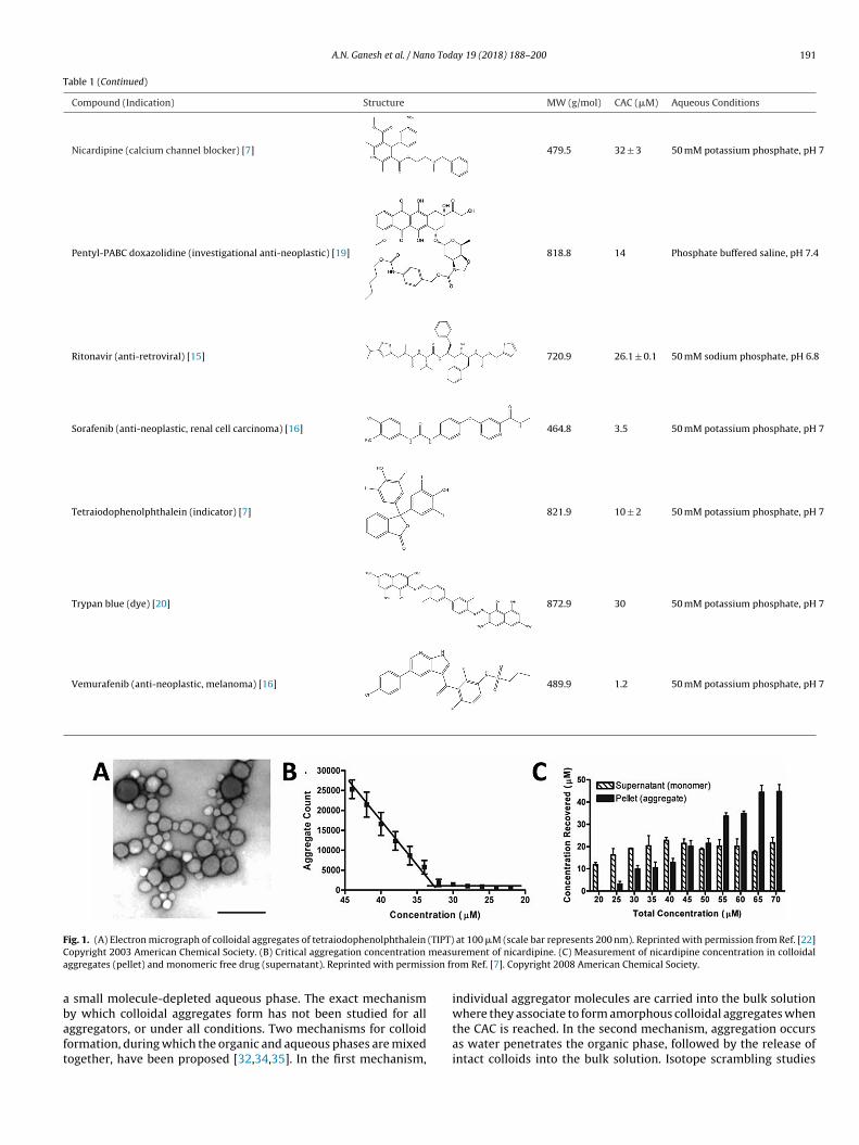
Nicardipine (calcium channel blocker) [7] 479.5 32 ± 3 50 mM potassium phosphate, pH 7
Pentyl-PABC doxazolidine (investigational anti-neoplastic) [19] 818.8 14 Phosphate buffered saline, pH 7.4
Ritonavir (anti-retroviral) [15] 720.9 26.1 ± 0.1 50 mM sodium phosphate, pH 6.8
Sorafenib (anti-neoplastic, renal cell carcinoma) [16] 464.8 3.5 50 mM potassium phosphate, pH 7
Tetraiodophenolphthalein (indicator) [7] 821.9 10 ± 2 50 mM potassium phosphate, pH 7
Trypan blue (dye) [20] 872.9 30 50 mM potassium phosphate, pH 7
Vemurafenib (anti-neoplastic, melanoma) [16] 489.9 1.2 50 mM potassium phosphate, pH 7
Fig. 1. (A) Electron micrograph of colloidal aggregates of tetraiodophenolphthalein (TIPT) at 100 �M (scale bar represents 200 nm). Reprinted with permission from Ref. [22]Copyright 2003 American Chemical Society. (B) Critical aggregation concentration measurement of nicardipine. (C) Measurement of nicardipine concentration in colloidalaggregates (pellet) and monomeric free drug (supernatant). Reprinted with permission from Ref. [7]. Copyright 2008 American Chemical Society.
a small molecule-depleted aqueous phase. The exact mechanismby which colloidal aggregates form has not been studied for allaggregators, or under all conditions. Two mechanisms for colloidformation, during which the organic and aqueous phases are mixedtogether, have been proposed [32,34,35]. In the first mechanism,
individual aggregator molecules are carried into the bulk solutionwhere they associate to form amorphous colloidal aggregates whenthe CAC is reached. In the second mechanism, aggregation occursas water penetrates the organic phase, followed by the release ofintact colloids into the bulk solution. Isotope scrambling studies

192 A.N. Ganesh et al. / Nano Today 19 (2018) 188–200
Fig. 2. Schematic phase diagram illustrating the formation of amorphous colloids,and their ultimate precipitation into crystals.
have supported the first mechanism for the dissolution of polymer-dispersed aggregator with 10% and 20% aggregator loadings [35];however, systems with higher loading behave in a manner moreconsistent with the second mechanism, where aggregation occurswithin the polymer matrix upon exposure to water and colloidsare released as the matrix dissolves [34]. In reality, these twomechanisms are extremes of a single continuum; phase separationwill occur whenever local supersaturation is achieved during massexchange between organic and aqueous phases. Since phase sep-aration will occur when the local composition enters the unstableregion, the aggregator concentration in the organic phase (point1 on phase diagram of Fig. 2) and the solubility parameters ofthe aggregator and organic matrix (which influence the shape ofthe unstable and metastable regions) likely dictate where phaseseparation will occur [34]. When the aggregator concentration islow, phase separation occurs only after a relatively large amount ofwater has penetrated the organic phase as aggregator-in-polymerformulations dissolve, resulting in phase separation that moreclosely follows the first mechanism [35]. Conversely, when theaggregator concentration is high, phase separation occurs after onlya small amount of water diffuses into the organic phase, and thesecond mechanism is more closely followed [34].
Factors that influence the critical aggregation concentration
The CAC of an aggregator is determined by both its intrinsicproperties and the conditions of the continuous phase. Eq. (1)estimates the CAC as a function of the crystal solubility (Csat),enthalpy of fusion (�Hf), melting temperature (Tm), temperature(T), gas constant (R), and a correction factor that accounts forthe aggregator-rich phase containing solutes such as water (exp(-I(a2))) [15,36]. This approach generally yields a good approximationof the CAC; more rigorous methods are more predictive when thedegree of undercooling (Tm − T) is high [29].
CAC = Csat exp
(�Hf (Tm − T)
RT2m
)exp (−I (a2)) (1)
For a single-aggregator system, the enthalpy of fusion, melt-ing temperature, and correction factor are static and intrinsic toeach aggregator. However, the CAC is also affected by extrinsicfactors. For example, the CAC can be indirectly altered by factorsthat affect the crystal solubility (Csat), as is evident from Eq. (1).This property, while heavily dependent on the nature of the aggre-gator, is strongly affected by the solvent conditions [3,15,32]. For
example, crystal solubility is increased with increasing tempera-ture, which increases the CAC; however, some systems exhibit aminimum CAC at temperatures between 20 and 40 ◦C [15], whichmay arise from the temperature term in the first exponential ofEq. (1). Additionally, adding salts to the aqueous phase reduces thesolubility of relatively non-polar aggregators, and the CAC will bereduced, similar to self-assembled micelle systems [37,38]. Con-versely, adding an organic solvent that is miscible with water (e.g.ethanol, DMSO or THF) will increase the aggregator solubility, andthe CAC will increase. Addition of solubilizing excipients such ascyclodextrins or surfactants can greatly increase the effective CACof an aggregator because the aggregator partitions with the solu-bilizer, reducing the concentration of aggregator in the continuousphase [22,26,35,39,40]. Similarly, changing the pH can appear tochange the CAC of an aggregator with ionizable groups [31,41]. Thisphenomenon is often due to conversion of the neutral aggregatorto a generally more soluble charged species. In pH conditions thatfavour the charged species, a higher overall aggregator concentra-tion is needed for the neutral aggregator to reach its CAC; however,the true value of the CAC is unaffected by pH [28]. Practically, thisbehavior means that the observed CAC of an aggregator dependson the composition and pH of the media.
Aggregation of multiple compounds
When multiple aggregators are present in a solution they mayco-aggregate to form multi-compound colloidal aggregates, if theyare miscible in the amorphous state [40,42]. Interestingly, thesemixtures can form aggregates even when each aggregator is belowits individual CAC; here, formation of aggregates occurs when the“compound load” exceeds a critical threshold [40,42,43]. Above thismixed CAC, different ratios of each aggregator will produce colloidswith different compositions. In colloids of multiple aggregators thatare completely miscible, the concentration of each compound in thecontinuous phase (i.e. the CAC) depends on their mole fraction inthe mixture, as given by Eq. (2) [42]:
CAC (x1) = CAC◦1x1 (2)
where the CAC1◦ is the CAC of the compound and x1 is the
mole fraction of the compound. For example, if an aggregatorcomprises 50 mol% of the colloidal phase (x1 = 0.5), its concen-tration in the continuous phase will be 50% of its individualCAC. Conversely, when multiple completely immiscible aggrega-tors are combined their concentration in the continuous phase(CAC) remains unchanged. These changes to the CAC of each aggre-gator likely originate from the correction term of Eq. (1), sincethis term accounts for other compounds that are incorporated intothe colloid. In some cases, hydrophobic non-aggregators can beincorporated into colloids of another compound, similar to a solutepartitioning between two immiscible liquid phases. For example,fluorescent dyes can be non-covalently incorporated into colloidaldrug aggregates to enable their characterization (discussed below)[15,19,44].
Macromolecule adsorption onto colloidal aggregates
At least partly due to their high surface area, and perhaps to theapolar nature of that surface, colloidal aggregates sequester macro-molecules such as proteins, as illustrated in the following examples.The interaction of colloids with proteins was first observed throughtheir non-specific and time-dependent enzyme inhibition [22].Enzymes adsorb onto the surface of colloids and are partiallyunfolded, resulting in a loss of enzymatic activity [8]. The bindingof proteins to the colloid is driven by surface interactions betweenthe colloid and proteins, and often has dissociation constants in the
A.N. Ganesh et al. / Nano Today 19 (2018) 188–200 193
picomolar range [5,45]. Intriguingly, adsorption to colloids is spe-cific to proteins, and has not been observed with other commonbiomolecules. Comparing fluorescently labeled proteins and DNA,only proteins were observed to be substantially adsorbed to col-loidal aggregates; all DNA (single- or double-stranded) remainedfully solubilized [30]. Full proteins also adsorbed more stronglythan peptide fragments. For example, in a competitive assay inwhich the full �-lactamase protein and its peptide fragments wereincubated with colloids, the presence of peptides had little impacton the inactivation of the enzyme by the colloids, demonstratingthe preferential binding of full proteins to the colloidal surface.While protein adsorption to nanoparticles is not unique to colloidalaggregates, their reversible formation provides a unique method forenzyme re-activation [46].
Detergent reversibility
A defining property of colloidal aggregates is their detergentreversibility. When detergents are added to the colloids, at con-centrations greater than the aggregator concentration, the colloidscan be disrupted [22,47]. We have typically used 0.01% (v/v) TritonX-100 and 0.025% (v/v) polysorbate 80 to disrupt aggregation inenzyme- and cell-based assays, respectively [16,48]. While lowerconcentrations can prevent enzyme inhibition [22], they can alsostabilize colloidal aggregates, likely through surface passivation[19]. The true effects of colloid-forming compounds in enzyme andcell-based assays can be probed using detergents to disrupt colloidformation [16,22].
Identifying and characterizing colloidal aggregates
The unique properties of colloidal aggregates allow several ana-lytical techniques to be used. These methods have been successfullyemployed to identify the presence of colloidal aggregates undermany experimental conditions. Colloidal aggregates have similar-ities to other nano-emulsions (e.g. polymeric nanoparticles, drugnanocrystals, etc.) such as their particulate nature, reversible andconcentration-dependent formation, and macromolecule adsorp-tion. However, their unique properties permit selective techniquesfor their identification and study. In this section, we highlight themost prevalent assays. These techniques have been used to deter-mine the presence of colloids, their respective CACs, and theirstability under physiologically relevant conditions.
Light scattering
Due to the particulate nature of colloidal aggregates, their pres-ence in solution is easily identified by light scattering techniques.The formation of colloidal aggregates leads to a significant increasein scattering intensity (at least one order of magnitude greaterthan background scattering), which increases beyond the CAC (Figs.1B, 3A and B ) [3,7]. This method has been used to identify thecolloid-forming tendencies of many compounds and their respec-tive CACs. As expected, when colloids are disrupted by detergentsscattering intensity is reduced; since detergents themselves canalso form micelles, their own scattering must be considered duringthese experiments. Background scattering can also be a limitationin studying colloidal aggregates in physiologically relevant condi-tions, such as serum-containing media, due to scattering by serumproteins themselves [49]. Scattering intensity can be used to studythe stability of colloids, as precipitated particles will settle out andresult in a decreased scattering intensity [50]. Wang and Matayoshideveloped a high-throughput light scattering assay to test com-pound aggregation and compared this method to commonly-usedsolubility tests [51].
In addition to identifying their presence, dynamic light scat-tering (DLS) can be used to measure the hydrodynamic diameterdistribution of suspended particles [52]. Most colloids are typi-cally a few hundred nanometers shortly after formation and atconcentrations close their CACs. After colloids form, dynamic lightscattering can be used to monitor changes in their diameter overtime [3,7,15,26].
Light scattering can also be used in combination with flowcytometry to quantify size and number of colloids formed [7]. Whena flow cytometer modified to measure light scattering was used,the number of colloidal particles increased linearly with increasingcompound concentration above the CAC [7]. Since the compoundconcentration and the size of the colloids were both known, assum-ing solid particles allowed for calculations of the aggregate speciesconcentration itself. For nicardipine and miconazole, which haveparticles diameters of 300–600 nm, the aggregate concentrationwas in the femtomolar range, while the concentration of drugmolecules was in the micromolar range. While this method waslimited to particles greater than 300 nm due to the limit of detec-tion of the instrument, the results provided important insights intothe CAC and stoichiometry of colloidal aggregation, which can beextended to many colloid-forming compounds.
Electron microscopy
Electron microscopy suggests that colloidal aggregates, at leastunder the required imaging conditions, are spherical (Fig. 1A)[3]. The distinct morphology of amorphous colloidal aggregatesversus those of crystalline nanoparticles can also be investigatedusing electron microscopy techniques [53]. Transmission electronmicroscopy (TEM) has been used to visualize the adsorption ofenzymes to the colloid surface as a mechanism of non-specificinhibition [22].
Electron microscopy has also aided in the study of colloidaldrug aggregates under physiologically relevant conditions wherelight scattering techniques are limited [16,19,46,50]. Using electronmicroscopy, measurement of colloidal diameters during incubationin media containing 10% serum proteins is possible (Fig. 3C, D) [19].Importantly, the dehydrated conditions of electron microscopylead to smaller diameters than those measured by light scattering ina hydrated state. Due to the organic nature of these particles, manystudies have used negative stains (e.g. uranyl acetate, ammoniummolybdate) to enhance their visualization by TEM, though studieswithout negative stains are also possible [3,19,22,31,46].
Fluorescence assays
Due to their amorphous state, colloidal aggregates can incorpo-rate hydrophobic fluorophores, which facilitates their visualizationand characterization. For example, Ilevbare et al. incorporatedpyrene into colloids, and used subsequent changes to its emis-sion properties, to measure the CAC of several lipophilic drugsincluding ritonavir and felodipine (Fig. 4) [15]. Specifically, the I1/I3emission peak intensity ratio of pyrene is sensitive to the polarityof the chemical environment (I1 at 373 nm and I3 at 383 nm). Asthe environment in which the pyrene is solubilized becomes morehydrophobic there is a decrease in the I1/I3 ratio. At concentrationsbelow the CAC, pyrene had an emission spectrum similar to thatof free pyrene. At the CAC of the compound and higher concentra-tions, there was a sharp decrease in the I1/I3 ratio, indicating thatthe dye is in the more hydrophobic environment of the colloid.These results were corroborated with DLS and the obtained CACsshowed good agreement between the two methods. In addition tosteady-state fluorescence measurements, the fluorescence lifetimeof incorporated fluorophores (i.e. a measure of the how long the flu-

194 A.N. Ganesh et al. / Nano Today 19 (2018) 188–200
Fig. 3. Representative auto-correlation functions for (A) colloid-forming compound and (B) non-aggregator. Reprinted with permission from Ref. [22]. Copyright 2002American Chemical Society. (C) Transmission electron micrographs of colloidal drug aggregates during incubation in serum-containing media. Colloids of pentyl-PABCdoxazolidine were incubated in 10% fetal bovine serum for 0, 24 and 48 h. Negative staining of colloids with uranyl acetate. (D) Quantification of colloid diameter based onelectron micrographs. Reprinted with permission from Ref. [19] Copyright 2017 American Chemical Society.
Fig. 4. Measurement of CAC by fluorescence properties of pyrene upon inclusioninto colloidal drug aggregates of ritonavir. The onset of aggregation corresponds tothe decrease in I1/I3 fluorescence ratio as the pyrene enters a more hydrophobicenvironment. Reprinted with permission from Ref. [15]. Copyright 2013 AmericanChemical Society.
orophore remains in the excited state) can also be used to study thestate of aggregates [54].
Since these fluorophores can only incorporate into particles ofamorphous nature, they can also be used to study the onset ofcrystallization [26]. As crystallization occurs, the probe becomesexcluded from the crystal lattice of the aggregator and returns tothe polar environment of the aqueous phase. The incorporationof fluorophores allows colloids to be studied in complex biologi-cal environments mimicking those in vivo [38,55]. The stability ofcolloidal aggregates in serum containing media and their interac-tions with cells has been studied using fluorescence measurements[19,50].
Nuclear magnetic resonance
Nuclear magnetic resonance (NMR) is commonly used to studythe chemical properties of many materials. Recently LaPlante et al.used 1H-NMR to study the onset of colloidal aggregation [56,57].While fully solubilized small molecules have sharp peaks due totheir fast tumbling, precipitates tumble too slowly to resonate [58].Colloidal aggregates are an intermediate state where tumbling isfast enough for resonance to occur, but their presence leads tochanges in local magnetic fields and chemical environments. Withsingle molecules, only the peak intensity changes as a function ofconcentration; however, once aggregation has occurred, increas-ing concentrations result in changes to peak number, shape, andshifts. As is characteristic of colloidal aggregation, upon the addi-tion of detergents, peak properties return to those of solubilizedsmall molecule solutions. 1H-NMR has also been used to study theinteractions between stabilizers and colloidal aggregates [59].
While these methods corroborate other methods, they are lim-ited in their application to aggregates of small sizes (<200 nmdiameters) as the tumbling of large aggregates is too slow to res-onate on NMR timescales. Additionally, the limit of detection is onlyaround 12 �M [57]; thus, this method is not useful for measuringthe CACs of compounds below this limit, but can still be useful foridentifying the presence of aggregates at higher concentrations.
Other methods
While the four techniques described above have been themost extensively used to study and characterize colloidal aggre-gates, other techniques have also been used. For example,surface plasmon resonance has been applied to study colloidalaggregation interactions with proteins [45]. Flow-cell surfacesmodified with proteins were used to study their interactionswith colloids and could reproduce concentration-dependence anddetergent-reversible properties of colloidal aggregates. Polarizedlight microscopy has been used to support the non-crystallinenature of phase-separated drug solutions where aggregates werefound to be non-birefringent [15,60]. Size exclusion chromatog-

A.N. Ganesh et al. / Nano Today 19 (2018) 188–200 195
Fig. 5. Centrifugation-based identification of colloid-bound protein. In the presenceof colloids (inhib), enrichment of the enzyme (�-lactamase) is observed in the pel-leted colloid. Protein enrichment is not observed when colloids are disrupted withdetergent or in the presence of a non-aggregating specific inhibitor. Reprinted withpermission from Ref. [22]. Copyright 2003 American Chemical Society.
raphy has been commonly used for the study of self-assembledpolymeric nanoparticle stability in physiologically relevant media[61,62]. This technique also allows for the separation of serumproteins from fluorophore-labelled colloidal particles to study thestability of colloidal drug aggregates in serum-containing media[19,50].
Implications of colloidal aggregates
In addition to their interesting physical properties, colloidalaggregates have unique interactions with biological environments.As previously mentioned,their interactions with proteins can resultin false hits in HTS. Furthermore, their stability in the presence ofhigh-protein milieus impacts both in vitro and in vivo analyses. Pro-teins are abundant in biological systems, where their interactionswith other macromolecules drive important biochemical pathwaysand physical transport phenomena. Understanding the interactionsof colloids with proteins and cells is key to understanding in vitroand in vivo data.
Interactions of colloidal aggregates with proteins
A characteristic of colloidal aggregates is their strong surfaceadsorption of proteins. Direct association of colloids with proteinshas been shown by centrifugation of solutions with both colloidsand proteins (Fig. 5), where the protein is concentrated in the col-loid pellet [22,46,50]. TEM imaging of the colloid-protein complexhas also confirmed their direct association [22]. Colloids are stablein high-protein milieus and even form in the presence of proteins[16,63].
In the case of enzymes, adsorption to the colloid surface typicallyleads to a loss of catalytic activity. This phenomenon is non-specificand colloid-forming compounds inhibit many unrelated enzymesat micromolar concentrations [3,14,48]. Enzyme inhibition is typ-ically time-dependent and partly reversible, as demonstrated byadsorption kinetics studies. Liu et al. showed that initial rates of thereaction were inhibited by aggregates, and were suggestive of non-competitive inhibition wherein the aggregate binds to both freeenzyme and the enzyme-substrate complex [64]. Upon disruptionof the colloids (either by dilution or solubilization with detergents),the majority of enzyme activity returns [22]. Intriguingly, colloidalaggregates can actually stably sequester enzymes, preserving theiractivity until the colloid is disrupted [46].
The origins of protein inhibition by colloidal aggregates appearsto be sequestration followed by partial unfolding, though theimportance of the second, unfolding step, remains to be fully
determined. The occurrence of partial enzyme unfolding hasbeen demonstrated in several ways [8,63]. First, incubation of a�-lactamase-colloid complex with the irreversible inhibitor mox-alactam led to no observable effect on the reactivated enzymeafter colloid disruption, indicating that there is no significantexchange between bound and free enzyme. Second, binding of �-lactamase to colloids followed by deuterium-hydrogen exchange,led to increased incorporation of deuterium into the enzyme,as measured by mass spectroscopy after protease digestion.Such increased incorporation into the peptide backbone suggestsincreased accessibility to the solvent due to at least local unfolding.Third, �-lactamase bound to colloids was much more susceptibleto trypsin degradation than was the free enzyme, further support-ing denaturation of the enzyme on colloid binding. Because theenzyme regains much of its activity rapidly on colloid disruption,within the dead-time of a spectrophotometric assay, it seems likelythat the unfolding that the enzyme suffers on the colloids surface islocal and transient. Consistent with this view, antibodies adsorbedto the surface of colloidal aggregates remain able to bind to theirtarget receptor, indicating that the antigen-binding region can stilladopt an active conformation [50].
Based on particle counting methods combined with enzymeactivity assays, Coan et al. concluded that colloidal particles hadsufficient surface area to adsorb all the protein used in their study,but this observation does not rule out protein absorption into thecolloid core [7]. While the molar ratio of protein to colloid-formingcompounds in these systems may be on the order of 1:1000, theratio of enzyme to colloidal particle is much higher since eachparticle contains millions of drug molecules. This higher ratio ofenzyme to colloid also makes it possible for the particle surfaceto become saturated. For example, pre-incubation of colloids withalbumin significantly reduces enzyme inhibition because albuminadsorbs to the colloid surface, leaving less surface area for enzymes[3]. The colloidal surface may also be saturated with other macro-molecules, such as surfactants or polymers; in these cases, proteinadsorption is also greatly reduced [19]. Notably, the addition of pro-tein to the already formed colloid-enzyme complex neither freesadsorbed protein nor restores catalytic activity [63], likely due tothe slow dissociation of already bound enzyme (picomolar dissoci-ation constants).
Membrane transport of colloidal drug aggregates
Drug transport across cell membranes is key to efficacy. Study-ing membrane transport of drugs using a standard diffusion cell,with the donor and receiver chambers separated by a semiper-meable membrane, Taylor et al. found that when colloidal drugaggregates form, there is an upper limit for flux across the syntheticmembrane (Fig. 6) [65]. When the donor chamber concentrationwas below the CAC, they found that the flux of felodipine increasedlinearly with increasing concentrations. However, when the donorcell concentration exceeded the CAC, the flux of drug remained con-stant over all concentrations tested. This observation supports theformation of colloidal drug aggregates, as any drug above the CACself-assembles into particles, which cannot cross the membrane.Thus, the effective drug concentration that drives diffusion is lim-ited at the CAC; only the non-colloidal drug amount is able to diffuseinto the receiver chamber, which leads to a constant flux when thetotal drug concentration is greater than the CAC. As diffusion occurs,the drug in the continuous phase is replenished by drug within thecolloidal aggregates and thus flux is maintained over time.
Due to the reversible nature of colloidal aggregation, the pres-ence of solubilizing excipients also influences the diffusive fluxacross membranes. When only low concentrations of micellardetergents are present, the thermodynamic activity of the drugand therefore the diffusive flux remain constant. When an excess of

196 A.N. Ganesh et al. / Nano Today 19 (2018) 188–200
Fig. 6. (A) Felodipine concentration in the receiver compartment over time and (B) diffusive flux profiles demonstrate significant transition upon aggregate formation at5 �g/mL. Reprinted with permission from Ref. [65]. Copyright 2014 Wiley-VCH.
detergents is used to disrupt colloids, drug molecules partition withthese detergents and there is a reduction in flux due to a decreasein the concentration of free, non-micelle-bound drug to below theCAC [39]. A similar observation is made when the aqueous environ-ment itself affects the aggregation properties of the compounds.For example, Raina et al. observed a difference in diffusive fluxof felodipine in phosphate buffer versus simulated intestinal fluid(SIF) [39]. Constant flux above a certain concentration was observedin both buffers; however, the concentration at which this plateauwas observed was significantly higher in SIF, which is indicative ofa higher CAC in this medium. While these studies have importantimplications in the context of oral drug delivery, the role of activetransport processes and presence of biomacromolecules, such asproteins and lipids, remain sparsely studied.
Colloidal drug aggregates in cell culture
Colloidal aggregates are stable in high-protein milieus, and itis perhaps unsurprising that their presence would have an impacton drug activity in cell culture assays [16,20]. For instance, whenaggregating chemotherapeutic compounds reach their CAC valuesand adopt a colloidal form in cell culture, a substantial decrease(in some cases a total loss) of cytotoxic activity is observed. Con-versely, when a free drug monomer population is maintained,through the addition of detergents, the activity of the drug returns.The concentration at which loss of drug activity occurs coin-cides with the CAC of the compounds [20]. In contrast to theexpected monotonic sigmoidal dose-response curve observed formany chemical inhibitors, a “bell-shaped” dose response curve wasobserved for many colloidal aggregators (Fig. 7A). “Bell-shaped”curves are common in the literature, and are typically explainedby the engagement of multiple cellular pathways. Undoubtedlythis explanation holds for many molecules, but for at least somemolecules these unusual curves will reflect the formation of col-loidal aggregates [20].
The interactions between colloid-forming dyes and cells wereinvestigated to further understand the mechanism of drug activityloss (Fig. 7B, C) [20]. When colloidal dye particles were incubatedwith cells, little to no fluorescence was observed within the cell,indicating that colloids were not internalized by cells. However,upon disruption of the dye colloids with detergents, solubilizeddye molecules could freely cross the cell membrane. Permeabilizedmembranes allowed both colloidal and monomeric dye solutionsto enter cells. This observation suggests that the loss of drug activ-ity in colloidal formulations is due to the inability of drug colloidsto cross cell membranes. Consistent with previous work investi-
Fig. 7. (A) “Bell-shaped” dose-response curve of the colloid-forming anti-neoplasticdrug, crizotinib. Upon colloid formation, loss of anti-proliferative activity isobserved. Intracellular fluorescence after incubation with (B) colloidal and (C)monomeric formulations of Evans blue shows inability of colloids to permeate cellmembrane. Reprinted with permission from Ref. [20]. Copyright 2014 AmericanChemical Society.
gating nanoparticle-cell interactions, these results suggest that, inhigh-protein milieus, the strong affinity of the colloid surface forproteins leads to immediate protein adsorption. This formation ofa protein corona prevents interactions between the colloid and cellsurface, thus limiting entry of the drug into cells [66–68].
In recent studies, we have found that colloids can be modifiedwith proteins that are recognized by cell surface receptors, therebypromoting cellular uptake [50]. Colloids were passivated with aprotein corona comprising the targeting antibody trastuzumab,which is specific to human epidermal growth factor receptor 2(HER2) antigens. Even in the presence of serum proteins, these col-loids were selectively internalized by HER2-overexpressing cancercells but not by cells that had low expression of HER2. Conversely,colloids coated with a non-specificIgG antibody were not internal-ized by either cell type.
Persistence of colloidal drug aggregates in vivo
Based on expected gastric drug concentrations and the diver-sity of conditions under which colloidal aggregation occurs, it isnot surprising that colloidal drug aggregates are in fact present invivo. Doak et al. observed that many compounds formed colloidsat concentration relevant to in vivo dosing regimens in simulatedgastric environments in vitro [69]. Work by Frenkel et al. suggeststhat not only do colloids form and persist in the gastrointestinaltract, but their presence also impacts the bioavailability of thesecompounds in vivo [31,70].
A.N. Ganesh et al. / Nano Today 19 (2018) 188–200 197
Frenkel et al. investigated the colloid-forming properties of anumber of non-nucleoside reverse transcriptase inhibitors (NNR-TIs) which have known pharmacokinetic parameters in rodentmodels and humans [31]. They identified a number of NNRTIsthat form colloidal particles in simulated gastric environments atconcentrations similar to those expected in the gastrointestinaltract after oral dosing. They classified these compounds into twogroups based on aggregate size: small particles (60–220 nm diam-eters) and large particles (>500 nm diameters). They found goodcorrelations between this classification of compounds and theirknown pharmacokinetic parameters. For example, compoundsthat formed aggregates with small diameters had good adsorp-tion/bioavailability parameters (AUC > 5 �g h/mL) while those thatformed large aggregates had poor bioavailability (AUC < 1 �g h/mL).The authors hypothesized that the smaller aggregates wereabsorbed by M cells of the Peyer’s patch and then entered sys-temic circulation via lymphatic circulation. This result is consistentwith other studies investigating nanoparticle formulations for oraldelivery [71,72]. In contrast, larger aggregates appeared to haveprecipitated, thus limiting the bioavailability of the drug.
Since changes in pH play an important role in gastrointestinaldrug absorption, Frenkel et al. also investigated the effect of pH onaggregation and subsequent bioavailability. For most of the com-pounds studied, an increase in pH led to an increase in aggregatesize; however, the degree to which the aggregate size changeddepended on the compound itself [31]. This pH-dependence onsize change was correlated to bioavailability of the compound; themost bioavailable compounds were found to be the least affectedby increasing pH while the least bioavailable compounds increasedin size the most with increasing pH values.
Stabilizing colloidal drug aggregates
While the phenomenon of colloidal aggregation has tradition-ally been considered a nuisance in drug screening assays, theirproperties make them attractive as intentional formulations. Theirdrug-rich composition, with diameters in the hundreds of nanome-ters range, is highly desirable for nanoparticle-based formulations.However, for any formulation to be useful, it must be control-lable and stable over the appropriate time frames and conditions.Colloidal drug aggregates have been stabilized with polymericexcipients, other aggregating compounds, and proteins.
Mechanisms of colloid destabilization
To formulate stable colloids, the forces that destabilize themmust be understood. As small, hydrophobic particles dispersedin water, non-stabilized colloidal aggregates are prone to furtheraggregating together in a process called flocculation. The drivingforce for flocculation is the reduction in colloid-water contact areawhen particles stick together, which reduces the total free energyof the system. As two colloidal particles in solution approach eachother, attractive and repulsive forces between them determinethe probability of flocculation. Some of these forces are describedby DLVO theory, which considers attractive van der Waals forcesbetween particles and the repulsive electrostatic forces betweentheir electrical double layers [73]. Under low ionic strength condi-tions, the double layer is disperse, giving the particle an effectivecharge which repels other similarly charged particles. However, insalt solutions, the double layer is compacted, the surface and dou-ble layer charges negate each other, and the colloids tend to furtheraggregate into large, polydisperse particles [3]. In addition to grow-ing through flocculation, colloidal aggregates can grow over timethrough ripening, wherein material dissolves from smaller particles(due to their higher surface energy) and is absorbed by larger parti-
cles [32]. While growth through ripening maintains a monomodalparticle size distribution with low size dispersity, flocculation isdistinguished by the emergence of a second population of largeflocs.
Due to their metastable amorphous nature, colloidal aggregatesare also prone to crystallization. The driving force for crystalliza-tion is measured by the supersaturation (S), which is the ratio of theconcentration of a species and its crystal solubility. Crystallization isthermodynamically favored for systems with supersaturation val-ues greater than 1, i.e. when the solute concentration exceeds itscrystal solubility [74]. In phase-separated colloidal systems, theconcentration of free aggregator is maintained at the CAC, and thussupersaturation is the ratio of the CAC and the crystalline solu-bility. Since the CAC is typically higher than the crystal solubility,colloid-containing media are supersaturated (S > 1); therefore, thedrug will eventually precipitate [17,26,50]. Furthermore, becauseboth phases are in equilibrium, the supersaturation of the colloid isthe same as the continuous phase [60]. Although crystals are ther-modynamically favored, there is a kinetic barrier to forming newcrystals as very small crystals tend to dissolve due to their highsurface area to volume ratio. Due to this barrier, a growing particlemust reach a critical size before it persists [74]. Classical nucleationtheory states that increasing supersaturation increases the rate atwhich these stable nuclei are formed [32].
Crystal nucleation is thought to occur at the colloid-water inter-face because of the reduced energy barrier for nucleation within thecolloid [17]. Once formed, the crystals likely grow into the colloids,since the interfacial tension between crystal nuclei and the drug-rich phase is minimal [26,59]. This mechanism is supported by thefact that crystallization is accelerated by the presence of colloids[26]; furthermore, crystallites have been directly imaged inside thesolute-rich phase of some phase-separated systems [75,76].
Though we currently cannot predict why some compoundspersist as amorphous aggregates while others rapidly crystallize,studies on supercooled drug melts suggest that molecules withhigh molecular weight, high flexibility, and diverse intermolecularhydrogen bonding patterns crystallize slowly due to the config-urational requirements to fit into the crystal lattice and the highviscosity of the drug-rich phase [41,77,78]. Thermodynamically,slow crystallizers have low enthalpies and entropies of fusion, andlow melting temperatures [79]. As demonstrated by Eq. (1), thesefactors contribute to a lower supersaturation at the CAC, ultimatelyreducing the crystallization rate and leading to good kinetic stabil-ity.
Stabilizing colloids with polymeric excipients
A common method of stabilizing colloidal aggregates uses poly-mers that produce steric repulsive surface forces [38,80]. Stericallystabilized particles are prevented from flocculating by unfavor-able mixing and compression of adsorbed stabilizer due to osmoticforces as two particles approach each other [73]. For effective stericstabilization, the stabilizer should completely cover the particlesurface, have segments that mix favorably with the solvent, andform a corona on the particle surface that is thicker than 5 nm[73]. Early work found that the use of detergents could preventprecipitation and maintain these aggregates in a colloidal state[31]. Moreover, colloids formulated with many polymeric excipi-ents have significantly improved stability [19]. Low concentrationsof the surfactants could stabilize colloidal aggregates of fulvestrantand the investigational drug pentyl-PABC doxazolidine, over 48 hin buffered solutions. Surfactants greatly reduced protein adsorp-tion to the colloids and rendered these formulations stable inserum-containing media as well. This enhanced serum stability isimportant for use of colloids in physiologically relevant conditions.
198 A.N. Ganesh et al. / Nano Today 19 (2018) 188–200
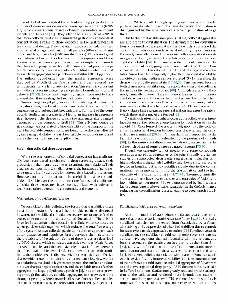
Fig. 8. (A) Stabilization of sorafenib through co-aggregation with Congo red. A minimum drug to dye ratio of 25:1 is required for stability. Reprinted with permission fromRef. [46]. Copyright 2016 American Chemical Society. (B) Size of colloids stabilized by protein coronas have a concentration-dependent relationship. Different drug-proteincombinations require different amounts of proteins for stability. Reprinted with permission from Ref. [50]. Copyright 2017 American Chemical Society.
In addition to providing steric stability, polymers can increasestability against crystallization, and low concentrations of poly-mers can increase the induction time before crystallization occurs[17,39,59,81,82]. Two mechanisms are thought to contribute tothis effect. First, macromolecular additives can increase the crys-talline solubility of the drug or decrease its CAC by altering thecorrection factor in Eq. (1) [17]. These changes reduce supersat-uration, lowering the crystal nucleation rate. Second, adsorbedmacromolecules are thought to alter the nucleation kinetics atthe water-colloid interface. Adsorbed species can either acceler-ate or retard drug crystallization, depending on the nature of bothspecies [26,83]. This non-uniform behavior may be due to the dif-ferent localizations of each polymer within the colloid, which arisefrom differences in polymer-drug interactions [39]. Some macro-molecules localize to the colloid surface, thereby interfering withheterogeneous nucleation; some may partition into the colloids,inhibiting crystal growth inside the particle [59]. Other macro-molecules do not adsorb significantly and have little effect onnucleation kinetics [84]. Another factor is differences in hydropho-bic interactions between the polymer and drug. For example, thestructure of the hydrophobic segments of a series of surfactantscorrelates with its ability to suppress or enhance the crystalliza-tion of amorphous celecoxib [83]. In cases where crystallization isenhanced, the macromolecule may reduce the activation barrierfor nucleation by stabilizing the crystal nucleus. However, poly-mers adsorbed onto pre-formed drug crystals can also inhibit theirgrowth to varying degrees [85,86], which could help sustain a pop-ulation of colloids even in the presence of a few crystals. In general,partially hydrophobic polymers, which can suppress nucleation atthe water-colloid interface, seem to best stabilize colloidal aggre-gates against crystallization.
Co-aggregation to stabilize drug colloids
Recently, co-formulation of aggregators with azo-dyes—whichthemselves form aggregates—has been shown to stabilize the col-loids (Fig. 8A) [46]. Stable colloids of sorafenib and vemurafenibwere formed with the aggregating dyes Congo red and Evans blue,and the resulting co-formulated colloids were unusually mono-dispersed with small diameters (50–100 nm). Drug to dye ratiosas high as 25:1 yielded colloidal aggregates that were stable over72 h in buffered solutions and even in serum-containing media[30]. The incorporation of the azo-dyes in these colloids resulted inhighly negative surface charges (zeta potentials of approximately
−40 mV), and the resultant electrostatic repulsion may be the driv-ing force of the colloidal stability of these formulations. In additionto having exceptional stability, dye-stabilized colloids preservedthe activity of adsorbed enzymes. Enzymes loaded onto these col-loids were active after 72 h once released back into solution, whilefree enzyme was only stable for 4 h.
Exploiting protein adsorption to stabilize colloidal aggregates
A decade of work has established that colloidal aggregatesbind strongly to proteins. This property was exploited to stabi-lize colloidal aggregates using a protein corona on the colloidsurface, which provided stabilizing steric repulsive forces [50].Protein corona formation could control colloidal aggregate size ina concentration-dependent manner (Fig. 8B). These colloids hadenhanced stability in buffered solutions (maintenance of colloidsize over 48 h) compared to non-stabilized colloids, which precip-itated over the course of 24 h. The protein-stabilized colloids werealso stable in serum-containing media. Importantly, this strategyis amenable to a range of proteins and compounds. However, eachdrug-protein combination likely has a different ratio for optimalstability, since the properties of the colloidal surface and the proteindrive these interactions.
Conclusions and outlook
Self-aggregation to form colloidal particles is a phenomenonexhibited by a wide range of compounds, including many drugmolecules and dyes; there is evidence that over 10,000 moleculescan form these species, and presumably many more can. Though thecolloidal particles are metastable, the colloid-forming compoundsdiscussed herein show delayed crystallization. To be classified as acolloidal aggregator, the following properties are required:
1. CAC-dependent formation of distinct colloidal particles;2. Non-specific protein adsorption and inhibition of enzymes
(model enzymes used to demonstrate this include �-lactamase,�-chymotrypsin, and malate dehydrogenase); and
3. Detergent-reversible formation (e.g. return of enzyme activityand return of small molecule activity in cell-based assays fol-lowing detergent addition).
Detergent reversibility provides a robust means of controllingfor aggregation in many enzyme- and cell-based assays. Different

A.N. Ganesh et al. / Nano Today 19 (2018) 188–200 199
detergents may be required for each assay, taking into account theeffects of the detergents themselves on the assay. We prefer Tri-ton X-100 for enzyme inhibition studies and polysorbate 80 forcell-based assays. With appropriate controls and under the rightconditions, one may now expect to control for the role of colloidalaggregation as an artifact in early drug discovery.
Due to the wide variety of conditions used in drug screeningand the persistence of colloids under different conditions, efforts topredict colloidal aggregation have had limited success [10,87,88].Aggregate Advisor (http://advisor.bkslab.org) is one such predictivetool that can aide in identifying potential colloid-forming com-pounds for further verification [89], though we caution that thetool is crude and suffers from false positives and false negatives; ithas the advantage, however, of categorizing and matching to morethan 10,000 observations of colloidal aggregation. As the numberof known colloid-forming compounds continues to rise, and theirphysical and chemical properties are further explored, predictivetechnologies may improve.
Although colloidal aggregates have been historically viewed asa nuisance artifact in drug screens, efforts to exploit their uniqueproperties are changing this outlook. The ability to stabilize col-loidal aggregates by using excipients such as polymers, proteins,and other small molecule aggregators has allowed not only theirfurther study, but also their potential use as intentional drugformulations [19,50]. Many studies have investigated the impactof colloidal drug aggregation on the dissolution profiles of oralformulations [31,33,90]. Colloidal drug aggregates may yield for-mulations with drug loadings that are an order of magnitudehigher than conventional nanoparticle formulations. While signifi-cant progress has been made in regard to identifying and studyingthese drug aggregates, key questions remain. For example, fewstudies have investigated the stability of colloidal drug aggregatesin physiologically relevant, protein-rich conditions; even fewerhave studied their interactions with cells or their fate in vivo. Fur-thermore, the ability to generalize current stabilization strategiesto all colloid-forming compounds is unknown and will require sys-tematic evaluation of these methods. Such studies are required toturn this phenomenon into a reliable formulations strategy.
Acknowledgements
This work was supported by funding from the Canadian CancerSociety Research Institute (to M.S.S. and B.K.S.), the US NationalInstitutes of General Medical Sciences (GM71630 to B.K.S andM.S.S.), the Natural Sciences and Engineering Research Council(NSERC Discovery to M.S.S. and Postgraduate Research Scholar-ship to A.N.G.) and QEII graduate scholarship (to E.N.D). We thankmembers of the Shoichet labs (in Toronto and San Francisco) forthoughtful discussion.
References
[1] K.H. Bleicher, H.J. Bohm, K. Muller, A.I. Alanine, Nat. Rev. Drug Discov. 2(2003) 369–378.
[2] B.K. Shoichet, Drug Discov. Today 11 (2006) 607–615.[3] S.L. McGovern, E. Caselli, N. Grigorieff, B.K. Shoichet, J. Med. Chem. 45 (2002)
1712–1722.[4] B.Y. Feng, A. Simeonov, A. Jadhav, K. Babaoglu, J. Inglese, B.K. Shoichet, et al., J.
Med. Chem. 50 (2007) 2385–2390.[5] B.K. Shoichet, J. Med. Chem. 49 (2006) 7274–7277.[6] C. Aldrich, C. Bertozzi, G.I. Georg, L. Kiessling, C. Lindsley, D. Liotta, et al., J.
Med. Chem. 60 (2017) 2165–2168.[7] K.E. Coan, B.K. Shoichet, J. Am. Chem. Soc. 130 (2008) 9606–9612.[8] K.E. Coan, D.A. Maltby, A.L. Burlingame, B.K. Shoichet, J. Med. Chem. 52 (2009)
2067–2075.[9] S.L. McGovern, B. Shoichet, J. Med. Chem. 46 (2003) 1478–1483.
[10] J. Seidler, S.L. McGovern, T.N. Doman, B.K. Shoichet, J. Med. Chem. 46 (2003)4477–4486.
[11] W. Ma, A.G. Cheetham, H. Cui, Nano Today 11 (2016) 13–30.[12] S.M. D’Addio, R.K. Prud’homme, Adv. Drug Deliv. Rev. 63 (2011) 417–426.
[13] S.Y. Qin, A.Q. Zhang, S.X. Cheng, L. Rong, X.Z. Zhang, Biomaterials 112 (2017)234–247.
[14] D. Duan, A.K. Doak, L. Nedyalkova, B.K. Shoichet, ACS Chem. Biol. 10 (2015)978–988.
[15] G.A. Ilevbare, L.S. Taylor, Cryst. Growth Des. 13 (2013) 1497–1509.[16] S.C. Owen, A.K. Doak, P. Wassam, M.S. Shoichet, B.K. Shoichet, ACS Chem. Biol.
7 (2012) 1429–1435.[17] M.J. Jackson, S.J. Toth, U.S. Kestur, J. Huang, F. Qian, M.A. Hussain, et al., Mol.
Pharm. 11 (2014) 3027–3038.[18] N. Li, C.J. Gilpin, L.S. Taylor, Mol. Pharm. 14 (2017) 1691–1705.[19] A.N. Ganesh, J. Logie, C.K. McLaughlin, B.L. Barthel, T.H. Koch, B.K. Shoichet,
et al., Mol. Pharm. 14 (2017) 1852–1860.[20] S.C. Owen, A.K. Doak, A.N. Ganesh, L. Nedyalkova, C.K. McLaughlin, B.K.
Shoichet, et al., ACS Chem. Biol. 9 (2014) 777–784.[21] L.I. Mosquera-Giraldo, L.S. Taylor, Mol. Pharm. 12 (2015) 496–503.[22] S.L. McGovern, B.T. Helfand, B. Feng, B.K. Shoichet, J. Med. Chem. 46 (2003)
4265–4272.[23] C.L. Zhao, M.A. Winnik, G. Riess, M.D. Croucher, Langmuir 6 (1990) 514–516.[24] M. Wilhelm, C.L. Zhao, Y.C. Wang, R.L. Xu, M.A. Winnik, J.L. Mura, et al.,
Macromolecules 24 (1991) 1033–1040.[25] E.D. Goddard, N.J. Turro, P.L. Kuo, K.P. Ananthapadmanabhan, Langmuir 1
(1985) 352–355.[26] G.A. Ilevbare, H. Liu, J. Pereira, K.J. Edgar, L.S. Taylor, Mol. Pharm. 10 (2013)
3392–3403.[27] H.S. Purohit, L.S. Taylor, Mol. Pharm. 12 (2015) 1623–1635.[28] A.S. Indulkar, K.J. Box, R. Taylor, R. Ruiz, L.S. Taylor, Mol. Pharm. 12 (2015)
2365–2377.[29] L. Almeida e Sousa, S.M. Reutzel-Edens, G.A. Stephenson, L.S. Taylor, Mol.
Pharm. 12 (2015) 484–495.[30] D. Duan, H. Torosyan, D. Elnatan, C.K. McLaughlin, J. Logie, M.S. Shoichet, et al.,
ACS Chem. Biol. 12 (2017) 282–290.[31] Y.V. Frenkel, A.D. Clark Jr, K. Das, Y.H. Wang, P.J. Lewi, P.A. Janssen, et al., J.
Med. Chem. 48 (2005) 1974–1983.[32] M.C. Brick, H.J. Palmer, T.H. Whitesides, Langmuir 19 (2003) 6367–6380.[33] A.S. Indulkar, Y. Gao, S.A. Raina, G.G. Zhang, L.S. Taylor, Mol. Pharm. 13 (2016)
2059–2069.[34] P. Harmon, K. Galipeau, W. Xu, C. Brown, W.P. Wuelfing, Mol. Pharm. 13
(2016) 1467–1481.[35] A.S. Indulkar, J.E. Waters, H. Mo, Y. Gao, S.A. Raina, G.G.Z. Zhang, et al., J.
Pharm. Sci. 106 (2017) 1998–2008.[36] S.B. Murdande, M.J. Pikal, R.M. Shanker, R.H. Bogner, J. Pharm. Sci. 99 (2010)
1254–1264.[37] M.F. Emerson, A. Holtzer, J. Phys. Chem. 71 (1967) 1898–1907.[38] S.C. Owen, D.P.Y. Chan, M.S. Shoichet, Nano Today 7 (2012) 53–65.[39] S.A. Raina, G.G. Zhang, D.E. Alonzo, J. Wu, D. Zhu, N.D. Catron, et al., Pharm.
Res. 32 (2015) 3350–3364.[40] B.Y. Feng, B.K. Shoichet, J. Med. Chem. 49 (2006) 2151–2154.[41] N.S. Trasi, L.S. Taylor, J. Pharm. Sci. 104 (2015) 2583–2593.[42] N.S. Trasi, L.S. Taylor, Int. J. Pharm. 496 (2015) 282–290.[43] A. Alhalaweh, C.A. Bergstrom, L.S. Taylor, J. Control Release 229 (2016)
172–182.[44] S.A. Raina, D.E. Alonzo, G.G. Zhang, Y. Gao, L.S. Taylor, Pharm. Res. 32 (2015)
3660–3673.[45] A.M. Giannetti, B.D. Koch, M.F. Browner, J. Med. Chem. 51 (2008) 574–580.[46] C.K. McLaughlin, D. Duan, A.N. Ganesh, H. Torosyan, B.K. Shoichet, M.S.
Shoichet, ACS Chem. Biol. 11 (2016) 992–1000.[47] A.J. Ryan, N.M. Gray, P.N. Lowe, C.W. Chung, J. Med. Chem. 46 (2003)
3448–3451.[48] B.Y. Feng, B.K. Shoichet, Nat. Protoc. 1 (2006) 550–553.[49] K. Rausch, A. Reuter, K. Fischer, M. Schmidt, Biomacromolecules 11 (2010)
2836–2839.[50] A.N. Ganesh, C.K. McLaughlin, D. Duan, B.K. Shoichet, M.S. Shoichet, ACS Appl.
Mater. Interfaces 9 (2017) 12195–12202.[51] J. Wang, E. Matayoshi, Pharm. Res. 29 (2012) 1745–1754.[52] S. Bhattacharjee, J. Control Release 235 (2016) 337–351.[53] L. Lindfors, P. Skantze, U. Skantze, J. Westergren, U. Olsson, Langmuir 23
(2007) 9866–9874.[54] F. Tres, S.D. Hall, M.A. Mohutsky, L.S. Taylor, J. Pharm. Sci. 107 (2018) 94–102.[55] A. Gaudin, M. Yemisci, H. Eroglu, S. Lepetre-Mouelhi, O.F. Turkoglu, B.
Donmez-Demir, et al., Nat. Nanotechnol. 9 (2014) 1054–1062.[56] S.R. LaPlante, N. Aubry, G. Bolger, P. Bonneau, R. Carson, R. Coulombe, et al., J.
Med. Chem. 56 (2013) 7073–7083.[57] S.R. LaPlante, R. Carson, J. Gillard, N. Aubry, R. Coulombe, S. Bordeleau, et al., J.
Med. Chem. 56 (2013) 5142–5150.[58] M. Pellecchia, D.S. Sem, K. Wuthrich, Nat. Rev. Drug Discov. 1 (2002) 211–219.[59] K. Ueda, K. Higashi, K. Moribe, Mol. Pharm. 14 (2017) 2314–2322.[60] S.A. Raina, B. Van Eerdenbrugh, D.E. Alonzo, H. Mo, G.G.Z. Zhang, Y. Gao, et al.,
J. Pharm. Sci. 104 (2015) 1981–1992.[61] X. Zhao, Z. Poon, A.C. Engler, D.K. Bonner, P.T. Hammond, Biomacromolecules
13 (2012) 1315–1322.[62] J. Logie, S.C. Owen, C.K. McLaughlin, M.S. Shoichet, Chem. Mater. 26 (2014)
2847–2855.[63] K.E. Coan, B.K. Shoichet, Mol. Biosyst. 3 (2007) 208–213.[64] H.Y. Liu, Z. Wang, C. Regni, X. Zou, P.A. Tipton, Biochemistry 43 (2004)
8662–8669.
200 A.N. Ganesh et al. / Nano Today 19 (2018) 188–200
[65] S.A. Raina, G.G.Z. Zhang, D.E. Alonzo, J. Wu, D. Zhu, N.D. Catron, et al., J. Pharm.Sci. 103 (2014) 2736–2748.
[66] F. Alexis, E. Pridgen, L.K. Molnar, O.C. Farokhzad, Mol. Pharm. 5 (2008)505–515.
[67] A. Lesniak, F. Fenaroli, M.P. Monopoli, C. Aberg, K.A. Dawson, A. Salvati, ACSNano 6 (2012) 5845–5857.
[68] C.D. Walkey, J.B. Olsen, F. Song, R. Liu, H. Guo, D.W. Olsen, et al., ACS Nano 8(2014) 2439–2455.
[69] A.K. Doak, H. Wille, S.B. Prusiner, B.K. Shoichet, J. Med. Chem. 53 (2010)4259–4265.
[70] Y.V. Frenkel, E. Gallicchio, K. Das, R.M. Levy, E. Arnold, J. Med. Chem. 52 (2009)5896–5905.
[71] M.A. Clark, M.A. Jepson, B.H. Hirst, Adv. Drug Deliv. Rev. 50 (2001) 81–106.[72] L.M. Ensign, R. Cone, J. Hanes, Adv. Drug Deliv. Rev. 64 (2012) 557–570.[73] T. Tadros, General principles of colloid stability and the role of surface forces,
in: Colloid Stability, Wiley-VCH Verlag GmbH & Co. KGaA, 2006, pp. 1–22.[74] J.W. Mullin, Crystallization, Elsevier Science, 2001.[75] N.D. Loh, S. Sen, M. Bosman, S.F. Tan, J. Zhong, C.A. Nijhuis, et al., Nat. Chem. 9
(2017) 77–82.[76] P.G. Vekilov, Cryst. Growth Des. 4 (2004) 671–685.[77] J.A. Baird, D. Santiago-Quinonez, C. Rinaldi, L.S. Taylor, Pharm. Res. 29 (2012)
271–284.[78] A. Kalra, P. Tishmack, J.W. Lubach, E.J. Munson, L.S. Taylor, S.R. Byrn, et al.,
Mol. Pharm. 14 (2017) 2126–2137.[79] J.A. Baird, B. Van Eerdenbrugh, L.S. Taylor, J. Pharm. Sci. 99 (2010) 3787–3806.[80] N. Anton, J.P. Benoit, P. Saulnier, J. Control Release 128 (2008) 185–199.[81] M.J. Jackson, U.S. Kestur, M.A. Hussain, L.S. Taylor, Pharm. Res. 33 (2016)
1276–1288.[82] G.A. Ilevbare, H.Y. Liu, K.J. Edgar, L.S. Taylor, Cryst. Growth Des. 13 (2013)
740–751.[83] J. Chen, J.D. Ormes, J.D. Higgins, L.S. Taylor, Mol. Pharm. 12 (2015) 533–541.[84] S.A. Raina, D.E. Alonzo, G.G. Zhang, Y. Gao, L.S. Taylor, Mol. Pharm. 11 (2014)
3565–3576.[85] C.J. Schram, L.S. Taylor, S.P. Beaudoin, Langmuir 31 (2015) 11279–11287.[86] C.J. Schram, S.P. Beaudoin, L.S. Taylor, Cryst. Growth Des. 16 (2016)
2094–2103.[87] H. Rao, Z. Li, X. Li, X. Ma, C. Ung, H. Li, et al., J. Comput. Chem. 31 (2010)
752–763.[88] B.Y. Feng, A. Shelat, T.N. Doman, R.K. Guy, B.K. Shoichet, Nat. Chem. Biol. 1
(2005) 146–148.[89] J.J. Irwin, D. Duan, H. Torosyan, A.K. Doak, K.T. Ziebart, T. Sterling, et al., J. Med.
Chem. 58 (2015) 7076–7087.[90] M.J. Jackson, U.S. Kestur, M.A. Hussain, L.S. Taylor, Mol. Pharm. 13 (2016)
223–231.
Ahil Ganesh is currently a PhD candidate in ProfessorMolly Shoichet’s laboratory at the University of Toronto.He received a BASc in Engineering Science, Majoring inBiomedical Engineering from the University of Toronto.His current research interests include strategies to stabi-lize colloidal drug aggregates and exploit their propertiesfor drug delivery.
Eric Donders is a PhD student in Professor MollyShoichet’s group at the University of Toronto. Previ-ously, he completed his BASc in Engineering Chemistryat Queen’s University in Kingston, Ontario. Currently, Ericresearches strategies to control the stability of colloidaldrug aggregates, with the goal of using these drug-richassemblies as vehicles for drug delivery.
Brian Shoichet, PhD is a professor of pharmaceuticalchemistry at the University of California - San Fran-cisco. He trained in computational chemistry with TackKuntz and in experimental protein biophysics with BrianMatthews. Research in his lab merges both streams,using model systems to test new docking methods bydetailed experiment. Successful new methods are appliedto novel reagent discovery for GPCRs. Some unexpectedobservations have flowed from this cycle of theory andexperiment, such as the discovery and subsequent mech-anistic characterization of colloidal aggregation, the focusof this paper.
Molly Shoichet, PhD is University Professor at the Uni-versity of Toronto and the Tier 1 Canada Research Chair inTissue Engineering. She is an Officer of the Order of Canadaand holds the Order of Ontario. Professor Shoichet leadsa lab of 30 researchers, has published over 575 papers,patents and abstracts, and given over 350 lectures world-wide. She cherishes this collaboration on colloidal drugaggregates with her brother, Brian Shoichet, and is cur-rently Chief Scientist, Ontario.