comparative genomic analysis of haemophilus …lrishishwar3/pdf/2016-jcm-haemophilus.pdf ·...
TRANSCRIPT

Comparative Genomic Analysis of Haemophilus haemolyticus andNontypeable Haemophilus influenzae and a New Testing Scheme forTheir Discrimination
Fang Hu,a Lavanya Rishishwar,b,c,d Ambily Sivadas,b Gabriel J. Mitchell,b,e I. King Jordan,b,c,d Timothy F. Murphy,f Janet R. Gilsdorf,g
Leonard W. Mayer,a Xin Wanga
Meningitis and Vaccine Preventable Diseases Branch, Division of Bacterial Diseases, National Center for Immunization and Respiratory Diseases, Centers for DiseaseControl and Prevention, Atlanta, Georgia, USAa; School of Biology, Georgia Institute of Technology, Atlanta, Georgia, USAb; Applied Bioinformatics Laboratory, Atlanta,Georgia, USAc; PanAmerican Bioinformatics Institute, Cali, Valle del Cauca, Colombiad; Institute of Science and Technology Austria, Klosterneuburg, Austriae; Division ofInfectious Diseases, Department of Medicine, Clinical and Translational Research Center, Department of Microbiology and Immunology, University at Buffalo, StateUniversity of New York, Buffalo, New York, USAf; Department of Pediatrics, University of Michigan, Ann Arbor, Michigan, USAg
Haemophilus haemolyticus has been recently discovered to have the potential to cause invasive disease. It is closely related tonontypeable Haemophilus influenzae (NT H. influenzae). NT H. influenzae and H. haemolyticus are often misidentified becausenone of the existing tests targeting the known phenotypes of H. haemolyticus are able to specifically identify H. haemolyticus.Through comparative genomic analysis of H. haemolyticus and NT H. influenzae, we identified genes unique to H. haemolyticusthat can be used as targets for the identification of H. haemolyticus. A real-time PCR targeting purT (encoding phosphoribosylg-lycinamide formyltransferase 2 in the purine synthesis pathway) was developed and evaluated. The lower limit of detection was40 genomes/PCR; the sensitivity and specificity in detecting H. haemolyticus were 98.9% and 97%, respectively. To improve thediscrimination of H. haemolyticus and NT H. influenzae, a testing scheme combining two targets (H. haemolyticus purT and H.influenzae hpd, encoding protein D lipoprotein) was also evaluated and showed 96.7% sensitivity and 98.2% specificity for theidentification of H. haemolyticus and 92.8% sensitivity and 100% specificity for the identification of H. influenzae, respectively.The dual-target testing scheme can be used for the diagnosis and surveillance of infection and disease caused by H. haemolyticusand NT H. influenzae.
Haemophilus haemolyticus is a human commensal that colo-nizes the respiratory tract. It shares high similarities in mor-
phology, biochemistry, and genetics with nontypeable Haemophi-lus influenzae (NT H. influenzae) (1–3). NT H. influenzae hasemerged as the major cause of invasive H. influenzae disease sincethe implementation of H. influenzae serotype b (H. influenzae b)vaccine (4). NT H. influenzae infections such as childhood otitismedia and respiratory tract infections in adults with chronic ob-structive pulmonary disease (COPD) result in an enormous socialand economic burden to societies (5). H. haemolyticus was re-cently reported to cause invasive disease (1). These cases werepreviously misidentified as NT H. influenzae due to the lack ofproper identification methods to discriminate between the twospecies (6). Reevaluation of NT H. influenzae cases from previousyears revealed that about 2% (7/374) of the NT H. influenzae in-vasive cases reported from Active Bacterial Core surveillance(ABCs) were in fact caused by H. haemolyticus (1).
Misidentification of H. haemolyticus as NT H. influenzae hasbeen repeatedly reported by multiple research groups, with up to40% of isolates being misidentified as NT H. influenzae using clas-sical phenotypic methods in clinical microbiology laboratories (1,7–11). The misidentification of H. haemolyticus has a potentialimpact on accurate assessment of the prevalence of antibiotic-resistant NT H. influenzae (12). Therefore, a rapid and accuratemethod is needed to distinguish H. haemolyticus from NT H. in-fluenzae and to better understand the epidemiology of H. haemo-lyticus infections. Testing schemes, including both standard mi-crobiological and molecular methods, have been proposed toimprove the identification of the two bacteria. These molecular
methods target a number of genes such as the lipo-oligosaccharidegene lgtC, the IgA protease gene iga, heme acquisition genes(hxuABC, hemR, and hup), the fuculose kinase gene fucK, and theHaemophilus protein D gene (hpd), with hpd-based PCR being thebest method for discriminating NT H. influenzae and H. haemo-lyticus (13–16). However, identification of H. haemolyticus relieson negative results, as all these genes are present in H. influenzaestrains and absent in the majority of H. haemolyticus strains. A testfor specific identification of H. haemolyticus is needed to betterunderstand the epidemiology of H. haemolyticus disease. In thisstudy, we conducted comparative genomic analysis to identifyunique target genes that are exclusively present in H. haemolyticusstrains and can be used to develop PCR assays for specific detec-tion and identification of H. haemolyticus.
Received 13 July 2016 Returned for modification 3 August 2016Accepted 26 September 2016
Accepted manuscript posted online 5 October 2016
Citation Hu F, Rishishwar L, Sivadas A, Mitchell GJ, Jordan IK, Murphy TF, GilsdorfJR, Mayer LW, Wang X. 2016. Comparative genomic analysis of Haemophilushaemolyticus and nontypeable Haemophilus influenzae and a new testing schemefor their discrimination. J Clin Microbiol 54:3010 –3017. doi:10.1128/JCM.01511-16.
Editor: N. A. Ledeboer, Medical College of Wisconsin
Address correspondence to Xin Wang, [email protected].
Supplemental material for this article may be found at http://dx.doi.org/10.1128/JCM.01511-16.
Copyright © 2016, American Society for Microbiology. All Rights Reserved.
crossmark
3010 jcm.asm.org December 2016 Volume 54 Number 12Journal of Clinical Microbiology
on Decem
ber 7, 2016 by GE
OR
GIA
INS
T O
F T
EC
HN
OLO
GY
http://jcm.asm
.org/D
ownloaded from

MATERIALS AND METHODSBacterial strains and growth conditions. Bacterial strains used in thisstudy included ATCC strains, clinical invasive isolates, and carriage iso-lates. Eighty-nine clinical invasive H. influenzae isolates were collected aspart of the Active Bacterial Core surveillance (ABCs) of the Centers forDisease Control and Prevention’s Emerging Infectious Program (http://www.cdc.gov/abcs/reports-findings/surv-reports.html). Seventy-eightH. influenzae isolates and 45 H. haemolyticus isolates were collected duringa carriage survey in Minnesota in 2009 (17). Additional H. haemolyticusisolates were kindly provided by University at Buffalo, State University ofNew York (n � 31), and University of Michigan Medical Center (n �104). The clinical and carriage isolates in this study have been character-ized and confirmed using standard microbiology and 16S rRNA genesequencing.
Whole-genome sequencing and comparison. Genome sequences ofthe five H. haemolyticus strains were used for comparison with those of H.influenzae. Details of whole-genome sequencing of the strains and theassembled sequences have been reported previously (18). Twenty com-plete and draft H. influenzae (19 NT H. influenzae and 1 H. influenzae bgenome) genome sequences were downloaded from the NCBI RefSeq andShotgun Assembly Sequence database (19) (Table 1). The evolutionaryrelationships among these H. haemolyticus and H. influenzae strains werecharacterized using analysis of 16S rRNA gene sequences and multilocussequence typing (MLST) locus sequences. 16S rRNA and MLST locussequences were obtained from the annotated GenBank entries at NCBI.Multiple copies of 16S rRNA loci were retained with postfix .a, .b, etc. 16S
rRNA and concatenated sequences of the seven MLST loci were alignedusing the program MUSCLE (20), and the resulting alignments were usedto reconstruct phylogenetic trees using the neighbor-joining method (21)as implemented in the program MEGA (22). Evolutionary relationshipsamong the H. haemolyticus and H. influenzae strains were also character-ized using whole-genome sequences by computing the average nucleotideidentity (ANI) (23, 24) between each pair of genomes using the programMUMmer (25).
Identification of H. haemolyticus-specific genes by comparativegenomics. Complete gene (protein-coding) sets from the H. haemolyticusand H. influenzae genome sequences were obtained from the annotatedGenBank entries at NCBI (Table 1). All-against-all nucleotide sequencecomparison of complete gene sets was performed using the BLASTCLUSTalgorithm (26) using default parameters. BLASTCLUST creates clusters ofgenes that share sequence similarity and coverage above a given thresholdusing single-linkage clustering. The resulting clusters are exclusive, with agene mapping to exactly one cluster. The results of the clustering proce-dure are visualized as a presence/absence matrix.
Sanger sequencing of target genes. Three genes (purT, coding forputative phosphoribosylglycinamide formyltransferase 2; hdg, coding forputative hydrogenase-2 small chain; and sod, coding for putative super-oxide dismutase [Cu-Zn]-like) from five additional Haemophilus haemo-lyticus isolates were sequenced using the Sanger sequencing method tovalidate their sequence conservation levels. Primers for amplification andsequencing of target genes are shown in Table 2. The PCR amplificationand DNA Sanger sequencing were performed as described previously us-
TABLE 1 H. haemolyticus and H. influenzae strains, phenotypes, and whole-genome sequences analyzed in this study
Species and strain NCBI accession no. Patha Hemolysisb
No. ofcontigsc
Total length(bases)d
Largest contig(bases)e N50 (bases)f
H. haemolyticusM19107 AFQN00000000.1 No Yes 115 1,909,361 81,211 30,635M19501 AFQO00000000.1 No No 22 1,884,911 364,637 238,571M21127 AFQP00000000.1 Yes Yes 34 1,943,222 319,487 121,923M21621 AFQQ00000000.1 Yes Yes 28 2,089,205 339,425 234,852M21639 AFQR00000000.1 Yes No 50 2,326,092 187,351 98,989
H. influenzaePittHH AAZH00000000.1 Yes No 51 1,831,290 164,041 95,398R3021 AAZE00000000.1 No No 45 1,875,022 314,724 79,72022.4-21 AAZJ00000000.1 No No 41 1,848,720 365,947 129,943PittAA AAZG00000000.1 Yes No 38 1,875,062 245,610 123,700M21709 Yes No 34 1,822,150 319,861 141,272NT127 ACSL00000000.1 Yes No 34 1,860,733 210,220 77,0846P18H1 ABWW00000000.1 Yes No 28 1,912,236 392,297 152,038PittII AAZI00000000.1 Yes No 22 1,952,226 385,065 135,7363655 AAZF00000000.1 Yes No 21 1,876,504 381,682 184,5457P49H1 ABWV00000000.1 Yes No 18 1,827,137 550,114 368,08122.1-21 AAZD00000000.1 No No 15 1,886,604 648,211 440,09210810 NC_016809 Yes No 1 1,981,535 1,981,535 1,981,53586-028NP NC_007146 Yes No 1 1,914,490 1,914,490 1,914,490F3031 NC_014920 Yes No 1 1,985,832 1,985,832 1,985,832F3047 NC_014922 Yes No 1 2,007,018 2,007,018 2,007,018PittEE NC_009566 Yes No 1 1,813,033 1,813,033 1,813,033PittGG CP000672 Yes No 1 1,887,192 1,887,192 1,887,192R2846 NC_017452 Yes No 1 1,819,370 1,819,370 1,819,370R2866 NC_017451 Yes No 1 1,932,306 1,932,306 1,932,306Rd-KW20 NC_000907 Yes No 1 1,830,138 1,830,138 1,830,138
a Whether or not strain was determined to be pathogenic (disease causing).b Whether or not a strain was shown to be capable of hemolysis.c Total number of contigs of �1 kbp (i.e., large contigs) produced in the assembly.d Total number of bases assembled into large contigs.e Size of the largest contig in the assembly.f Size of the contig at which contigs of that length or longer cover at least half of the assembly.
Detection of Haemophilus haemolyticus
December 2016 Volume 54 Number 12 jcm.asm.org 3011Journal of Clinical Microbiology
on Decem
ber 7, 2016 by GE
OR
GIA
INS
T O
F T
EC
HN
OLO
GY
http://jcm.asm
.org/D
ownloaded from

ing chromosomal DNA template extracted from five additional isolates(27). The gene sequences were assembled using DNAStar Lasergene 9(DNAStar, Inc., Madison, WI).
RT-PCR. Chromosomal DNA and crude cell lysates were prepared asdescribed previously (28). Primer Express 3.0 (Applied Biosystems) wasused to design appropriate primers and probes for detecting purT. Allprimers and probes used in this study were optimized by testing in therange of 100 to 900 nM and 100 to 300 nM, respectively. Real-time PCR(RT-PCR) was performed as described previously (28). A chromosomalDNA or crude cell lysate was considered positive if the cycle threshold(CT) value was equal to or less than 35 and negative if the CT value wasgreater than 40. If a CT value was between 35 and 40, the sample wasdiluted 10-fold and retested to determine if PCR inhibitors were present.The specimen was considered positive if the CT value of the diluted spec-imen was equal to or less than 35 and negative if the CT value was greaterthan 35. To determine the lower limit of detection (LLD), genomic DNAwas extracted from H. haemolyticus isolates (28). The DNA concentrationwas determined using a NanoDrop spectrophotometer (NanoDropTechnologies, Wilmington, DE), adjusted to 20 ng/�l, and then 10-foldserially diluted in PCR-grade water. Each dilution was tested by PCR intriplicate. DNA concentration was converted to genome equivalents permicroliter on the basis of 1.8 Mb (carriage isolate) and 2.0 Mb (invasiveisolate) per H. haemolyticus genome (AFQN00000000, AFQO00000000,AFQP00000000, AFQQ00000000, AFQR00000000). The LLD for an RT-PCR assay was defined as the DNA concentration that yielded a CT valueof 35. Confidence intervals for the H. haemolyticus and H. influenzae sen-sitivity and specificity values, calculated based on the results of the RT-PCR assays, were determined using standard validation analyses. The val-idation analyses were performed using SAS 9.2 (SAS Institute Inc., Cary,NC), and exact binomial 95% confidence intervals were estimated usingStata v. 9.2 (StataCorp).
RESULTSComparative genomic analysis of H. haemolyticus and H. influ-enzae. Comparative analyses of 25 whole-genome sequences of H.haemolyticus (5) and H. influenzae (20) were performed in aneffort to assess whether strains from these two species, which areoften confused using classical phenotypic or biochemical meth-ods, can be clearly distinguished at the genomic level. Phyloge-netic analysis using individual (16S rRNA) or multiple (MLST)gene loci clearly distinguishes H. haemolyticus and H. influenzaeevolutionary lineages (Fig. 1A and B). Comparison of whole-ge-nome sequences using the ANI technique also yields unambigu-ous discrimination between the H. haemolyticus and H. influenzaestrains (Fig. 1C). These results suggested the possibility that theremay be individual gene loci with presence/absence patterns thatdistinguish H. haemolyticus from H. influenzae. Such loci wouldrepresent ideal targets for a real-time PCR (RT-PCR) typingscheme.
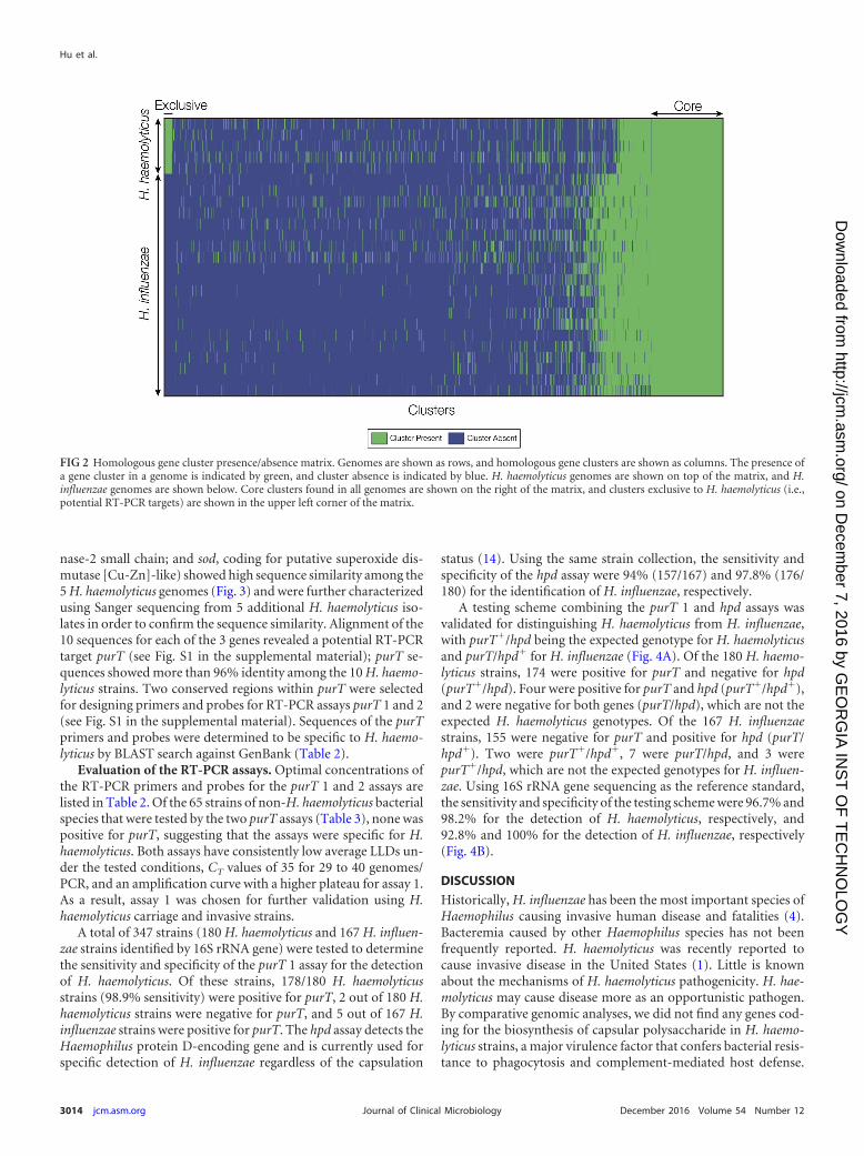
Identification of H. haemolyticus-specific RT-PCR gene tar-gets. All-against-all comparison of complete gene sets from the 25genomes analyzed here was used to define clusters of homologousgenes that are exclusive to the H. haemolyticus lineage. A total of 93clusters were found with homologous genes that were present inall 5 of the H. haemolyticus genomes and absent in all 20 of the H.influenzae genomes (Fig. 2). All of these clusters represent poten-tial gene targets for H. haemolyticus-specific RT-PCR, and clusterswith conserved flanking regions (for primer and probe bindingsites) were considered for the development of RT-PCR assays.Three genes (purT, coding for putative phosphoribosylglyci-namide formyltransferase 2; hdg, coding for putative hydroge-
TABLE 2 Primers and probes used for DNA sequencing and real-time PCR assaysa
Purpose and target genePrimerdesignation 5=-3= nucleotide sequenced
Ampliconlength (bp)
Final concn(nM)
DNA amplification and sequencing primerspurTb purT Fw1 CACTTGGCACAGCATTAACC 1,145 400
purT Fw TAACCCCTAATGCAACCAAAGT 1,130 400purT Rv CTTGTTTTACTTTTTCCAACGC 400purT seq CCAATAGGTCATCGTCAAGAAGA 160purT seq1R TCTTCTTGACGATGACCTATTGG 160
hdg hdg Fw TTATGAAATTATGTACCGCACTTG 1,084 400hdg Fw1 TGGCGTTTTTTCTGCRTTG 1,125 400hdg Rv TCCTTGATCTTGATTGGCTTG 400hdg seq AACMGTTGCCTATATCATCACTT 160hdg seq1R AAGTGATGATATAGGCAACKGTT 160
sod sod Fw GTTAGTGCGGTATGTTCAGTTGGT 502 400sod Rv CCAAGTGGMGCTGGATGATC 400sod seq Fw GTTAGTGCGGTATGTTCAGTTGGT 160sod seq Rv CCAAGTGGMGCTGGATGATC 160
RT-PCR primers and probespurT 1 purT Fw1 ATTAACCCCTAATGCAACCAAAGT 91 100
purT Rv1 CTTCAACGCCTAAACGTTGCA 900purT pb1c ATGTTGGGTTCTGGTGAA 300
purT 2 purT Fw2 TTAATGTTGTGCCGACTGCAA 78 100purT Rv2 AACTCTTCTGAAGCTAGACGACGAAT 600purT pb2c TCAACTTACGATGAATCGTGA 300
a All primers and probes were synthesized at the CDC Biotechnology Core Facility.b Primers for amplification and sequencing of purT.c All probes were labeled with 6-carboxyfluorescein at the 5= end and black hole quencher at the 3= end.d K � G�T; M � A�C; R � A�G.
Hu et al.
3012 jcm.asm.org December 2016 Volume 54 Number 12Journal of Clinical Microbiology
on Decem
ber 7, 2016 by GE
OR
GIA
INS
T O
F T
EC
HN
OLO
GY
http://jcm.asm
.org/D
ownloaded from

FIG 1 Comparison of H. haemolyticus versus H. influenzae evolutionary lineages. H. haemolyticus and H. influenzae strains analyzed here are labeled with theirspecies abbreviations and the strain names shown in Table 1. (A and B) Phylogenetic trees based on 16S rRNA gene (A) and concatenated MLST loci (B) showingthe evolutionary relationships of the H. haemolyticus and H. influenzae genome sequences analyzed here. Percent bootstrap values indicate support for internalnodes on the trees, and the branch length scale bars show P distances. (C) Results of whole-genome sequence comparisons among H. haemolyticus and H.influenzae based on ANI analysis. ANI values (percentages) between pairs of genomes are color coded as shown in the key, and the relationships among thegenomes based on these values are shown as dendrograms on both axes.
Detection of Haemophilus haemolyticus
December 2016 Volume 54 Number 12 jcm.asm.org 3013Journal of Clinical Microbiology
on Decem
ber 7, 2016 by GE
OR
GIA
INS
T O
F T
EC
HN
OLO
GY
http://jcm.asm
.org/D
ownloaded from
nase-2 small chain; and sod, coding for putative superoxide dis-mutase [Cu-Zn]-like) showed high sequence similarity among the5 H. haemolyticus genomes (Fig. 3) and were further characterizedusing Sanger sequencing from 5 additional H. haemolyticus iso-lates in order to confirm the sequence similarity. Alignment of the10 sequences for each of the 3 genes revealed a potential RT-PCRtarget purT (see Fig. S1 in the supplemental material); purT se-quences showed more than 96% identity among the 10 H. haemo-lyticus strains. Two conserved regions within purT were selectedfor designing primers and probes for RT-PCR assays purT 1 and 2(see Fig. S1 in the supplemental material). Sequences of the purTprimers and probes were determined to be specific to H. haemo-lyticus by BLAST search against GenBank (Table 2).
Evaluation of the RT-PCR assays. Optimal concentrations ofthe RT-PCR primers and probes for the purT 1 and 2 assays arelisted in Table 2. Of the 65 strains of non-H. haemolyticus bacterialspecies that were tested by the two purT assays (Table 3), none waspositive for purT, suggesting that the assays were specific for H.haemolyticus. Both assays have consistently low average LLDs un-der the tested conditions, CT values of 35 for 29 to 40 genomes/PCR, and an amplification curve with a higher plateau for assay 1.As a result, assay 1 was chosen for further validation using H.haemolyticus carriage and invasive strains.
A total of 347 strains (180 H. haemolyticus and 167 H. influen-zae strains identified by 16S rRNA gene) were tested to determinethe sensitivity and specificity of the purT 1 assay for the detectionof H. haemolyticus. Of these strains, 178/180 H. haemolyticusstrains (98.9% sensitivity) were positive for purT, 2 out of 180 H.haemolyticus strains were negative for purT, and 5 out of 167 H.influenzae strains were positive for purT. The hpd assay detects theHaemophilus protein D-encoding gene and is currently used forspecific detection of H. influenzae regardless of the capsulation
status (14). Using the same strain collection, the sensitivity andspecificity of the hpd assay were 94% (157/167) and 97.8% (176/180) for the identification of H. influenzae, respectively.
A testing scheme combining the purT 1 and hpd assays wasvalidated for distinguishing H. haemolyticus from H. influenzae,with purT�/hpd being the expected genotype for H. haemolyticusand purT/hpd� for H. influenzae (Fig. 4A). Of the 180 H. haemo-lyticus strains, 174 were positive for purT and negative for hpd(purT�/hpd). Four were positive for purT and hpd (purT�/hpd�),and 2 were negative for both genes (purT/hpd), which are not theexpected H. haemolyticus genotypes. Of the 167 H. influenzaestrains, 155 were negative for purT and positive for hpd (purT/hpd�). Two were purT�/hpd�, 7 were purT/hpd, and 3 werepurT�/hpd, which are not the expected genotypes for H. influen-zae. Using 16S rRNA gene sequencing as the reference standard,the sensitivity and specificity of the testing scheme were 96.7% and98.2% for the detection of H. haemolyticus, respectively, and92.8% and 100% for the detection of H. influenzae, respectively(Fig. 4B).
DISCUSSION
Historically, H. influenzae has been the most important species ofHaemophilus causing invasive human disease and fatalities (4).Bacteremia caused by other Haemophilus species has not beenfrequently reported. H. haemolyticus was recently reported tocause invasive disease in the United States (1). Little is knownabout the mechanisms of H. haemolyticus pathogenicity. H. hae-molyticus may cause disease more as an opportunistic pathogen.By comparative genomic analyses, we did not find any genes cod-ing for the biosynthesis of capsular polysaccharide in H. haemo-lyticus strains, a major virulence factor that confers bacterial resis-tance to phagocytosis and complement-mediated host defense.
FIG 2 Homologous gene cluster presence/absence matrix. Genomes are shown as rows, and homologous gene clusters are shown as columns. The presence ofa gene cluster in a genome is indicated by green, and cluster absence is indicated by blue. H. haemolyticus genomes are shown on top of the matrix, and H.influenzae genomes are shown below. Core clusters found in all genomes are shown on the right of the matrix, and clusters exclusive to H. haemolyticus (i.e.,potential RT-PCR targets) are shown in the upper left corner of the matrix.
Hu et al.
3014 jcm.asm.org December 2016 Volume 54 Number 12Journal of Clinical Microbiology
on Decem
ber 7, 2016 by GE
OR
GIA
INS
T O
F T
EC
HN
OLO
GY
http://jcm.asm
.org/D
ownloaded from

However, genes encoding factors for colonization and invasionsuch as pili and IgA1 protease are present in these strains.
Unambiguous discrimination of the closely related species H.influenzae and H. haemolyticus is important for laboratory diag-nosis and surveillance of H. influenzae and H. haemolyticus diseaseand carriage evaluations. Over the last decade, considerable re-search effort has focused on identifying molecular targets andsuitable methodologies to differentiate NT H. influenzae from H.haemolyticus. One of the principal phenotypic differences betweenH. haemolyticus and NT H. influenzae is hemolysis by H. haemo-lyticus. However, this difference is often unreliable as H. haemo-lyticus can lose the defining hemolytic phenotype upon in vitropassage (1–3, 11, 29–31). A number of genetic targets (hpd,ompP2, opmP6, lgtC, 16S rRNA, fucK, and iga) have been evalu-
ated for differentiation of NT H. influenzae from H. haemolyticus(15). However, no single gene target tested was able to unequivo-cally differentiate NT H. influenzae and H. haemolyticus. For ex-ample, 16S rRNA gene PCR permitted identification of H. haemo-lyticus and NT H. influenzae for only 90% of strains (6), while theompP6-based assay detects about 97% of NT H. influenzae strainsbut also detects 12% of H. haemolyticus strains (2). Identificationof H. haemolyticus-specific targets was challenging due to the lackof available H. haemolyticus genome sequences.
Whole-genome sequencing (WGS) provides a valuable tool forbetter understanding H. haemolyticus genetics and for identifyingunique genetic targets for assay development. Recently, five H.haemolyticus strains were sequenced and annotated by our group(18). Comparative analysis revealed several H. haemolyticus-spe-cific genes, including the purT gene; these genes were found to bepresent exclusively among 5 H. haemolyticus genomes and absentfrom 20 H. influenzae genomes. purT sequences are highly con-served among all of the H. haemolyticus strains examined in thisstudy, which makes it an ideal target for RT-PCR development.While the purT assay was found to be highly sensitive and specificfor H. haemolyticus detection, the addition of testing for the pres-ence of hpd might provide enhanced discrimination for the detec-tion of both organisms and could be used to enhance diagnosisand surveillance of H. haemolyticus and H. influenzae infections.
A few exceptions were observed (Fig. 4A): four H. haemolyticusand two H. influenzae strains were positive for both purT and hpdgenes. Because of the high frequency of horizontal gene transfer
TABLE 3 Non-H. haemolyticus bacterial species tested in this study
Organism No. of isolates
Branhamella catarrhalis 1Corynebacterium diphtheriae 1Cryptococcus neoformans 1Haemophilus influenzae biogroup aegyptius 7Haemophilus aphrophilusa 2Haemophilus influenzae a, b, c, d, e, f, and NT 1 eachHaemophilus parahaemolyticus 1Haemophilus parainfluenzae 6Haemophilus paraphrophilusa 1Haemophilus spp. 1
Neisseria meningitidis serogroupsA, C, W, X, Y, Z, NG, and E 1 eachB and Z 2 each
Bordetella pertussis 1Escherichia coli strain O16:K1 1Escherichia coli strain O7:K1 1Neisseria cinerea 2Neisseria gonorrhoeae 2Neisseria lactamica 12Neisseria sicca 1Neisseria subflava 1Pseudomonas aeruginosa 1Staphylococcus aureus 1Streptococcus agalactiae 1Streptococcus pneumoniae 1
Total 65a Both H. aphrophilus and H. paraphrophilus have been classified as Aggregatibacteraphrophilus (37).
FIG 3 H. haemolyticus-specific potential RT-PCR target genes. The presence(green) and absence (blue) of 3 H. haemolyticus-specific genes—hdg, purT, andsod—are shown for the H. haemolyticus (n � 10) and H. influenzae (n � 20)strains analyzed here. Strain names are as shown in Table 1. The percent iden-tities of H. haemolyticus-specific genes to reference genes from strain M19107are color coded as shown in the key. The sequences of hdg, purT, and sod of sixH. haemolyticus strains (marked with asterisks) were determined by Sangersequencing. Five H. haemolyticus strains were sequenced by WGS. TheM19107 strain was characterized by both methods.
Detection of Haemophilus haemolyticus
December 2016 Volume 54 Number 12 jcm.asm.org 3015Journal of Clinical Microbiology
on Decem
ber 7, 2016 by GE
OR
GIA
INS
T O
F T
EC
HN
OLO
GY
http://jcm.asm
.org/D
ownloaded from

among and between Haemophilus species, it is conceivable that,during pharyngeal colonization with both species, H. haemolyticusstrains can acquire H. influenzae genes and vice versa. Two H.haemolyticus and seven H. influenzae strains were negative forboth purT and hpd genes. The results seen with purT and hpdPCR-negative isolates may have been due to deletions at that locusor to sequence variation at any of the primer or probe binding sites(30, 32).
Multiple studies have shown that single-target-based tests arenot ideal for discriminating bacterial species, particularly forclosely related species, or for classifying the variant strains thathave diverged from their species, such as fuzzy species of Neisseria(2, 15, 33). While multitarget-based approaches can improve thesensitivity and specificity, developing multiple tests for each bac-terial species is a huge undertaking. WGS can potentially serve as auniversal high-throughput method to improve the accuracy ofspecies delineation and has been proven accurate for bacterialspecies classification (34). In addition, WGS provides much-en-riched genetic information for strain subtyping and will be veryuseful for disease surveillance and outbreak investigations. Ma-trix-assisted laser desorption ionization–time of flight mass spec-trometry (MALDI-TOF MS) is increasingly used for species clas-sification in diagnostic microbiology laboratories. However, itmay not be able to provide sufficient resolution for subtyping ofbacterial pathogens, which is often performed in public healthmicrobiology laboratories for disease surveillance and other largeepidemiological surveys. The power of MALDI-TOF for identifi-cation of H. haemolyticus and H. influenzae is highly dependent ona well-defined reference spectrum for H. haemolyticus in speciesdatabases, which varies between laboratories (35, 36). Its utility indiscriminating between H. haemolyticus and H. influenzae re-mains to be further validated.
As the laboratory bioinformatics capacity increases and costsof system acquisition decrease, the advanced molecular detectiontools such as WGS and MALDI-TOF may be widely utilized indiagnostic and public health microbiology laboratories. However,
rapid and less expensive methods with high throughput, such asPCR, remain valuable today in these laboratories for diagnosis andsurveillance of infectious diseases.
ACKNOWLEDGMENTS
We are grateful to ABCs for providing strains and the Bacterial MeningitisLaboratory for technical support.
REFERENCES1. Anderson R, Wang X, Briere EC, Katz LS, Cohn AC, Clark TA,
Messonnier NE, Mayer LW. 2012. Haemophilus haemolyticus isolatescausing clinical disease. J Clin Microbiol 50:2462–2465. http://dx.doi.org/10.1128/JCM.06575-11.
2. McCrea KW, Xie J, LaCross N, Patel M, Mukundan D, Murphy TF,Marrs CF, Gilsdorf JR. 2008. Relationships of nontypeable Haemophilusinfluenzae strains to hemolytic and nonhemolytic Haemophilus haemolyti-cus strains. J Clin Microbiol 46:406 – 416. http://dx.doi.org/10.1128/JCM.01832-07.
3. Kilian M. 1976. A taxonomic study of the genus Haemophilus, with theproposal of a new species. J Gen Microbiol 93:9 – 62. http://dx.doi.org/10.1099/00221287-93-1-9.
4. MacNeil JR, Cohn AC, Farley M, Mair R, Baumbach J, Bennett N,Gershman K, Harrison LH, Lynfield R, Petit S, Reingold A, SchaffnerW, Thomas A, Coronado F, Zell ER, Mayer LW, Clark TA, MessonnierNE. 2011. Current epidemiology and trends in invasive Haemophilus in-fluenzae disease—United States, 1989-2008. Clin Infect Dis 53:1230 –1236. http://dx.doi.org/10.1093/cid/cir735.
5. Murphy TF. 2015. Vaccines for nontypeable Haemophilus influenzae: thefuture is now. Clin Vaccine Immunol 22:459 – 466. http://dx.doi.org/10.1128/CVI.00089-15.
6. Murphy TF, Brauer AL, Sethi S, Kilian M, Cai X, Lesse AJ. 2007.Haemophilus haemolyticus: a human respiratory tract commensal to bedistinguished from Haemophilus influenzae. J Infect Dis 195:81– 89. http://dx.doi.org/10.1086/509824.
7. Chang A, Adlowitz DG, Yellamatty E, Pichichero M. 2010. Haemophilusinfluenzae outer membrane protein P6 molecular characterization maynot differentiate all strains of H. influenzae from H. haemolyticus. J ClinMicrobiol 48:3756 –3757. http://dx.doi.org/10.1128/JCM.01255-10.
8. Hare KM, Binks MJ, Grimwood K, Chang AB, Leach AJ, Smith-Vaughan H. 2012. Culture and PCR detection of Haemophilus influenzaeand Haemophilus haemolyticus in Australian Indigenous children with
FIG 4 Results of RT-PCR assays for the discrimination of H. haemolyticus versus H. influenzae strains. (A) Four possible results for combined purT and hpdRT-PCR assays are evaluated: purT�/hpd, purT/hpd�, purT�/hpd�, and purT/hpd. The numbers of H. haemolyticus (red) and H. influenzae (blue) strains witheach combination are shown. (B) Sensitivity and specificity values for discrimination of H. haemolyticus and H. influenzae based on purT or hpd RT-PCR assaysalone compared to combined RT-PCR assays for H. haemolyticus (purT�/hpd) and H. influenzae (purT/hpd�).
Hu et al.
3016 jcm.asm.org December 2016 Volume 54 Number 12Journal of Clinical Microbiology
on Decem
ber 7, 2016 by GE
OR
GIA
INS
T O
F T
EC
HN
OLO
GY
http://jcm.asm
.org/D
ownloaded from

bronchiectasis. J Clin Microbiol 50:2444 –2445. http://dx.doi.org/10.1128/JCM.00566-12.
9. Kirkham LA, Wiertsema SP, Mowe EN, Bowman JM, Riley TV, Rich-mond PC. 2010. Nasopharyngeal carriage of Haemophilus haemolyticus inotitis-prone and healthy children. J Clin Microbiol 48:2557–2559. http://dx.doi.org/10.1128/JCM.00069-10.
10. Pickering J, Binks MJ, Beissbarth J, Hare KM, Kirkham LA, Smith-Vaughan H. 2014. A PCR-high-resolution melt assay for rapid differen-tiation of nontypeable Haemophilus influenzae and Haemophilus haemo-lyticus. J Clin Microbiol 52:663– 667. http://dx.doi.org/10.1128/JCM.02191-13.
11. Norskov-Lauritsen N. 2009. Detection of cryptic genospecies misidenti-fied as Haemophilus influenzae in routine clinical samples by assessment ofmarker genes fucK, hap, and sodC. J Clin Microbiol 47:2590 –2592. http://dx.doi.org/10.1128/JCM.00013-09.
12. Witherden EA, Kunde D, Tristram SG. 2013. PCR screening for theN526K substitution in isolates of Haemophilus influenzae and Haemophi-lus haemolyticus. J Antimicrob Chemother 68:2255–2258. http://dx.doi.org/10.1093/jac/dkt189.
13. Pickering J, Richmond PC, Kirkham LA. 2014. Molecular tools fordifferentiation of non-typeable Haemophilus influenzae from Haemophi-lus haemolyticus. Front Microbiol 5:664. http://dx.doi.org/10.3389/fmicb.2014.00664.
14. Wang X, Mair R, Hatcher C, Theodore MJ, Edmond K, Wu HM,Harcourt BH, Carvalho MDG, Pimenta F, Nymadawa P, AltantsetsegD, Kirsch M, Satola SW, Cohn A, Messonnier NE, Mayer LW. 2011.Detection of bacterial pathogens in Mongolia meningitis surveillance witha new real-time PCR assay to detect Haemophilus influenzae. Int J MedMicrobiol 301:303–309. http://dx.doi.org/10.1016/j.ijmm.2010.11.004.
15. Binks MJ, Temple B, Kirkham LA, Wiertsema SP, Dunne EM, Rich-mond PC, Marsh RL, Leach AJ, Smith-Vaughan HC. 2012. Molecularsurveillance of true nontypeable Haemophilus influenzae: an evaluation ofPCR screening assays. PLoS One 7:e34083. http://dx.doi.org/10.1371/journal.pone.0034083.
16. Hariadi NI, Zhang L, Patel M, Sandstedt SA, Davis GS, Marrs CF,Gilsdorf JR. 2015. Comparative profile of heme acquisition genes in dis-ease-causing and colonizing nontypeable Haemophilus influenzae andHaemophilus haemolyticus. J Clin Microbiol 53:2132–2137. http://dx.doi.org/10.1128/JCM.00345-15.
17. Theodore MJ, Anderson RD, Wang X, Katz LS, Vuong JT, Bell ME,Juni BA, Lowther SA, Lynfield R, MacNeil JR, Mayer LW. 2012. Eval-uation of new biomarker genes for differentiating Haemophilus influenzaefrom Haemophilus haemolyticus. J Clin Microbiol 50:1422–1424. http://dx.doi.org/10.1128/JCM.06702-11.
18. Jordan IK, Conley AB, Antonov IV, Arthur RA, Cook ED, Cooper GP,Jones BL, Knipe KM, Lee KJ, Liu X, Mitchell GJ, Pande PR, Petit RA,Qin S, Rajan VN, Sarda S, Sebastian A, Tang S, Thapliyal R, VargheseNJ, Ye T, Katz LS, Wang X, Rowe L, Frace M, Mayer LW. 2011. Genomesequences for five strains of the emerging pathogen Haemophilus haemo-lyticus. J Bacteriol 193:5879–5880. http://dx.doi.org/10.1128/JB.05863-11.
19. Tatusova T, Ciufo S, Federhen S, Fedorov B, McVeigh R, O’Neill K,Tolstoy I, Zaslavsky L. 2015. Update on RefSeq microbial genomes re-sources. Nucleic Acids Res 43:D599 –D605. http://dx.doi.org/10.1093/nar/gku1062.
20. Edgar RC. 2004. MUSCLE: multiple sequence alignment with high accu-racy and high throughput. Nucleic Acids Res 32:1792–1797. http://dx.doi.org/10.1093/nar/gkh340.
21. Saitou N, Nei M. 1987. The neighbor-joining method: a new method forreconstructing phylogenetic trees. Mol Biol Evol 4:406 – 425.
22. Kumar S, Stecher G, Tamura K. 2016. MEGA7: Molecular EvolutionaryGenetics Analysis version 7.0 for bigger datasets. Mol Biol Evol http://dx.doi.org/10.1093/molbev/msw054.
23. Margulies M, Egholm M, Altman WE, Attiya S, Bader JS, Bemben LA,Berka J, Braverman MS, Chen YJ, Chen Z, Dewell SB, Du L, Fierro JM,Gomes XV, Godwin BC, He W, Helgesen S, Ho CH, Irzyk GP, JandoSC, Alenquer ML, Jarvie TP, Jirage KB, Kim JB, Knight JR, Lanza JR,Leamon JH, Lefkowitz SM, Lei M, Li J, Lohman KL, Lu H, MakhijaniVB, McDade KE, McKenna MP, Myers EW, Nickerson E, Nobile JR,
Plant R, Puc BP, Ronan MT, Roth GT, Sarkis GJ, Simons JF, SimpsonJW, Srinivasan M, Tartaro KR, Tomasz A, Vogt KA, Volkmer GA,Wang SH, Wang Y, Weiner MP, Yu P, Begley RF, Rothberg JM. 2005.Genome sequencing in microfabricated high-density picolitre reactors.Nature 437:376 –380.
24. Konstantinidis KT, Tiedje JM. 2005. Genomic insights that advance thespecies definition for prokaryotes. Proc Natl Acad Sci U S A 102:2567–2572. http://dx.doi.org/10.1073/pnas.0409727102.
25. Kurtz S, Phillippy A, Delcher AL, Smoot M, Shumway M, Antonescu C,Salzberg SL. 2004. Versatile and open software for comparing large ge-nomes. Genome Biol 5:R12. http://dx.doi.org/10.1186/gb-2004-5-2-r12.
26. Altschul SF, Madden TL, Schaffer AA, Zhang J, Zhang Z, Miller W,Lipman DJ. 1997. Gapped BLAST and PSI-BLAST: a new generation ofprotein database search programs. Nucleic Acids Res 25:3389 –3402. http://dx.doi.org/10.1093/nar/25.17.3389.
27. Wang X, Cohn A, Comanducci M, Andrew L, Zhao X, MacNeil JR,Schmink S, Muzzi A, Bambini S, Rappuoli R, Pizza M, Murphy E,Hoiseth SK, Jansen KU, Anderson AS, Harrison LH, Clark TA, Mes-sonnier NE, Mayer LW. 2011. Prevalence and genetic diversity of candi-date vaccine antigens among invasive Neisseria meningitidis isolates in theUnited States. Vaccine 29:4739 – 4744. http://dx.doi.org/10.1016/j.vaccine.2011.04.092.
28. Wang X, Theodore MJ, Mair R, Trujillo-Lopez E, du Plessis M, WolterN, Baughman AL, Hatcher C, Vuong J, Lott L, von Gottberg A, SacchiC, McDonald JM, Messonnier NE, Mayer LW. 2012. Clinical validationof multiplex real-time PCR assays for detection of bacterial meningitispathogens. J Clin Microbiol 50:702–708. http://dx.doi.org/10.1128/JCM.06087-11.
29. Mukundan D, Ecevit Z, Patel M, Marrs CF, Gilsdorf JR. 2007. Pharyn-geal colonization dynamics of Haemophilus influenzae and Haemophilushaemolyticus in healthy adult carriers. J Clin Microbiol 45:3207–3217.http://dx.doi.org/10.1128/JCM.00492-07.
30. Norskov-Lauritsen N, Overballe MD, Kilian M. 2009. Delineation of thespecies Haemophilus influenzae by phenotype, multilocus sequence phy-logeny, and detection of marker genes. J Bacteriol 191:822– 831. http://dx.doi.org/10.1128/JB.00782-08.
31. Pittman M. 1953. A classification of the hemolytic bacteria of the genusHaemophilus: Haemophilus haemolyticus Bergey et al. and Haemophilusparahaemolyticus nov spec. J Bacteriol 65:750 –751.
32. Ridderberg W, Fenger MG, Norskov-Lauritsen N. 2010. Haemophilusinfluenzae may be untypable by the multilocus sequence typing schemedue to a complete deletion of the fucose operon. J Med Microbiol 59:740 –742. http://dx.doi.org/10.1099/jmm.0.018424-0.
33. Hanage WP. 2013. Fuzzy species revisited. BMC Biol 11:41. http://dx.doi.org/10.1186/1741-7007-11-41.
34. Varghese NJ, Mukherjee S, Ivanova N, Konstantinidis KT, Mavrom-matis K, Kyrpides NC, Pati A. 2015. Microbial species delineation usingwhole-genome sequences. Nucleic Acids Res 43:6761– 6771. http://dx.doi.org/10.1093/nar/gkv657.
35. Frickmann H, Christner M, Donat M, Berger A, Essig A, Podbielski A,Hagen RM, Poppert S. 2013. Rapid discrimination of Haemophilus influ-enzae, H. parainfluenzae, and H. haemolyticus by fluorescence in situ hy-bridization (FISH) and two matrix-assisted laser-desorption-ionizationtime-of-flight mass spectrometry (MALDI-TOF-MS) platforms. PLoSOne 8:e63222. http://dx.doi.org/10.1371/journal.pone.0063222.
36. Zhu B, Xiao D, Zhang H, Zhang Y, Gao Y, Xu L, Lv J, Wang Y, ZhangJ, Shao Z. 2013. MALDI-TOF MS distinctly differentiates nontypableHaemophilus influenzae from Haemophilus haemolyticus. PLoS One8:e56139. http://dx.doi.org/10.1371/journal.pone.0056139.
37. Norskov-Lauritsen N, Kilian M. 2006. Reclassification of Actinobacillusactinomycetemcomitans, Haemophilus aphrophilus, Haemophilus para-phrophilus and Haemophilus segnis as Aggregatibacter actinomycetemcomi-tans gen. nov., comb. nov., Aggregatibacter aphrophilus comb. nov. andAggregatibacter segnis comb. nov., and emended description of Aggregati-bacter aphrophilus to include V factor-dependent and V factor indepen-dent isolates. Int J Syst Evol Microbiol 56:2135–2146. http://dx.doi.org/10.1099/ijs.0.64207-0.
Detection of Haemophilus haemolyticus
December 2016 Volume 54 Number 12 jcm.asm.org 3017Journal of Clinical Microbiology
on Decem
ber 7, 2016 by GE
OR
GIA
INS
T O
F T
EC
HN
OLO
GY
http://jcm.asm
.org/D
ownloaded from