comparative study of vaginal lactobacillus phages …cvi.asm.org/content/8/1/31.full.pdfcomparative...
TRANSCRIPT
CLINICAL AND DIAGNOSTIC LABORATORY IMMUNOLOGY,1071-412X/01/$04.0010 DOI: 10.1128/CDLI.8.1.31–39.2001
Jan. 2001, p. 31–39 Vol. 8, No. 1
Copyright © 2000, American Society for Microbiology. All Rights Reserved.
Comparative Study of Vaginal Lactobacillus Phages Isolatedfrom Women in the United States and Turkey: Prevalence,
Morphology, Host Range, and DNA HomologyALI O. KILIC,1 SYLVIA I. PAVLOVA,2 SENGUL ALPAY,1 S. SIRRI KILIC,3 AND LIN TAO2*
Department of Microbiology and Clinical Microbiology, Faculty of Medicine, Karadeniz Technical University, 61080Trabzon,1 and Department of Clinical Bacteriology and Infectious Diseases, Faculty of Medicine,
Firat University, Elazig,3 Turkey and Department of Oral Biology, College of Dentistry,University of Illinois at Chicago,Chicago, Illinois2
Received 1 May 2000 /Returned for modification 21 August 2000 /Accepted 10 October 2000
Lactobacilli play an important role in maintaining vaginal health. However, during bacterial vaginosislactobacilli decrease for unknown reasons. Our preliminary study showed that phages could infect vaginallactobacilli. Therefore, the aim of this study was to analyze the distribution, virulence, and types of vaginalLactobacillus phages isolated from women of two countries: the United States and Turkey. A total of 209 vaginallactobacilli were isolated from reproductive-aged women in the United States (n 5 107) and Turkey (n 5 102).By analysis of 16S rRNA gene sequence and by comparison of protein profiles, most lactobacilli were identifiedas L. crispatus, L. gasseri, and L. jensenii. After mitomycin C induction, 28% of American lactobacilli and 36%of Turkish lactobacilli released phages. A total of 67 phages were isolated and further characterized by theirhost range, electron microscopy, and DNA homology. All 67 phages were infective against lactobacilli from bothcollections. The host ranges of most phages were broad, including multiple Lactobacillus species. Even thoughthe phages were all temperate, they were able to cause lytic infection in various strains. The electron micro-graphs of these phages showed a hexagon-shaped head and a long tail with or without a contractile tail sheath.Based on their morphology, these phages belonged to Bradley’s phage groups A and B, and could be furtherclassified into four morphotypes. All four types were found among American phages, but only three were foundamong Turkish isolates. DNA hybridization with labeled probes of the four types of phages revealed thatadditional genetic types existed within each morphotype among these phages. The phage genomic sizes rangedbetween 34 and 55 kb. Many of the lysogenic Lactobacillus strains released phages spontaneously at a highfrequency of 1023 to 1024 PFU/cell. In conclusion, lysogeny in vaginal lactobacilli is widely spread. Somelysogenic lactobacilli spontaneously release phages with a broad host range, which can be lytic against othervaginal lactobacilli regardless of their geographic origin.
Lactobacilli indigenous to the human vagina are beneficialto women’s health (35). These bacteria can inhibit other po-tentially harmful microorganisms by producing lactic acid, hy-drogen peroxide (H2O2), and antimicrobial substances (12, 23,43). In most healthy women, lactobacilli are the dominantspecies in the vagina. Theoretically, the anaerobic bacteria aresuppressed by lactobacilli (12, 23) and cannot replace lactoba-cilli unless the latter is first diminished. However, the group ofanaerobic bacteria commonly outnumber lactobacilli, causinga microbial imbalance called bacterial vaginosis (BV) (3, 9, 10,15, 38, 40).
BV is a clinical condition that is characterized by decreasedlactobacilli and an increased number of anaerobic gram-nega-tive rods, Gardnerella species, and genital mycoplasmas (10, 38,40). Women who suffer from BV may have an increased dis-charge that often has an unpleasant fishy odor. BV has beenassociated with many health risks, including preterm birth oflow-birth-weight infants, midtrimester pregnancy loss, amni-
otic fluid infection, postpartum endometritis, pelvic inflamma-tory disease, and gynecologic postoperative infections (14, 16,17, 28, 29). Recently, a lack of vaginal lactobacilli or the pres-ence of BV was found to promote human immunodeficiencyvirus transmission (8, 27, 37).
The cause of BV is currently unknown, and it is unclear whatcauses the decrease of vaginal lactobacilli. Several possiblemechanisms by which vaginal lactobacilli decrease have beenproposed. These include douching (13); the use of spermicide,such as nonoxynol-9 (18); and treatment with antibiotics forother infections. It is important to examine the possibility thatvaginal lactobacilli may decrease due to natural causes, such asphages or viruses.
Lactobacillus phages have been isolated from varioussources, including dairy products (22), sausage (30), humanintestines (34), and sewage (24). Recently, we reported theisolation of phages from human vaginal lactobacilli and docu-mented their infectivity in vitro against lactobacilli isolatedfrom the same and/or different women (32, 41). This suggestedthat reduction of vaginal lactobacilli may be caused by phages.It is important to further study and characterize these phages.In this study, we analyzed 67 vaginal Lactobacillus phages iso-lated from women in the United States and in Turkey based ontheir morphology, host range, spontaneous induction rate,DNA homology, and prevalence.
* Corresponding author. Mailing address: University of Illinois atChicago, College of Dentistry, M/C 690, 801 South Paulina St., Chi-cago, IL 60612. Phone: (312) 355-4077. Fax: (312) 996-6044. E-mail:[email protected].
31
MATERIALS AND METHODS
Bacterial strains and growth media. Vaginal samples were obtained fromreproductive-aged women visiting obstetrics and gynecology clinics at the Tru-man Medical Center in Kansas City, Mo., and at the medical schools of Kara-deniz Technical University, Trabzon, Turkey, and Firat University, Elazig, Tur-key. These included healthy women and women with vaginal infections, such asBV and candidiasis. Both the Amsel criteria (3) and Nugent scoring system (31)were used for diagnosis of vaginosis. Vaginal pH was measured with pH paper(Fisher Scientific). Microscopic examination of the Gram-stained vaginal sampleslides was used to confirm the initial clinical diagnosis. During sampling, twosterile cotton swabs were inserted into the vagina, rotated a few turns along thevaginal sidewall, and allowed to absorb for a few seconds before being with-drawn. One swab was used for Gram staining. The other swab was placed into atest tube containing the RTF-glycerol transport buffer and sent to the laboratoryfor analysis. The transport buffer included (wt/vol) 0.045% K2HPO4, 0.045%KH2PO4, 0.09% NaCl, 0.09% (NH4)SO4, 0.018% MgSO4(or MgCl2), 0.038%EDTA, 0.04%Na2CO3, 0.02% dithiothreitol, and 10% glycerol. Samples wereeither analyzed immediately or kept at 220°C for several weeks before process-ing. To isolate lactobacilli, the samples were streaked onto Lactobacillus Rogosa(Difco, Detroit, Mich.) agar plates (pH 5.2) and incubated at 37°C for 48 h underanaerobic conditions. The MRS medium (Difco) was subsequently used to growlactobacilli. Lactobacilli were initially identified by their ability to grow on theselective Rogosa agar, gram-positive staining, rod shape, and catalase-negativephenotype. Purified cultures were maintained at 280°C in MRS broth with 10%glycerol. Biochemical analyses, including sugar fermentation profile and gasproduction in MRS broth, were conducted as described in Bergey’s Manual ofSystematic Bacteriology (21). Lactobacillus type strains used in the study includedLactobacillus acidophilus ATCC 4356 and 4357, Lactobacillus brevis ATCC14869, Lactobacillus buchneri ATCC 4005, Lactobacillus casei subsp. casei ATCC393 and 27139, Lactobacillus crispatus ATCC 33197 and 33820, Lactobacillusfermentum ATCC 14931 and 23271, Lactobacillus gasseri ATCC 9857, Lactoba-cillus jensenii ATCC 25258, Lactobacillus johnsonii ATCC 33220, Lactobacillusplantarum ATCC 8014 and 14917, Lactobacillus reuteri ATCC 23272, Lactoba-cillus rhamnosus ATCC 7489, Lactobacillus ruminis ATCC 25644, Lactobacillussalivarius subsp. salivarius ATCC 11741, and Lactobacillus vaginalis ATCC 49540.
Whole-cell protein analysis. Sodium dodecyl sulfate-polyacrylamide gel elec-trophoresis (SDS-PAGE) of whole-cell proteins of lactobacilli was performed tohelp identify bacterial species. Approximately 50 mg of cells (wet weight)/ml waslysed by boiling in SDS sample buffer (25) for 10 min and then centrifuged at10,000 3 g for 15 min to remove any precipitates. The gel system of Laemmli (25)was used. Proteins were visualized by staining with Coomassie blue. Markerproteins were obtained from Sigma (St. Louis, Mo.).
16S rRNA gene sequence analysis. The extraction of the genomic DNA oflactobacilli was performed as described by Chassy et al. (6). The amplification ofthe 16S ribosomal DNA (rDNA) by PCR and the determination of the sequenceswere described previously (Pavlova et al., Abstr. 100th Gen. Meet. Am. Soc.Microbiol., abstr. C-94, 2000). Analysis of genes encoding 16S rRNA of vaginallactobacilli from women in different countries reveals multiple novel species(unpublished data). The sequences were used for comparison with data fromGenBank.
Phage induction. Mitomycin C (Sigma) was used to induce phages fromvaginal lactobacilli as previously described (22, 32). The induction of Lactoba-cillus prophages was indicated by the lysis of a Lactobacillus culture 4 to 7 h afterthe addition of mitomycin C. These lysates were then centrifuged, filteredthrough a 0.45-mm-pore-size filter, and maintained at 4°C with a drop of chlo-roform.
Spontaneous phage induction. Each lysogenic vaginal Lactobacillus strain wasgrown in 2 ml of MRS broth to mid-exponential phase without mitomycin C
treatment. One milliliter of the culture was diluted and plated on MRS agarplates for cell count. Another 1 ml was centrifuged to harvest the supernatant,which was filtered through a sterile 0.45-mm-pore-size filter. The supernatant wasdiluted and used to infect its indicator strain by the soft-agar overlay method asdescribed before (32). Plaques were enumerated after 24 h of incubation at 37°C.The frequency of spontaneous phage induction was calculated as the total num-ber of phage plaques per milliliter of culture divided by the number of CFU andthe burst size of the phage, which was calculated by one-step growth curves asdescribed before (22, 32).
Phage infectivity assay. Phage infectivity was determined by the agar spotmethod as previously described (32). All of the 67 phages were used to infect thetwo collections of vaginal Lactobacillus strains of a total of 209 isolates. Thepositive results were verified by single plaque formation.
Electron microscopy. One drop of the purified phage in 0.1 M ammoniumacetate (pH 7.0) was spotted on grids with a carbon-coated Formvar film (LaddResearch Industry, Burlington, Vt.). After drying for 30 s, the sample wasnegatively stained with 2% uranyl acetate (pH 4.2). Electron microscopy wasperformed with the CM12 transmission electron microscope (Philips ElectronicInstruments, Inc., Mahwah, N.J.) at 80 kV.
Phage DNA isolation and restriction analysis. The Lactobacillus phages werepurified from 1 liter of mitomycin-induced lysate by a procedure described byManiatis et al. (26). The phage DNA was extracted with the QIAGEN (Chats-worth, Calif.) lambda phage DNA isolation kit. Restriction enzyme (EcoRI)digests of the phage DNA were subjected to gel electrophoresis on a 0.8%agarose gel at 40 V for 3 h. The gel was stained with ethidium bromide andphotographed under a UV light.
Phage genomic DNA hybridization. The genomic DNA from representativephages was isolated and labeled with the nonradioactive biotinylated labeling kitfrom GIBCO-BRL as probes (Life Technologies, Inc., Rockville, Md.). TheDNA from target phages was processed by two methods. The first method was todigest the DNA with restriction enzymes. The digested DNA was then subjectedto agarose gel electrophoresis and Southern hybridization with the labeledprobes. The second method was to perform a simple dot hybridization withundigested DNA.
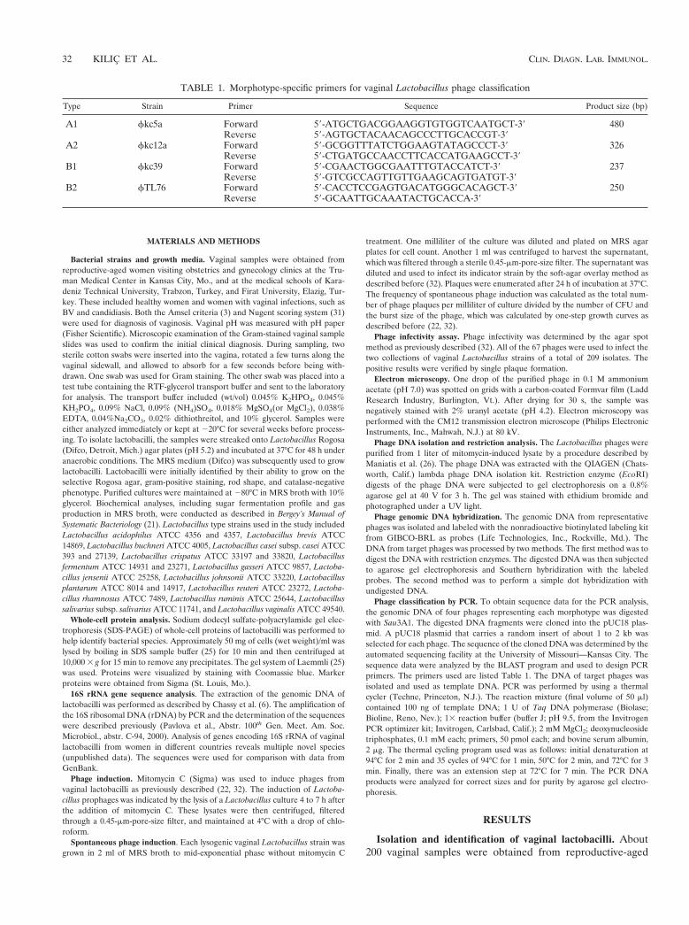
Phage classification by PCR. To obtain sequence data for the PCR analysis,the genomic DNA of four phages representing each morphotype was digestedwith Sau3A1. The digested DNA fragments were cloned into the pUC18 plas-mid. A pUC18 plasmid that carries a random insert of about 1 to 2 kb wasselected for each phage. The sequence of the cloned DNA was determined by theautomated sequencing facility at the University of Missouri—Kansas City. Thesequence data were analyzed by the BLAST program and used to design PCRprimers. The primers used are listed Table 1. The DNA of target phages wasisolated and used as template DNA. PCR was performed by using a thermalcycler (Techne, Princeton, N.J.). The reaction mixture (final volume of 50 ml)contained 100 ng of template DNA; 1 U of Taq DNA polymerase (Biolase;Bioline, Reno, Nev.); 13 reaction buffer (buffer J; pH 9.5, from the InvitrogenPCR optimizer kit; Invitrogen, Carlsbad, Calif.); 2 mM MgCl2; deoxynucleosidetriphosphates, 0.1 mM each; primers, 50 pmol each; and bovine serum albumin,2 mg. The thermal cycling program used was as follows: initial denaturation at94°C for 2 min and 35 cycles of 94°C for 1 min, 50°C for 2 min, and 72°C for 3min. Finally, there was an extension step at 72°C for 7 min. The PCR DNAproducts were analyzed for correct sizes and for purity by agarose gel electro-phoresis.
RESULTS
Isolation and identification of vaginal lactobacilli. About200 vaginal samples were obtained from reproductive-aged
TABLE 1. Morphotype-specific primers for vaginal Lactobacillus phage classification
Type Strain Primer Sequence Product size (bp)
A1 fkc5a Forward 59-ATGCTGACGGAAGGTGTGGTCAATGCT-39 480Reverse 59-AGTGCTACAACAGCCCTTGCACCGT-39
A2 fkc12a Forward 59-GCGGTTTATCTGGAAGTATAGCCCT-39 326Reverse 59-CTGATGCCAACCTTCACCATGAAGCCT-39
B1 fkc39 Forward 59-CGAACTGGCGAATTTGTACCATCT-39 237Reverse 59-GTCGCCAGTTGTTGAAGCAGTGATGT-39
B2 fTL76 Forward 59-CACCTCCGAGTGACATGGGCACAGCT-39 250Reverse 59-GCAATTGCAAATACTGCACCA-39
32 KILIC ET AL. CLIN. DIAGN. LAB. IMMUNOL.
women in Turkey and about 100 were obtained from theUnited States. While the Turkish women were all Caucasian,the American group included black (55%), white (35%), Asian(5%), Hispanic (3%), and Native American (2%) women.Some Turkish isolates did not survive the oversea shipping, soonly 102 Lactobacillus strains were obtained. From Americanwomen, 107 strains were obtained. Among the Turkish women,43 cases of BV were diagnosed, but only 22 had culturablelactobacilli. Among the American women, 14 cases of BV werediagnosed, but only 4 had culturable lactobacilli. Storage ofsamples in the RTF-glycerol buffer at 220 to 270°C did notresult in loss of Lactobacillus viability. Each collection had 10obligate anaerobic strains (about 10%). All of the remainingstrains were facultative anaerobes (Table 2).
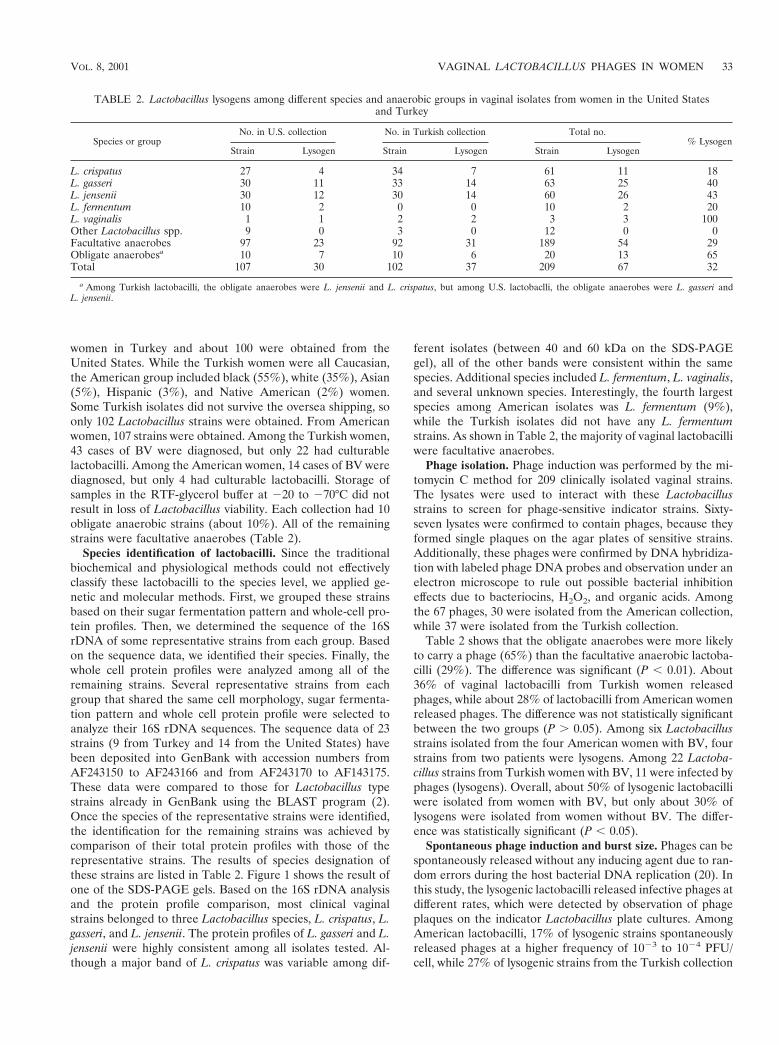
Species identification of lactobacilli. Since the traditionalbiochemical and physiological methods could not effectivelyclassify these lactobacilli to the species level, we applied ge-netic and molecular methods. First, we grouped these strainsbased on their sugar fermentation pattern and whole-cell pro-tein profiles. Then, we determined the sequence of the 16SrDNA of some representative strains from each group. Basedon the sequence data, we identified their species. Finally, thewhole cell protein profiles were analyzed among all of theremaining strains. Several representative strains from eachgroup that shared the same cell morphology, sugar fermenta-tion pattern and whole cell protein profile were selected toanalyze their 16S rDNA sequences. The sequence data of 23strains (9 from Turkey and 14 from the United States) havebeen deposited into GenBank with accession numbers fromAF243150 to AF243166 and from AF243170 to AF143175.These data were compared to those for Lactobacillus typestrains already in GenBank using the BLAST program (2).Once the species of the representative strains were identified,the identification for the remaining strains was achieved bycomparison of their total protein profiles with those of therepresentative strains. The results of species designation ofthese strains are listed in Table 2. Figure 1 shows the result ofone of the SDS-PAGE gels. Based on the 16S rDNA analysisand the protein profile comparison, most clinical vaginalstrains belonged to three Lactobacillus species, L. crispatus, L.gasseri, and L. jensenii. The protein profiles of L. gasseri and L.jensenii were highly consistent among all isolates tested. Al-though a major band of L. crispatus was variable among dif-
ferent isolates (between 40 and 60 kDa on the SDS-PAGEgel), all of the other bands were consistent within the samespecies. Additional species included L. fermentum, L. vaginalis,and several unknown species. Interestingly, the fourth largestspecies among American isolates was L. fermentum (9%),while the Turkish isolates did not have any L. fermentumstrains. As shown in Table 2, the majority of vaginal lactobacilliwere facultative anaerobes.
Phage isolation. Phage induction was performed by the mi-tomycin C method for 209 clinically isolated vaginal strains.The lysates were used to interact with these Lactobacillusstrains to screen for phage-sensitive indicator strains. Sixty-seven lysates were confirmed to contain phages, because theyformed single plaques on the agar plates of sensitive strains.Additionally, these phages were confirmed by DNA hybridiza-tion with labeled phage DNA probes and observation under anelectron microscope to rule out possible bacterial inhibitioneffects due to bacteriocins, H2O2, and organic acids. Amongthe 67 phages, 30 were isolated from the American collection,while 37 were isolated from the Turkish collection.
Table 2 shows that the obligate anaerobes were more likelyto carry a phage (65%) than the facultative anaerobic lactoba-cilli (29%). The difference was significant (P , 0.01). About36% of vaginal lactobacilli from Turkish women releasedphages, while about 28% of lactobacilli from American womenreleased phages. The difference was not statistically significantbetween the two groups (P . 0.05). Among six Lactobacillusstrains isolated from the four American women with BV, fourstrains from two patients were lysogens. Among 22 Lactoba-cillus strains from Turkish women with BV, 11 were infected byphages (lysogens). Overall, about 50% of lysogenic lactobacilliwere isolated from women with BV, but only about 30% oflysogens were isolated from women without BV. The differ-ence was statistically significant (P , 0.05).
Spontaneous phage induction and burst size. Phages can bespontaneously released without any inducing agent due to ran-dom errors during the host bacterial DNA replication (20). Inthis study, the lysogenic lactobacilli released infective phages atdifferent rates, which were detected by observation of phageplaques on the indicator Lactobacillus plate cultures. AmongAmerican lactobacilli, 17% of lysogenic strains spontaneouslyreleased phages at a higher frequency of 1023 to 1024 PFU/cell, while 27% of lysogenic strains from the Turkish collection
TABLE 2. Lactobacillus lysogens among different species and anaerobic groups in vaginal isolates from women in the United Statesand Turkey
Species or groupNo. in U.S. collection No. in Turkish collection Total no.
% LysogenStrain Lysogen Strain Lysogen Strain Lysogen
L. crispatus 27 4 34 7 61 11 18L. gasseri 30 11 33 14 63 25 40L. jensenii 30 12 30 14 60 26 43L. fermentum 10 2 0 0 10 2 20L. vaginalis 1 1 2 2 3 3 100Other Lactobacillus spp. 9 0 3 0 12 0 0Facultative anaerobes 97 23 92 31 189 54 29Obligate anaerobesa 10 7 10 6 20 13 65Total 107 30 102 37 209 67 32
a Among Turkish lactobacilli, the obligate anaerobes were L. jensenii and L. crispatus, but among U.S. lactobaclli, the obligate anaerobes were L. gasseri andL. jensenii.
VOL. 8, 2001 VAGINAL LACTOBACILLUS PHAGES IN WOMEN 33
released phages at this level. About one-third of both collec-tions released phages at an intermediate frequency (about1026 PFU/cell). Approximately one-half of the culture collec-tions from both countries spontaneously released phages at afrequency of less than 1028 PFU/cell. These data were re-peated observations, and the frequency of phage release fromeach strain was highly stable. The burst sizes were between 60and 300 phages per cell.
Phage host ranges and infection characteristics. All 67 tem-perate phages isolated from vaginal lactobacilli infected vagi-nal lactobacilli in vitro by forming clear plaques on agar plates.As shown in Table 3, the 30 phages from the United States and37 phages from Turkey infected most vaginal lactobacilli fromboth collections, including lysogenic strains. Overall, fewer lac-tobacilli isolated from Turkish women resisted phage infection
than lactobacilli isolated from U.S. women. A group of vaginallactobacilli sensitive to multiple phages was identified. Theywere used as indicator strains to display clear single plaquesafter the infection and used to screen for new phages. Therewere no apparent differences in phage sensitivity between lac-tobacilli isolated from healthy women and those from womenwith vaginal infections.
Many phages had a broad host range and infected vaginalLactobacillus strains of multiple species, including L. crispatus,L. jensenii, L. gasseri, L. fermentum, and L. vaginalis. Amongthe obligate anaerobic lactobacilli, the American collectionhad mostly L. gasseri strains, while the Turkish collection hadmostly L. jensenii strains. They were equally high in the rate ofphage lysogeny. After infection of 100 million Lactobacilluscells by these phages (multiplicity of infection, 1:10), no sur-vival colonies or lysogens could be observed, indicating lyticinfection. Nearly all temperate phages in the two collectionslytically infected other sensitive lactobacilli.
Phage morphology. The electron micrograph (Fig. 2)showed two major morphotypes, Bradley (5) type A and B,among the 67 phages studied to date. Bradley type A is char-acterized by a hexagonal head and a tail with a contractilesheath. The first type, represented by fkc21T and fkc12a,belongs to Bradley phage type A (5), because both phages hada contractile tail sheath. However, there was a difference be-tween the two phages in the head size and tail length. Addi-tionally, fkc12a had a tail plate. Bradley type B is character-ized by a hexagonal head and a tail without a contractilesheath. The second type, represented by fkc39 and fkc7a,
FIG. 1. Protein profiles of some representative Lactobacillus strains on SDS-PAGE(10% polyacrylamide). Lane M contains protein molecularweight markers. Lanes 1 to 17 contain the indicated vaginal Lactobacillus strains: 1, KC23T; 2, TL152; 3, TL145a; 4, TL143b; 5, TL114; 6, TL127a;7, TL109b; 8, TL60a; 9, TL27; 10, TL23b; 11, TL23a; 12, TL33a; 13, TL13; 14, TL102; 15, TL76; 16, TL74c; 17, TL34c. At the bottom of the gel,the species identification of each strain is indicated by a letter. C; L. crispatus; G; L. gasseri; J; L. jensenii.
TABLE 3. Infection of vaginal lactobacilli by 67 phages from theUnited States and Turkey
Infection category
No. of vaginallactobacillus strains
froma: Total
U.S.women
Turkishwomen
Infected by both phage collections 71 (20) 86 (25) 157 (45)Infected only by American phages 7 (5) 2 (0) 9 (5)Infected only by Turkish phages 3 (1) 13 (11) 16 (12)Resisted all phages 26 (4) 1 (1) 27 (5)Total 107 (30) 102 (37) 209 (67)
a The number in parentheses represents lysogenic strains in each group.
34 KILIC ET AL. CLIN. DIAGN. LAB. IMMUNOL.
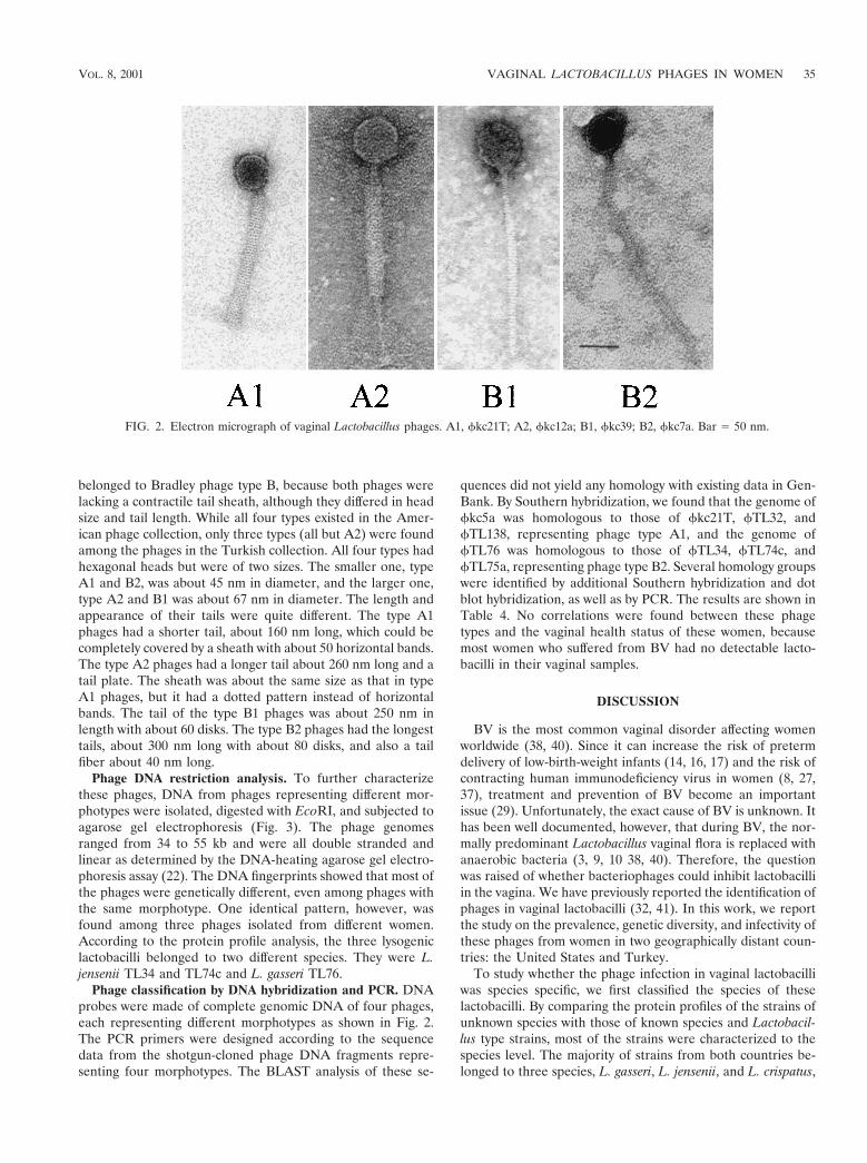
belonged to Bradley phage type B, because both phages werelacking a contractile tail sheath, although they differed in headsize and tail length. While all four types existed in the Amer-ican phage collection, only three types (all but A2) were foundamong the phages in the Turkish collection. All four types hadhexagonal heads but were of two sizes. The smaller one, typeA1 and B2, was about 45 nm in diameter, and the larger one,type A2 and B1 was about 67 nm in diameter. The length andappearance of their tails were quite different. The type A1phages had a shorter tail, about 160 nm long, which could becompletely covered by a sheath with about 50 horizontal bands.The type A2 phages had a longer tail about 260 nm long and atail plate. The sheath was about the same size as that in typeA1 phages, but it had a dotted pattern instead of horizontalbands. The tail of the type B1 phages was about 250 nm inlength with about 60 disks. The type B2 phages had the longesttails, about 300 nm long with about 80 disks, and also a tailfiber about 40 nm long.
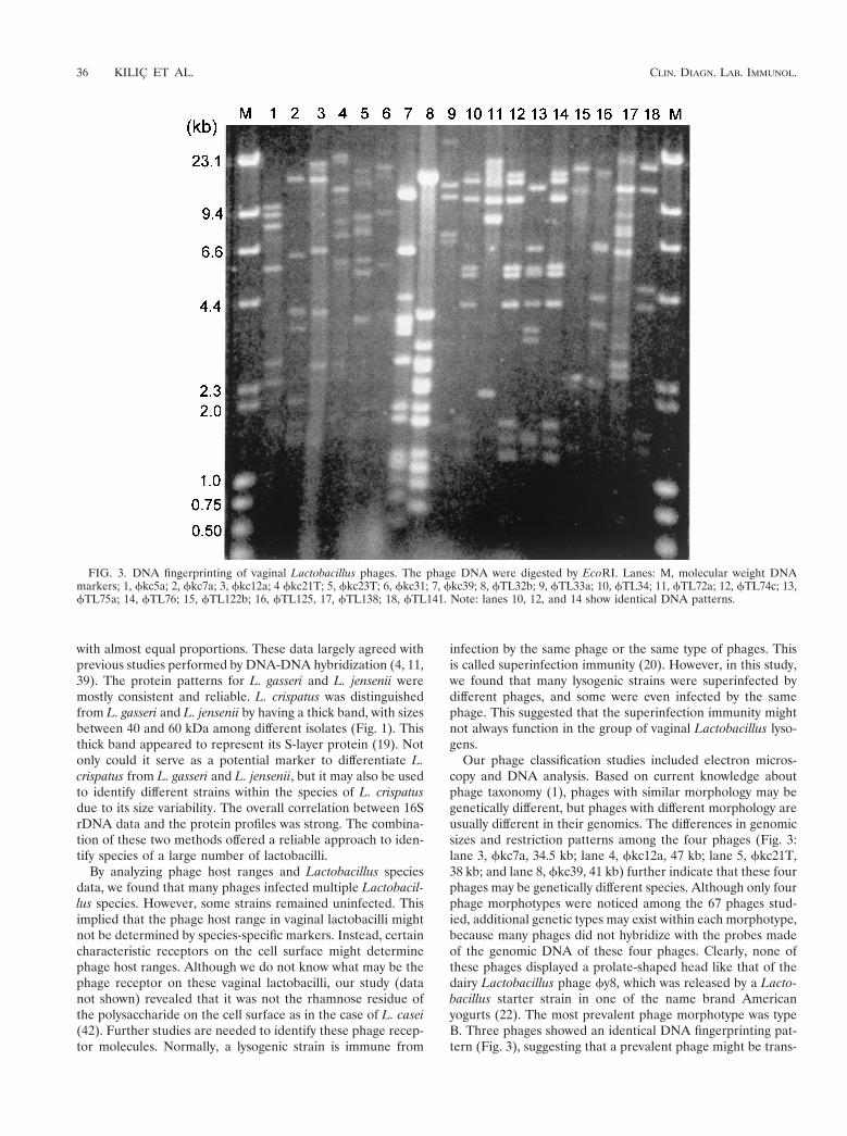
Phage DNA restriction analysis. To further characterizethese phages, DNA from phages representing different mor-photypes were isolated, digested with EcoRI, and subjected toagarose gel electrophoresis (Fig. 3). The phage genomesranged from 34 to 55 kb and were all double stranded andlinear as determined by the DNA-heating agarose gel electro-phoresis assay (22). The DNA fingerprints showed that most ofthe phages were genetically different, even among phages withthe same morphotype. One identical pattern, however, wasfound among three phages isolated from different women.According to the protein profile analysis, the three lysogeniclactobacilli belonged to two different species. They were L.jensenii TL34 and TL74c and L. gasseri TL76.
Phage classification by DNA hybridization and PCR. DNAprobes were made of complete genomic DNA of four phages,each representing different morphotypes as shown in Fig. 2.The PCR primers were designed according to the sequencedata from the shotgun-cloned phage DNA fragments repre-senting four morphotypes. The BLAST analysis of these se-
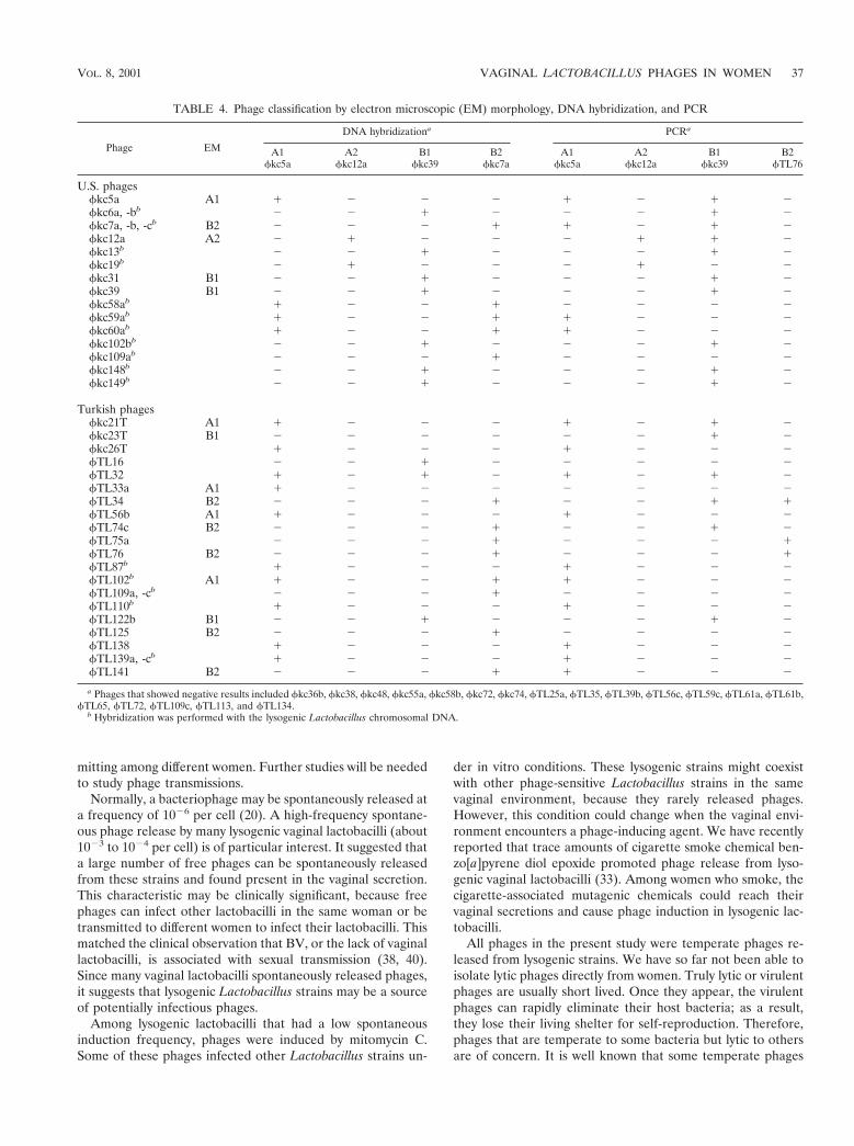
quences did not yield any homology with existing data in Gen-Bank. By Southern hybridization, we found that the genome offkc5a was homologous to those of fkc21T, fTL32, andfTL138, representing phage type A1, and the genome offTL76 was homologous to those of fTL34, fTL74c, andfTL75a, representing phage type B2. Several homology groupswere identified by additional Southern hybridization and dotblot hybridization, as well as by PCR. The results are shown inTable 4. No correlations were found between these phagetypes and the vaginal health status of these women, becausemost women who suffered from BV had no detectable lacto-bacilli in their vaginal samples.
DISCUSSION
BV is the most common vaginal disorder affecting womenworldwide (38, 40). Since it can increase the risk of pretermdelivery of low-birth-weight infants (14, 16, 17) and the risk ofcontracting human immunodeficiency virus in women (8, 27,37), treatment and prevention of BV become an importantissue (29). Unfortunately, the exact cause of BV is unknown. Ithas been well documented, however, that during BV, the nor-mally predominant Lactobacillus vaginal flora is replaced withanaerobic bacteria (3, 9, 10 38, 40). Therefore, the questionwas raised of whether bacteriophages could inhibit lactobacilliin the vagina. We have previously reported the identification ofphages in vaginal lactobacilli (32, 41). In this work, we reportthe study on the prevalence, genetic diversity, and infectivity ofthese phages from women in two geographically distant coun-tries: the United States and Turkey.
To study whether the phage infection in vaginal lactobacilliwas species specific, we first classified the species of theselactobacilli. By comparing the protein profiles of the strains ofunknown species with those of known species and Lactobacil-lus type strains, most of the strains were characterized to thespecies level. The majority of strains from both countries be-longed to three species, L. gasseri, L. jensenii, and L. crispatus,
FIG. 2. Electron micrograph of vaginal Lactobacillus phages. A1, fkc21T; A2, fkc12a; B1, fkc39; B2, fkc7a. Bar 5 50 nm.
VOL. 8, 2001 VAGINAL LACTOBACILLUS PHAGES IN WOMEN 35
with almost equal proportions. These data largely agreed withprevious studies performed by DNA-DNA hybridization (4, 11,39). The protein patterns for L. gasseri and L. jensenii weremostly consistent and reliable. L. crispatus was distinguishedfrom L. gasseri and L. jensenii by having a thick band, with sizesbetween 40 and 60 kDa among different isolates (Fig. 1). Thisthick band appeared to represent its S-layer protein (19). Notonly could it serve as a potential marker to differentiate L.crispatus from L. gasseri and L. jensenii, but it may also be usedto identify different strains within the species of L. crispatusdue to its size variability. The overall correlation between 16SrDNA data and the protein profiles was strong. The combina-tion of these two methods offered a reliable approach to iden-tify species of a large number of lactobacilli.
By analyzing phage host ranges and Lactobacillus speciesdata, we found that many phages infected multiple Lactobacil-lus species. However, some strains remained uninfected. Thisimplied that the phage host range in vaginal lactobacilli mightnot be determined by species-specific markers. Instead, certaincharacteristic receptors on the cell surface might determinephage host ranges. Although we do not know what may be thephage receptor on these vaginal lactobacilli, our study (datanot shown) revealed that it was not the rhamnose residue ofthe polysaccharide on the cell surface as in the case of L. casei(42). Further studies are needed to identify these phage recep-tor molecules. Normally, a lysogenic strain is immune from
infection by the same phage or the same type of phages. Thisis called superinfection immunity (20). However, in this study,we found that many lysogenic strains were superinfected bydifferent phages, and some were even infected by the samephage. This suggested that the superinfection immunity mightnot always function in the group of vaginal Lactobacillus lyso-gens.
Our phage classification studies included electron micros-copy and DNA analysis. Based on current knowledge aboutphage taxonomy (1), phages with similar morphology may begenetically different, but phages with different morphology areusually different in their genomics. The differences in genomicsizes and restriction patterns among the four phages (Fig. 3:lane 3, fkc7a, 34.5 kb; lane 4, fkc12a, 47 kb; lane 5, fkc21T,38 kb; and lane 8, fkc39, 41 kb) further indicate that these fourphages may be genetically different species. Although only fourphage morphotypes were noticed among the 67 phages stud-ied, additional genetic types may exist within each morphotype,because many phages did not hybridize with the probes madeof the genomic DNA of these four phages. Clearly, none ofthese phages displayed a prolate-shaped head like that of thedairy Lactobacillus phage fy8, which was released by a Lacto-bacillus starter strain in one of the name brand Americanyogurts (22). The most prevalent phage morphotype was typeB. Three phages showed an identical DNA fingerprinting pat-tern (Fig. 3), suggesting that a prevalent phage might be trans-
FIG. 3. DNA fingerprinting of vaginal Lactobacillus phages. The phage DNA were digested by EcoRI. Lanes: M, molecular weight DNAmarkers; 1, fkc5a; 2, fkc7a; 3, fkc12a; 4 fkc21T; 5, fkc23T; 6, fkc31; 7, fkc39; 8, fTL32b; 9, fTL33a; 10, fTL34; 11, fTL72a; 12, fTL74c; 13,fTL75a; 14, fTL76; 15, fTL122b; 16, fTL125, 17, fTL138; 18, fTL141. Note: lanes 10, 12, and 14 show identical DNA patterns.
36 KILIC ET AL. CLIN. DIAGN. LAB. IMMUNOL.
mitting among different women. Further studies will be neededto study phage transmissions.
Normally, a bacteriophage may be spontaneously released ata frequency of 1026 per cell (20). A high-frequency spontane-ous phage release by many lysogenic vaginal lactobacilli (about1023 to 1024 per cell) is of particular interest. It suggested thata large number of free phages can be spontaneously releasedfrom these strains and found present in the vaginal secretion.This characteristic may be clinically significant, because freephages can infect other lactobacilli in the same woman or betransmitted to different women to infect their lactobacilli. Thismatched the clinical observation that BV, or the lack of vaginallactobacilli, is associated with sexual transmission (38, 40).Since many vaginal lactobacilli spontaneously released phages,it suggests that lysogenic Lactobacillus strains may be a sourceof potentially infectious phages.
Among lysogenic lactobacilli that had a low spontaneousinduction frequency, phages were induced by mitomycin C.Some of these phages infected other Lactobacillus strains un-
der in vitro conditions. These lysogenic strains might coexistwith other phage-sensitive Lactobacillus strains in the samevaginal environment, because they rarely released phages.However, this condition could change when the vaginal envi-ronment encounters a phage-inducing agent. We have recentlyreported that trace amounts of cigarette smoke chemical ben-zo[a]pyrene diol epoxide promoted phage release from lyso-genic vaginal lactobacilli (33). Among women who smoke, thecigarette-associated mutagenic chemicals could reach theirvaginal secretions and cause phage induction in lysogenic lac-tobacilli.
All phages in the present study were temperate phages re-leased from lysogenic strains. We have so far not been able toisolate lytic phages directly from women. Truly lytic or virulentphages are usually short lived. Once they appear, the virulentphages can rapidly eliminate their host bacteria; as a result,they lose their living shelter for self-reproduction. Therefore,phages that are temperate to some bacteria but lytic to othersare of concern. It is well known that some temperate phages
TABLE 4. Phage classification by electron microscopic (EM) morphology, DNA hybridization, and PCR
Phage EM
DNA hybridizationa PCRa
A1fkc5a
A2fkc12a
B1fkc39
B2fkc7a
A1fkc5a
A2fkc12a
B1fkc39
B2fTL76
U.S. phagesfkc5a A1 1 2 2 2 1 2 1 2fkc6a, -bb 2 2 1 2 2 2 1 2fkc7a, -b, -cb B2 2 2 2 1 1 2 1 2fkc12a A2 2 1 2 2 2 1 1 2fkc13b 2 2 1 2 2 2 1 2fkc19b 2 1 2 2 2 1 2 2fkc31 B1 2 2 1 2 2 2 1 2fkc39 B1 2 2 1 2 2 2 1 2fkc58ab 1 2 2 1 2 2 2 2fkc59ab 1 2 2 1 1 2 2 2fkc60ab 1 2 2 1 1 2 2 2fkc102bb 2 2 1 2 2 2 1 2fkc109ab 2 2 2 1 2 2 2 2fkc148b 2 2 1 2 2 2 1 2fkc149b 2 2 1 2 2 2 1 2
Turkish phagesfkc21T A1 1 2 2 2 1 2 1 2fkc23T B1 2 2 2 2 2 2 1 2fkc26T 1 2 2 2 1 2 2 2fTL16 2 2 1 2 2 2 2 2fTL32 1 2 1 2 1 2 1 2fTL33a A1 1 2 2 2 2 2 2 2fTL34 B2 2 2 2 1 2 2 1 1fTL56b A1 1 2 2 2 1 2 2 2fTL74c B2 2 2 2 1 2 2 1 2fTL75a 2 2 2 1 2 2 2 1fTL76 B2 2 2 2 1 2 2 2 1fTL87b 1 2 2 2 1 2 2 2fTL102b A1 1 2 2 1 1 2 2 2fTL109a, -cb 2 2 2 1 2 2 2 2fTL110b 1 2 2 2 1 2 2 2fTL122b B1 2 2 1 2 2 2 1 2fTL125 B2 2 2 2 1 2 2 2 2fTL138 1 2 2 2 1 2 2 2fTL139a, -cb 1 2 2 2 1 2 2 2fTL141 B2 2 2 2 1 1 2 2 2
a Phages that showed negative results included fkc36b, fkc38, fkc48, fkc55a, fkc58b, fkc72, fkc74, fTL25a, fTL35, fTL39b, fTL56c, fTL59c, fTL61a, fTL61b,fTL65, fTL72, fTL109c, fTL113, and fTL134.
b Hybridization was performed with the lysogenic Lactobacillus chromosomal DNA.
VOL. 8, 2001 VAGINAL LACTOBACILLUS PHAGES IN WOMEN 37
can become virulent due to genetic mutations (36), but it isunknown why so many temperate phages from vaginal lacto-bacilli can become lytic against other vaginal Lactobacillusstrains. Probably, certain differences in the bacterial host back-ground prohibit these phages from integrating their DNA intothe chromosome of their new hosts to form lysogens (7).
In conclusion, we studied phages from vaginal lactobacilli ofwomen in Turkey and the United States. We have determinedthat most of these Lactobacillus strains belonged to three spe-cies, L. crispatus, L. gasseri, and L. jensenii. Phages isolatedfrom vaginal lactobacilli of some women lytically infected vag-inal lactobacilli of other women regardless of their countries oforigin. Four morphotypes were identified among these phages,and their host range was broad and beyond any particularLactobacillus species. Most lysogenic lactobacilli spontane-ously released phages into the environment at varied frequen-cies. This suggested that lysogenic lactobacilli could be asource of infective phages. Although the phage infection ob-served in vitro may not necessarily indicate that the samesituation could happen in vivo, the results imply that vaginallactobacilli may be eliminated or repressed by phages. Thisimplication may be important for studying the etiology of BVdue to its association with a decrease in vaginal lactobacilli.Apparently, further studies with an increased number of clin-ical samples will be needed to associate phage infections invaginal lactobacilli with women’s vaginal health.
ACKNOWLEDGMENTS
We are grateful to Susan Mou for her assistance in obtaining sam-ples from American women. We also thank S. Robinson and D. Sacku-vich for assisting with electron microscopy.
This work was supported in part by grant 02069-15-RG from theConcerned Parents for AIDS Research-AmFAR, grant K-3-40532from the University of Missouri Research Board, Public Health Ser-vice grant R03 AI45127 from the National Institute of Allergy andInfectious Diseases, and grant 2-2-25521 from the Center for Researchon Women and Gender, University of Illinois at Chicago.
REFERENCES
1. Ackermann H. W., M. S. DuBow, A. W. Jarvis, L. A. Jones, V. N. Krylov, J.Maniloff, J. Rocourt, R. S. Safferman, J. Schneider, L. Seldin, T. Sozzi, P. R.Stewart, M. Werquin, and L. Wunsche. 1992. The species concept and itsapplication to tailed phages. Arch. Virol. 124:69–82.
2. Altschul, S. F., W. Gish, W. Miller, E. W. Myers, and D. J. Lipman. 1990.Basic local alignment search tool. J. Mol. Biol. 215:403–410.
3. Amsel, R., P. A. Totten, C. A. Spiegel, K. S. C. Chen, D. A. Eschenbach, andK. K. Holmes. 1983. Nonspecific vaginitis. Diagnostic criteria and microbialand epidemiologic associations. Am. J. Med. 74:14–22.
4. Antonio, M. A., S. E. Hawes, and S. L. Hillier. 1999. The identification ofvaginal Lactobacillus species and the demographic and microbiologic char-acteristics of women colonized by these species. J. Infect. Dis. 180:1950–1956.
5. Bradley, D. E. 1967. Ultrastructure of bacteriophage and bacteriocin. Bac-teriol. Rev. 31:230–314.
6. Chassy, B., M. Gibson, and A. Guifrida. 1976. Evidence for extrachromo-somal elements in Lactobacillus. J. Bacteriol. 127:1576–1578.
7. Cluzel, P. J., J. Serio, and J. P. Accolas. 1987. Interaction of Lactobacillusbulgaricus temperate bacteriophage 0448 with host strains. Appl. Environ.Microbiol. 53:1850–1854.
8. Cohen, C. R., A. Duerr, N. Pruithithada, S. Rugpao, S. L. Hillier, P. Garcia,and K. Nelson. 1995. Bacterial vaginosis and HIV seroprevalence amongfemale commercial sex workers in Chiang Mai, Thailand. AIDS 9:1093–1097.
9. Eschenbach, D. A., P. R. Davick, B. L. Williams, S. J. Klebanoff, K. Young-Smith, C. M. Critchlow, and K. K. Holmes. 1989. Prevalence of hydrogenperoxide-producing Lactobacillus species in normal women and women withbacterial vaginosis. J. Clin. Microbiol. 27:251–256.
10. Eschenbach, D. A., S. L. Hillier, C. Critchlow, C. Stevens, T. DeRousen, andK. K. Holmes. 1988. Diagnosis and clinical manifestations of bacterial vagi-nosis. Am. J. Obstet. Gynecol. 158:819–828.
11. Giorgi, A., S. Torriani, F. Dellaglio, G. Bo, E. Stola, and L. Bernuzzi. 1987.Identification of vaginal lactobacilli from asymptomatic women. Microbio-logica 10:377–384.
12. Hallen, A., C. Jarstrand, and C. Påhlson. 1992. Treatment of bacterialvaginosis with lactobacilli. Sex. Transm. Dis. 19:146–148.
13. Harwood, B., R. Mittendorf, D. Judge, S. Dayal, and C. Walker. 1996.Patterns of vaginal douching and their association with vaginal bacteriosis.Infect. Dis. Obstet. Gynecol. 4:51.
14. Hay, P. E., R. F. Lamont, D. Taylor-Robinson, D. J. Morgan, C. A. Ison, andJ. Pearson. 1994. Abnormal bacterial colonization of the genital tract andsubsequent preterm delivery and late miscarriage. Br. Med. J. 308:295–298.
15. Hill, G. B. 1993. The microbiology of bacterial vaginosis. Am. J. Obstet.Gynecol. 169:450–454.
16. Hillier, S. L., M. A. Krohn, E. Cassen, T. R. Easterling, L. K. Rabe, and D. A.Eschenbach. 1995. The role of bacterial vaginosis and vaginal bacteria inamniotic fluid infection in women in preterm labor with intact fetal mem-branes. Clin. Infect. Dis. 20(Suppl. 2):S276–278.
17. Hillier, S. L., R. P. Nugent, D. A. Eschenbach, M. A. Krohn, R. S. Gibbs,D. H. Martin, M. F. Cotch, R. Edelman, J. G. Pastorek II, A. V. Rao, D.McNellis, J. A. Regan, J. C. Carey, M. A. Klebanoff, and the Vaginal Infec-tions and Prematurity Study Group. 1995. Association between bacterialvaginosis and preterm delviery of a low-birth-weight infant. N. Engl. J. Med.333:1737–1742.
18. Hooton, T. M., C. I. Fennell, A. M. Clark, and W. E. Stamm. 1991. Nonoxy-nol-9: differential antibacterial activity and enhancement of bacterial adher-ence to vaginal epithelial cells. J. Infect. Dis. 164:1216–1219.
19. Johnson, M. C., B. Ray, and T. Bhowmik. 1987. Selection of Lactobacillusacidophilus strains for use in “acidophilus products.” Antonie Leeuwenhoek53:215–231.
20. Joklik, W. K., H. P. Willett, and D. B. Amos. 1980. Zinsser microbiology,17th ed, p. 1178. Appleton-Century-Crofts, New York; N.Y.
21. Kandler, O., and N. Weiss. 1986. Genus Lactobacillus, p. 1209–1234. In P.Sneath (ed.), Bergey’s manual of systematic bacteriology, vol. 2. Williams, &Wilkins, Baltimore; md.
22. Kilic, A. O., S. I. Pavlova, W. Ma, and L. Tao. 1996. Analysis of Lactobacillusphages and bacteriocins in American dairy products and characterization ofa phage isolated from yogurt. Appl. Environ. Microbiol. 62:2111–2116.
23. Klebanoff, S. J., S. L. Hillier, D. A. Eschenbach, and A. M. Waltersdorph.1991. Control of the microbial flora of the vagina by H2O2-generating lac-tobacilli. J. Infect. Dis. 164:94–100.
24. Kopeloff, N. 1934. Dissociation and filtration of Lactobacillus acidophilus.J. Infect. Dis. 55:368–372.
25. Laemmli, U. K. 1970. Cleavage of structural proteins during the assembly ofthe head of the bacteriophage T4. Nature 227:684–685.
26. Maniatis, T., E. F. Fritsch, and J. Sambrook. 1982. Molecular cloning: alaboratory manual. Cold Spring Harbor Laboratory, Cold Spring Harbor,N.Y.
27. Martin, H. L., B. A. Richardson, P. M. Nyange, L. Lavreys, S. L. Hillier, B.Chohan, K. Mandaliya, J. O. Ndinya-Achola, J. Bwayo, and J. Kreiss. 1999.Vaginal lactobacilli, microbial flora, and risk of human immunodeficiencyvirus type 1 and sexually transmitted disease acquisition. J. Infect. Dis.180:1863–1868.
28. Martius, J., M. A. Krohn, S. L. Hillier, W. E. Stamm, K. K. Holmes, andD. A. Eschenbach. 1988. Relationship of vaginal Lactobacillus species, cer-vical Chlamydia trachomatis, and vaginosis to preterm birth. Obstet. Gy-necol. 71:89–95.
29. McGregor, J. A., J. I. French, R. Parker, D. Draper, E. Patterson, W. Jones,K. Thorsgard, and J. McFee. 1995. Prevention of premature birth by screen-ing and treatment for common genital tract infections. Results of a prospec-tive controlled evaluation. Am. J. Obstet. Gynecol. 173:157–167.
30. Nes, I. F., and O. Sorheim. 1984. Effect of infection of a bacteriophage in astarter culture during the production of salami dry sausage: a model study. J.Food. Sci. 49:337–340.
31. Nugent, R. P., M. A. Krohn, and S. L. Hillier. 1991. Reliability of diagnosingbacterial vaginosis is improved by a standardized method of Gram staininterpretation. J. Clin. Microbiol. 29:297–301.
32. Pavlova, S. I., A. O. Kilic, S. M. Mou, and L. Tao. 1997. Phage infection invaginal Lactobacillus: an in vitro study. Infect. Dis. Obstet. Gynecol. 5:36–44.
33. Pavlova, S. I., and L. Tao. 2000. Induction of vaginal Lactobacillus phages bythe cigarette smoke chemical benzol[a]pyrene diol epoxide. Mutat. Res.466:57–62.
34. Raya, R. R., E. G. Kleeman, J. B. Luchansky, and T. R. Klaenhammer. 1989.Characterization of the temperate bacteriophage fadh and plasmid trans-duction in Lactobacillus acidophilus ADH. Appl. Environ. Microbiol. 55:2206–2213.
35. Redondo-Lopez, V., R. L. Cook, and J. D. Sobel. 1990. Emerging role oflactobacilli in the control and maintenance of the vaginal bacterial micro-flora. Rev. Infect. Dis. 12:856–872.
36. Sechaud, L., P. J. Cluzel, M. Pousseau, A. Baumgartner, and J. P. Accolas.1988. Bacteriophages of lactobacilli. Biochimie 70:401–410.
37. Sewankambo, N., R. H. Gray, M. J. Wawer, L. Paxton, D. McNairn, F.Wabwire-Mangen, D. Serwadda, C. Li, N. Kiwanuka, S. L. Hillier, L. Rabe,
38 KILIC ET AL. CLIN. DIAGN. LAB. IMMUNOL.

C. A. Gaydos, T. C. Quinn, and J. Konde-Lule. 1997. HIV-1 infection asso-ciated with abnormal vaginal flora morphology and bacterial vaginosis. Lan-cet 350:546–550.
38. Sobel, J. D. 1997. Vaginitis. N. Engl. J. Med. 337:1896–1903.39. Song, Y. L., N. Kato, Y. Matsumiya, C. X. Liu, H. Kato, and K. Watanabe.
1999. Identification of and hydrogen peroxide production by fecal and vag-inal lactobacilli isolated from Japanese women and newborn infants. J. Clin.Microbiol. 37:3062–3064.
40. Spiegel, C. A. 1991. Bacterial vaginosis. Clin. Microbiol. Rev. 4:485–502.
41. Tao, L., S. Pavlova, S. Alpay, and A. Kilic. 1999. Initial evidence for phageinfection and transmission in vaginal lactobacilli. Int. J. Gynecol. Obstet.67(Suppl):S49.
42. Watanabe, K., and S. Takesue. 1975. Use of L-rhamnose to study irreversibleadsorption of bacteriophage PL-1 to a strain of Lactobacillus casei. J. Gen.Virol. 28:29–35.
43. Zheng, H., T. M. Alcorn, and M. S. Cohen. 1994. Effects of H2O2-producinglactobacilli on Neisseria gonorrhoeae growth and catalase activity. J. Infect.Dis. 170:1209–1215.
VOL. 8, 2001 VAGINAL LACTOBACILLUS PHAGES IN WOMEN 39