comparison between set index - caal - international
TRANSCRIPT

- 1 -
EFFECT OF THERAPEUTIC RIDING ON THE COORDINATION OF MOVEMENTS OFDOWN-
SYNDROME CHILDREN
Henriette Steiner
International Children Safety Service, Budapest, Hungary
and
Institute of Behavioural Sciences, Semmelweis University, Budapest, HUNGARY
Tibor Szilágyi
Biomechanics laboratory, Department of Biomechanics, Institute of Kinesiology and Sport Medicine,
Semmelweis University, Budapest, HUNGARY
ABSTRACT
Purpose. We examined the effect of therapeutic riding on development of Down-syndrome children.
Examination of walking is appropriate for assessing the coordination of movement and for following the
changes. We found therapeutic riding should be considered as a new form of habilitation. Pupils of an auxiliary
school participated in therapeutic riding. This raised the question how therapeutic riding affects the development
of motion, so we researched it.
Methods. We selected riding and non-riding children and conducted walk analysis one month before and after
therapeutic riding to follow the changes in their coordination. We chose to analyse walking. We used four video
cameras from four different views. We processed the data by the APAS video analyser system. We used the
Dempster model. For the construction of the model we conducted anthropometric measurements. This method
made it possible to follow the movements of the selected points of the body in three dimensions. Statistical
analysis was based on T-probe.
Results. We found significant differences between test and control groups. In the case of the children who
participated in therapeutic riding several parameters were similar to the same parameters of healthy children.
Their gait asymmetry and hip-motion asymmetry was decreased. We made several movement analyses of the
same group and these data were compared.
Conclusion. According to our results therapeutic riding may be successfully used as an additional therapy for
Down-syndrome children and it may present a form of habilitation in cases when other means of therapy are not
successful.
Keywords. Therapeutic riding, Down-syndrome, hip - motion asymmetry, asymmetry, coordination of
movements.
1. INTRODUCTION
Before the description of the measurements, it must be writes about biomechanics of gait , and Down syndrome
Hip
The hip in Down syndrome is retroverted, with excessive external rotation both in flexion and extension,
resulting in out-toe gait. Five percent of children with Down syndrome develop a dislocatable or dislocated hip.
These children are usually delayed in walking; their hips are hypermobile but not dislocatable until at two to four
years of age, the affected hip spontaneously becomes dislocated and relocated. Presenting complains are a click
in the hip, an increasing limp or "giving way," and refusal to walk With recurrent dislocation, physical activity
diminishes. The dislocations are not painful. If untreated, eventually subluxation or dislocation may become
fixed. The recurring dislocated hip is usually treated surgically.
Atlantoaxial Instability
Atlantoaxial instability is an established entity in Down syndrome
It occurs in 10% to 20% of these patients. Atlantoaxial instability in Down syndrome is caused by ligamentous
laxity of the transverse ligament that holds the odontoid process close to the anterior arch of the atlas. This
instability results in loose joints, where the cervical vertebrae slip forward and the spinal cord is vulnerable to
compression.
The neurologic manifestations of spinal compression are fatigue in walking, gait disturbance, progressive
clumsiness, incoordination, spasticity, hyperfiexion, clonus, and toe-extensor reflex. Onset of neck pain,
headache and torticollis are indicative of malposition of the odontoid..
Motor Development and Gait

- 2 -
Children with Down syndrome show a longer period of stance than independent walkers, comparable to that of
the supported walking of infants. There is a decrease in hip extension and early hip extension near the end of
swing. This is seen as an attempt to make a flatfoot contact instead of the initial heel contact. There is a decrease
in ankle sagittal plane rotation, and exaggerated abduction of the swing limb appears to be necessary for foot
clearance. There also appears to be a relationship between sitting patterns and gait patterns of children with
Down syndrome. Clinical observation suggests that children with Down syndrome usually have excessive
external rotation and abduction of the hip, demonstrated by their sitting with widespread legs, and this excessive
external rotation and abduction is displayed when they learn to sit. The wide-angled gait is caused by marked hip
retroversion, genu valgum of the knee, external tibial torsion, and excessively pronated feet. It has been proven
that ambulation performance, including balance and jumping, can be significantly improved in children with
Down syndrome with even minimal physical therapy sessions, such as jumping classes. This type of therapy
should be encouraged.
Considerations In the Correction of Congenital Deformities
Early detection and treatment of congenital pedal deformities is important in a child with Down syndrome. Since
these children are subject to a multiplicity of orthopedic problems, an aggressive program to maintain proper
skeletal alignment can significantly decrease the severity of these problems and allow the individual to function
much more efficiently. In a report on management of foot and knee deformities in the mentally retarded, Lindsey
and Drennan asserted that "proper alignment of the immature foot will frequently decrease the external rotation
of the limb and result in development of a more appropriate gait pattern." Rather than being aggressively treated,
many of the common congenital deformities, such as metatarsus adductus and tibial torsion, are overlooked
because of the patients' many other medical and orthopedic problems, and no treatment is rendered. The
treatment modalities used in the correction of congenital foot and torsional abnormalities in the child with Down
syndrome are the same that would be used in the normal patient. These include serial immobilization casting,
corrective shoes and splints, and surgery. Due to the prolonged excessive ligamentous laxity and the relatively
slower foot growth, corrective modalities often are required for longer periods of time in the child with Down
syndrome. Immobilization modalities that impede walking, such as plaster casts or restrictive splinting, should
be avoided in the older child, since these can further delay the progression of neuromotor development in a child
that already will exhibit a significant delay in learning to walk. The use of properly modified corrective shoes
should be encouraged when correcting foot pathology in children with Down syndrome who have progressed
beyond the states of sitting independently and crawling. Reduction of Out-Toe Gait and Genu Valgum
2. METHODOLOGY
Our work hypothesis was that if riding changes the coordination of movements in these children it will exert
effects on their walking as well Walking requires coordinated work of many muscles, virtually the whole body.
Execution of these complex movements requires not only appropriate development of the muscular and the
osseous systems but it requires adequate control of movements (e.g. faultless working of flexor and extensor
reflexes) as well. We started our examinations (with methods learned from foreign experiences) in Budapest.
Criteria of investigation. Subjects had to have the presence of Down-syndrome and their age had to be in the
range of 10-13 years. We examined 30 children, but four of them put out of the groups, because of illness
(staying in hospital).
Features Children participating in
therapeutic riding
Control group
Disease Down syndrome Down syndrome
Age 10-13 years 10-13 years
Number of children 13 13
Therapy Therapeutic riding
Exercises:
physioball, sitting up,
leg raising on wall-bars
Duration 1 month 1 month
1 The two examined groups
Subjects of investigation. We selected riding and non-riding children and conducted walk analysis before and
after therapeutic riding to follow the changes in their coordination. We chose the gait analysis. The pupils of the
auxiliary school were examined by the physician of the school and all of them were advised to participate in our
investigation. The parents of the 30 children signed an agreement that they would accept the result of the random
drawing that formed the riding and the non-riding groups. While the riding group participated in therapeutic
riding the other group did their habitual exercises (as before). The physician of the school advised every child to
participate in therapeutic riding. Orthopedic doctors radiographed functional cervical radiogram. The parents of

- 3 -
the children decided whether their child should participate in therapeutic riding or not. Our examination is a
comparative study of the two therapeutic methods.
Before testing subject signed an informed consent form approved By the Policy and Review Committee on
Human Research of Semmelweis University, children’s guardians and school.
Therapeutic methods. We analyzed two different methods to compare the effectiveness of rehabilitational
techniques. The investigated population was divided into two groups. For the control group (group 1) three times
a week 1-1 hour of classical kinesitherapy was conducted. In the test group (group 2) once a week the children
were trained by special riding for 15 minutes.
Type of special riding. As for some aspects western style riding fits best for therapeutic riding. In case of
backward children special attention has to be paid to the safety of the riders during therapeutic riding. This
requires increased attention and tolerance of the trainer and unconditional obedience of the horse. Equipment
used in western style riding
Gait analysis. We made video recordings of the walk of the children one month before and after the therapy.
The walk was recorded from four views (front, rear, left and right). The video recordings were processed by the
APAS (Ariel Performance Analysis System). This system made it possible to present three dimensional
kinematics parameters by the analysis of video recordings made in everyday situations
Equipment for analysis. We used four video cameras (type: Panasonic M10) from four different views.
Sampling frequency was 50 frames/second (sampling rate 0.02 second) with shutter speed of 1/250 second. We
processed the data by the APAS video analyzer system (VCR is Panasonic AG-7350, Computer is AST Bravo
486/25). Digitalization of landmarks was made manually (relative digitalization error 3-5%). The system
generated the 3D database with direct linear transformation (DLT). We used the software version APAS rev.
6.73. We made smoothing (noise filtering) through quintal spline algorithm (built in the APAS).
2 The equipment for analysis

- 4 -
The validation system. After setting the cameras calibration was performed. For our investigations we used an
eight-point calibration parallelepipedon like cubic structure. After the measurement situation was set and
recorded the parallelepipedon was removed so as not to bother the children in moving. The coordinates of the
eight control points are in the table. The coordinates of the first point are not zero in all three dimensions because
we shifted the coordinates in relation to a virtual origo to enable easier visualization of the data (none of the
coordinates fall below zero). The eight points determine the three dimensions of space. The two bolded black
marks indicate a 200 cm long section in which the two gait cycle were analysed.
The body model. We used a body model to follow the movements of several points of the body. On processing
the data we applied a modified form of the Dempster's body model that consists of eighteen points and several
lines that connect them. The nineteenth point is the centre of mass of the body. The Dempster's model was
modified in the following way. We defined the landmarks, segments, and interconnections of the model. For the
construction of the model we conducted anthropometric measurements. The global and local anthropometric data
of every child were taken into consideration (segment lengths, relative body masses, radiuses of gyration).
Height and body weight were measured and the partial centres of mass of the body segments were calculated.
The final model was constructed after using these data. This method made it possible to follow the movements
of the selected points of the body in three dimensions.
3 Control points and the Dempster Body-model
Statistical analysis. The basic of statistical analysis was T-probe. We compared the differences between mean
values of parameters of selected groups (length of gait cycle). The equality of standard deviation in the statistical
populations was controlled by F-probe
7 (x = 250, y = 150, z = 100)
y
startline
2
1 (x = 50, y = 0, z = 50)
4
3
5
6
8
endline
fixed point
x
z
right hand – 1
right wrist – 2
right elbow – 3
right shoulder – 4
right hip – 5
right knee – 6
right ankle – 7
right foot – 8
16 – left hand
15 – left wrist
14 – left elbow
13 – left shoulder
12 – left hip
11 – left knee
10 – left ankle
9 – left foot
17
19
18 – head
17 – chin,
19 – center of
gravity

- 5 -
3. RESULTS
Data on the coordination of movements of "riding" and "non-riding" children were compared. The data of only
13-13 children were processed because 2-2 children of the groups could not participate in the second
measurement series. In the next section the coordination of movements of 1-1 child from both groups will be
analyzed.
The headway (x-axis) motion of both legs of the "riding" child was plotted against time (Figure 4-5). The first
Figure shows that the two legs were exposed to unequal loading. The child made shorter steps with the right leg
and the foot spent longer time on the ground (the straight sections of the curves are longer). The enclosed areas
of the two curves are not equal because of unequal loading. The vertical lines indicate the sections of the curve
when both feet are on the ground at the same time (double supporting). Asymmetry of motion can be observed.
The right leg of the child was weaker than the left. The second Figure indicates that the influence of riding was
that asymmetry of the motion of the two legs decreased (the areas enclosed by the curves are nearly equal) and
the speed of walking increased (the duration of double supporting decreased).
The headway (x-axis) motion of both legs of the "non-riding" child was similarly plotted against time (Figure 6-
7). The areas enclosed by the curves are not equal due to asymmetric loading. The vertical lines indicate double
supportings. Asymmetry of motion is clearly visible. No betterment was observed in the walking technique of
these children after the treatment. During the second series of measurements higher speed was observed, but the
duration of double supporting showed significant increase. There is a seeming contradiction between the higher
speed and the longer duration of double supporting (that slow walking) but this can be construed by the
worsening of balance and that this balance is corrected by conscious motory control (longer double supporting).
The swing phase of the step became quicker and the resultant of these two factors is higher speed.
Figure A
0
50
100
150
200
250
300
1 8
15
22
29
36
43
50
57
64
71
78
85
92
99
106
113
120
127
134
141
Time
Cm
RIGHT FOOT LEFT FOOT
Figure B
0
50
100
150
200
250
300
1 4 7
10
13
16
19
22
25
28
31
34
37
40
43
46
49
52
55
Time
Cm
RIGHT FOOT LEFT FOOT

- 6 -
Figure C
0
50
100
150
200
250
300
1 5 9
13
17
21
25
29
33
37
41
45
49
53
57
61
65
69
73
Time
Cm
RIGHT FOOT LEFT FOOT
Figure D
0
50
100
150
200
250
300
1 5 9
13
17
21
25
29
33
37
41
45
49
53
57
61
65
69
73
Time
cm
RIGHT FOOT LEFT FOOT
Figure E
-50
0
50
100
150
200
250
300
1 7
13
19
25
31
37
43
49
55
61
67
73
79
85
91
97
103
109
115
Time
cm
RIGHT FOOT LEFT FOOT
4 Motion of both legs
Figure A riding child before therapeutic riding
Figure B riding child after therapeutic riding
Figure C of non- riding child before therapy
Figure D non- riding child after therapy
Figure E Normal child
In the next step we analyzed the motion of the hip during walking (Figure 5). The three dimensional motion of
the hip before the treatment can be characterized in the following way: the motion is performed more or less in

- 7 -
opposite phase but the left side of the hip moves with much greater amplitude during walking. Asymmetry is
clearly visible. The three phases of the healthy motion of the hip (supporting, rolling and pushing off) cannot be
found because the hip is stiff.
In Figure the motion of the two hips is compared after therapeutic riding. The two sides moved more
symmetrically than before. The shape of the curve changed because one component of the healthy step cycle
appeared in the motion of the hips. Figure shows the motion of the hip of a healthy child of similar age. The
three phases of motion can be followed and identified (0.5–1.0 sec). Its appearance is like the inverse of the first
Figure because a Down-syndrome child does not step on the ground quickly like healthy people do. This
difference is compensated with a quick pushing off.
The analysis of the motion of centerogravity shows the balance of the gait . In two dimenson measurements ( x
and z ) the centerogravity often moves with bigger amplitude

- 8 -
Figure A
-40
-20
0
20
40
60
80
100
1201 7
13
19
25
31
37
43
49
55
61
67
73
79
85
91
97
103
109
115
121
127
133
139
Time
cm
cgx cgz
Figure B
-50
0
50
100
150
1 4 7
10
13
16
19
22
25
28
31
34
37
40
43
46
49
52
55
Time
cm
cgx cgz
Figure C
-40
-20
0
20
40
60
80
1 5 9
13
17
21
25
29
33
37
41
45
49
53
57
61
65
69
73
Time
cm
cgx cgz

- 9 -
Figure D
0
20
40
60
80
100
120
140
1 5 9 13 17 21 25 29 33 37 41 45 49 53 57 61 65 69 73
time
cgx cgy
Figure E
-40
-20
0
20
40
60
80
100
120
140
1 7 13 19 25 31 37 43 49 55 61 67 73 79 85 91 97 103 109 115
Time
cm
cgx cgz
5 The motion of centerogravity
Figure A riding child before therapeutic riding
Figure B riding child after therapeutic riding
Figure C of non- riding child before therapy
Figure D non- riding child after therapy
Figure E Normal child
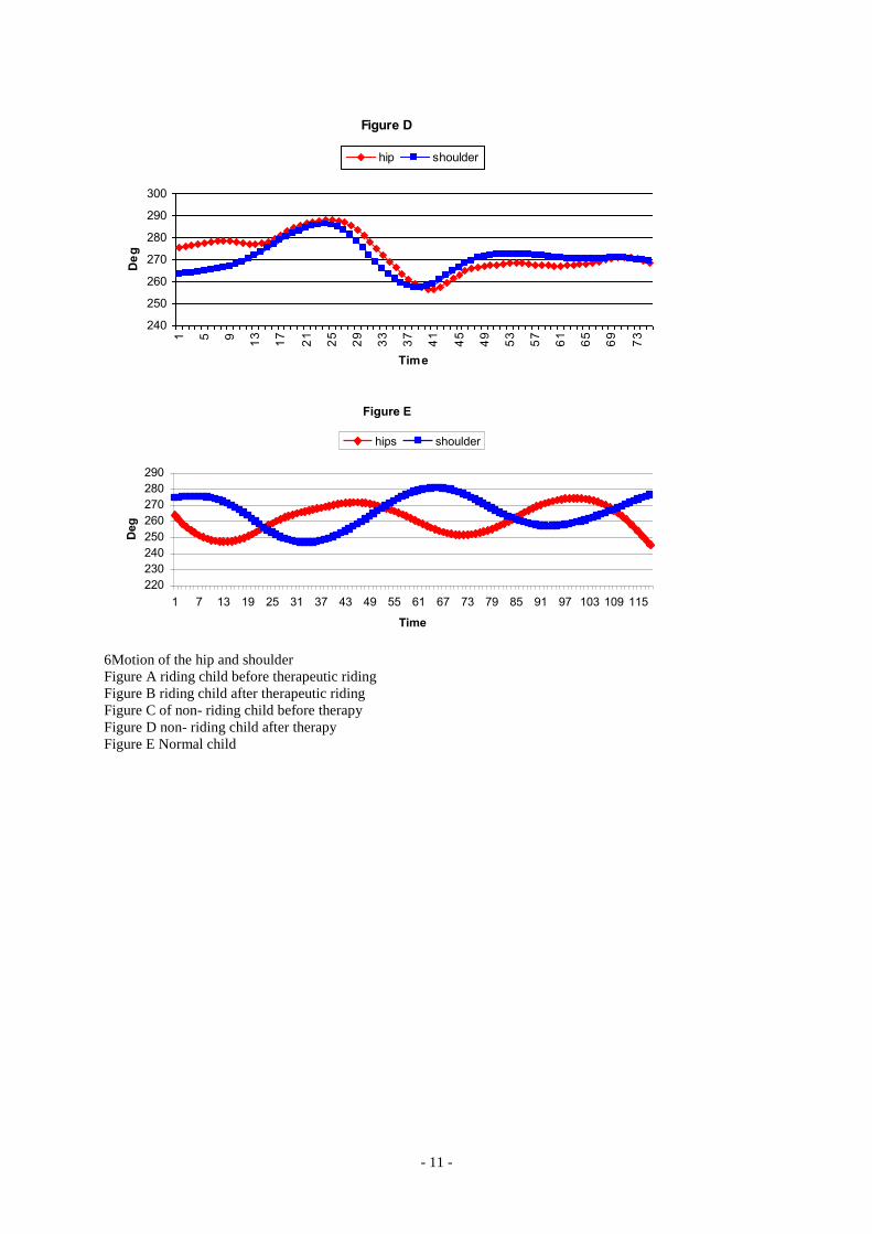
Analysis of the motion of the shoulder and the hip axes gives insight into the motion of the spine and the motion
of the hip and the shoulder of the same side (Figure 11-13). Before treatment the hips moved stiffly while the
shoulders made significant excursions during the step cycle. The stiffness of the hips is compensated with the
movements of the shoulders (0.5–1.0 sec). After therapeutic riding we observed that the hips and the shoulders
moved in the same phase (the hips and shoulders of the same side moved together) because the turning of the
trunk that helped walking. The amplitude of the excursion of the two hips increased and were nearly equal.
The hips and shoulders of a healthy child move in the opposite phase. With therapeutic riding a transitional
condition was achieved that may be further developed.

- 10 -
Figure A
0
20
40
60
80
100
120
1 8
15
22
29
36
43
50
57
64
71
78
85
92
99
106
113
120
127
134
141
Time
De
g
Hips Soulders
Figure B
230
240
250
260
270
280
290
300
310
1 3 5 7 9
11
13
15
17
19
21
23
25
27
29
31
33
35
Time
De
g
hips shoulders
Figure C
240
250
260
270
280
290
300
1 5 9
13
17
21
25
29
33
37
41
45
49
53
57
61
65
69
73
Time
De
g
hips shoulders
- 11 -
Figure D
240
250
260
270
280
290
300
1 5 9
13
17
21
25
29
33
37
41
45
49
53
57
61
65
69
73
Time
De
g
hip shoulder
Figure E
220
230
240
250
260
270
280
290
1 7 13 19 25 31 37 43 49 55 61 67 73 79 85 91 97 103 109 115
Time
Deg
hips shoulder
6Motion of the hip and shoulder
Figure A riding child before therapeutic riding
Figure B riding child after therapeutic riding
Figure C of non- riding child before therapy
Figure D non- riding child after therapy
Figure E Normal child

- 12 -
Figure A
-100
0
100
200
300
4001 8
15
22
29
36
43
50
57
64
71
78
85
92
99
106
113
120
127
134
141
Time
cm
/s
right foot left foot
Figure B
-100
0
100
200
300
400
1 8
15
22
29
36
43
50
57
64
71
78
85
92
99
106
113
120
127
134
141
Time
cm
/s
right foot left foot
Figure C
-100
0
100
200
300
1 5 9
13
17
21
25
29
33
37
41
45
49
53
57
61
65
69
73
Time
Cm
/s
right foot left foot

- 13 -
Figure D
-50
0
50
100
150
200
250
300
1 4 7
10
13
16
19
22
25
28
31
34
37
40
43
46
49
52
55
58
61
64
67
70
73
Time
cm
/s
right foot left foot
Figure E
-100
0
100
200
300
400
500
1 7
13
19
25
31
37
43
49
55
61
67
73
79
85
91
97
103
109
115
Time
cm
/s
right foot left foot
7 Velocity of feet e
Figure A riding child before therapeutic riding
Figure B riding child after therapeutic riding
Figure C of non- riding child before therapy
Figure D non- riding child after therapy
Figure E Normal child
Analysis of the Velocity of feet was given new information from the asszimmetry. The right side is weaker ,
but after the therapeutic riding the assimetry is neraer the normal gait. ( in normal gait we measured a little
asimmetry, but statisticaly is non significant
When we compared the "riding" and the "non-riding" groups several considerations had to be taken. For the
analysis of the coordination of movements we had to choose a parameter that adequately indicates the occurring
changes: improvement or relapse had to be followed by consistent changes of the parameter. The parameter had
to give information of the extent of hip - motion asymmetry, asymmetry and balance, altogether: the
coordination of movements. The length of the steps is considered to be a parameter of this kind (increase of the
length of the steps indicates higher speed, better balance and better coordination of movements). As the motion
of a Down-syndrome child is asymmetric we measured the length of steps between two supporting phases of one
leg by using the selected points of the body model (in cm). The measurements were performed one month before
and one month after the therapy. Means and coefficients of variance were calculated. With dual T-probe we
checked whether significant changes can be found in the motion of the two sides.
Significant increase was detected in the length of the steps of both legs, which indicates better coordination of
movements and decrease of hip-motion asymmetry. We measured the same groups for two years and searched
the asymmetry of two sides. There are no differences between the sides, like in healthy children (Figure 14-15).

- 14 -
14 Length of the steps in the riding group
15 Length of the steps in the non-riding group
4. CONCLUSIONS
Thus, as we have shown previously, coordination of movements is controlled by several factors. For the
development of motion the extent of asymmetry and muscular atrophy/hypotony have to be diminished and
appropriate working of muscles and coordination of movements have to be developed. For this purpose special
exercises are used to cure muscular hypotony, to help the establishment of normal muscular tension and help to
strengthen the "less developed side" of the children. Our parameters help us to control the changes of motion. As
you can see in results, therapeutic riding makes better balance, smaller asymmetry, and hip-motion asymmetry.
Last but not least it is important to examine the costs of therapeutic riding and how it compares to the costs of
Therapeutic riding
Costs of therapeutic riding Costs of habilitation
Therapeutic riding Costs (USD) Habilitation Cost (USD)OEP
One child/hour 100 Fiziotherapeutist with one child
one hour in hospital rehabilitation
Costs per year 52 x 100 = 5200 Costs per year 52 x
16 Costs of therapies (therapeutic riding vs.other habilitation) OEP
According to our results therapeutic riding may be successfully used as an additional therapy for Down-
syndrome children and it may present a form of habilitation in cases when other means of therapy are not
applicable (e.g. because of psychical and physical conditions).
5. REFERENCES
1. Bennet GC, Rang M, Roye DP, Aprin H. Dislocation of the hip in trisomy 21. Bone Joint Surg Br 1982;
64(3): 289-94.
2. Millar AL, Fernhall B, Burkett LN. Effects of aerobic training in adolescents with Down syndrome.
Med Sci Sports Exerc. 1993 Feb; 25(2): 270-4.
3. Takanashi J, Sugita K, Honda A, Niimi H. Moyamoya syndrome in a patient with Down syndrome
presenting with chorea. Pediatr. Neurol. 1993 Sep-Oct; 9(5): 396-8.
4. Turner ML. Rebecca's ride. Am J Nurs. 1994 Jan; 94(1): 96.
5. APAS System Description and User's Guide Ariel Life System Incorporated, San Diego, CA, USA,
1993
6. Barton, J.: Bevezetés a biomechanikába (Introduction to Biomechanics) Egészségügyi Főiskolák
tankönyve, AESCULART, Budapest, Hungary, 1996
LENGTH OF THE STEPS IN THE NON-RIDING GROUP (Down-syndrome)
(Averages of four measurements)
0
5
10
15
20
25
30
35
1 2 3 4
RIGHT side
LEFT side
Tright1.2 = 0,00816*
Tright3.4 = 0,3559
T3 left1.2 = 0,00002712*
T4 left3.4 = 0,2428
*SIGNIFICANT
LENGTH OF THE STEPS IN THE RIDING GROUP (Down-symdrome)
(Averages of four measurements)
05
101520253035404550
1 2 3 4
RIGHT side
LEFT side
Tright1.2 = 0,00000259*
Tright3.4 = 0,0000404*
T3 left1.2 = 0,000064*
T4 left3.4 = 0,005*
*SIGNIFICANT

- 15 -
7. Szilágyi T., Bartos G., Tóth Sz., Szántó M.: A mozgásanalízis felhasználásának lehetőségei a klinikai
gyakorlatban (Utilization of Motion Analysis in Clinical Practice), Mozgásterápia IV., 1996/4, pp. 3-10
8. Aprin H, Zink WP, Hall JE: Management of dislocation of the hip in Down syndrome. J Pediatr Orthop
5: 428, 1985.
9. Mark Castelli Biomechanical management of children and adolescents with down syndrome: proper
diagnosis of biomechanical abnormalities allows for more effective treatment of this condition -
Biomechanics & Orthotics 2003
10. Aprin H, Zink WP, Hall JE: Management of dislocation of the hip in Down syndrome. J Pediatr Orthop
5: 428, 1985.
11. Can J: Mental and motor development in young Mongol children. J Ment Defic Res 14: 205, 1970.
12. Caselli MA, Cohen-Sobel E, Thompson J, Adler J, Gonzalez L: Biomechanical management of
children and adolescents with Down syndrome. JAPMA 81:119, 1991.
13. Committee on Sports Medicine: Atlantoaxial instability in Down syndrome. Pediatrics 74: 152,1984.
14. Diamond LS Management of inherited disorders of the skeleton. Course Led 25: 107, 1976.
15. Diamond LS, Lynne D, Sigman B: Orthopedic disorders in patients with Down's syndrome. Orthop
Clin North Am 12: 57, 1981.
16. Dugdale TW, Renshaw TS: Instability of the patellofemoral joint in Down syndrome. J Bone Joint
Surg 68A: 405, 1986.
17. Hreidarsson S, Magran G, Singer H: Symptomatic atlantoaxial dislocation in Down syndrome.
Pediatrics 69: 568,1982.
18. Parker AW, Bronks R: Gait of children with Down syndrome. Arch Phys Med Rehabil 61:343, 1980.
19. Parker AW, Bronks R, Snyder CW: Walking patterns in Down's syndrome. J Ment Defic Res 30: 317,
1986.
20. Pueschel SM, Scola FH: Atlantoaxial instability in individuals with Down syndrome: epidemiologic
radiographic, and clinical studies. Pediatrics 80: 555, 1987.
21. Tachdjian MO: Pediatric Orthopedics, 2nd Ed, WB Saunders Company, Philadelphia, 1990.
22. Wang WY, Ju YH: Promoting balance and jumping skills in children with Down syndrome. Percept
Mot Skills 94:443,2002.
23. Dr. Caselli Is Staff Podiatrist at the VA Hudson Valley Health Care System and Adjunct Professor,
Department of Orthopedic Sciences, New York College of Podiatric Medicine.
AFFILLATION
Corresponding author: Henriette Steiner Human – Biologist
International Children Safety Service
Budapest, Hungary
1066 Teréz krt 24
Phone: +36 1 4757000
E-mail: [email protected]
Tibor Szilágyi
Biomechanics laboratory, Department of Biomechanics, Institute of Kinesiology and Sport Medicine,
Semmelweis University, Budapest, Alkotás út 44., HUNGARY