conformational analysis of a polyconjugated protein …original paper conformational analysis of a...
TRANSCRIPT
ORIGINAL PAPER
Conformational analysis of a polyconjugated protein-bindingligand by joint quantum chemistry and polarizable molecularmechanics. Addressing the issues of anisotropy, conjugation,polarization, and multipole transferability
Elodie Goldwaser & Benoit de Courcy & Luc Demange & Christiane Garbay &
Françoise Raynaud & Reda Hadj-Slimane & Jean-Philip Piquemal & Nohad Gresh
Received: 10 March 2014 /Accepted: 21 September 2014# Springer-Verlag Berlin Heidelberg 2014
Abstract We investigate the conformational propertiesof a potent inhibitor of neuropilin-1, a protein involvedin cancer processes and macular degeneration. This in-hibitor consists of four aromatic/conjugated fragments: abenzimidazole, a methylbenzene, a carboxythiourea, anda benzene-linker dioxane, and these fragments are alllinked together by conjugated bonds. The calculationsuse the SIBFA polarizable molecular mechanics proce-dure. Prior to docking simulations, it is essential toensure that variations in the ligand conformational en-ergy upon rotations around its six main-chain torsionalbonds are correctly represented (as compared to high-
level ab initio quantum chemistry, QC). This is done intwo successive calibration stages and one validationstage. In the latter, the minima identified followingindependent stepwise variations of each of the sixmain-chain torsion angles are used as starting pointsfor energy minimization of all the torsion angles simul-taneously. Single-point QC calculations of the mini-mized structures are then done to compare their relativeenergies ΔEconf to the SIBFA ones. We compare threedifferent methods of deriving the multipoles and polar-izabilities of the central, most critical moiety of theinhibitor: carboxythiourea (CTU). The representationthat gives the best agreement with QC is the one thatincludes the effects of the mutual polarization energyEpol between the amide and thioamide moieties. Thisagain highlights the critical role of this contribution.The implications and perspectives of these findings arediscussed.
Keywords Polarizable force fields .Multipoles . Conjugation
Introduction
Our laboratories are involved in the design of inhibitorsof the neuropilin-1 (NRP-1) protein. NRP-1 is a co-receptor of vascular endothelial growth factor (VEGF),which promotes angiogenesis and is involved in severaldiseases such as cancer and macular denegeracy [1, 2],so inhibitors of NRP-1 could block the development ofthose pathologies. Virtual screening studies have recent-ly identified a potent inhibitor of NRP-1 which was
Electronic supplementary material The online version of this article(doi:10.1007/s00894-014-2472-5) contains supplementary material,which is available to authorized users.
E. Goldwaser : L. Demange :C. Garbay : F. Raynaud :N. Gresh (*)Laboratoire de Chimie et Biochimie Pharmacologiques etToxicologiques, UMR 8601, UFR Biomédicale, UniversitéParisDescartes, 45, rue des Saints-Pères, 75006 Paris, Francee-mail: [email protected]
E. Goldwaser :B. de Courcy : J.<P. Piquemal (*)Laboratoire de Chimie Théorique, Sorbonne Universités, UPMC,Paris 6, case courrier 137, 4, place Jussieu, F75252 Paris, Francee-mail: [email protected]
R. Hadj-SlimaneTragex Pharma, 29, rue Marcel Dassault, 92100 BoulogneBillancourt, France
Present Address:L. DemangeInstitut de Chimie de Nice (ICN), UMR 7272 CNRS, Université deNice Sophia-Antipolis, Parc Valrose, 06000 Nice, France
J Mol Model (2014) 20:2472DOI 10.1007/s00894-014-2472-5
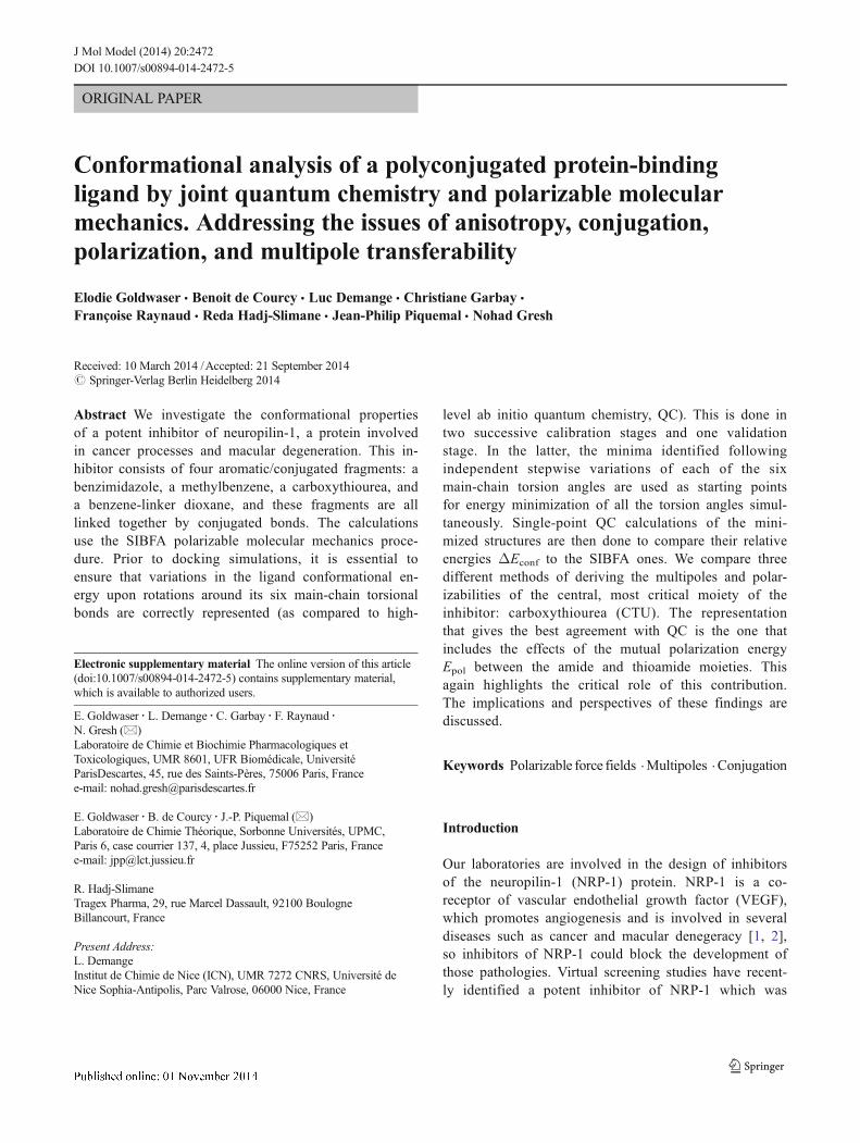
denoted ‘lig-47’ and shown to possess submicromolaractivity on several cancer cell lines. Its structure (Fig. 1)contains the following chemical groups: a benzimid-azole, a methylbenzene, a carboxythiourea, and abenzene-substituted dioxane. Lig-47 is a highly conju-gated drug, and the connections between its four groupsall involve unsaturated atoms with sp or sp2 hybridiza-tion. In the present work, we investigate the conforma-tional features of lig-47 in detail, as knowledge of thesefeatures is required in order to understand its complex-ation to NRP-1.
Unraveling the structural and energetic factors in-volved in NRP-1–lig-47 complexation is a necessarystep in the design of new derivatives with enhancedaffinities. However, with the exception of complexesof NRP-1 with the naturally occurring peptide tuftsin[3] and small peptide-like ligands [4], no high-resolutionstructural studies of complexes of NRP-1 with inhibitorsor ligands are available. Two such NRP-1 complexescould be used as candidate starting points for lig-47docking. However, there is a major issue relating tothe conformational flexibility of the drug. On the onehand, it could adopt an extended conformation, similarto tuftsin; alternatively, it could adopt a compact con-formation, as found from initial studies performed withthe SURFLEX software [5] (Borriello et al. is Cancer Letts.,submitted). Whereas molecular dynamics (MD) and en-ergy minimization (EM) are likely to identify alternativeposes with intermediate drug conformations, it is neces-sary to be able to correctly rank the energies of all candidate
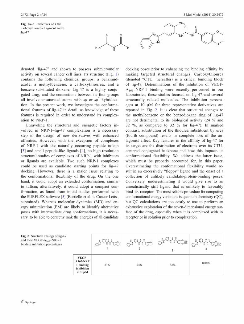
docking poses prior to enhancing the binding affinity bymaking targeted structural changes. Carboxythiourea(denoted “CTU” hereafter) is a critical building blockof lig-47. Determinations of the inhibition of VEGF-A165–NRP-1 binding were recently performed in ourlaboratories; these studies focused on lig-47 and severalstructurally related molecules. The inhibition percent-ages at 10 μM for three representative derivatives arereported in Fig. 2. It is clear that structural changes tothe methylbenzene or the benzodioxane ring of lig-47are not detrimental to its biological activity (24 % and32 %, as compared to 32 % for lig-47). In markedcontrast, substitution of the thiourea substituent by urea(fourth compound) results in complete loss of the an-tagonist effect. Key features in the affinity of lig-47 forits target are the distribution of electrons over its CTU-centered conjugated backbone and how this impacts itsconformational flexibility. We address the latter issue,which must be properly accounted for, in this paper.Overestimating the confomational flexibility would re-sult in an excessively “floppy” ligand and the onset of acollection of unlikely candidate-protein-binding poses.Conversely, underestimating it would give rise to anunrealistically stiff ligand that is unlikely to favorablybind its receptor. Themost reliable procedure for computingconformational energy variations is quantum chemistry (QC),but QC calculations are too costly to use to perform anexhaustive exploration of the seven-dimensional energy sur-face of the drug, especially when it is complexed with itsreceptor or in solution prior to complexation.
H2N
S
N
H
O
H
Fig. 1a–b Structures of a thecarboxythiourea fragment and blig-47
1 23
4
VEGF-A165/NRP1 binding inhibition at 10µM
33% 24% 32%0.00%
Fig. 2 Sructural analogs of lig-47and their VEGF-A165–NRP-1binding inhibition percentages
2472, Page 2 of 24 J Mol Model (2014) 20:2472
In our laboratories, we are involved in the devel-opment of an accurate, QC-grounded molecularmechanics/dynamics potential denoted SIBFA (sumof interactions between fragment ab initio computed),which is used to calculate inter- and intramolecularinteractions [6, 7]and has been applied to ligand–pro-tein complexes (reviewed in [8–10]). The vast major-ity of the conformation changes dealt with so far inthis context have involved rotations around singlebonds, for which a single threefold torsion barrierwith amplitudes in the 0–2.2 kcal/mol range couldsuffice. By contrast, all of the torsion angles in lig-47 are associated with conjugated bonds. It is thusclear that a threefold (n=3, see Eq. 2 below) barrieris inadequate and that barriers with n=1 and n=2should be calibrated instead. These barriers alonecould ensure coplanarity of the two connected frag-ments in order to account for conjugation. In theirabsence, steric repulsions between the connected frag-ments would result in a mutually perpendicular ar-rangement. However, a reliable conformation studyof lig-47 involves more than just calibrating torsionbarriers. This is because atoms in all four of thebuilding blocks of lig-47 can move closer togetherduring EM or MD as a result of simultaneous, andpossibly concerted, changes in the conformation of itsseven rotatable bonds. In some cases, such proxim-ities may be imposed because the interacting frag-ments are connected by chemical bonds and are notfree to relax fully, unlike in “true” intermolecularinteractions between free fragments. This also impliesthat the intramolecular interfragment interactions mustbe accurately calibrated and validated. Such calibra-tion should focus on the set of atoms (C, N, O, andS) that belong to the constitutive fragments of lig-47,and should be done for a range of interaction dis-tances, including distances that are shorter than theequilibrium distance (which can occur during EM orMD).
Therefore, the present investigation is carried out inthree steps. The CTU fragment (“a”) is first representedby considering the fragment in its entirety.
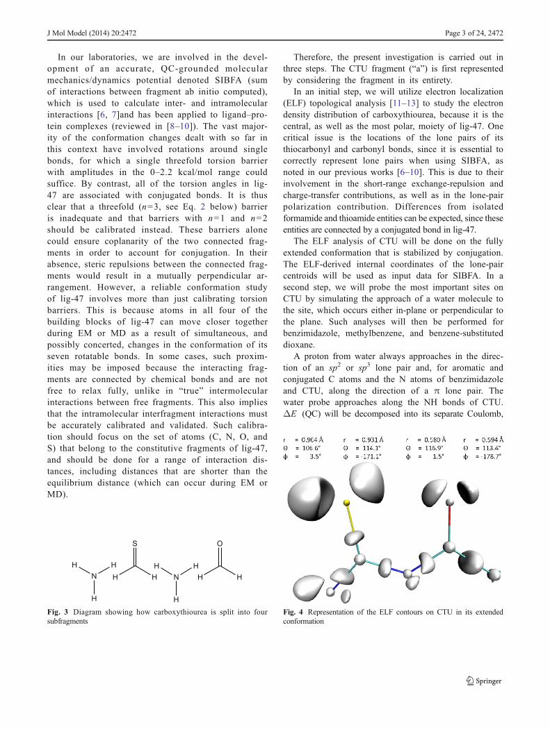
In an initial step, we will utilize electron localization(ELF) topological analysis [11–13] to study the electrondensity distribution of carboxythiourea, because it is thecentral, as well as the most polar, moiety of lig-47. Onecritical issue is the locations of the lone pairs of itsthiocarbonyl and carbonyl bonds, since it is essential tocorrectly represent lone pairs when using SIBFA, asnoted in our previous works [6–10]. This is due to theirinvolvement in the short-range exchange-repulsion andcharge-transfer contributions, as well as in the lone-pairpolarization contribution. Differences from isolatedformamide and thioamide entities can be expected, since theseentities are connected by a conjugated bond in lig-47.
The ELF analysis of CTU will be done on the fullyextended conformation that is stabilized by conjugation.The ELF-derived internal coordinates of the lone-paircentroids will be used as input data for SIBFA. In asecond step, we will probe the most important sites onCTU by simulating the approach of a water molecule tothe site, which occurs either in-plane or perpendicular tothe plane. Such analyses will then be performed forbenzimidazole, methylbenzene, and benzene-substituteddioxane.
A proton from water always approaches in the direc-tion of an sp2 or sp3 lone pair and, for aromatic andconjugated C atoms and the N atoms of benzimidazoleand CTU, along the direction of a π lone pair. Thewater probe approaches along the NH bonds of CTU.ΔE (QC) will be decomposed into its separate Coulomb,
H
N
H
H
H
S
H
H
N
H
H
H
O
H
Fig. 3 Diagram showing how carboxythiourea is split into foursubfragments
Fig. 4 Representation of the ELF contours on CTU in its extendedconformation
J Mol Model (2014) 20:2472 Page 3 of 24, 2472

short-range repulsion, polarization, and charge-transfercontributions.
We will accordingly readjust the effective van derWaals (vdW) radii used in SIBFA for the respectiveenergy contribution and their possible increments alongthe angular directions corresponding to the orientationsof the individual lone pairs. This is possible becausethe input includes the internal coordinates and vdWincrement (default value is null) of each individuallone pair.
As mentioned below, with the exception of the π lone pairsof sulfur, the vdW increments are readjusted by limitedamounts. This should not impair transferability because suchincrements can be considered to be fragment-specific.
Following this calibration, we will perform stepwise (15°)variations in torsion angle around each of the rotatable bonds,first for CTU and then for the entire lig-47, and calculate theresulting variations in QC conformational energy. For each ofthe seven angles, we will then calibrate the amplitude of thetorsional potential V0 for both the one- and the two-fold barriers
a b
c d
e
Fig. 5a–n Studied directions of approach of water to the sites of lig-47. aApproach perpendicular to the H-linked C atom of the pentacyclic ring ofbenzimidazole. b Approach perpendicular to the purine-like nitrogen ofthe pentacycle of benzimidazole. c Approach via oxygen 11 of thebenzodioxane. d Approach perpendicular to carbon 2 of thebenzodioxane. e Approach perpendicular to carbon 2 of methylbenzene.fApproach via the outer lone pair of the sulfur atom of CTU. gApproach
via the inner lone pair of the sulfur atom of CTU. h Approach via theinner lone pair of the oxygen atom of CTU. iApproach via the outer lonepair of the oxygen atom of CTU. j Approach via the π lone pair of thesulfur atom of CTU. kApproach via the π lone pair of the oxygen atom ofCTU. l Approach via the π lone pair of the primary nitrogen atom ofCTU.m Approach via the π lone pair of the secondary nitrogen atom ofCTU. n Approach via the hydrogen on the secondary nitrogen of CTU
2472, Page 4 of 24 J Mol Model (2014) 20:2472

in SIBFA to ensure a good match with the corresponding QCconformational curves.
In the fourth step, we will evaluate the transferability of theapproach. To achieve this, we will select minima of the sixcurves and use them as starting points to perform SIBFAenergy minimization. We will then rank the minimized con-formations in terms of their relative stabilities and compare therankings to the corresponding rankings obtained from single-point QC calculations. The influence of the polarization con-tribution on the energy separations between the minimizedconformations will be assessed, and other, more comprehen-sive, methods of incorporating this contribution will beevaluated.
Finally, we will assess the impact of the correlation/dispersion on the conformations of lig-47 calculated at the
QC level, and the extent to which we can account for thisusing the Edisp contribution in SIBFA.
Procedure
Quantum chemistry
The conformational energy variations are obtained via the cc-pvtz(−f) basis set [14, 15] using the G09 software [16].Analyses of the interaction energies of the water probe withthe sites of lig-47 are done with the reduced variational spaceanalysis (RVS) procedure [17], as implemented inGAMESS [18]. The electron distribution is analyzed usingthe ELF procedure [11] with the TopMod package [12].
h i
f g
j k
Fig. 5 (continued)
J Mol Model (2014) 20:2472 Page 5 of 24, 2472

Further details about the applications of ELF to biologicalsystems can be found in [12].
SIBFA
In the SIBFA procedure [6–10], the conformation energy in aflexible molecule is computed as the sum of the intermolecu-lar interactions ΔE between the fragments that comprise themolecule. ΔE is computed as a sum of five contributions:
ΔE ¼ EMTP � þ Erep þ Epol þ Ect þ Edisp; ð1Þ
which are, respectively, the electrostatic multipolar,the short-range repulsion, the polarization, the charge-transfer, and the dispersion contributions. EMTP* is com-puted with distributed multipoles derived from theccpvtz(−f) QC molecular orbitals (MOs) of the individ-ual fragments of lig-47 using Stone’s analysis [19, 20],and is augmented with a short-range penetration correc-tion [21]. Erep is the sum of the bond–bond, bond–lonepair, and lone pair–lone pair interactions. Epol is
computed using distributed anisotropic polarizabilitytensors located on the chemical bonds and the localizedlone-pair hybrids of the fragments, as derived from theprocedure reported by Garmer and Stevens [22]. Thepolarizing field is calculated with the same multipolesas for EMTP* and is screened by a Gaussian function.Ect is a short-range charge-transfer contribution andEdisp is the dispersion contribution. A periodic torsionalenergy contribution is included via the expression
V ¼ V 0 1ð Þ2
cosΦþ 1ð Þ þ V 0 2ð Þ2
cos2Φþ 1ð Þ; ð2Þ
where φ is the torsion angle. The two terms on the right-hand side express the periodicity, which corresponds to n=1and 2, respectively, for the conjugated bonds.
Lig-47 is constructed from four distinct fragments:benzimidazole, methylbenzene, CTU, and benzene-linked dioxane. It should be recalled that, in thecontext of SIBFA [6–10], the multipoles of the twojunction bonds X–H and H–Y that make up an X–Y
n
l m
Fig. 5 (continued)
2472, Page 6 of 24 J Mol Model (2014) 20:2472

bond are redistributed on atoms X and Y and at themidpoint of the created bond X–Y. EMTP* is notcomputed between the atoms and bonds belonging toany connecting bond and the two fragments it con-nects. To allow for rotations around its successive N–C,C–N, and N–C bonds, CTU is split into four fragments(Fig. 3). The first and third are sp2 amines, the secondis thioaldehyde, and the fourth is aldehyde. In thisprocess, fictitious connecting H atoms are created andgiven null multipoles, and each of the two connectingbonds that make up a junction is given half the multi-pole and polarizability of the original bond prior tosplitting. Thus, the multipoles of the N–H and H–Cbonds of the first junction have half the values of theinitial N–C bond and are located at its middle, etc. CTUtherefore retains its net initial charge of 0. An importantpoint relates to Epol and Ect: these contributions arecalculated between all polarizability centers of theinteracting fragments, but they are not calculatedbetween the four split subfragments that make upCTU. This is because mutual polarization of thesesubfragments is considered to have initially taken place duringthe SCF iterations, which resulted in the MOs from which themultipoles were derived. Furthermore, such mutual polariza-tions would take place between subfragments with non-netcharges (0, −1, +1, etc.), which was observed by us to greatlyamplify the magnitude and weight of Epol. In addition, in thegeneral case, the multipoles used for Epol and the polarizabil-ities of the junctions are not redistributed but carried back onthe X and Y atoms of X–Y bonds, and the X–H and Y–Hbonds are collapsed onto X and Y, respectively. This enableseach fragment to retain its original net charge and also pre-vents two connecting fragments from polarizing one anotherat distances that would be shorter than the length of the actualX–Y bond. This overall procedure has been found to beeffective when compared with QC calculations focusing onten conformers of Ala tetrapeptides [23] that were designed tobenchmark the accuracy of molecular mechanics force fields[24]. A similar treatment is applied to methylbenzene, whichis split into a benzene-like and a methane-like fragment.
Energyminimization of selected conformers was donewiththe Merlin [25] minimizer.
Results and discussion
ELF analysis of CTU
Figure 4 highlights the ELF function distribution aroundthe sp2 lone pairs of CTU. The internal coordinates that pro-vide the positions of their attractors (analogous to the
centroids in the localized MO picture) with respect tothe atoms that bear them are listed in this figure.
Probing lig-47 and CTU using one water molecule
The different approach directions of the water probe areillustrated in Fig. 5. Water approaches via a proton perpendic-ular to a C atom of benzimidazole, methylbenzene, or benzene-linked dioxane; via the N atom of benzimidazole; or via any ofthe N, O, or S atoms of CTU. It approaches in-plane through itsH atom to the external bisector of one ether O of benzene-linkeddioxane and along two possible directions to the C=S and C=Obonds of CTU. Upon binding to the “inner” S or O sp2 lonepair, the angle θ (C=X–Hw) was set to 150°. It was set at 120°upon approaching the “outer” lone pairs. A larger value of θ forthe inner approach to one C=X bond allows the steric repulsionbetween water and the other bond to be reduced. Finally, waterapproaches in-plane along the external bisector of its O atom tothe central amide H of CTU.
Table 1 Values (distances in Å, angles in degrees) of the vdW parame-ters for the H, C, N, O, and S atoms and of the internal coordinates andvdW increments/decrements of the inner sp2, outer sp2, and π lone pairs
Atom VdW (repulsion) Atom VdW (penetration)
C (benzene) 1.82 C (benzene) 1.6
O 1.40 O 1.41
O (ether) 1.448 O (ether) 1.1
N (sp2) 1.48 N (sp2) 1.61
S (CTU) 1.85 S (CTU) 1.6
O (carbonyl) 1.41 O (carbonyl) 1.44
H 1.24 H 1.1
H (amide) 1.30 H (amide) 1.3
N (sp3) 1.72 N (sp3) 1.45
θ φ r Increment
CTU O (inner) 116.9 0.00 0.59 −0.03CTU O (outer) 113.4 180.00 0.58 −0.03CTU O (π) 90.00 90.00 0.5 0.00
CTU O (π) 90.00 90.00 0.5 0.00
CTU S (outer) 106.6 0 0.93 0.08
CTU S (inner) 114.1 180.00 0.93 −0.05CTU S (π) 90.00 90.00 0.5 0.60
CTU S (π) 90.00 90.00 0.5 0.60
CTU Nprim (π) 90.00 90.00 0.25 −0.15CTU Nprim (π) 90 −90 0.25 −0.15CTU Nsec (π) 90.00 90.00 0.25 −0.20CTU Nsec (π) 90 −90 0.25 −0.20
J Mol Model (2014) 20:2472 Page 7 of 24, 2472

For the SIBFA calculations, the centroids of the sp2 lonepairs were located at positions consistent with the ELF results.The vdW radii for all atoms and contributions are listed inTable 1, as are the internal coordinates of the lone pairs andincrements. As mentioned above, we have calibrated theincrements/decrements of the S and O vdW radii along
each individual lone pair so that Erep (SIBFA) matchesthe radial behavior of Eexch-rep (RVS). This was done bydecreasing the vdW radius of the inner sp2 lone pair ofS by 0.05 Å while increasing that of the outer lone pairby 0.08 Å, yielding a difference of 0.13 Å. For O, bothlone pairs underwent similar decreases in their vdW
Table 2 Values (kcal/mol) of the intermolecular interaction energies at the equilibrium distance and their contributions to the binding of the probe watermolecules with the lig-47 fragments
Benzimidazole C7 d=3.4 Å EC/Emtp Eexc/Erep Epol Ect Etot
RVS −0.2 0.1 −0.1 0 −0.2SIBFA −0.2 0.1 −0.1 0 −0.2
Benzimidazole N6 d=2.7 Å EC/Emtp Eexc/Erep Epol Ect Etot
RVS −1.4 1 −0.4 −0.1 −0.9SIBFA −1.4 0.9 −0.1 −0.1 −0.7
Dioxane O11 d=2.2 Å EC/Emtp Eexc/Erep Epol Ect Etot
RVS −3.9 2.7 −0.6 −0.4 −2.2SIBFA −4.2 2.6 −0.4 −0.2 −2.2
Dioxane C2 d=2.8 Å EC/Emtp Eexc/Erep Epol Ect Etot
RVS −1.9 1.1 −0.3 −0.2 −1.3SIBFA −1.5 1.1 −0.2 0 −0.6
Methylbenzene C2 d=2.8 Å EC/Emtp Eexc/Erep Epol Ect Etot
RVS −1.8 1.1 −0.3 −0.2 −1.2SIBFA −1.6 1.1 −0.2 0 −0.7
CTU O (inner) d=2.9 Å O (outer) d=2.1 Å O (π) d=2.6 Å S (outer) d=2.7 Å S (inner) d=3.0 Å S (π) d=2.7 Å
EC −3.9 −6.7 −2.1 −5.2 −1.8 −4.4Emtp −4.2 −6.1 −2 −4.9 −2 −4.2Eexc 2.2 4 0.9 3.1 0.6 2.7
Erep 2.2 3.8 0.8 2.8 0.6 2.6
Epol −0.4 −1 −0.4 −0.6 −0.3 −0.6Epol −0.4 −0.9 −0.2 −0.7 −0.3 −0.3Ect −0.3 −0.5 −0.2 −0.5 −0.1 −0.6Ect −0.2 −0.3 −0.1 −0.3 −0.1 −0.2ΔERVS −2.4 −4.3 −1.8 −3.3 −1.6 −2.9Etot −2.6 −3.5 −1.5 −3.1 −1.8 −2.1
CTU Nprim EC/Emtp Eexc/Erep Epol Ect EtotRVS −1.3 0.7 −0.3 −0.1 −1SIBFA −1.3 0.8 −0.2 −0.1 −0.8
CTU Nsec EC/Emtp Eexc/Erep Epol Ect EtotRVS −0.5 0.1 −0.1 0 −0.6SIBFA −0.5 0.2 −0.1 0 −0.4
CTU NH EC/Emtp Eexc/Erep Epol Ect EtotRVS −5.8 2.5 −0.8 −0.3 −4.6SIBFA −5.9 2.2 −1 −0.4 −5.1
2472, Page 8 of 24 J Mol Model (2014) 20:2472

radii, amounting to 0.03 Å. To correctly account for themagnitudes of Erep and Ect in the vertical complexes ofwater with the C=S bond, large increments (0.60 Å)were found to be necessary for the vdW radii of the πlone pairs on S, while no increments were necessary forthe corresponding complex with the C=O bond.
Table 2 lists the values at the equilibrium distance ofthe intermolecular interaction energies and their contri-butions to the binding of probe water molecules to thefollowing non-CTU sites: benzimidazo C7 and N6; di-oxane O and C; methylbenzene C. It also lists thevalues for the CTU sites, i.e., the carbonyl O for inner
H2N
S
NH
O
H
n=2 n=1
-22 0
-23 8
6 -22 -4
Fig. 6 Variations in the conformational energy of CTU as functions of the torsion angles φ4, φ5, and φ6
J Mol Model (2014) 20:2472 Page 9 of 24, 2472

and outer in-plane approaches by the water (on thesame side and trans to the C=S bond, respectively);the carbonyl O for a perpendicular approach; thethiocarbonyl S for inner, outer, and perpendicular ap-proaches; the first and second amide nitrogens for aperpendicular approach; and the amide H for an in-plane approach. The results for the binding of water toCTU at a range of distances shorter than the equilibriumdistance are given in Table S1 of the “Electronic sup-plementary material,” ESM. Table S1 shows that theoverall agreement between the SIBFA and RVS valuesis satisfactory, but needs to be validated at the level ofeach contribution.
Monitoring the conformation behavior of CTU and lig-47
Having calibrated the vdW radii on the basis of theQC energy decomposition analyses for intermolecularinteractions, we then calibrated the amplitudes of thetorsional barriers to intramolecular interactions. Thiswas first done on the sole CTU moiety by varying thethree torsion angles shown in Fig. 6 in a stepwisemanner such that the variations in the sum of theSIBFA energy without dispersion plus the torsionalcontribution match those obtained from QC calcula-tions done at the HF level. For consistency with thesubsequent lig-47 conformation study, we denote thetorsion angles around the N1–C2, C2–N2, and N2–C3bonds (the atoms are numbered in succession from left toright) as φ4, φ5, and φ6, respectively. The φ angles arenumbered in ascending order along the main chain. Thestarting conformation is the fully extended one, so the φvalues are 180°. The fitted V0 values corresponding to n=1and 2 are also given in Fig. 6. The SIBFA curves are seen toclosely match the QC ones, with deep minima correspondingto the extended conformations.
We then considered lig-47 in its entirety. The labelsfor the eight torsional angles and the fitted V0 values aregiven in Fig. 7. φ1 denotes the torsion angle around the
bond separating the benzimidazole and methylbenzenerings. No rotations are considered around φ2 for themethyl substituent because of free rotation. The startingvalue of φ3 was 240° instead of 180° because stericrepulsion occurs between the sulfur and the benzene Hatom ortho to both junctions connecting benzene withbenzimidazole and CTU when φ3 is 180°. Retaining aφ3 value of 180° when performing all eight stepwisevariations would shift all of the curves other than thatfor φ3 to higher energies—a bias that could mask thepossible occurrence of energy-relevant conformations insuch curves. The curves (see Fig. 8) are drawn assum-ing that the energy of the lowest-lying conformer, whichoccurred with φ5=180° (curve 4), has an energy ofzero. There is a good match between the QC andSIBFA curves in all three plots, and all six curves areconsistent with each other. The most conspicuous devi-ations between the QC and SIBFA curves, correspond-ing to errors of >5 kcal/mol, occur at the rightmostparts of the φ3 and φ5 curves and at the leftmost partof the φ6 one, but these relate to unstable conformationsfor which δEconf >20 kcal/mol. On the other hand, it isimportant to note that the minima in the QC and SIBFAcurves for φ1–3 and φ5–6 are shifted by similar amounts withrespect to the global minimum of curve φ4.
Energy minimization of the lowest-lying conformers
The previous curves were drawn by varying each tor-sion angle individually while the other angles weremaintained at values corresponding to an extended con-formation. This obviously prevents lig-47 from foldingand thus attractive intramolecular interactions from oc-curring between well-separated atoms in lig-47. It wastherefore critical to evaluate if the good match betweenthe SIBFA and QC curves still holds when all of thetorsion angles are relaxed simultaneously namely, uponperforming energy minimization. Note that free rotationaround the methyl group should not impact the main-chain conformations. EM was done starting from select-ed minima in the conformational energy curves. Tomake the evaluation more comprehensive, we also in-cluded energy-minimized conformers that were obtainedat an earlier stage of this study, with a less refined setof vdW and V0 values.
The three-dimensional structures resulting from min-imization of the four most relevant conformers areshown in Fig. 9a–d. The values of the torsion anglesof the 15 conformers are reported in Table S2 of theESM. Their conformational energies and their
Fig. 7 Diagram of the torsion angles of lig-47
2472, Page 10 of 24 J Mol Model (2014) 20:2472

contributions are listed in Table 3, where the energy ofthe most stable conformer was taken to be zero.
Single-point QC calculations were performed to allowfor a comparison of the relative SIBFA and QC
2 1
-10 -1
-17 -5
-28 -5
-23 5
-24 5
-19 0
Fig. 8 Variations in the conformational energy of lig-47 as functions of the torsion angles φ1 and φ3–φ7
J Mol Model (2014) 20:2472 Page 11 of 24, 2472

conformational energy. Two related conformers, A andA′, were found to be the most stable in both theSIBFA and QC calculations, while all the remainingconformers have comparable relative conformational
energies, whichever method (SIBFA or QC) isemployed. However, A and A′ differ in energy fromthe other conformers by a much smaller amount inSIBFA than in QC (12 kcal/mol as compared to 20).
1b (topleft), 2a (topright), 0a (below left), 4a (below right)
Fig. 9a–d Three-dimensionalstructures of the four mostrelevant conformations (a–d) oflig-47
Table 3 Relative QC and SIBFA conformational energies (kcal/mol) ofall conformers along with the various energies that contribute to theSIBFA energy. 0a and 0b are the conformations resulting from the energyminimization of the initial conformation used for the curve. The otherconformations result from energy minimizations of the structures corre-sponding to the minima of the conformational curves. 1a and 1b result
from energy minimization of the minimum of the φ1 curve and corre-spond to φ1=150°. 2a and 2b correspond to an initial angle φ3 of 180°.3a and 3b correspond to an initial angleφ4 of 135°. For 3c,φ4=315°. For4a and 4b, φ5=165°. For 5a, φ6=260°. For 6a, φ7=30°. For 6b and 6c,φ7=150°
Conformer HF E1order+E2order+Etor Emtp Erep E1order Epol Ect E1order+Epol+Ect Etor
0a 20.97 12.40 9.70 4.1 13.80 −3.40 0.00 10.30 2.10
0b 21.22 13.30 12.00 0.30 12.30 −4.40 0.10 7.80 5.50
1a 0.17 2.10 −2.20 8.20 6.00 0.40 0.00 6.30 −4.201b 0.00 0.00 0.00 0.00 0.00 0.00 0.00 0.00 0.00
2a 17.80 15.30 7.80 −40.50 −32.70 −5.90 −2.40 6.50 8.80
2b 19.49 11.50 11.20 −1.40 9.80 −3.60 0.00 6.10 5.40
3a 18.42 11.20 7.80 −40.50 −32.70 −8.10 −2.60 2.70 8.50
3b 18.53 11.50 7.80 4.40 12.20 −8.00 −2.60 2.80 8.70
3c 20.97 12.40 9.70 −38.60 −28.90 −3.40 0.00 10.30 2.10
4a 22.39 17.20 4.90 −43.40 −38.50 −1.30 0.30 1.70 15.50
4b 21.36 17.50 4.90 −7.30 −2.40 −2.30 0.10 −4.80 22.30
5a 21.93 15.70 3.90 −44.40 −40.50 −1.40 0.30 0.60 15.10
6a 16.78 11.20 7.30 −41.00 −33.70 −6.80 −2.40 2.50 8.70
6b 20.12 13.30 9.30 3.20 12.50 −1.90 0.00 10.50 2.80
6c 19.22 13.00 11.00 −0.30 10.70 −1.90 0.10 8.70 4.30
2472, Page 12 of 24 J Mol Model (2014) 20:2472

The match between δEconf (SIBFA) and QC is illus-trated by the regression curve shown in Fig. 10a,which has a slope of 0.63 and yields a correlation ofr2=0.88. The evolution of δEconf when either SIBFA or
QC is used, as well as the evolutions of some SIBFAcontributions, are shown as functions of the conformerconsidered in Fig. 10b. In Fig. 10, as well as inFig. 12, the fifteen conformers are connected by a
Fig. 10 a Regression curve illustrating how closely the curves for δEconf (SIBFA) and δEconf (HF) match. b Comparison of the evolutions of δEconf(QC), δEconf (SIBFA), and the components of δEconf (SIBFA) as functions of the conformer
J Mol Model (2014) 20:2472 Page 13 of 24, 2472

curve in order to guide the eye. The underestimation ofδEconf by SIBFA could have adverse consequencesupon docking lig-47 to its target protein. Indeed, thehigh-lying conformers can be stabilized more by favor-able intermolecular interactions with the receptor thanby the energy minimum when the differences betweenconformers are in the range 12–20 kcal/mol. In thiscase, the high-lying conformers are erroneously pre-dicted to bind more favorably than conformers relatedto the lowest-lying ones. Imbalanced inter- and intra-molecular interactions is a caveat common to alldocking procedures [26–32].
It should be noted that the two energy minima arethe only ones that are stabilized by an intramolecularH-bond between the amide H of CTU connected tomethylbenzene and its carbonyl oxygen. This stabili-zation could be underestimated since the mutual
polarization of these two groups is prevented withinCTU. We must therefore consider alternative methodsof constructing CTU. One involves assembling CTUfrom thioamide and amide fragments, which are thensplit again into sp2 amine, thioaldehyde, and aldehydesubfragments. This enables the mutual polarization ofthioamide and amide to be calculated, but it couldmisrepresent the electronic distribution (and henceEMTP*) as the conjugation that is actually present in CTU ismissing. A second alternative method involves assemblingCTU using the multipoles and polarizabilities of the amine,thioaldehyde, and aldehyde fragments, all of which are calcu-lated independently. This enables the polarization between allfour fragments to be computed, but at the cost of a further lossof accuracy in the representation of the electron density. Weassess the advantages and limitations of these two approachesbelow.
Fig. 10 (continued)
2472, Page 14 of 24 J Mol Model (2014) 20:2472

Assembling CTU from thioamide and amide fragments(representation “b”)
The binding energies of water with CTU at the equilibriumdistance are reported in Table 4. For completeness, they arealso given in Table S3 of the ESM for a range of distances thatare shorter than the equilibrium distance. Not unexpectedly,the agreement between these results and the QC results is lessthan the agreement between the QC results and those obtainedwhen treating CTU as an integral unit. The errors in ΔE canexceed 1 kcal/mol in some cases, for instance wheninner water binds to the carbonyl O or to the innerNH group. These errors can be traced back essentiallyto the electrostatic contribution. This may imply thatalternative ways to derive the multipolar expansion onhighly conjugated and flexible fragments while retainingthe net charge on the subfragments must be sought (see[33] for an example). Work will have to be consideredalong these lines. It should be noted that this issue doesnot apply in the general case of the SIBFA library offragments, which essentially consists of saturated frag-ments or rigid conjugated or aromatic ones.
Using this set of multipoles, we recalibrated the V0 valuesfor the eight torsion angles of lig-47. Those values are shownin Fig. 11, which displays the new conformational energycurves. The V0 values are seen to vary by only small amountsfrom the values obtained for the integral CTU, with thenotable exception of n=1 for φ5 (V0 for n=1 is −15 kcal/mol, as compared to 6 when calculated for the integral CTU).This is an interesting case of the possible impact of therepresentation of conjugation on EMTP*, which can becorrected for by using a different sign and amplitude of V0.The eight new curves have similar overlaps to the QC ones,with the possible exception of φ5, for which the 50–100°region has δEconf (SIBFA) values that are up to 5 kcal/molhigher than the QC ones (i.e., they are in the 15–20 kcal/molrange, in contrast to the δEconf (QC) plateau at 15 kcal/mol).
The corresponding values of the conformationalenergy of lig-47 are reported in Table 5, along withthe individual contributions to the SIBFA energy.The regression graph and the evolutions of δEconf
(QC), δEconf (SIBFA), and the various contributionsto δEconf (SIBFA) as functions of the conformer arereported in Fig. 12. It should be noted that these
Table 4 Construction of CTU by assembling thioamide and amide fragments. Values of the intermolecular interaction energies (kcal/mol) at theequilibrium distances and their contributions to the binding of probe water molecules with the sites on CTU are shown
CTU O (inner) d=2.9 Å O (outer) d=2.1 Å O (π) d=2.6 Å S (outer) d=2.7 Å S (inner) d =3.0 Å S (π) d=2.7 Å
EC −3.9 −6.7 −2.1 −5.2 −1.8 −4.4Emtp −4.9 −6.9 −2.9 −4.4 −2.3 −4Eexc 2.2 4 0.9 3.1 0.6 2.7
Erep 2.2 3.9 0.9 2.8 0.6 2.6
Epol −0.4 −1 −0.4 −0.6 −0.3 −0.6Epol −0.5 −0.8 −0.3 −0.4 −0.3 −0.4Ect −0.3 −0.5 −0.2 −0.5 −0.1 −0.6Ect −0.2 −0.3 −0.1 −0.3 −0.1 −0.2ΔERVS −2.4 −4.3 −1.8 −3.3 −1.6 −2.9Etot −3.4 −4.1 −2.4 −2.3 −2.1 −2
CTU Nprim d=2.8 Å EC/Emtp Eexc/Erep Epol Ect Etot
RVS −1.3 0.7 −0.3 −0.1 −1SIBFA −0.7 0.7 −0.2 −0.1 −0.3
CTU Nsec d=3.3 Å EC/Emtp Eexc/Erep Epol Ect Etot
RVS −0.5 0.1 −0.1 0 −0.6SIBFA −0.8 0.1 −0.1 0 −0.8
CTU NH d=2.3 Å EC/Emtp Eexc/Erep Epol Ect Etot
RVS −5.8 2.5 −0.8 −0.3 −4.6SIBFA −6.6 2.1 −1.2 −0.4 −6.1
J Mol Model (2014) 20:2472 Page 15 of 24, 2472

results show very significant improvements in bothther2 value (0.96 as compared to 0.88) and the slope
(0.94 as compared to 0.63) compared to the resultsfor the integral CTU. Table 5 shows that the δEconf
-10 -1
-17 -5
-32 0
-19 -15
-29 5
-17 0
Fig. 11 Construction of CTU by assembling thioamide and amide fragments. Variations in the conformational energy of lig-47 as functions of thetorsion angles φ1 and φ3–φ7 are shown
2472, Page 16 of 24 J Mol Model (2014) 20:2472

(SIBFA) values are very close to the correspondingQC ones, and the energy separation between thelowest-lying minima and the other minima is now inthe correct range of 17–24 kcal/mol, instead of 11-15kcal/mol as previously. Epol, now involving the NHgroup of thioamide and the CO group of formamide,is the decisive contributor that sets the two minimaapart from the rest.
Assembling CTU from isolated amine, thioaldehyde,and aldehyde fragments (representation “c”)
A “minimalist” representation of CTU using the multi-po les and pola r i zab i l i t i e s of i so la ted amine ,thioaldehyde, and aldehyde fragments further degradesthe agreement with the QC results for the interaction ofthe water probe with the CTU sites (the RVS andSIBFA results are reported in Table S4 of the ESM).This is a consequence of the loss of conjugation, asreflected in some poor values of EMTP* as compared toEC, particularly upon binding to the S atom. The sixconformational energy curves of lig-47 are shown inTable S5 of the ESM. They are similar to their QCcounterparts (as also seen for the previous representa-tions of CTU). However, this was only true following asignificant recalibration of V0 for φ4, φ5, and φ6. Thus,for n=1, the V0 values are −22, −2, and −11 kcal/mol,respectively; they were 0, −15, and 5 kcal/mol for the
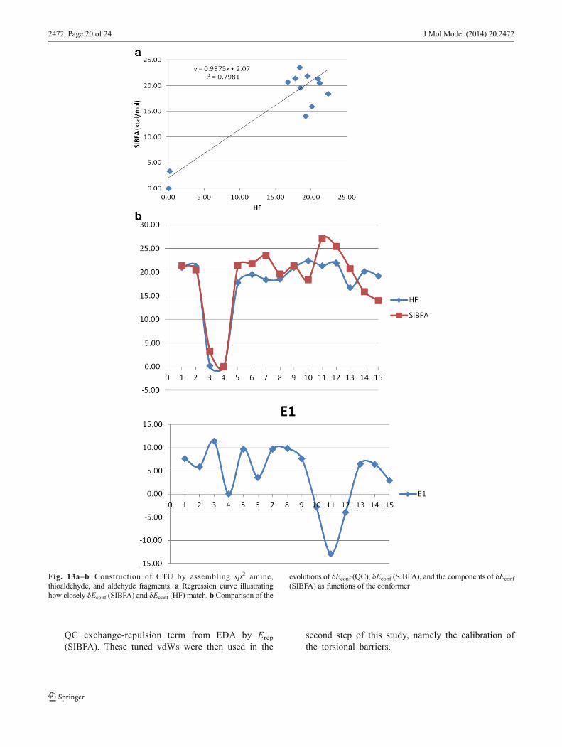
previous representation. The conformational energy re-sults are given in Table 6. Figure 13a and b show theregression graph and the evolution of δEconf as a func-tion of conformer number, respectively. The r2 value isless (0.80) than in the previous case (0.96), whereas theslope is the same (0.94). Generally speaking, δEconf
(SIBFA) has comparable values to δEconf (QC), butthere are two conspicuous outliers with values of 27.8and 29.6 kcal/mol as compared to 21.5 and 21 kcal/mol,respectively, from QC.
It can be concluded from this section that, even though itgives relative δEconf values that generally agree with thoseafforded by QC, the minimalist approach is unreliable, asindicated by the presence of outliers and a poor match toΔE (RVS) in some cases.
Possible impact of correlation/dispersion
The previous results were obtained at the Hartree–Fock level, since we were seeking a “proof of prin-ciple” procedure to justify the construction of lig-47.The impact of correlation/dispersion was further eval-uated by recomputing δEconf (QC) at correlatedlevels, namely MP2, B3LYP [34, 35], and B97-D[36]. These results are reported in Table 7, along withthe SIBFA ones from representations a–c, which nowinclude Edisp. MP2 is the QC approach that leads tothe largest reduction in δEconf. This reduction is in
Table 5 Construction of CTU by assembling thioamide and amide fragments. The relative QC and SIBFA conformational energies (kcal/mol) of allconformers are shown, along with the contributions to the SIBFA energy
Conformer HF E1order+E2order+Etor Emtp Erep E1order Epol Ect E1order+Epol+Ect Etor
0a 20.97 19.50 20.30 −3.9 16.40 15.50 0.80 32.7 −13.20b 21.22 19.70 22.50 −7.7 14.80 14.40 0.90 29.9 −10.21a 0.17 0.00 0.00 0 0.00 0.00 0.00 0 0
1b 0.00 1.20 3.40 −8 −4.60 1.60 0.20 −2.8 4
2a 17.80 19.80 16.00 −1.7 14.30 18.00 −0.50 31.7 −11.92b 19.49 18.90 21.70 −9.3 12.40 16.00 0.80 29 −10.13a 18.42 18.10 15.90 −3.2 12.70 18.10 −0.60 30 −11.93b 18.53 18.30 16.00 −3.2 12.80 18.10 −0.70 30.1 −11.83c 20.97 19.50 20.30 −3.9 16.40 15.50 0.80 32.7 −13.24a 22.39 21.70 10.80 −10.1 0.70 19.20 1.10 21 0.7
4b 21.36 23.90 10.60 −15.3 −4.70 19.70 0.90 15.9 8
5a 21.93 19.30 10.00 −10.2 −0.20 18.40 1.10 19.2 0.1
6a 16.78 17.00 14.80 −4.3 10.50 18.50 −0.50 28.5 −11.56b 20.12 19.50 19.50 −4.8 14.70 16.30 0.90 31.7 −12.26c 19.22 18.80 21.10 −8.2 12.90 16.10 0.90 29.8 −11
J Mol Model (2014) 20:2472 Page 17 of 24, 2472

fact more pronounced than the reduction which occursupon including Edisp in SIBFA. MP2 was previouslyshown to overestimate the stability of folded conformersvia intramolecular basis set superposition error calcula-tions [37, 38] (e.g., for 1a, 1b, 2b, 3a, and 6b), and thiscan explain their excessive decreases in δEconf. The
results from B3LYP, which includes correlation but doesnot incorporate dispersion effects, are very close to theδEconf (SIBFA) results for representation b (r2=0.98),even though the multipoles and polarizabilities werederived at the HF level. δEconf (SIBFA) also agreesclosely with B97-D, which includes an explicit disper-sion term, although this agreement is not as strong as iti s w i t h B3LYP ( r 2 = 0 . 9 5 i n s t e a d o f 0 . 9 8 ) .Representations a and c lead to significantly reducedagreement with the QC calculations. In this context,several papers have showed the importance of includingcorrelation effects when estimating the rotational bar-riers around conjugated bonds [39–43]. This led us torecalibrate V0 (n=1) and V0 (n=2) against B97-D con-formation–energy curves. A comparison of the evolu-tions of the B97-D and SIBFA conformational energiesis provided in Fig. 14, which also shows therecalibrated V0 barriers. The most important changesconcern the two central torsion angles, φ4 and φ5 ofthe thioamide moiety. A substantial improvement with re-spect to B97-D is apparent. The conformational energiesare listed in the penultimate column of Table 7. Thoseresults imply that in the context of polarizable MM, the
Fig. 12a–b Construction of CTU by assembling thioamide and amidefragments. a Regression graph showing how closely δEconf (SIBFA) andδEconf (HF) match. b Comparison of the evolutions of δEconf (QC), δEconf(SIBFA), and the components of δEconf (SIBFA) as functions of the conformer
Fig. 12 (continued)
2472, Page 18 of 24 J Mol Model (2014) 20:2472

inclusion of an explicit dispersion contribution couldallow the changes in δEconf (QC) upon passing fromthe HF to the correlated level to be accounted for, withan additional improvement being afforded by a prelim-inary recalibration of V0. However, this approach couldbe more problematic when using classical molecularmechanics potentials in which the 1/R6 vdW contribu-tion contains more than simply the dispersion.
Conclusions
Rotations around bonds connecting unsaturated atomsgovern the conformations of numerous pharmacologicalligands. This is exemplified by lig-47, a molecule thatwas selected via virtual screening and is active bothin vitro and in cellulo against neovascularization [1,2]. Lig-47 is constructed from a benzimidazole, amethylbenzene, a carboxythiourea, and a benzene-linked dioxane. In the context of molecular mechanics/dynamics, it is essential to reliably account for thevariations in conformational energy that result fromrotations around its connecting bonds in order to pre-serve the proper balance of intramolecular interactionson the one hand and intermolecular interactions (i.e.,ligand–receptor and ligand–water) on the other. Bothof these types of interactions can only be validated
through comparison with the results from high-level abinitio QC computations. Calibrating the one- and two-fold rotational barriers of lig-47 is obviously necessarybut is also far from sufficient. The presence of a highlyc on j u g a t e d po l a r i z a b l e y e t f l e x i b l e c en t r a lcarboxythiourea (CTU) entity led us to address thefollowing issues in succession. To our knowledge, thereis no precedent for such analyses.
– Anisotropy. The energy contributions to SIBFA are allanisotropic (see [7–10] for discussions). The anisotropyof the overlap-dependent terms Erep and Ect stems from theexplicit representation of the heteroatom lone pairs.However, there is an extra amount of anisotropythat could be due to the unequal spatial extensionsand lone pairs that are centered on a given atom.Regarding the ‘inner ’ sp2 lone pairs of thethiocarbonyl and carbonyl bonds, this could bedue to the mutual repulsion of these lone pairs.We have accordingly modified the vdW radii ofthe CTU S, O, and N lone pairs. QC energy de-composition analysis (EDA) of the interaction of awater probe with CTU enabled us to calibrate theamounts by which the vdW radii of the inner lonepairs decrease and the outer lone pairs and π lonepairs increase. The modified vdW radii led to asignificant improvement in the reproduction of the
Table 6 Construction of CTU by assembling sp2 amine, thioaldehyde, and aldehyde fragments. Relative QC and SIBFA conformational energies(kcal/mol) of all conformers are shown, along with the contributions to the SIBFA energy
Conformer HF E1order+E2order+Etor Emtp Erep E1order Epol Ect E1order+Epol+Ect Etor
0a 20.97 21.30 3.80 3.80 7.60 20.70 0.30 28.7 −7.40b 21.22 20.50 6.10 −0.20 5.90 18.50 0.30 24.8 −4.31a 0.17 3.30 3.00 8.40 11.40 −0.70 0.10 10.7 −7.41b 0.00 0.00 0.00 0.00 0.00 0.00 0.00 0 0
2a 17.80 21.40 3.30 6.40 9.70 −6.10 −0.20 3.5 17.9
2b 19.49 21.80 5.50 −1.90 3.60 22.60 0.30 26.5 −4.73a 18.42 23.50 4.80 4.90 9.70 −4.30 −0.20 8.3 15.2
3b 18.53 19.60 4.90 4.90 9.80 −4.20 −1.40 4.2 15.4
3c 20.97 21.30 3.80 3.80 7.60 20.70 0.30 28.7 −7.44a 22.39 18.40 −0.30 −2.50 −2.80 −3.50 0.30 −5.9 24.3
4b 21.36 27.10 −5.00 −7.90 −12.90 3.70 0.10 −9.2 36.3
5a 21.93 25.40 −1.30 −2.70 −4.00 7.40 0.30 3.6 21.8
6a 16.78 20.70 2.70 3.80 6.50 −1.10 −0.10 5.3 15.4
6b 20.12 15.90 3.50 2.90 6.40 13.20 0.30 19.9 −46c 19.22 14.00 5.50 −2.60 2.90 13.70 0.30 16.9 −2.9
J Mol Model (2014) 20:2472 Page 19 of 24, 2472
QC exchange-repulsion term from EDA by Erep
(SIBFA). These tuned vdWs were then used in thesecond step of this study, namely the calibration ofthe torsional barriers.
Fig. 13a–b Construction of CTU by assembling sp2 amine,thioaldehyde, and aldehyde fragments. a Regression curve illustratinghow closely δEconf (SIBFA) and δEconf (HF) match. b Comparison of the
evolutions of δEconf (QC), δEconf (SIBFA), and the components of δEconf(SIBFA) as functions of the conformer
2472, Page 20 of 24 J Mol Model (2014) 20:2472

– Conjugation. In the context of MM/MD, conjuga-tion raises two issues. The first is the need tocalibrate onefold (n=1) and twofold (n=2) V0 ro-tation barriers around each bond connecting sp/sp2
atoms. The second is the requirement of a satis-factory representation of the electronic distribu-tion of an “integral” conjugated fragment suchas CTU. Using the multipolar distribution andthe distributed polarizabilities of CTU split intofour non-mutually-polarizable subfragments en-abled us to calibrate the two rotation barriersaround each of the three connecting bonds ofCTU and to accurately reproduce the QC confor-mational energy curve. The study was then extend-ed to lig-47 by calibrating the barriers of the threeadditional torsional bonds. For all six torsion an-gles, the δEconf (SIBFA) values again closely
matched their QC counterparts. Could such a rep-resentation of lig-47 be used in actual simulations?This led us to consider the next issue.
– Polarization and multipole transferability. The energyof lig-47 was subsequently minimized by simulta-neously relaxing its six main-chain torsion anglesusing selected minima from the six conformation–energy curves as starting points. The two moststrongly bound minima were stabilized by an in-tramolecular H-bond within CTU between the car-bonyl O and the thioamide NH proton. However,their relative stabilizations were underestimatedwith respect to QC. Noting the prominent roleof Epol in stabilizing H-bonded networks [44, 45],and in an attempt to facilitate its involvement inbonding between the thioamide and amidemoeities, we considered an alternative method of
Fig. 13 (continued)
J Mol Model (2014) 20:2472 Page 21 of 24, 2472

const ruct ing CTU: assembl ing i t f rom thethioamide and amide entities, which were thenfurther split into amine and thioaldehyde on theone hand and amine and aldehyde on the other.There is thus a trade-off between the lost conjuga-tion between these two entities (which is present inintegral CTU) and the increase in their mutualpolarization. Following a new recalibration stepfor the vdWs and the torsional barriers, the δEconf
values at the energy minima were recomputed.This led to a marked improvement in the δEconf
values such that they were now in near-quantitativeagreement with the QC ones, with an r2 regressioncoefficient of 0.97 and a slope of 0.98. Epol wasobserved to be the main contributor to the δEconf
(SIBFA) values. When the mutual polarization ofthioamide and amide entities of CTU was accountedfor, the evolution of Epol as a function of conformernumber was found to give the closest match to thecorresponding evolution of δEconf (QC). Finally, aminimalist representation in which CTU was con-structed from the multipoles and polarizabilities ofindependent sp2 amine, thioaldehyde, and aldehyde
fragments was analyzed. It presented significantlyweaker agreement with the QC values in terms ofboth CTU interactions with a water probe andδEconf.
Along these lines, several papers have mentioned the needto introduce coupling between the torsional degrees of free-domwhen treating polyconjugated ligands [46, 47]. However,those studies were performed in the context of nonpolarizablemolecular mechanics. It is likely that such couplings indirectlyaccounted for many-body effects that are otherwise absentfrom such approaches, while they are inherently included inthe Epol contribution.
The present results again underline the essential roleof polarization, as found in our previous conformationalstudies of peptides [23], conformation-dependent diva-lent cation binding [48–50], ligand–macromolecule com-plexes [51–53], and networks of H-bonded complexes[44, 45]. They are fully consistent with the conclusionsdrawn about another polarizable multipolar force field,AMOEBA [54–57], although AMOEBA does not incor-porate anisotropy at the level of the short-range repul-sion contribution.
Table 7 Comparison of δEconf (QC) values obtained at the MP2, B3LYP, and B-97D levels with the corresponding δEconf (SIBFA) values thatincorporate the dispersion contribution. r2 regression coefficients are shown at the bottom of the table. Energies are in kcal/mol
Conformer MP2 B97d B3LYP Etot (SIBFA) a Etot (SIBFA) b SIBFA (B97D) Etot (SIBFA) c
0a 18.4 19.1 19.5 11.6 19.4 19 41.4
0b 19.1 19.9 19.9 12.9 19.9 20 19
1a 0 0 0 1.4 0 0 23.5
1b 1.11 1.2 1 0 2 2.5 0
2a 13.9 14.8 16.8 13.5 18.6 16.1 40.5
2b 18.2 19 18.9 11.3 19.4 20 20.7
3a 15.1 15.8 17.6 9.7 17.3 14.6 39.9
3b 15.2 15.9 17.7 10 17.5 14.8 17.7
3c 18.4 19.1 19.5 11.6 19.4 18.9 41.4
4a 15 16.9 20.7 15.5 20.6 15.7 37.6
4b 12.5 14.3 20.2 14.5 21.5 17.2 23
5a 14.2 16.4 20.8 13.1 17.4 13.5 43.7
6a 13.8 14.4 16.2 10 16.4 13.7 40.4
6b 17.3 17.9 18.4 12.8 19.6 18.8 36.3
6c 17.4 18 18 12.8 19.2 19.1 34.7
MP2 B97D B3LYP
a 0.84 0.88 0.95
b 0.93 0.95 0.98
c 0.52 0.54 0.57
2472, Page 22 of 24 J Mol Model (2014) 20:2472

2 1
-10 -1
-20 -9
-32 3
-16 -15
-25 8
-17 0
Fig. 14 Construction of CTU by assembling thioamide and amide fragments. Variations in the conformational energy of lig-47 as functions of thetorsion angles φ1 and φ3–φ7
J Mol Model (2014) 20:2472 Page 23 of 24, 2472
We are presently using the minimized conformers of lig-47as starting points for docking simulations of the target proteinneuropilin-1, and the results will be reported separately.
Acknowledgments We wish to thank the Grand Equipement Nationalde Calcul Intensif (GENCI): Institut duDeveloppement et des Ressourcesen Informatique Scientifique (IDRIS), Centre Informatique del’Enseignement Superieur (CINES), France, project no. x2009-075009),and the Centre de Ressources Informatiques de Haute Normandie(CRIHAN, Rouen, France), project 1998053.
We wish to acknowledge a CIFRE grant allotted to Elodie Goldwaserin the course of her Ph.D. thesis.
We are pleased to thank Drs. Lucia Borriello and Pascal Dao forenriching discussions during the course of this work.
References
1. Djordjevic S, Driscoll PC (2013) Drug Discov Today 18:442. Allain B, Jarray R, Borriello L, Leforban B, Dufour S, Liu W,
Pamonsinlapatham P, Bianco S, Larghero J, Hadj-Slimane R,Garbay C, Raynaud F, Lepelletier Y (2012) Cell Signal 24:214
3. Van der Kooi CW, Jusino MA, Perman B, Neau DB, Bellamy HD,Leahy D (2007) J Proc Natl Acad Sci USA 104:6152
4. Jarvis A, Allerston CK, Jia H, Herzog B, Garza-Garcia A,Winfield N, Ellard K, Aqil R, Lynch R, Chapman C,Hartzoulakis B, Nally J, Stewart M, Cheng L, Menon M,Tickner M, Djordjevic S, Driscoll PC, Zachary I, SelwoodDL (2010) J Med Chem 53:2215
5. Jain A (2003) J Med Chem 46:4996. Gresh N, Claverie P, Pullman A (1984) Theor Chim Acta 66:17. Gresh N (1995) J Comput Chem 16:8568. Gresh N (2006) Curr Pharm Des 12:21219. Gresh N, Cisneros GA, Darden TA, Piquemal J (2007) J Chem
Theory Comput 3:196010. Piquemal J-P, Chevreau H, Gresh N (2007) J Chem Theory Comput
3:82411. Silvi B, Savin A (1994) Nature 371:68312. Piquemal J-P, Pilme J, Parisel O, Gerard H, Fourre I, Berges J,
Gourlaouen C, De La Lande A, Van Severen M-C, Silvi B (2008)Int J Quantum Chem 108:1951
13. Chaudret R, Gresh N, Cisneros GA, Scemama A, Piquemal J-P(2013) Can J Chem 91:1
14. Dunning TH (1989) J Chem Phys 90:100715. Feller D (1996) J Comput Chem 17:157116. Frisch MJ, Trucks GW, Schlegel HB et al. (2009) Gaussian 09,
revision A.1. Gaussian, Inc., Wallingford17. Stevens WJ, Fink W (1987) Chem Phys Letts 139:1518. Schmidt MW, Baldridge KK, Boatz JA, Elbert ST, Gordon MS,
Jensen JH, Koseki S, Matsunaga N, Nguyen KA, Su S, WindusTL, Dupuis M, Montgomery JA (1993) J Comput Chem 14:1347
19. Stone A (1981) J Chem Phys Letts 83:233
20. Stone AJ, Alderton M (1985) Mol Phys 56:104721. Piquemal J-P, Gresh N, Giessner-Prettre C (2003) J Phys Chem A
107:1035322. Garmer DR, Stevens WJ (1989) J Phys Chem A 93:826323. Gresh N, Kafafi SA, Truchon J-F, Salahub DR (2004) J Comput
Chem 25:82324. Beachy MD, Chasman D, Murphy RB, Halgren TA, Friesner RA
(1997) J Am Chem Soc 119:590825. Evangelakis GA, Rizos JP, Lagaris IE, Demetropoulos IN (1987)
Comput Phys Comm 46:40126. McInnes C (2007) Curr Op Chem Biol 11:49427. Cavasotto CN, Orry A (2007) J Curr Top Med Chem 7:100628. Kroemer RT (2007) Curr Protein Pept Sci 8:31229. Irwin J (2008) J Comp-Aided Mol Des 22:19330. Sotriffer CA, Sanschagrin P,Matter H, Klebe G (2008) Proteins 73:39531. Gilson MK, Zhou HX (2007) Ann Rev Biophys Biomol Struct 36:2132. Schneider G (2010) Nat Rev Drug Discov 9:27333. Ren P, Ponder JW (2003) J Phys Chem B 107:593334. Lee C, Yang W, Parr RG (1988) Phys Rev B37:78535. Becke A (1993) J Chem Phys 98:564836. Grimme S (2006) J Comput Chem 27:178737. Gloaguen E, de Courcy B, Piquemal J-P, Pilme J, Parisel O, Pollet R,
Biswal HS, Piuzzi F, Tardivel B, Broquier M, Mons M (2010) J AmChem Soc 132:11860
38. van Mourik T (2008) J Chem Theory Comput 4:161039. Head-Gordon M, Pople JA (1993) J Phys Chem 97:114740. Head-Gordon M, Pople JA (1993) J Phys Chem 97:1025041. Meier RJ (1993) J Phys Chem 97:1024842. Meier RJ (2011) J Phys Chem 115:360443. Klug R, Burcl R (2010) J Phys Chem A 114:640144. Guo H, Gresh N, Roques BP, Salahub DR (2000) J Phys Chem B
104:974645. Piquemal J-P, Chelli R, Procacci P, Gresh N (2007) J Phys Chem A
111:817046. Zheng J, Yu T, Papajak E, Alecu IM, Mielke S, Truhlar DG (2011)
Phys Chem Chem Phys 13:1088547. Tafipolsky M, Schmid R (2005) J Comput Chem 26:157948. Rogalewicz F, Ohanessian G, Gresh N (2000) J Comput Chem 21:
96349. Tiraboschi G, Fournié-Zaluski M-C, Roques B-P, Gresh N (2001) J
Comput Chem 22:103850. Gresh N, Shi GB (2004) J Comput Chem 25:16051. Antony J, Piquemal J-P, Gresh N (2005) J Comput Chem 26:113152. Courcy B, Piquemal J-P, Garbay C, Gresh N (2010) J Am Chem Soc
132:331253. Gresh N, Courcy B, Piquemal J-P, Foret J, Courtiol-Legourd, Salmon
L (2011) J Phys Chem B 115:830454. Jiao D, Golubkov PA, Darden TA, Ren P (2008) Proc Natl Acad Sci
USA 105:629055. Jiao D, Zhang JJ, Duke RE, Li GH, Schnieders MJ, Ren P (2009) J
Comput Chem 30:170156. Ren P, Wu C, Ponder JW (2011) J Chem Theory Comput 7:302757. Zhang J, Yang W, Piquemal J-P, Ren P (2012) J Chem Theory
Comput 8:1314
2472, Page 24 of 24 J Mol Model (2014) 20:2472