conformation−function relationship of vitamin d: conformational analysis predicts potential...
TRANSCRIPT
Conformation-Function Relationship of Vitamin D: Conformational AnalysisPredicts Potential Side-Chain Structure
Sachiko Yamada,* Keiko Yamamoto, Hiroyuki Masuno, and Masateru Ohta†
Institute for Medical and Dental Engineering, Tokyo Medical and Dental University, 2-3-10 Surugadai Kanda,Chiyoda-ku, Tokyo 101, Japan
Received November 10, 1997
In previous studies, we have grouped regions in space occupied by the vitamin D side chaininto four: A, G, EA, and EG. We showed that the receptor (VDR) affinity of 1R,25-dihydroxyvitamin D3 derivatives increases, in terms of side-chain region, in the order EG, G,A, and EA. We called this the active space group concept. In the present study, we used thisactive space group concept to analyze the conformation-activity relationship of about 40representative potent 1R,25-dihydroxyvitamin D3 analogues. We initially listed structuralmodifications in the side chain of potent vitamin D analogues and estimated their potencyfactor. Possible side-chain conformations of representative analogues were calculated by themolecular mechanics method and plotted on a dot map compared with the regions A, G, EA,and EG. The cell-differentiating potency of the analogues was correlated with our active spacegroup concept with few exceptions. Among potent analogues with a natural configuration atC(20), the side chains of those with a 22-oxa, 22-ene, 16-ene, or a 18-nor modification werelocated in front of region EA (termed F). The side chains of the most potent 20-epi-22-oxa-24-homovitamin D analogues were concentrated at the left side of the EA region (L-EA). Thus,the side chains of almost all potent analogues were distributed around the EA region, andpotency increased in the order A, F, EA, and L-EA.
Introduction
Binding of a ligand to the receptor is the initial andmost crucial step in activating gene transcription medi-ated by a ligand-dependent nuclear transcription factor.How molecular recognition occurs at the interface of aligand and the receptor is of interest to chemists.Control of this process is also key to developing potentand selective clinical agents.Vitamin D exerts its function by binding to its nuclear
receptor (VDR),1 which is a member of the nuclearreceptor superfamily.2 In binding to VDR, the threehydroxyl groups of 1R,25-dihydroxyvitamin D3 (1,25-(OH)2D3, 1) play important roles. In particular, thoseof the 1R- and 25-hydroxyl groups are critical, as theirremoval diminishes the VDR affinity by 1/500-1/1000.3
Over 300 active vitamin D analogues have beensynthesized4 since the discovery of 1,25-(OH)2D3 (1),5and some are in clinical use to treat metabolic bonediseases and skin disorders such as psoriasis.6 Nearly100 analogues with higher activities than that of theparent natural ligand (1) have been identified, and mostof them are modified around the side chain.4 In ourinitial structure-function studies of vitamin D,7 wefocused our attention on the side chain. Vitamin D sidechains are highly flexible as they possess the full carbonstructure of cholesterol. Therefore the 25-hydroxylgroup, which apparently plays a crucial role in anchor-ing vitamin D to the VDR, can occupy wide regions ofspace. The 20-epimer 2 of 1,25-(OH)2D3 (1) is muchmore potent than the natural vitamin,8a although theside chain of these epimers appears to occupy different
regions of space. Okamura and Midland9 analyzed theconformational mobility of the side chain of theseepimeric vitamins (1 and 2) and relatives by plottingthe 25-oxygen position of possible conformations on adot map. In these studies, they discriminated thecontribution of the conformers using the energy windowconcept. To determine the VDR-bound side-chain con-formation, we initially analyzed possible side-chainconformations of 1,25-(OH)2D3 (1) and its 20-epimer 2by molecular mechanics and grouped the regions oc-cupied by their 25-oxygen into four: A, G, EA, and EG(Figure 1).7a,b We then designed and synthesized ana-logues in which the side-chain mobility was restrictedin one of these regions, the diastereomers 3-6 at C(20)and C(22) of 22-Me-1,25-(OH)2D3.7a,b The VDR affinityof these conformationally restricted analogues increasedin the order 6, 3, 4, and 5 (1/100, 1/60, 1/3, and 20,respectively, relative to 1,25-(OH)2D3, 1). In terms ofthe region, the potency increased in the order EG, G,A, and EA.7a From these results, we postulated that1,25-(OH)2D3 (1) binds to VDR when its side chain islocated in the A region10 and that 20-epi-1,25-(OH)2D3(2) binds to VDR when its side chain is located in theEA region.We next studied whether our active space group
concept can predict the potency of known vitamin Danalogues. In this study, we (1) listed structuralmodifications in the side chain (including C(17)) ofvitamin D that elevate potency, (2) calculated thepossible side-chain conformations of these analoguesthat were displayed on dot maps, and (3) investigatedthe relationship between the side-chain region andactivity. The results not only support our idea of activeareas of the side chain but also demonstrate that† Present address: Chugai Pharmaceutical Co., Ltd.
1467J. Med. Chem. 1998, 41, 1467-1475
S0022-2623(97)00761-9 CCC: $15.00 © 1998 American Chemical SocietyPublished on Web 04/03/1998
conformational analysis can predict the potency ofvitamin D analogues.
MethodsPotent Vitamin D Analogues and Activation Factor.
We compared the potency of vitamin D derivatives in termsof their cell-differentiating activity (throughout this paper,potency indicates cell-differentiating activity unless otherwisenoted). Affinity for VDR may be more appropriate for predict-ing receptor-bound conformation, but IC50 values determinedby competitive binding assays are not always reproducible forcompounds having higher affinity than 1,25-(OH)2D3 (1). Thisis because attaining equilibrium among the receptor and twosuch ligands (the sample and radioactive standard) is difficultunder assay conditions.11Table 1 lists structural modifications that enhance potency,
together with representative compounds and activation factorsestimated on the basis of cell-differentiation activity. For
simplicity, the analogues were restricted to those derived from1,25-(OH)2D3 (1). The modifications that increase potency are(1) epimerization at the 20-position,8 (2) replacement of C(22)-H2 by oxygen,12 (3) introduction of double or triple bond(s) tothe side chain13 or the 16-position,14 (4) removal of the angularC(18) methyl group,15 (5) elongation of the side chain,16 (6)methylation of the terminal C(26) and -(27) methyl groups,16(7) perfluorination of C(24) methylene17 or the terminal gem-dimethyl groups,18 and (8) methylation at C(20).19 Combina-tions of these modifications enormously enhance potency asthe compounds in Table 2 show. In most cases, the potencyof compounds with multimodifications can be estimated bymultiplying by the activation factor shown in Table 1.Conformational Analysis. We analyzed the relationship
between the potency of the analogues and the areas occupiedby the 25-oxygen. (1) The possible side-chain conformationsof each analogue were searched using the molecular mechanicsmethod. (2) These possible conformations were plotted bypositioning the 25-oxygen on a dot map where one dotcorresponds to one-side chain conformation. (3) The dot mapof each analogue was overlaid with those of 1,25-(OH)2D3 (1)and 20-epi-1,25(OH)2D3 (2), and then we analyzed the rela-tionship between the side-chain area and cell-differentiatingactivity.The conformations were analyzed using the software SYBYL
(version 6.3, Tripos)20 as reported.7a,b Briefly, (1) analoguestructures were constructed by modifying the crystallographicvitamin D3 structure in the SYBYL database; then the globalminimum-energy conformation of each analogue was gener-ated by repeating rotations (360° at 60° intervals) of rotatableC-C and C-O bonds in the side chain (grid search in SYBYL)followed by optimization. (2) Using this minimum-energyconformation as the initial structure, all possible side-chainconformations were searched by rotating (360° at 30° intervals)the bonds in the side chain (indicated by numbers in thestructures in Chart 1) (systematic search in SYBYL). In thesystematic search, the van der Waals bump coefficient wasset at 0.95 to eliminate infeasible conformations suffered fromvan der Waals repulsion. (3) All possible conformations were
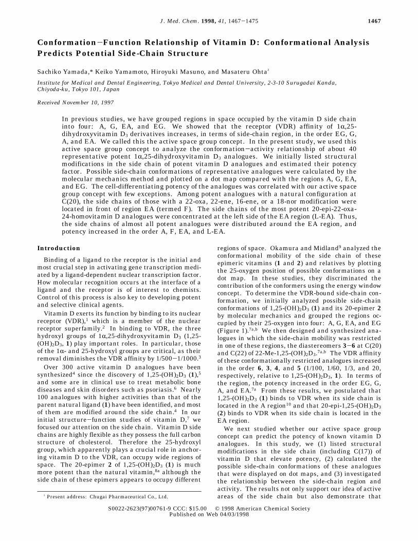
Figure 1. Grouping of the side-chain regions of 1,25-(OH)2D3
(1) and its 20-epimer (2) (stereoview). The mobile areas of theside chain of 1 (yellow) and 2 (cyan) are shown in a dot mapand are grouped into regions: A and G regions in 1 and EAand EG regions in 2 (see ref 7a).
Table 1. Side-Chain Modifications That Elevate Vitamin D Potency
modification abbrev activation factor representative compound
20-epimerization EP 20-30 20-epi-1,25-(OH)2D3 (2)8a22-oxygen 22O 5-10 22-oxa-1,25-(OH)2D3 (7)12unsaturation [yne (Y) or ene (E)] 22E 1-2 22-ene-1,25-(OH)2D3 (8)13a
23Y 2-3 23-yne-1,25-(OH)2D3 (10)13a16E 2-5 16-ene-1,25-(OH)2D3 (9)13a,14a
18-nor 18N 5-10 18-nor-1,25-(OH)2D3 (11)15elongation [homo (H) or dihomo (DH)] 24H 5-10 24-homo-1,25-(OH)2D3 (13)16a-c
24DH 2-5 24-dihomo-1,25-(OH)2D3 (14)16c26,27DH 2-5 26,27-dihomo-1,25-(OH)2D3 (15)16f,g
fluorination F2 5-10 24-F2-1,25-(OH)2D3 (16)17a,bF6 10 26,27-F6-1,25-(OH)2D3 (17)18
20-methyl 20Me 7 20-Me-1,25-(OH)2D3 (38)19
Table 2. Combination of Active Modifications
entry modification compound cell diffa
1 24H + 26,27DH 24,26,27-trihomo-1,25-(OH)2D3 (24)8a 5 (u)2 16E + 23Y 16-ene-23-yne-1,25-(OH)2D3 (12)13a,14a 10 (h)3 22E + 24H 22-ene-24-homo-1,25-(OH)2D3 (20)16a 10 (h)4 F2 + 24H 24-F2-24-homo-1,25-(OH)2D3 (25)16e,f 10 (h)5 22E + 24DH 22-ene-24,24-dihomo-1,25-(OH)2D3 (21)16c 10 (h)6 22O + 24H + 26,27DH 22-oxa-24,26,27-trihomo-1,25-(OH)2D3 (19)8a 20 (u)7 F6 + 22E 26,27-F6-22-ene-1,25-(OH)2D3 (26)13a 30 (h)8 22E + 26,27DH 22-ene-26,27-dihomo-1,25-(OH)2D3 (22)13c 60 (h)9 22E + 24E + 24H + 26,27DH 22,24-diene-24,26,27-trihomo-1,25-(OH)2D3 (23)13b 67 (u)10 16E + 23Y + F6 16-ene-23-yne-26,27-F6-1,25-(OH)2D3 (27)14b 80 (h)11 EP + 24H 20-epi-24-homo-1,25-(OH)2D3 (31)8a 200 (u)12 EP + 24H + 26,27DH 20-epi-24,26,27-trihomo-1,25-(OH)2D3 (32)8a 200 (u)13 EP + 22E + 24H + 26,27DH 20-epi-22-ene-24,26,27-trihomo-1,25-(OH)2D3 (33)8d 1000 (u)14 EP + 22O + 24H 20-epi-22-oxa-24-homo-1,25-(OH)2D3 (34)8a 1176 (u)15 EP + 22O + 24H + 26,27DH 20-epi-22-oxa-24,26,27-trihomo-1,25-(OH)2D3 (35)8a,4d 20000 (u)
a Relative differentiating activity of U 937 (u) or HL-60 (h) cells when the reference value of 1,25-(OH)2D3 (1) is defined as 1.
1468 Journal of Medicinal Chemistry, 1998, Vol. 41, No. 9 Yamada et al.
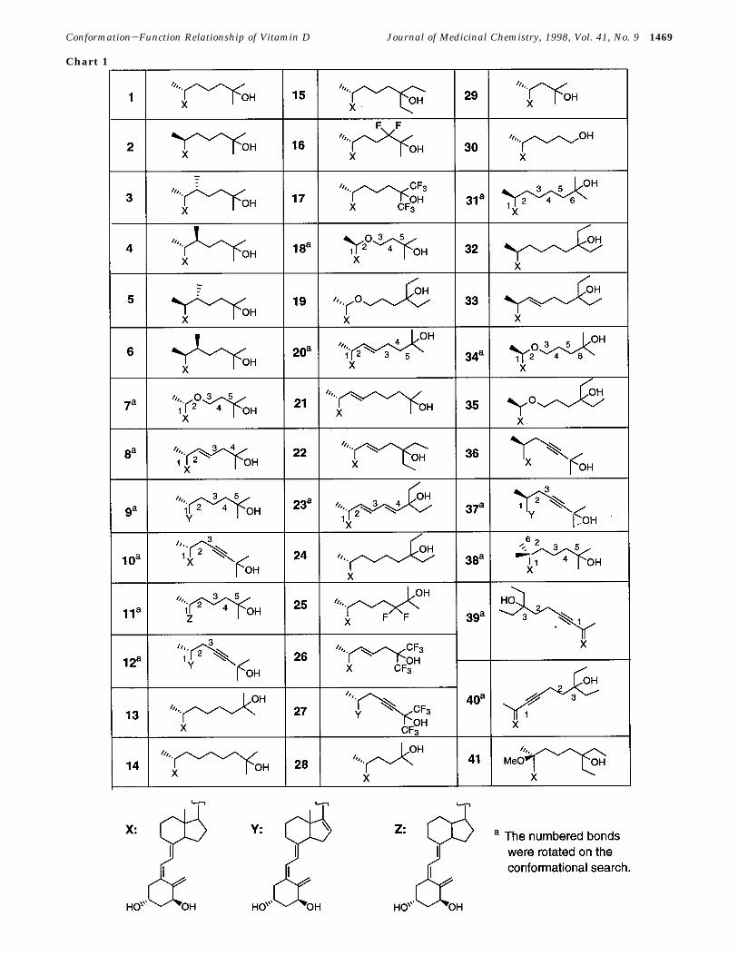
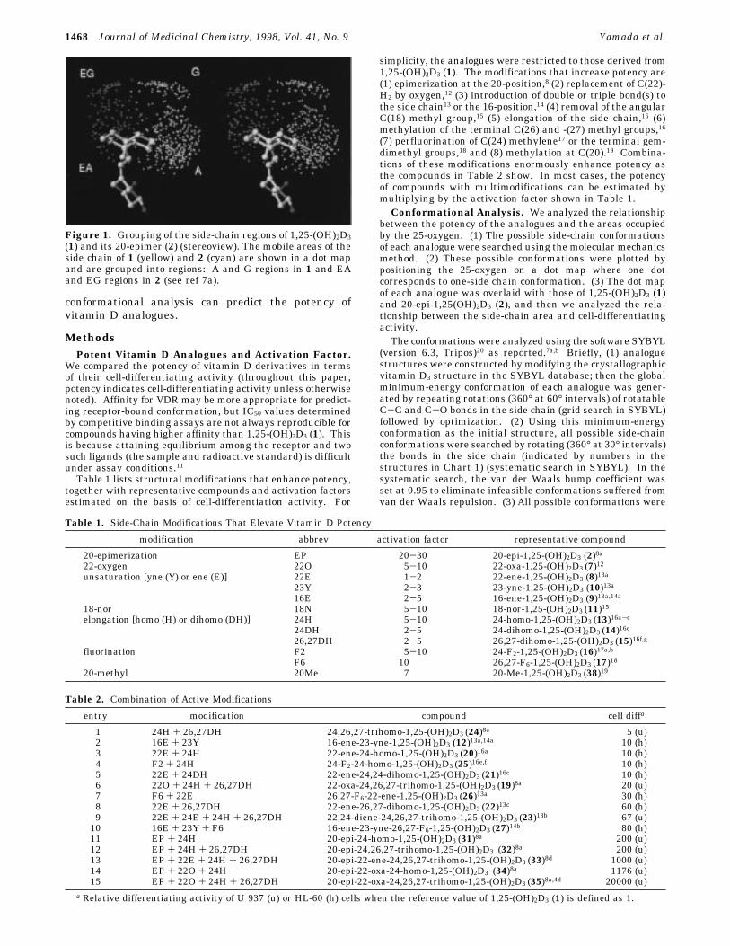
Chart 1
Conformation-Function Relationship of Vitamin D Journal of Medicinal Chemistry, 1998, Vol. 41, No. 9 1469
1470 Journal of Medicinal Chemistry, 1998, Vol. 41, No. 9 Yamada et al.

plotted on a dot map (graph sweep in SYBYL) in which theskeleton (the A, seco-B, C, and D ring moieties) is shown witha ball-and-stick model and the terminal oxygen position of eachpossible conformation is shown as a dot. (4) The mobile areasof the analogue’s side chain were compared with the areas A,G, EA, and EG, by superimposing the dot maps of theanalogues with those of 1,25-(OH)2D3 (1) and 20-epi-1,25-(OH)2D3 (2) at the skeleton.
Results
Compounds with a Side Chain at the F (Front)Region. We have shown that vitamin D compoundsare highly potent when the 25-hydroxyl group is locatedin the A or EA region.7a,b We found a group ofcompounds in which the 25-oxygen occupies an area infront of the EA region. We call this area F. Compoundswhich occupy this area have 22-oxa, 16-ene, 22-ene, or18-nor modifications, such as 22-oxa-1,25-(OH)2D3 (7),1222-ene-1,25-(OH)2D3 (8),13a 22,24-diene-24,26,27-trihomo-1,25-(OH)2D3 (23),13b 16-ene-1,25-(OH)2D3 (9),13a,14a and18-nor-1,25-(OH)2D3 (11).1522-Oxa-1,25-(OH)2D3 (7). 22-Oxa-1,25-(OH)2D3 (7)
was the first compound in which cell-differentiating andcalcemic activities of vitamin D were separated. Com-pound 7 has about 10-fold higher cell-differentiatingactivity than 1,25-(OH)2D3 (1), but it has low calcemicactivity.12 It has not been explained in structural termswhy replacing C(22)H2 with oxygen dramatically en-hances potency. Conformational analysis provided oneexplanation. Replacing C(22)H2 with oxygen signifi-cantly changes the mobility of the 25-oxygen. The 25-oxygen of 7 populates in the F region which lies in frontof EA (Figure 2a), whereas the 25-oxygen of 1,25-(OH)2D3 (1) occupies A and G.The C(22)H2 and 22-oxa compounds differ in their
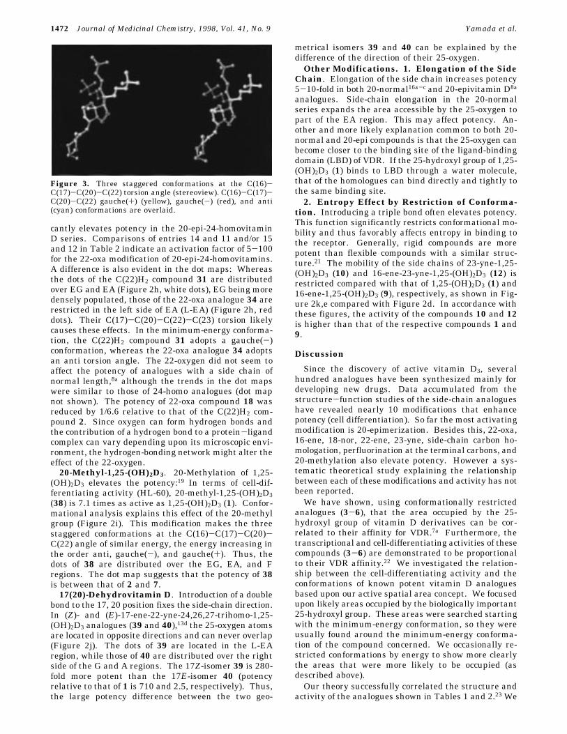
minimum-energy conformation. These two compoundsdiffer in their C(16)-C(17)-C(20)-C(22) torsion anglein the minimum-energy conformation. The C(22)H2compound adopts a C(16)-C(17)-C(20)-C(22) gauche-(+) conformation (Figure 3, yellow), whereas the 22-oxacompound has a gauche(-) conformation (Figure 3, red).In the gauche(-) conformation, C(22)H2 suffers severesteric repulsion from the C(18)H3 group. However inthe 22-oxa analogue this repulsion is reduced becausethe oxygen bears no hydrogen, so it adopts a C(16)-C(17)-C(20)-C(22) gauche(-) conformation. Whenvitamin D adopts a C(16)-C(17)-C(20)-C(22) gauche-(+), the 25-oxygen tends to reside in the A and Gregions, whereas when it adopts a gauche(-) conforma-tion the 25-oxygen tends to locate in the F region.22-Dehydro-1,25-(OH)2D3 derivatives. Introduc-
tion of a π-bond at C(22) similarly reduces the stericrepulsion between the C(22) part and the C(18)H3 group.Therefore, the 25-oxygen of 22-ene-1,25-(OH)2D3 (8)13aoccupies wide areas of the F-A regions (Figure 2b). 22,-24-Diene-24,26,27-trihomo-1,25-(OH)2D3 (23),13b which
is undergoing clinical trials as a drug to treat cancer,occupies similar regions (Figure 2c, red crosses). Sincethis compound has an elongated and rigid conjugateddiene structure, the area occupied by its 25-oxygenmoves further toward the front and the mobile area isrestricted compared with the more flexible 22-ene-24-homo-1,25-(OH)2D3 (20) (Figure 2c, white dots).16-Ene-1,25-(OH)2D3 Derivatives.14 The high po-
tency of these vitamin D analogues has been known fora long time, but the reasons were not understood.Introducing a double bond to C(16) also directs the sidechain to the front region F. Figure 2d shows that thearea occupied by the 25-oxygen of 16-ene-1,25-(OH)2D3(9) is similar to that occupied by the 22-oxa (Figure 2a)and 22-ene (Figure 2b) analogues. In the minimum-energy conformation, the calculated C(16)-C(17)-C(20)-C(22) torsion angle of 16-ene-1,25-(OH)2D3 (9) was -54°.Introducing a triple bond to C(23) and perfluorinationat C(26) and C(27) create analogues with increasedpotency, 12 (Figure 2e) and 27.18-Nor-1,25-(OH)2D3 (11).15 Removal of the angular
C(18)H3 group eliminates congestion between the meth-yl group and C(22)H2. The side chain therefore canrotate nearly freely around the 17,20 bond. Thus, whenall possible conformations were plotted, the area oc-cupied by the 25-oxygen of 11 extended over a widerange (Figure 2f, white dots). However, when 20%stable conformations are selected, the 25-oxygen ishighly concentrated in the F region (Figure 2f, red dots).Thus, we explain that this compound has high potencybecause its side-chain oxygen is located in the active Fregion.Compounds with Side Chains in the EA and Left
EA (L-EA) Regions. So far the 20-epimerization hasthe highest effect on increasing vitamin D potency. TheHL-60 differentiating activity is enhanced 25-30-fold.8We reasoned that 20-epi-1,25-(OH)2D3 (2) has highpotency because its 25-oxygen can occupy the mostactive EA region.7a In the minimum-energy conforma-tion, 20-epi-1,25-(OH)2D3 analogues adopt the C(16)-C(17)-C(20)-C(22) anti torsion angle (Figure 3, cyan).20-Epi-16-ene-1,25-(OH)2D3 Derivatives. The 20-
epimerization of 16-ene analogues has the reverse effect:14d,e 20-Epi-16-ene-23-yne-1,25-(OH)2D3 (37) is one-halfas active as 1,25-(OH)2D3 (1).14d Therefore, the 20-epimerization of 16-ene-23-yne 12 decreased potency by1/20. This difference in potency can be understood bycomparing the dot map of 16-ene-23-yne-1,25-(OH)2D3(12) (Figure 2e) with that of 20-epimer 37 (Figure 2g).While the dots of 12 are found over the active frontregions of A, F, and EA, those of 37 are distributed overthe inactive rear regions of EG-G.20-Epi-22-oxa-1,25-(OH)2D3 derivatives. We ex-
amined the effect of replacing C(22)H2 with oxygen inthe 20-epivitamin D series. Such replacement signifi-
Figure 2. Side-chain mobile area of potent 1,25-(OH)2D3 analogues compared with the four regions, A, G, EA, and EG, of 1(yellow) and 2 (cyan) (stereoviews). Dot maps of each analogue were overlaid with those of 1 and 2, the skeleton beingsuperimposed: (a, row 1, left) 22-oxa-1,25-(OH)2D3 (7) (red); (b, row 1, right) 22-ene-1,25-(OH)2D3 (8) (red); (c, row 2, left) 22,24-diene-24,26,27-trihomo-1,25-(OH)2D3 (23) (red crosses) and 22-ene-24-homo-1,25-(OH)2D3 (20) (white dots); (d, row 2, right) 16-ene-1,25-(OH)2D3 (9) (red); (e, row 3, left) 16-ene-23-yne-1,25-(OH)2D3 (12) (red); (f, row 3, right) 18-nor-1,25-(OH)2D3 (11) (whitedots, all possible conformations; red dots, 20% stable conformations); (g, row 4, left) 20-epi-16-ene-23-yne-1,25-(OH)2D3 (37) (red);(h, row 4, right) 20-epi-24-homo-1,25-(OH)2D3 (31) (white) and 20-epi-22-oxa-24-homo-1,25-(OH)2D3 (34) (red); (i, row 5, left) 20-methyl-1,25-(OH)2D3 (38) (red); (j, row 5, right) (17E)- and (17Z)-ene-22-yne-24,26,27-trihomo-1,25-(OH)2D3 (40 and 39) (rightand left red dots, respectively); (k, row 6) 23-yne-1,25-(OH)2D3 (10) (red).
Conformation-Function Relationship of Vitamin D Journal of Medicinal Chemistry, 1998, Vol. 41, No. 9 1471
cantly elevates potency in the 20-epi-24-homovitaminD series. Comparisons of entries 14 and 11 and/or 15and 12 in Table 2 indicate an activation factor of 5-100for the 22-oxa modification of 20-epi-24-homovitamins.A difference is also evident in the dot maps: Whereasthe dots of the C(22)H2 compound 31 are distributedover EG and EA (Figure 2h, white dots), EG being moredensely populated, those of the 22-oxa analogue 34 arerestricted in the left side of EA (L-EA) (Figure 2h, reddots). Their C(17)-C(20)-C(22)-C(23) torsion likelycauses these effects. In the minimum-energy conforma-tion, the C(22)H2 compound 31 adopts a gauche(-)conformation, whereas the 22-oxa analogue 34 adoptsan anti torsion angle. The 22-oxygen did not seem toaffect the potency of analogues with a side chain ofnormal length,8a although the trends in the dot mapswere similar to those of 24-homo analogues (dot mapnot shown). The potency of 22-oxa compound 18 wasreduced by 1/6.6 relative to that of the C(22)H2 com-pound 2. Since oxygen can form hydrogen bonds andthe contribution of a hydrogen bond to a protein-ligandcomplex can vary depending upon its microscopic envi-ronment, the hydrogen-bonding network might alter theeffect of the 22-oxygen.20-Methyl-1,25-(OH)2D3. 20-Methylation of 1,25-
(OH)2D3 elevates the potency:19 In terms of cell-dif-ferentiating activity (HL-60), 20-methyl-1,25-(OH)2D3(38) is 7.1 times as active as 1,25-(OH)2D3 (1). Confor-mational analysis explains this effect of the 20-methylgroup (Figure 2i). This modification makes the threestaggered conformations at the C(16)-C(17)-C(20)-C(22) angle of similar energy, the energy increasing inthe order anti, gauche(-), and gauche(+). Thus, thedots of 38 are distributed over the EG, EA, and Fregions. The dot map suggests that the potency of 38is between that of 2 and 7.17(20)-Dehydrovitamin D. Introduction of a double
bond to the 17, 20 position fixes the side-chain direction.In (Z)- and (E)-17-ene-22-yne-24,26,27-trihomo-1,25-(OH)2D3 analogues (39 and 40),13d the 25-oxygen atomsare located in opposite directions and can never overlap(Figure 2j). The dots of 39 are located in the L-EAregion, while those of 40 are distributed over the rightside of the G and A regions. The 17Z-isomer 39 is 280-fold more potent than the 17E-isomer 40 (potencyrelative to that of 1 is 710 and 2.5, respectively). Thus,the large potency difference between the two geo-
metrical isomers 39 and 40 can be explained by thedifference of the direction of their 25-oxygen.Other Modifications. 1. Elongation of the Side
Chain. Elongation of the side chain increases potency5-10-fold in both 20-normal16a-c and 20-epivitamin D8a
analogues. Side-chain elongation in the 20-normalseries expands the area accessible by the 25-oxygen topart of the EA region. This may affect potency. An-other and more likely explanation common to both 20-normal and 20-epi compounds is that the 25-oxygen canbecome closer to the binding site of the ligand-bindingdomain (LBD) of VDR. If the 25-hydroxyl group of 1,25-(OH)2D3 (1) binds to LBD through a water molecule,that of the homologues can bind directly and tightly tothe same binding site.2. Entropy Effect by Restriction of Conforma-
tion. Introducing a triple bond often elevates potency.This function significantly restricts conformational mo-bility and thus favorably affects entropy in binding tothe receptor. Generally, rigid compounds are morepotent than flexible compounds with a similar struc-ture.21 The mobility of the side chains of 23-yne-1,25-(OH)2D3 (10) and 16-ene-23-yne-1,25-(OH)2D3 (12) isrestricted compared with that of 1,25-(OH)2D3 (1) and16-ene-1,25-(OH)2D3 (9), respectively, as shown in Fig-ure 2k,e compared with Figure 2d. In accordance withthese figures, the activity of the compounds 10 and 12is higher than that of the respective compounds 1 and9.
Discussion
Since the discovery of active vitamin D3, severalhundred analogues have been synthesized mainly fordeveloping new drugs. Data accumulated from thestructure-function studies of the side-chain analogueshave revealed nearly 10 modifications that enhancepotency (cell differentiation). So far the most activatingmodification is 20-epimerization. Besides this, 22-oxa,16-ene, 18-nor, 22-ene, 23-yne, side-chain carbon ho-mologation, perfluorination at the terminal carbons, and20-methylation also elevate potency. However a sys-tematic theoretical study explaining the relationshipbetween each of these modifications and activity has notbeen reported.We have shown, using conformationally restricted
analogues (3-6), that the area occupied by the 25-hydroxyl group of vitamin D derivatives can be cor-related to their affinity for VDR.7a Furthermore, thetranscriptional and cell-differentiating activities of thesecompounds (3-6) are demonstrated to be proportionalto their VDR affinity.22 We investigated the relation-ship between the cell-differentiating activity and theconformations of known potent vitamin D analoguesbased upon our active spatial area concept. We focusedupon likely areas occupied by the biologically important25-hydroxyl group. These areas were searched startingwith the minimum-energy conformation, so they wereusually found around the minimum-energy conforma-tion of the compound concerned. We occasionally re-stricted conformations by energy to show more clearlythe areas that were more likely to be occupied (asdescribed above).Our theory successfully correlated the structure and
activity of the analogues shown in Tables 1 and 2.23 We
Figure 3. Three staggered conformations at the C(16)-C(17)-C(20)-C(22) torsion angle (stereoview). C(16)-C(17)-C(20)-C(22) gauche(+) (yellow), gauche(-) (red), and anti(cyan) conformations are overlaid.
1472 Journal of Medicinal Chemistry, 1998, Vol. 41, No. 9 Yamada et al.

confirmed that the 25-oxygen of highly potent analogueslocates around the EA region. We found that the 25-oxygen of potent 20-normal vitamin D analogues, whichhave 22-oxa, 16-ene, 22-ene, or 18-nor structures, isfound in front of the most active EA region (F) (Figure2a-f). This finding explains why these compounds arehighly potent, although they do not have the 20-epiconfiguration. We argue that 20-epi-22-oxa-24-homo-vitamin D analogues 34 and 35 have high potencybecause their side chain is located in the left EA region(Figure 2h). Analogues with a fixed 17,20 bond, 39 and40, show that the left regions are more active than theright regions (Figure 2j). Also a comparison of 16-ene-23-yne-1,25-(OH)2D3 (12) with its 20-epimer (37) indi-cated that the front regions are more potent than therear regions (Figure 2e,g). The relationship between theactivity and 25-oxygen area is summarized in Table 3.Modifications that do not significantly affect the
conformational mobility of the 25-oxygen are side-chainelongation, methylation of the terminal geminal methylgroups, and perfluorination of the terminal methyls andC(24)H2. Elongation of the side chain increases potencyprobably because the ligand and the protein can becomecloser. Conversely, truncating the side chain reducedthe potency of 24-nor- (28) and 23,24-dinor-1,25-(OH)2D3(29) to differentiate HL-60 cells by factors of 1/13 and1/220, respectively, compared with 1,25-(OH)2D3.16bFluorination and methylation of the terminal positionsincrease hydrophobicity of the terminal regions andincrease the lipophilic contact area, which yields highbinding energy.21,24 Exposing the 25-hydroxyl group byremoving the terminal gem-dimethyl groups lowers thecell-differentiating activity of 26,27-dinor-1,25-(OH)2D3(30) 100-fold relative to 1,25-(OH)2D3.16b
The results of the present study showed that thebiological activity of vitamin D derivatives is correlatedwith the area occupied by the 25-oxygen. Since thisarea is found around the minimum-energy conformationof vitamin D, these results indicate that the potency ofvitamin D derivatives is correlated with their minimum-energy conformations. It is generally accepted that aligand binds more tightly to the receptor when theligand adopts a more stable conformation.11,21 Weconfirmed that this is also true of vitamin D analogues.
These studies also allowed us to image a topologicalfeature of the LBD of VDR facing the 25-hydroxyl groupof the ligand. Figure 4 shows the surface structures of1,25-(OH)2D3 (1) and 20-epi-1,25-(OH)2D3 (2) in whichthe side chains are located in the A and EA regions,respectively (colors show lipophilic properties, brownand blue being the colors of the most and the leastlipophilic nature, respectively).In conclusion, we showed that the conformational
search of vitamin D side chains helps predict thepotency of vitamin D analogues. A similar approachwould be useful for predicting the potency of analogueswith seco-B and A ring modifications. These studiesmay also be used to discriminate the various activitiesof vitamin D, although this would require further study.
References(1) Darwish, H. M.; DeLuca, H. F. Recent Advances in the Molecular
Biology of Vitamin D Action. Prog. Nucleic Acid Res. Mol. Biol.1996, 53, 321-344. Pike, J. W. Vitamin D3 Receptors: Structureand Function in Transcription. Annu. Rev. Nutr. 1991, 11, 189-216. DeLuca, H. F. Mechanism of Action of 1,25-Dihydroxyvi-tamin D3: 1990 Version. J. Bone Miner. Metab. 1990, 8, 1-9.DeLuca, H. F.; Krisinger, J.; Darwish, H. The Vitamin DSystem: 1990. Kidney Int. (Suppl.) 1990, 38, S2-S8. Minghetti,P. P.; Norman, A. W. 1,25(OH)2-Vitamin D3 Receptors: GeneRegulation and Genetic Circuitry. FASEB J. 1988, 2, 3043-3053.
(2) Evans, R. M. The Steroid and Thyroid Hormone ReceptorSuperfamily. Science 1988, 240, 889-895. Beato, M. GeneRegulation by Steroid Hormones. Cell 1989, 56, 335-344.
(3) DeLuca, H. F. The vitamin D system in the regulation of calciumand phosphorus metabolism. Nutr. Rev. 1979, 37, 161-193.Norman, A. W.; Roth, J.; Orci, L. The Vitamin D EndocrineSystem: Steroid Metabolism, Hormone Receptors, and BiologicalResponse (Calcium Binding Proteins). Endocrine Rev. 1982, 3,331-366.
(4) a) Ikekawa, N. Structures and biological activities of vitamin Dmetabolites and their analogues. Med. Res. Rev. 1987, 7, 333-366. (b) DeLuca, H. F.; Ostrem, V. K. Analogues of the hormonalform of vitamin D and their possible use in leukemia. Prog. Clin.Biol. Res. 1988, 259, 41-55. (c) Dai, H.; Posner, G. H. SyntheticApproaches to Vitamin D. Synthesis 1994, 1383-1398. (d)Bouilion, R.; Okamura, W. H.; Norman, A. W. Structure-Function Relationships in the Vitamin D Endocrine System.Endocrine Rev. 1995, 16, 200-257. (e) Zhu, G. D.; Okamura,W. H. Synthesis of Vitamin D (Calciferol). Chem. Rev. 1995, 95,1877-1952. (f) See also the eight volumes of the consecutiveProceedings of Vitamin D Workshop; Walter de Gruyter: Berlin,1974-1994.
Table 3. Spatial Region and Activity
regionVDR
affinityacelldiffb representative compound
EG 1/100 (p) NDc 22S-Me-20-epi-1,25-(OH)2D3 (6)7a1/250 (b)
G 1/60 (p) 1/80 (h) 22R-Me-1,25-(OH)2D3 (3)7a,b1/50 (b)
A + G 1 (p) 1 (h) 1,25-(OH)2D3 (1)1 (b)
A 1/3 (p) 1 (h) 22S-Me-1,25-(OH)2D3 (4)7a,b1/3 (b)
F NDc 10 (h) 22-oxa-1,25-(OH)2D3 (7)12EA + EG 5 (b) 25-30 (u) 20-epi-1,25-(OH)2D3 (2)8aEA 20 (p) 200 (h) 22R-Me-20-epi-1,25-(OH)2D3 (5)7a
11 (b)L-EA NDc 1000 (u) 20-epi-22-oxa-24-homo-
1,25-(OH)2D3 (34)8a
a Relative VDR affinity determined in our laboratory usingporcine intestinal VDR (p) and bovine thymus VDR (b), thereference value of 1,25-(OH)2D3 (1) being defined as 1. b Relativedifferentiating activity of HL-60 (h) or U 937 (u) cells when thereference value of 1,25-(OH)2D3 (1) is defined as 1. c Not deter-mined in our laboratory.
Figure 4. Surface structures of 1,25-(OH)2D3 (1) and 20-epi-1,25-(OH)2D3 (2) in which the side chains are directed to theA and EA regions, respectively. Colors show lipophilic proper-ties, brown and blue being the colors of the most and the leastlipophilic nature, respectively.
Conformation-Function Relationship of Vitamin D Journal of Medicinal Chemistry, 1998, Vol. 41, No. 9 1473

(5) DeLuca, H. F.; Schnoes, H. K. Metabolism and Mechanism ofAction of Vitamin D. Annu. Rev. Biochem. 1976, 45, 631-666.DeLuca, H. F.; Schnoes, H. K. Vitamin D: Recent Advances.Annu. Rev. Biochem. 1983, 52, 411-439.
(6) DeLuca, H. F. The vitamin D story: a collaborative effort of basicscience and clinical medicine. FASEB J. 1988, 2, 224-236.Reichel, H.; Koeffler, H. P.; Norman, A. W. The role of thevitamin D endocrine system in health and disease. N. Engl. J.Med. 1989, 320, 980-991. See also the eight volumes of theconsecutive Proceedings of Vitamin D Workshop; Walter deGruyter: Berlin, 1974-1994.
(7) (a) Yamamoto, K.; Sun, W.-Y.; Ohta, M.; Hamada, K.; DeLuca,H. F.; Yamada, S. Conformationally Restricted Analogues of1R,25-Dihydroxyvitamin D3 and Its 20-Epimer: Compounds forStudy of the Three-Dimensional Structure of Vitamin D Re-sponsible for Binding to the Receptor. J. Med. Chem. 1996, 39,2727-2737. (b) Yamamoto, K.; Ohta, M.; DeLuca, H. F.; Yamada,S. On the Side Chain Conformation of 1R,25-DihydroxyvitaminD3 Responsible for Binding to the Receptor. BioMed. Chem. Lett.1995, 5, 979-984. (c) Ishida, H.; Shimizu, M.; Yamamoto, K.;Iwasaki, Y.; Yamada, S.; Yamaguchi, K. Syntheses of 1-Alkyl-1,25-dihydroxyvitamin D3. J. Org. Chem. 1995, 60, 1828-1833.(d) Yamamoto, K.; Takahashi, J.; Hamano, K.; Yamada, S.;Yamaguchi, K.; DeLuca, H. F. Stereoselective Syntheses of(22R)- and (22S)-22-Methyl-1R,25-dihydroxyvitamin D3: ActiveVitamin D3 Analogues with Restricted Side Chain Conformation.J. Org. Chem. 1993, 58, 2530-2537.
(8) (a) Binderup, L.; Latini, S.; Binderup, E.; Bretting, C.; Calverley,M.; Hansen, K. 20-Epi-vitamin D3 analogues: A novel class ofpotent regulators of cell growth and immune responses.Biochem.Pharmacol. 1991, 42, 1569-1575. (b) Binderup, L. Immunologi-cal properties of vitamin D analogues and metabolites. Biochem.Pharmacol. 1992, 43, 1885-1892. (c) Calverley, M.; Binderup,L. Synthesis and biological evaluation of MC 1357, a new 20-epi-23-oxa-1R,25-dihydroxyvitamin D3 analogue with potentnonclassical effects. BioMed. Chem. Lett. 1993, 3, 1845-1848.(d) Calverley, M. J.; Binderup, E.; Binderup, L. The 20-Epimodification in the vitamin D series; selective enhancement of“nonclassical” receptor mediated effects. In Vitamin D: GeneRegulation, Structure-Function Analysis and Clinical Applica-tion; Norman, A. W., Bouillon, R., Thomasset, M., Eds.; Walterde Gruyter: Berlin, 1991; pp 163-164. (e) Gniadecki, R.; Serup,J. Stimulation of epidermal proliferation in mice with 1R,25-dihydroxyvitamin D3 and receptor-active 20-epi analogues of1R,25-dihydroxyvitamin D3. Biochem. Pharmacol. 1995, 49,621-624.
(9) Okamura, W. H.; Palenzuela, J. A.; Plumet, J.; Midland, M. M.Vitamin D: Structure-Function Analyses and the Design ofAnalogues. J. Cell. Biochem. 1992, 49, 10-18. Midland, M. M.;Plumet, J.; Okamura, W. H. Effect of C20 Stereochemistry onthe Conformational Profile of the Side Chains of Vitamin DAnalogues. BioMed. Chem. Lett. 1993, 3, 1799-1804. See alsoref 4.
(10) Ikekawa’s group obtained the same conclusion using 22-hydroxy-and 22-methoxyvitamin D3 analogues. Eguchi, T.; Yoshida, M.;Ikekawa, N. Synthesis and Biological Activities of 22-Hydroxyand 22-Methoxy Derivatives of 1R,25-Dihydroxyvitamin D3:Importance of Side Chain Conformation for Biological Activities.Bioorg. Chem. 1989, 17, 294-307.
(11) Ajay; Murcko, M. A. Computational Method to Predict BindingFree Energy in Ligand-Receptor Complexes. J. Med. Chem.1995, 38, 4953-4967.
(12) (a) Murayama, E.; Miyamoto, K.; Kubodera, N.; Mori, T.;Matsunaga, I. Synthetic studies of vitamin D3 analogues. VIII.Synthesis of 22-oxavitamin D3 analogues. Chem. Pharm. Bull.1986, 34, 4410-4413. (b) Abe, J.; Morikawa, M.; Miyamoto, K.;Kaiho, S.; Fukushima, M.; Miyaura, C.; Abe, E.; Suda, T.; Nishii,Y. Synthetic analogues of vitamin D3 with an oxygen atom inthe side chain skeleton. FEBS Lett. 1987, 226, 58-62. (c) Abe,J.; Nakano, T.; Nishii, Y.; Matsumoto, T.; Ogata, E.; Ikeda, K.A Novel vitamin D3 Analogue, 22-Oxa-1,25-dihydroxyvitamin D3,Inhibits the Growth of Human Breast Cancer in Vitro and inVivo without Causing Hypercalcemia. Endocrinology 1991, 129,832-837. (d) Pernalete, N.; Mori, T.; Nishii, Y.; Slatopolsky, E.;Brown, A. J. The Activity of 22-Oxacalcitriol in Osteoblast-Like(ROS 17/2.8) Cells. Endocrinology 1991, 129, 778-784.
(13) (a) Chen, T. C.; Persons, K.; Uskokovic, M. R.; Horst, R. L.;Holick, M. F. An evaluation of 1,25-dihydroxyvitamin D3 ana-logues on the proliferation and differentiation of cultured humankeratinocytes, calcium metabolism and the differentiation ofhuman HL-60 cells. J. Nutr. Biochem. 1993, 4, 49-57. (b)Binderup, E.; Calverley, M. J.; Binderup, L. Synthesis andbiological activity of 1R-hydroxylated vitamin D analogues withpoly-unsaturated side chains. In Vitamin D: Gene Regulation,Structure-Function Analysis and Clinical Application; Norman,A. W., Bouillon, R., Thomasset, M., Eds.; Walter de Gruyter:Berlin, 1991; pp 192-193. (c) Ishizuka, S.; Honda, A.; Mori, Y.;Kurihara, N.; Tatsumi, J.; Anal, K.; Ikeda, K.; Norman, A. W.
Effects of vitamin D-binding proteins on biological functions of1R,25-dihydroxyvitamin D3 analogues. In Vitamin D, a Pluri-potent Steroid Hormone: Structural Studies, Molecular Endo-crinology and Clinical Applications; Norman, A. W., Bouillon,R., Thomasset, M., Eds.; Walter de Gruyter: Berlin, 1994; pp109-110. (d) Bretting, C.; Hansen, C. M.; Andersen, N. R.Chemistry and biology of 22,23-yne analogues of calcitriol. InVitamin D, a Pluripotent Steroid Hormone: Structural Studies,Molecular Endocrinology and Clinical Applications; Norman, A.W., Bouillon, R., Thomasset, M., Eds.; Walter de Gruyter:Berlin, 1994; pp 73-74.
(14) (a) Uskokovic, M. R.; Baggiolini, E.; Shiuey, S. J.; Jacobelli, J.;Hennessy, B.; Kiegiel, J.; Daniewski, A. R.; Pizzolato, G.;Coustney, L. F.; Horst, R. L. The 16-ene-analogues of 1,25-dihydroxycholecalciferol synthesis and biological activity. InVitamin D: Gene Regulation, Structure-Function Analysis andClinical Application; Norman, A. W., Bouillon, R., Thomasset,M., Eds.; Walter de Gruyter: Berlin, 1991; pp 139-145. (b) Zhou,J. Y.; Norman, A. W.; Akashi, M.; Chen, D. L.; Uskokovic, M.R.; Aurrecoechea, J. M.; Dauben, W. G.; Okamura, W. H.;Koeffler, H. P. Development of a novel 1,25-(OH)2-vitamin D3analogue with potent ability to induce HL-60 cell differentiationwithout modulating calcium metabolism. Blood 1991, 78, 75-82. (c) Norman, A. W.; Zhou, J.; Henry, H. L.; Uskokovic, M. R.;Koeffler, H. P. Structure-function studies on analogues of 1R,25-dihydroxyvitamin D3: differential effects on leukemic cell growth,differentiation, and intestinal calcium absorption. Cancer Res.1990, 50, 6857-6864. (d) Uskokovic, M. R.; Studzinski, G. P.;Gardner, J. P.; Reddy, S. G.; Campbell, M. J.; Koeffler, H. P.The 16-Ene Vitamin D Analogues. Curr. Pharm. Des. 1997, 3,99-123. (e) Daehne, W.; Hansen, C. M.; Hansen, D.; Mathiasen,I. S. Synthesis and biological activity of new side chain analoguesof 16-dehydrocalcitriol and its 20-epimer. In Vitamin D, Chem-istry, Biology and Clinical Applications of the Steroid Hormone;Norman, A. W., Bouillon, R., Thomasset, M., Eds.; Printing andReprographics University of California: Riverside, 1997; pp 81-82.
(15) Sicinski, R. R.; Perlman, K. L.; Prahl, J.; Smith, C.; DeLuca, H.F. Synthesis and Biological Activity of 1R,25-Dihydroxy-18-nor-vitamin D3 and 1R,25-Dihydroxy-18,19-dinor-vitamin D3. J. Med.Chem. 1996, 39, 4497-4506.
(16) (a) Ostrem, V. K.; Tanaka, Y.; Prahl, J.; DeLuca, H. F.; Ikekawa,N. 24- and 26-homo-1,25-dihydroxyvitamin D3: preferentialactivity in inducing differentiation of human-leukemia cell HL-60 in vitro. Proc. Natl. Acad. Sci. U.S.A. 1987, 84, 2610-2614.(b) Ostrem, V. K.; Lau, W. F.; Lee, S. H.; Perlman, K.; Prahl, J.;Schnoes, H. K.; DeLuca, H. F. Induction of Monocytic Dif-ferentiation of HL-60 Cells by 1,25-dihydroxyvitamin D Ana-logues. J. Biol. Chem. 1987, 262, 14164-14171. (c) Perlman, K.;Kutner, A.; Prahl, J.; Smith, C.; Inaba, M.; Schnoes, H. K.;DeLuca, H. F. 24-Homologated 1,25-dihydroxyvitamin D3 com-pounds; separation of calcium and cell differentiation activities.Biochemistry 1990, 29, 190-196. (d) Krisinger, J.; Strom, M.;Darwish, H. D.; Perlman, K.; Smith, C.; DeLuca, H. F. Inductionof Calbindin-D 9k mRNA but Not Calcium Transport in RatIntestine by 1,25-Dihydroxyvitamin D3 24-Homologues. J. Biol.Chem. 1991, 266, 1910-1913. (e) Ikekawa, N.; Eguchi, T.; Hara,N.; Takatsuto, S.; Honda, A.; Mori, T.; Otomo, S. 26,27-Diethyl-1R,25-dihydroxyvitamin D3 and 24,24-difluoro-24-homo-1R,25-dihydroxyvitamin D3. Chem. Pharm. Bull. 1987, 35, 4362-4365.(f) Ikekawa, N. Chemical synthesis of vitamin D analogues withselective biological activities. In Vitamin D Molecular, Cellularand Clinical Endocrinology; Norman, A. W., Schaefer, K.,Grigoleit, H. G., v Herrath, D., Eds.; Walter de Gruyter: Berlin,1988; pp 25-33. (g) Honda, A.; Mori, Y.; Otomo, S.; Ishizuka,S.; Ikekawa, N. Effects of novel 26,27-dialkyl analogues of 1R,25-dihydroxyvitamin D3 on differentiation-inducing activity ofhuman promyelocytic leukemia (HL-60) cells in serum-supple-mented or serum-free culture. Steroid 1991, 56, 142-147.
(17) (a) Yamada, S.; Ohmori, M.; Takayama, H. Synthesis of 24,24-difluoro-1R,25-dihydroxyvitamin D3. Chem. Pharm. Bull. 1979,27, 3196-3198. (b) Shiina, Y.; Abe, E.; Miyaura, C.; Tanaka, H.;Yamada, S.; Ohmori, M.; Nakayama, K.; Takayama, H.; Mat-sunaga, I.; Nishii, Y.; DeLuca, H. F.; Suda, T. Biological activityof 24,24-difluoro-1R,25-dihydroxyvitamin D3 and 1R,25-dihy-droxyvitamin D3 26,23-lactone in inducing differentiation ofhuman myeloid leukemia cells. Arch. Biochem. Biophys. 1983,220, 90-94. (c) Ikekawa, N.; Eguchi, T.; Hara, N.; Takatsuto,S.; Honda, A.; Mori, Y.; Otomo, S. 26,27-Diethyl-1R,25-dihydrox-yvitamin D3 and 24,24-difluoro-24-homo-1R,25-dihydroxyvitaminD3. Chem. Pharm. Bull. 1987, 35, 4362-4365.
(18) (a) Inaba, M.; Okuno, S.; Nishizawa, Y.; Yukioka, K.; Otani, S.;Matsui-Yuasa, I.; Morisawa, S.; DeLuca, H. F.; Morii, H.Biological activity of fluorinated vitamin D analogues at C-26and C-27 on human promyelocytic leukemia cells, HL-60. Arch.Biochem. Biophys. 1987, 258, 421-425. (b) Yukioka, K.; Otani,S.; Matsui-Yuasa, I.; Goto, H.; Morisawa, S.; Okuno, S.; Inaba,M.; Nishizawa, Y.; Morii, H. Biological activity of 26,26,26,27,
1474 Journal of Medicinal Chemistry, 1998, Vol. 41, No. 9 Yamada et al.

27,27-hexafluorinated analogues of vitamin D3 in inhibitinginterleukin-2 production by peripheral blood mononuclear cellsstimulated by phytohemagglutinin. Arch. Biochem. Biophys.1988, 260, 45-50.
(19) Danielsson, C.; Nayeri, S.; Wiesinger, H.; Thieroff-Ekerdt, R.;Carlberg, C. Potent Gene Regulatory and AntiproliferativeActivities of 20-Methyl Analogues of 1,25-Dihydroxyvitamin D3.J. Cell. Biochem. 1996, 63, 199-206. Neef, G.; Kirsch, G.;Schwarz, K.; Wiesinger, H.; Menrad, A.; Fahnrich, M.; Thieroff-Ekerdt, R.; Steinmeyer, A. 20-Methylvitamin D analogues. InVitamin D, a Pluripotent Steroid Hormone: Structural Studies,Molecular Endocrinology and Clinical Applications; Norman, A.W., Bouillon, R., Thomasset, M., Eds.; Walter de Gruyter:Berlin, 1994; pp 97-98.
(20) TRIPOS Inc., 1699 South Hanley Rd., Suite 303, St. Louis, MO63144-2913.
(21) Bohm, H. J.; Klebe, G. What Can We Learn from Molecular
Recognition in Protein-Ligand Complexes for the Design of NewDrugs? Angew. Chem., Int. Ed. Engl. 1996, 35, 2588-2614.
(22) Yamamoto, K.; et al. Unpublished results.(23) There are a few exceptions. Our space group concept failed to
explain the high potency of 20-alkoxy-1,25-(OH)2D3 derivatives(such as 41): Hansen, K.; Hansen, C. M. Synthesis and biologicalactivity of 20-hydroxylated vitamin D analogues. In Vitamin D,a Pluripotent Steroid Hormone: Structural Studies, MolecularEndocrinology and Clinical Applications; Norman, A. W., Bouil-lon, R., Thomasset, M., Eds.; Walter de Gruyter: Berlin, 1994;pp 95-96. The difference in activity between 20-epi-22-oxa-1,-25-(OH)2D3 (18) and 20-epi-1,25-(OH)2D3 (2) cannot be explainedsolely by conformational properties as described above.
(24) O’Hagan, D.; Rzepa, H. S. Some influences of fluorine inbioorganic chemistry. J. Chem. Soc., Chem. Commun. 1997,645-652.
JM970761L
Conformation-Function Relationship of Vitamin D Journal of Medicinal Chemistry, 1998, Vol. 41, No. 9 1475