congenital anomalies ofthe anusand rectum* · vaginal fistula, but the more important part, the...
TRANSCRIPT

Personal Practice
Archives of Disease in Childhood, 1972, 47, 960.
Congenital Anomalies of the Anus and Rectum*ANDREW W. WILKINSON
From the Institute of Child Health, University of London, and The Hospitalfor Sick Children, Great Ormond Street,London
There is confusion in the minds ofmany cliniciansabout the exact anatomical features of the individuallesions of anorectal anomalies and therefore aboutthe way in which they should be treated. Thisarises partly because some, like the covered anus,are visible in the perineum, whereas in others partof the anomaly is visible, for example, in a lowvaginal fistula, but the more important part, theblind end of the rectum, lies deep in the pelvisseparated from the skin by the levator ani sheetof muscle. The indiscriminate use of the term'imperforate anus' adds to the confusion because itimplies that only the anus is abnormal and that therest of the anal canal and rectum are unaffected.This term suggests that by incising the skin overthe anus and cutting deep enough into the midlineof the perineum the bowel can be opened and theobstruction relieved. As will be seen later, thisterm can properly be applied only to the so-called'covered anus', and the other types should beclassified anatomically according to their nature.
Confusion also arises because of the misleadingclassification of these anomalies into 'high' and 'low'types, the 'low' types including all those in whichthere is a visible external fistula, and the 'high' thosein which the lesion is believed to lie above the levatorani which forms the muscular sheet of the pelvicfloor. In fact only covered anus and anorectalstenosis are 'low' lesions in the sense that theabnormality lies below the levator ani. All theothers are 'high' lesions in the sense that the rectumends blindly above the levator ani muscular dia-phragm, though it may communicate with theexterior by a fistulawhich passes between the limbs ofthe puborectalis sling ofthe levator ani to the urethra,the posterior fornix or lower end of the vagina, orto the perineum. The passage of meconiumthrough such a fistula may be another source ofconfusion, since even when the perineum is carefully
*In the Personal Practice series of articles an author is invited togive his own views on some current practical problem.
inspected the abnormal source of meconium maynot always be noticed. Finally, in the rare atresiaof the middle third of the rectum, the normal anus,anal canal, and lower rectum may give the impressionthat the whole anorectal region is normal and thatthe cause of the obstruction lies much higher inthe bowel.
Congenital anomalies of the anus and rectumwere classified by Ladd and Gross (1934) into 4main groups (Types I to IV). The advantage oftheir classification is that it is based on the normaland pathological anatomy and is simple. I use amodification of it (Table I). In this there are 4main types of lesion: anorectal stenosis; covered anus;anorectal atresia involving the lower third of therectum with or without an associated fistula; andrectal atresia or stenosis affecting the middle thirdof the rectum.
Anorectal Stenosis
Anorectal stenosis is usually limited to a narrowfibrous ring at any point in the anal canal but mostoften near the anorectal junction; the anal canaland rectum are otherwise normal (Fig. la, b, c).This fibrosis in the wall of the anal canal mayrarely be much more extensive and involve a largepart or the whole of the anal canal. When thestenosis is simply a narrow ring this can usuallybe dilated and an anal canal of normal calibreobtained, though dilatation may have to be repeated.If the anorectal stenosis is very extensive it may benecessary to excise the fibrous tissue and mobilizethe rectum, so that it can be brought down andsutured to the lower part of the anal canal or theperineal skin. Anorectal stenosis does not pre-vent the passage of some meconium and the anuslooks normal. It is only when perhaps days,weeks, or months after birth the baby is noticedto be straining hard to pass a stool that digitalexamination of the anal canal reveals a severe degreeof stenosis or else inspection of the stool reveals
960
Congenital Anomalies of the Anus and Rectum 961TABLE I
Classification of Anorectal Anomalies with Number of Patients, Sex, and Deaths in Each Type
Total Cases DeathsLesion Male Female
No. % No. No. %
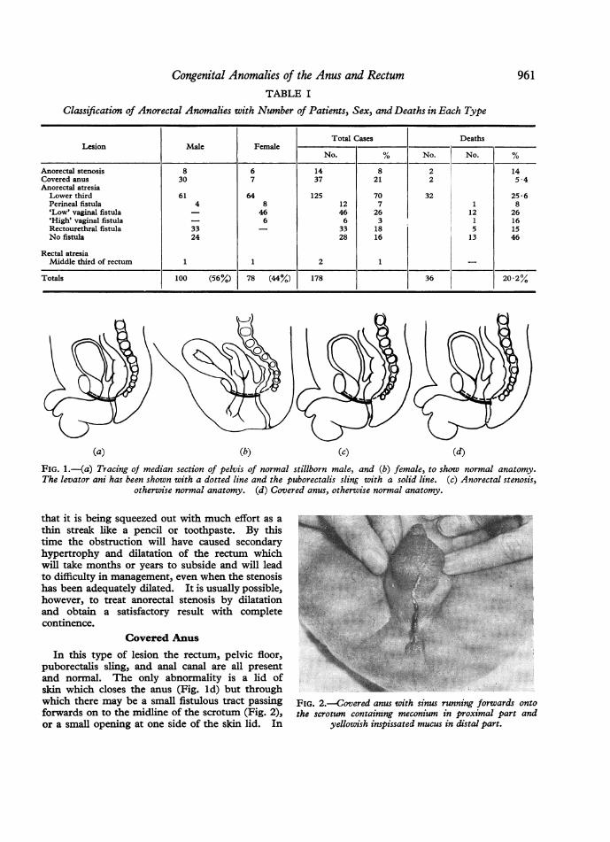
Anorectal stenosis 8 6 14 8 2 14Covered anus 30 7 37 21 2 5-4Anorectal atresiaLower third 61 64 125 70 32 25-6Perineal fistula 4 8 12 7 1 8'Low' vaginal fistula - 46 46 26 12 26'High' vaginal fistula - 6 6 3 1 16Rectourethral fistula 33 - 33 18 5 15No fistula 24 28 16 13 46
Rectal atresiaMiddle third of rectum 1 1 2 1
Totals 100 (56%) 78 (44%) 178 36 20-2%
(a) (b) (c) (d)FIG. l.-(a) Tracing of median section of pelvis of normal stillborn male, and (b) female, to show normal anatomy.The levator ani has been shown with a dotted line and the puborectalis sling with a solid line. (c) Anorectal stenosis,
otherwise normal anatomy. (d) Covered anus, otherwise normal anatomy.
that it is being squeezed out with much effort as athin streak lke a pencil or toothpaste. By thistime the obstruction will have caused secondaryhypertrophy and dilatation of the rectum which.will take months or years to subside and will leadto difficulty in management, even when the stenosishas been adequately dilated. It is usually possible,however, to treat anorectal stenosis by dilatationand obtain a satisfactory result with completecontinence. 0
Covered Anus
In this type of lesion the rectum, pelvic floor,puborectalis sling, and anal canal are all presentand normal. The only abnormality is a lid ofskin which closes the anus (Fig. ld) but throughwhich there may be a small fistulous tract passing FIG. 2.-Covered anus with sinus running forwards ontoforwards on to the midline of the scrotum (Fig. 2), the scrotum containing meconium in proximal part andor a small opening at one side of the skin lid. In yellowish inspissated mucus in distal part.

A. W. Wilkinson
(a) (b)FIG. 3.-(a) Covered anus with roll of skin and sinus on the right side of it through which meconium has escaped.
(b) 21 months later to show patulous anus (from Wilkinson, 1969).
other patients there may be a rolled ridge of skinacross the centre of the skin lid with or without afistula (Fig. 3a). This is the only anorectal anomalywhich could properly be called 'imperforate anus'since the rectum and the anal canal are normalapart from the cover of skin at the lowest part ofthe anal canal. Unfortunately, when the skin coverhas been excised, whether or not the mucousmembrane of the anal canal and the skin of theperineum at the site of the anus are sewn together,the superficial and subcutaneous anal sphinctersdo not function normally. The child is continentbut the anus is patulous and there is usually aleakage of faecal stained mucus from the lining ofthe anal canal (Fig. 3b); the resultant staining ofthe underclothing may cause embarrassment whenthe child has to undress in public at school. Afterexcision of the skin lid the orifice of the anus andthe anal canal should be repeatedly dilated in orderto maintain an adequate passage.
Anorectal AtresiaThis involves the lower third of the rectum and
the anal canal and the rectum ends blindly abovethe pelvic floor (Fig. 4). In addition, in somepatients a fistula runs from the blind end of therectum, through the limbs of the puborectalis slingof the levator ani to enter the upper end of the
vagina (high rectovaginal fistula, Fig. 5), or thelower end of the vagina (low rectovaginal fistula,Fig. 6). In either sex the fistula may open in theperineum in front of the anal dimple which overliesthe superficial and subcutaneous parts ofthe externalanal sphincter (rectoperineal fistula, Fig. 7, 8).In the male a fistula may open into the prostaticor membranous urethra, or very rarely into the
FIG. 4.-Characteristic appearance of the perineum ina boy uith anorectal atresia without a fistula. The siteof the anal sphincter can be seen in the centre of the perineumand a little white discharge at the urethral orifice but no
meconinm.
962

Congenital Anomalies of the Anus and Rectum
Fig. 5FIG. 5.-Tracing of median section of female pelvis as inFig. 1(b). The paths of high and low rectovaginal
fistulae and of a rectoperineal fistula are superimposed.FIG. 6.-Low level rectovaginalfistula with anorectal atresia
(from Wilkinson, 1969).FIG. 7.-Rectoperineal fistula with anorectal atresia inneonate with imperforate hymen (from Wilkinson, 1969).FIG. 8.-Rectoperineal fistula in girl aged 9 months toshow patulous opening without sphincters separated by
bridge of normal skin from vagina. Fig. 6m m ~ ~~~~~~~ _.-w > .......^'.':..:.-' .':.!..
Fig..... 1_ .. .<. ...... 8.,
Fig. 7. Fig. 8.9

A. W. Wilkinson
FIG. 9.-Tracing of median section of pelvis of a maleinfant with anorectal atresia of the lower third of rectumand anal canal to show relation of blind end of bowel tolevator ani (dotted line) and puborectalis (solid line).The paths of two types of rectourethral fistula and a
rectoperineal fistula are superimposed.
bladder (rectourethral or rectovesical fistula, Fig. 9and 10). In both sexes, in the absence of a fistula,there is complete low intestinal obstruction whichrequires relief by some type of surgical operation.The rectoperineal fistula, like the other fistulas,
passes between the limbs of the puborectalis muscleand provided it can be dilated sufficiently does notrequire any other treatment; the child is continentbecause of the relation of the fistula to the puborec-talis sling. The presence of any type of fistulain the female is apt to be misleading becausemeconium is passed and it is not always appreciatedat the outset that the meconium is coming from anabnormal opening. Diagnosis depends mainly ona careful inspection of the perineum and on aninsistence in seeing the orifice from which themeconium is being discharged.
It is very important to recognize that a fistulaconsists of a fibrous wall with an epithelial lining,and that there is no striated muscle in its wall andno external sphincter associated with its orifice.It is therefore anatomically wrong to call such afistulous opening an 'ectopic anus' since anusimplies that there is a sphincteric muscular ringround the orifice which can close it by contraction.
In the past it has been strongly recommendedthat the low fistula opening somewhere near thelower end of the vagina should be dealt with by alocal operation. In the original so-called 'cutbackoperation' one blade of a scissors was placed in thefistula and the other across the perineum; on closingthe scissors the lower orifice was enlarged by a cutbackwards in a midline of the perineum. Thisenlarged opening was subsequently kept wide by
FIG. 10-Male neonate with anorectal atresia in lowerthird of rectum above levator ani, with slight protrusionof most distal part of blind end of rectum through puborec-talis sling, in incontinuity with rectourethral fistula joiningthe prostatic urethra, and showing some escape of opaquemedium upwards into bladder as well as distally into urethra.
dilatation. Because the fistula passed through thelimbs of the puborectalis sling, if a sufficiently widechannel was made by dilatation, the child wascontinent except when the rectum filled withfaeces and faecal impaction led to spurious diarr-hoea. This operation was not always successfulin the long term; in some girls faeces were dischargedat least partially into the vagina, and while this mightbe tolerated in the young child it became offensiveto older children, especially after puberty. It wasan operation which was designed at a time when therisk of a more major procedure, such as anabdominoperineal dissection, was greater becauseof poorer anaesthesia and postoperative treatment.The preferred abdominoperineal operation is nowsufficiently safe to justify the abandonment of thistype of cutback operation. (Some surgeons havemodified this original cutback operation. Theymake a more careful and detailed dissection of thelower end of the fistula in the perineum whichavoids any major division of even the superficialmuscles, but I have not used this operation.)The common belief that lesions might be 'high'
964

Congenital Anomalies of the Anus and Rectumor 'low' implies that there may be a difference inthe level of the blind end of the rectum. Thoughduring straining the blind end of the rectum mayappear to be pushed closer to the perineal skin, itis still above the pelvic floor as represented by thesheet of the levator ani muscle. It is this idea ofthe blind end of the rectum being above the pelvicfloor which is so important to the future continenceof the child. Any attempt to reach the blind endof the rectum from the perineum by cuttingupwards in the midline is almost certain to damagethe puborectalis sling and the levator ani muscles,and in this way to destroy the future continence ofthe child, which depends on the bowel passingnormally through the limbs of the puborectalissling and then down to the perineum. It followsthat the procedure suggested by Wangensteen andRice (1930) of judging the type of lesion accordingto the level of the gas shadow in the blind end ofrectum when the child was x-rayed in the invertedposition is based on a misconception of the anatomyand is misleading. Moreover, gas does not alwaysreach the blind end of the rectum within 6 hoursof birth as was supposed (Wilkinson, 1944).
Atresia in Middle Third of RectumAtresia affecting the middle third of the rectum
is the fourth and much the rarest type of anorectalanomaly. In this the anus, the anal canal, andthe lower third of the rectum are normal and thereis a gap at the site of the middle third of the rectum,-the upper third ending blindly in the hollow of thesacrum. Fistulas are not associated with this type-of atresia (Fig. 11).
Associated AnomaliesThe pelvis must be x-rayed in all patients with
anorectal anomalies. Abnormalities of the sacrumand coccyx are commonly associated with anorectalanomalies and may be accompanied by neuro-muscular defects in the pelvic floor and anomaliesof the urinary tract. It should be recognized alsothat in the presence of several bony abnormalitiesof the pelvis the chances of continence being goodare reduced.
Intravenous pyelography should always be carriedout before the child is discharged from the firstadmission to hospital.
Between September 1958 and December 1970,178 patients with anorectal anomalies were treatedin the Professorial surgical unit at The Hospitalfor Sick Children, Great Ormond Street (Table I).In two-thirds of the total the rectum ended blindlyabove the levator ani, and in most of these therewas some type of fistula. The distribution betweenthe various types in this series is similar to what
FIG. 11.-Atresia of middle third of the rectum showingnormal anal canal and lower third of rectum.
has been found in a number of other publishedseries in which there is enough anatomical detail toallow them to be reclassified in the same way.There are associated congenital abnormalities in
about half of the patients in any large series ofanorectal anomalies, the genitourinary (26%),skeletal (21%) and cardiovascular systems (17%)being most commonly affected (Cozzi andWilkinson, 1968). These associated anomalies areoften severe in degree and threaten survival inabout half the patients who have them. In thisseries oesophageal atresia occurred in 21 patients(12%) and only 4 survived. In 118 neonates inthe series the risk to survival was graded accordingto birthweight, complications, and other associatedanomalies (Table II). The mortality was low ingroups A and B and most of the deaths occurred ingroup C. There was little to choose between thesevere associated anomaly and low birthweight, asfactors contributing to death; and of 5 babies whoweighed less than 1-8 kg (4 lb) at birth, 4 died.Of 12 babies with rectal atresia who died aftercolostomy, 5 also had oesophageal atresia; 2 died ofrespiratory failure associated with prematurity,3 of cardiac failure secondary to congenital heartlesions, 1 of Esch. coli meningitis and peritonitis,and 1 of Esch. coli gastroenteritis and a congenitalheart lesion. 5 died before any treatment of therectal atresia could be started, 2 after the firststage of treatment for oesophageal atresia, and 3 sosoon after admission that no treatment was possible.None of the deaths was due directly to the making ofa colostomy, though there is no doubt that some-times this procedure contributed to death.
TreatmentAnorectal stenosis was treated by repeated
965

TABLE IIClassification of Risk in Neonates with Anorectal Anomalies and Their Mortality Rates
Anorectal Covered Anorectal Atresia Rectal Atresia TotalBirthweight Stenosis Anus Lower Third Middle Third Total Deaths
Group* CasesCases Died Cases Died Cases Died Cases Died No. %
A 2 - 13 - 25 2 - - 40 2 5B 1 - 7 24 1 - - 32 1 3*1C 2 2 5 - 39 24 2 - 46 28 608
*Group A. Birthweight more than 2 -5 kg (51 lb) and no other abnormality or complication. Group B. Birthweight between 1-8 and2-5 kg (4-5 lb) with no other abnormality or complication; or Group A by weight plus complication or associated anomaly of moderateseverity. Group C. Birthweight less than 1-8 kg (4 lb); or Group A with severe, or Group B with moderately severe, associated anomalyor complication.
dilatation, at first under general anaesthesia.Covered anus was treated by excision of the lidof skin over the anus and any associated folds orfistulous tracts leading to the skin on the midlineof the scrotum. The mucous membrane was sewnto the cut edge of the skin with interrupted stitchesof fine black silk, and subsequently dilatation wascarried out as necessary. Rectoperineal fistulawas treated by repeated dilatation. It is mypractice to treat all the other lesions by a colostomyin the neonatal period to relieve the intestinalobstruction, followed by an abdominoperinealtype of pull-through operation at the age of 1 yearor when the child has attained a weight of about9 kg (20 lb). Earlier attempts at dissecting thelow type of vaginal or vestibular fistula and trans-planting it backwards across the perineum to thenormal site of the anus, the anal dimple, resulted insuch frequent breakdown of the new attachmentof the fistula to the skin with recurrence of thefistula in the lower end of the vagina that thisprocedure has now been given up.A transverse colostomy is preferable to a left
iliac colostomy because it allows much greaterfreedom during the pull-through operation, atwhich it may often be necessary to mobilize therectum fairly extensively before it can be broughtto the perineum; this is hindered by the presence ofa left iliac colostomy and it may be impossible toget sufficient viable rectum to the perineumwithout first taking down the left iliac colostomy.Whether the colostomy is opened at the time it ismade or 24 hours later depends entirely on thedegree of distension of the bowel and the abdomenand the effect this has on diaphragmatic respiration.Interference with respiratory exchange and theconsequent increased tendency to respiratoryacidosis is of especial importance in babies withpneumonia or associated cardiovascular anomalies.Sometimes it is necessary to aspirate the gas and
meconium from the colon before the colostomycan be completed.The introduction of plastic bags, worn on a
flange and held in position by adhesive and a belthas considerably reduced the complications anddifficulties formerly associated with a transversecolostomy in an infant. A colostomy at this levelresults in a discharge of thin and often very irritantfaeces compared with those which come from a leftiliac colostomy, though in a child even these aremore liquid and more irritant than in the adult.In the past, severe irritation of the skin and repeatedminor damage to the mucous membrane exposed atthe colostomy have resulted in secondary anaemia,which has required the administration of ironthroughout the life of the colostomy. Moreoverthe irritation of the skin becomes sufficiently severeto cause the child considerable discomfort andrequires the use of large quantities of ointment anddressings which must be changed every time thecolostomy is cleaned. The introduction of plasticbags has almost entirely prevented both the excoria-tion of the skin and secondary anaemia, and it isalmost easier to look after a colostomy in this waythan to care for a normal bowel opening during thefirst year of life. The cost, however, is high, about£95 per annum for bags and flanges, but when thisis set against the reduction in discomfort, excoria-tion of the skin, and anaemia, and the difficultiesthese and the care of the colostomy present to thechild's mother, as well as the hours of nappywashing which she is saved, the benefit is cheap atthe price. Moreover, the use of bags greatlyreduces the emotional disturbance which somewomen find unavoidably associated with thecare of a colostomy in a small baby. This may beso severe that the pull-through operation has tobe undertaken earlier than would otherwise be thecase because of emotional distress of the mother.Once the colostomy has been made and is working
966 A. W. WilEnson

Congenital Anomalies of the Anus and Rectumsatisfactorily, the bowel beyond the colostomyshould be washed out in order to remove all meco-nium and prevent the formation of hard scybalousmasses which will distend the lower end of therectum. When there is an associated rectourethralfistula, washout of the distal bowel is essential toprevent recurrent urinary infection. Even so,urinary infection is occasionally so persistent thatit is necessary to undertake the pull-throughoperation before the child reaches the age of 1year or 9 kg (20 lb) in weight.
In the past many surgeons have carried out theabdominoperineal operation soon after birth andsome still do. It should be remembered, however,that while the mortality rate for colostomy alonein the neonatal period is 20 to 25%, which compareswith the overall mortality rate of 22 4% in 1970in this hopsital for all emergency surgical admis-sions within 4 weeks of birth (J. A. S. Dickson,personal communication, 1971), primary ab-dominoperineal operation at this time probablycarries a mortality rate of well over 30%. More-over, because of the size of the tissues it ismuch more difficult to make a clean dissectiondeep in the pelvis and to be certain that the bowelhas been pulled through the puborectalis sling onwhich in the end continence will so largely depend.Only 2 children in this series have died after 78abdominoperineal operations at the age of 1 year;1 on the third postoperative day of cardiac failuresecondary to Fallot's tetralogy for which a Blalockoperation had been done at the age of 6 months;the other 5 days after operation for a persistentcirculatory failure for which no explanation, eitherclinical or at necropsy, could be found.The abdominoperineal operation is unavoidable
in patients with high level rectovaginal, recto-urethral, or rectovesical fistulas. These cannot besatisfactorily dealt with by any form of perinealapproach. Several patients have been treated who hadhad a perineal operation in other hospitals for suchfistulas, and in the case of boys with rectourethralfistulas the damage to the puborectalis sling andthe median raphe of the levator ani muscle was suchthat they were incontinent of both urine and faeces.In dividing the rectourethral fistulas it is importantto do so flush with the posterior surface of the ure-thra, so that there is no remaining pouch from theurethra which could become infected and bethe source of repeated bouts of urinary infection.To emphasize the futility of the perineal operation
for rectal atresia it should be said that it is oftennecessary, after mobilizing the rectum completelyout of the pelvis, to divide the superior rectalvessels to obtain sufficient length to bring the rectum
to the perineum; and sometimes it is necessary tosacrifice a third or a half of the rectum becausemobilization and division of the superior rectalvessels results in impairment of the blood supplyto the distal rectum. This may not, however,be much of a disadvantage because if the atreticrectum is similar to the atretic small bowel, thenthe dilated hypertrophied blind end has abnormalperistaltic function in any case and would be betterexcised. Indeed, much of the difficulty in gettingthe child to move the bowel satisfactorily after theabdominoperineal mobilization might be avoided ifthe whole of the dilated hypertrophied rectum wasexcised and the upper third of rectum or the distalend of the pelvic colon was brought down to theperineum instead.The colostomy should be closed as soon as possible
after a satisfactory new opening has been obtainedin the perineum. It is probably better to delayclosure of the colostomy, however, until this openingis in its final stage. Prolapse of mucosa at the newanus is not uncommon and should be trimmedbefore the colostomy is closed. Similarly theopening is sometimes too narrow and it shouldbe widened before closing the colostomy. Closureof the colostomy may be associated with a gooddeal of bleeding and blood must be crossmatchedand available for transfusion. It is usually betterto dissect out the colostomy and excise it completelyand make a formal end-to-end anastamosis of thetwo stumps of colon with a single layer of inter-rupted fine black mattress sutures than to attemptany less radical closure which is much more likely tobreak down.
Functional ResultsThe results of treating anorectal stenosis by
dilatation are usually good (Tables III, IV).Control of faeces is ultimately normal thoughthere may be some difficulty in the early stages;sensation of fullness of the rectum, the warningperiod, and the control of flatus are all normal.There is seldom any difficulty with urinary functionapart from what one might expect to occur in theproportion of children who are temporarily enuretic.If medicine is needed to move the bowel, thatsuggests the stenosis may have recurred.
In covered anus, the control of faeces is usuallygood but there is commonly a leakage of faecal-stained mucus from the anal canal which slightlystains the underclothing. This usually persistsfor years. Sensation is usually good, the warningperiod is normal, and so is the control of flatusand urine.
In rectoperineal fistula the picture is much the
967

968 A. W. WilkinsonTABLE III
Functional Results of Treatment of Anorectal Anomalies; Control of Faeces, Flatus, and Urine in 76 Patients
Faeces Flatus UrinePrimary Lesion
Normal Partial None Normal Partial None Normal Partial None
Anorectal stenosis 7 3 _ 8 _ 2 8 2Covered anus 12 2 - 10 - 2 13 - 1Rectal atresia lower third 18 29 4 13 6 23 42 3 6Rectal atresia middle third 1 - - 1 - - 1 - -
Total (76) 38 34 4 32 6 27 64 5 7
same as for covered anus. Most of the functionsare present and normal unless there is someassociated defect in the sacrum or urinary tract.
Anorectal atresia treated by the abdomino-perineal pull-through operation gives variableresults, but when allowance is made in a fewpatients for associated abnormalities such as ectopicbladder, sacral defects, or myelomeningocele, theresults are surprisingly good, but in any particularindividual depend on the age of the child at whichthe situation is assessed. Comparatively fewchildren achieve control of urine or faeces afterthis operation as soon as normal children. Thesensation of fullness of the bowel though present isnot normal, and it takes most children some timeto realize the significance of what sensation theyhave. Even then, and no matter how willing theymay be to keep themselves clean, the warningperiod is often so short that they are unable alwaysto reach the lavatory in time. In some children,however, for several years there seems to be anaversion to cleanliness, and others, preoccupiedwith their play or with work, do not act soon enoughon what warning they have and are persistentlydirty.
After the pull-through operation slight soiling ofthe underclothing with faecal-stained mucus iscommon because the superficial and subcutaneous
parts of the external sphincter do not close off thenew anal opening. Though these two parts of theexternal sphincter can be shown to function bystimulating the perineal skin at the centre of theanal dimple before the pull-through operation isdone, these two sphincteric rings are seldom ableto close the opening after the rectum has beenbrought to the perineal skin.Even in the best patients there is variation in
function from time to time. These children aremore liable to disturbance and malfunction as aresult of injudicious consumption of fruit andvegetables, and are also more liable to severe consti-pation. In some, constipation is most persistentand is mainly related to the abnormal dilated andhypertrophied rectum in which peristalsis is abnor-mal and allows faeces to accumulate. Goodfunction is not related particularly to the type oflesion and occurs as often with rectourethralfistula as it does with high or low rectovaginalfistula.
In the past the treatment of these anomalies of theanus and rectum has received less than its propershare of respect and care from surgeons. Onlythose whose bowel is not continent, or who havestained underclothing, or are the parents of a childin this state have any real idea of the social andeconomic burdens these entail. Successful treat-
TABLE IVFunctional Results of Treatment of Anorectal Anomalies; Warning Period, Sensation or Fullness, and Need
for Laxative in 76 Patients
Warning Period Sensation MedicinePrimary Lesion _
Normal Short None Normal rartial None No Yes
Anorectal stenosis 10 - - 10 - - 2Covered anus 12 - - 13 1 - 12 2Rectal atresia lower third 18 25 5 34 14 - 38 13Rectal atresia middle third 1 - - 1 - 1 -
Total (76) 41 25 5 58 15 - 51 17

Congenital Anomalies of the Anus and Rectum 969ment of these anomalies depends on a sufficientdegree of anatomical certainty in diagnosis andsurgical care in treatment. In anorectal atresia ofthe lower third of the rectum, as defined earlier,the dissection of the rectum and its transference tothe perineum through the limbs of the undamagedpuborectalis sling of the levator ani is essentialfor continence. When subsequently a good pubo-rectalis sling is palpable a confident assurance thatcontinence will be attained can be given to theanxious and often disappointed mother, who alreadyhas waited far longer than she had ever expectedfor her child to be ordinarily clean. The effects ofmaternal disappointment and frustration on thesuccessful long-term management of these childrenmay be profound. The child, the victim of severalmajor operations, weeks or months in hospital,with painful dilatations by one or both of theparents of a stenosed anus, of pain from the passageof constipated stools, or bleeding from mucosaprolapsing through the new 'anus', can hardly beexpected to perform as a normal child would.Indeed, it is surprising that such nearly normalbowel function is achieved as often and as soon asit is by many of these children.The management of such children and their
parents after the pull-through operation is difficult,prolonged, and delicate. Provided one is certainat the pull-through operation, and subsequently ondigital examination of the new 'anal canal' andrectum, that the bowel has been brought downthrough a good puborectalis sling and pelvicfloor, confidence in ultimate continence and goodfunction is justified and should be plainly, firmly,and repeatedly expressed. It is essential for bothmother and child and their relationship that mal-function and disappointment should be containedby confidence in future good control.
There is much controversy about the treatmentof those children in whom partial absence of thesacrum suggests that innervation of the pelvicfloor or the musculature may be abnormal. It ispossible that the best course may be to carry outthe pull-through operation in the hope that it maygive a satisfactory result, but in such patients it isimportant first to warn the parents that control maybe imperfect and function poor. This is probablybetter than to advise an iliac colostomy as an initialand permanent treatment.When the pelvic floor has been damaged at either
a primary perineal operation in an attempt to bringthe rectum to the surface or during a pull-throughoperation, it is often still possible, and nearlyalways worth while, to attempt to improve the situa-tion by a pelvic floor repair rather than to abandonall hope of improvement and advise a permanentiliac colostomy.
I am indebted to Mr. J. A. S. Dickson and Mr. A. J.Dougall for their help during the preparation of thispaper; and to the Editor, Proceedings of the Royal Societyof Medicine, for Fig. 3(a), (b), 6, and 7.
REFERENCESCozzi, F., and Wilkinson, A. W. (1968). Congenital abnormalities
of anus and rectum: mortality and function. British MedicalJournal, 1, 144.
Ladd, W. E., and Gross, R. E. (1934). Congenital malformationsof anus and rectum: report of 162 cases. American Journal ofSurgery, 23, 167.
Wangensteen, 0. H., and Rice, C. 0. (1930). Imperforate anus:method of determining surgical approach. Annals of Surgery,92, 77.
Wilkinson, A. W. (1944). Imperforate anus: some observationsregarding diagnosis of the level of obstruction. Archives ofDisease in Childhood, 19, 138.
Wilkinson, A. W. (1969). Anorectal abnormality in children.Proceedings of the Royal Society of Medicine, 62, 1234.
Correspondence to Professor A. W. Wilkinson,Institute of Child Health, 30 Guilford Street, LondonWC1N IEH.