congenital heart disease. chd chd = abnormalities of the heart or great vessels present from birth...
TRANSCRIPT

Congenital Heart Congenital Heart DiseaseDisease
CHDCHD
CHD = Abnormalities of the heart or great CHD = Abnormalities of the heart or great vessels present from birthvessels present from birth
Most – faulty embryogenesis during the 3Most – faulty embryogenesis during the 3rdrd--88thth week when the CVS form and begin week when the CVS form and begin functioningfunctioning
Worst ones don’t survive to termWorst ones don’t survive to termThose who do usually have only discrete Those who do usually have only discrete
regions of the heart affected e.g. septal regions of the heart affected e.g. septal defect or valvular defectdefect or valvular defect
CHDCHD
DxDx
Some when change from fetal to postnatal Some when change from fetal to postnatal circulationcirculation
50% diagnosed by one year of life50% diagnosed by one year of life
Mild forms - adulthoodMild forms - adulthood
CHDCHD
IncidenceIncidence1% of all live births1% of all live birthsCV defects among most common malformations and are the most CV defects among most common malformations and are the most
common cause of heart disease in childrencommon cause of heart disease in childrenHigher in premies and stillbornsHigher in premies and stillbornsTable 12-2 Table 12-2
VSD most common VSD most common Tetralogy of Fallot most common cyanoticTetralogy of Fallot most common cyanotic
Many survive into adulthood - repairsMany survive into adulthood - repairsarrhythmiasarrhythmiasadditional surgeryadditional surgeryventricular dysfunctionventricular dysfunctionuse of prostheticsuse of prostheticsrisk of childbearingrisk of childbearing
CHDCHD
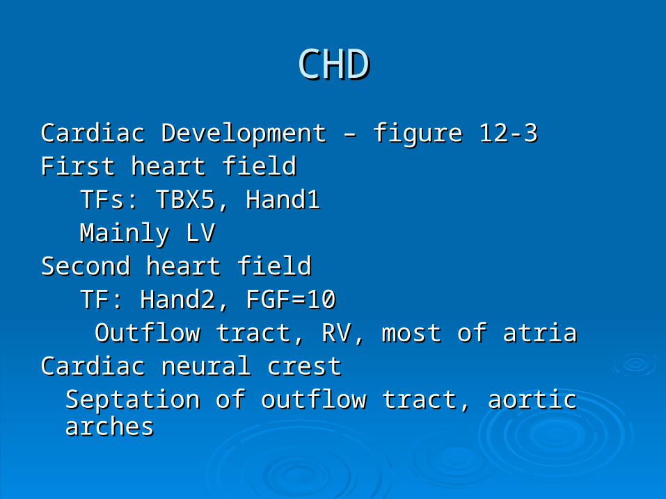
Cardiac Development – figure 12-3Cardiac Development – figure 12-3First heart field First heart field
TFs: TBX5, Hand1TFs: TBX5, Hand1 Mainly LVMainly LV
Second heart field Second heart field TF: Hand2, FGF=10TF: Hand2, FGF=10 Outflow tract, RV, most of atriaOutflow tract, RV, most of atria
Cardiac neural crestCardiac neural crestSeptation of outflow tract, aortic archesSeptation of outflow tract, aortic arches

CHDCHD
ECM – swellings – endocardial cushionsECM – swellings – endocardial cushionsFuture valve developmentFuture valve development
Day 50 – 4 chambered heartDay 50 – 4 chambered heartSignaling pathways regulating TFsSignaling pathways regulating TFs
WntWntVEgfVEgfbone morphogenetic factorbone morphogenetic factorTGF-betaTGF-betaFGFFGFNotchNotch
Heart – mechanical organ – exposed to flowing blood from earliest Heart – mechanical organ – exposed to flowing blood from earliest stages – hemodynamic forces play a rolestages – hemodynamic forces play a role
Specific micro RNAs – critical role- patterns and levels of TF Specific micro RNAs – critical role- patterns and levels of TF expressionexpression
CHDCHD
AD mutations – partial loss of function in one AD mutations – partial loss of function in one or more required factors, TFs usuallyor more required factors, TFs usually
““The main known cause of CHD consist of The main known cause of CHD consist of sporadic genetic abnormalities.”sporadic genetic abnormalities.”single gene mutationssingle gene mutationssmall chromosome deletionssmall chromosome deletionsadditions or deletions of whole additions or deletions of whole chromosomeschromosomes
Table 12-3Table 12-3
CHDCHD
Heterozygotes = 50% reduction in activity = Heterozygotes = 50% reduction in activity = deranged cardiac developmentderanged cardiac development
Factors work together- large protein Factors work together- large protein complexes – different single gene complexes – different single gene mutations produce similar defectsmutations produce similar defects
Signaling pathways or structural rolesSignaling pathways or structural rolesNOTCH1 – bicuspid AVNOTCH1 – bicuspid AVNOTCH2, JAGGED1 – TOFNOTCH2, JAGGED1 – TOFFibrillin – Marfan’sFibrillin – Marfan’s
CHDCHD
DiGeorge SyndromeDiGeorge SyndromeSmall deletion of 22q11.2 in 50%Small deletion of 22q11.2 in 50%44thth branchial arch and 3 branchial arch and 3rdrd and 4 and 4thth pharyngeal pouches pharyngeal pouches Thymus, parathyroids, heartThymus, parathyroids, heartTBX1TBX1
Chromosomal aneuploidiesChromosomal aneuploidiesTurner SyndromeTurner SyndromeTrisomies 13,18, 21Trisomies 13,18, 21
21- most common genetic cause of CHD21- most common genetic cause of CHDendocardial cushion defectsendocardial cushion defects
CHDCHD
First-degree relatives of affected patients are at increased First-degree relatives of affected patients are at increased of CHD – subtle forms of genetic variationof CHD – subtle forms of genetic variation
Environmental factors?Environmental factors?+/- genetic factors+/- genetic factorscongenital rubella infectioncongenital rubella infectiongestational diabetesgestational diabetesexposure to teratogensexposure to teratogensnutritional factors? nutritional factors? transient environmental stresses during 1transient environmental stresses during 1stst trimester? trimester?
CHDCHD
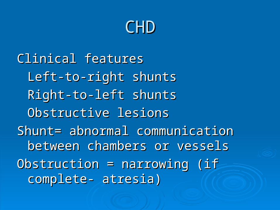
Clinical featuresClinical features
Left-to-right shuntsLeft-to-right shunts
Right-to-left shuntsRight-to-left shunts
Obstructive lesionsObstructive lesions
Shunt= abnormal communication between Shunt= abnormal communication between chambers or vesselschambers or vessels
Obstruction = narrowing (if complete- Obstruction = narrowing (if complete- atresia)atresia)
CHDCHD
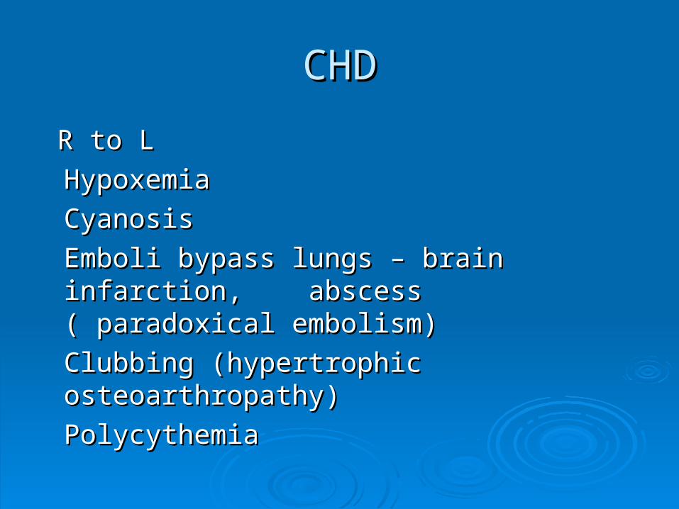
R to LR to L
HypoxemiaHypoxemia
CyanosisCyanosis
Emboli bypass lungs – brain infarction, Emboli bypass lungs – brain infarction, abscess ( paradoxical embolism)abscess ( paradoxical embolism)
Clubbing (hypertrophic osteoarthropathy)Clubbing (hypertrophic osteoarthropathy)
PolycythemiaPolycythemia

CHDCHDL to RL to R
Normally low-pressure, low-resistance pulmonary circulation now sees high flow Normally low-pressure, low-resistance pulmonary circulation now sees high flow volumes and pressuresvolumes and pressuresRVHRVHAtherosclerosis of pulmonary vesselsAtherosclerosis of pulmonary vessels
medial hypertrophymedial hypertrophyvasoconstrictionvasoconstrictionirreversible obstructive intimal lesionsirreversible obstructive intimal lesions
Pulm pressures reach systemic levelsPulm pressures reach systemic levelsR to L shuntR to L shuntEisenmenger SyndromeEisenmenger Syndrome
Altered hemodynamics of CHDAltered hemodynamics of CHDDilation, hypetrophy or bothDilation, hypetrophy or bothDecreased volume and muscle mass – hypoplasia – before birth, atrophy – Decreased volume and muscle mass – hypoplasia – before birth, atrophy – postnatallypostnatally
CHDCHD
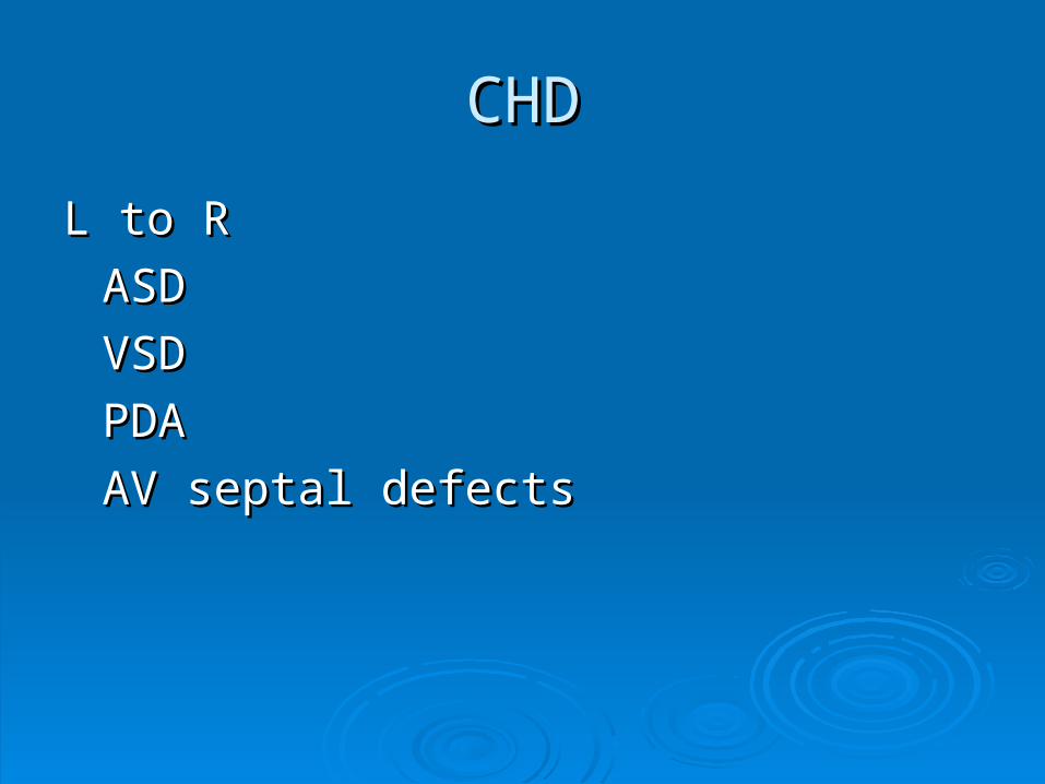
L to RL to R
ASDASD
VSDVSD
PDAPDA
AV septal defectsAV septal defects
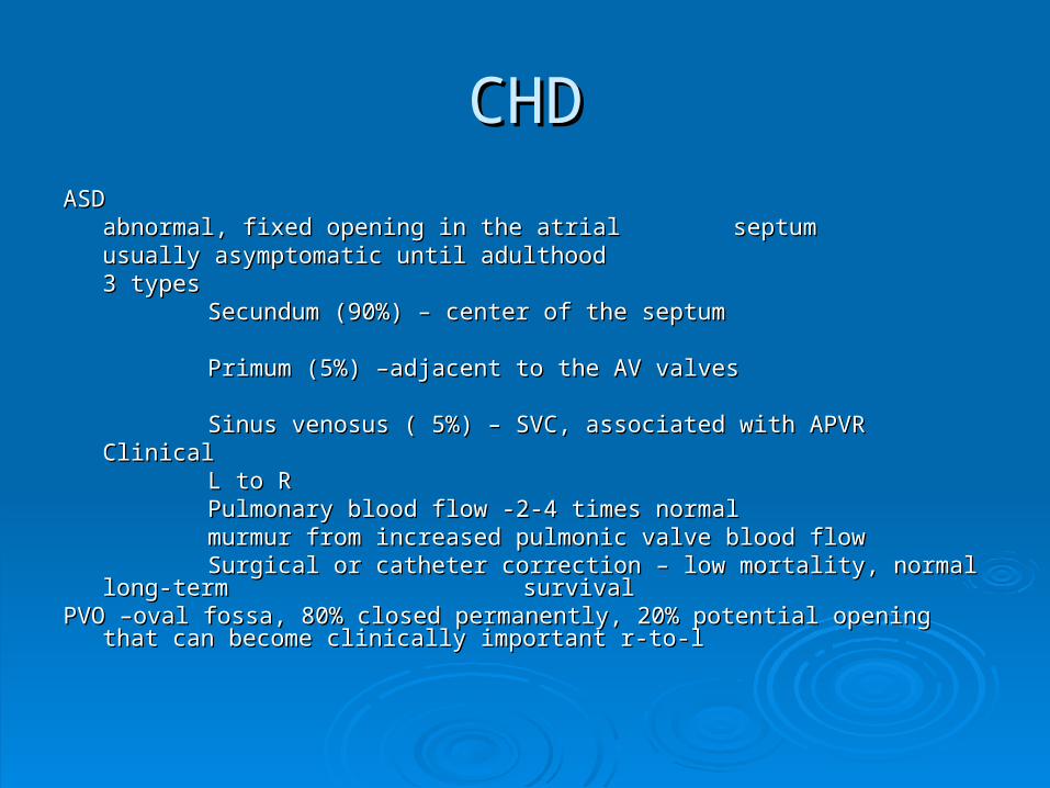
CHDCHDASDASD
abnormal, fixed opening in the atrial abnormal, fixed opening in the atrial septumseptumusually asymptomatic until adulthoodusually asymptomatic until adulthood3 types3 types
Secundum (90%) – center of the septumSecundum (90%) – center of the septum
Primum (5%) –adjacent to the AV valvesPrimum (5%) –adjacent to the AV valves
Sinus venosus ( 5%) – SVC, associated with APVRSinus venosus ( 5%) – SVC, associated with APVRClinicalClinical
L to RL to RPulmonary blood flow -2-4 times normalPulmonary blood flow -2-4 times normalmurmur from increased pulmonic valve blood flowmurmur from increased pulmonic valve blood flowSurgical or catheter correction – low mortality, normal long-term Surgical or catheter correction – low mortality, normal long-term
survivalsurvivalPVO –oval fossa, 80% closed permanently, 20% potential opening that can become PVO –oval fossa, 80% closed permanently, 20% potential opening that can become
clinically important r-to-lclinically important r-to-l
CHDCHD
VSDVSDMost common congenital anomalyMost common congenital anomaly20-30% isolated finding20-30% isolated findingMost are associated with other cardiac anomaliesMost are associated with other cardiac anomaliesClassified by size and locationClassified by size and location90% membranous90% membranousRest are infundibular ( below the PV) or muscularRest are infundibular ( below the PV) or muscularMuscular can be multiple ( “Swiss-cheese”)Muscular can be multiple ( “Swiss-cheese”)ClinicalClinical
Large – problems from birth, RVH, pulmonary Large – problems from birth, RVH, pulmonary hypertension, hypertension, correct correct before irreversible changesbefore irreversible changes
Smaller – well-toleratedSmaller – well-tolerated
CHDCHD
PDAPDADA stays open, allowing L to R shunt from DA stays open, allowing L to R shunt from aorta to pulmonary arteryaorta to pulmonary artery90% isolated anomaly90% isolated anomaly““machinery-like” murmurmachinery-like” murmurclose as soon as possible to prevent irreversible close as soon as possible to prevent irreversible
PHPHSome congenital lesions are ductus dependent Some congenital lesions are ductus dependent
and there by need to keep the DA open-and there by need to keep the DA open-e.g. aortic atresia, use prostaglandin E)e.g. aortic atresia, use prostaglandin E)
CHDCHD
AV septal defectAV septal defectComplete atrioventricular canal defectComplete atrioventricular canal defectPartial – primum ASD with mitral Partial – primum ASD with mitral insufficiencyinsufficiencyComplete – large combined AV septal Complete – large combined AV septal defect defect and a common AV valve – all 4 and a common AV valve – all 4 chambers chambers communicate, all have communicate, all have hypertrophyhypertrophy1/3 have Down syndrome1/3 have Down syndromeSurgically correctibleSurgically correctible
CHDCHD
R to LR to L
Tetralogy of FallotTetralogy of Fallot
Transposition of the Great ArteriesTransposition of the Great Arteries
Truncus arteriosusTruncus arteriosus
Tricupsid AtresiaTricupsid Atresia
Total Anomalous Venous Connection Total Anomalous Venous Connection
CHDCHDTetralogy of FallotTetralogy of Fallot
4 cardinal features4 cardinal featuresVSDVSDObstruction of the right ventricular outflow tract (subpulmonary stenosis)Obstruction of the right ventricular outflow tract (subpulmonary stenosis)An aorta that overrides the VSDAn aorta that overrides the VSDRVHRVH
Embryoloigcally – anterosuperior displacement of the infundibular septumEmbryoloigcally – anterosuperior displacement of the infundibular septum““Boot-shaped” heart – marked apical RVHBoot-shaped” heart – marked apical RVHSometimes PVS, PV atresiaSometimes PVS, PV atresia
Sometines AV insufficiency, ASD Sometines AV insufficiency, ASD 25% right aortic arch25% right aortic archClinical – Classic TOF – r-to-l shuntClinical – Classic TOF – r-to-l shunt
Pink TOF – l to r shunt because of mild subpulmonary stenosisPink TOF – l to r shunt because of mild subpulmonary stenosisAs child grows obstuction becomes worseAs child grows obstuction becomes worseStenosis protects pulmnary arteries from overload and RV failure rare because RV Stenosis protects pulmnary arteries from overload and RV failure rare because RV
decompressed by the VSDdecompressed by the VSD

CHDCHD
TGATGA
Ventriuloarterial discordVentriuloarterial discord
Aorta from RVAorta from RV
PA from LVPA from LV
Separation of the systemic and pulmonary Separation of the systemic and pulmonary
circulations – incompatible with life circulations – incompatible with life unless a shunt exists VSD or PFO or PDA or unless a shunt exists VSD or PFO or PDA or artificial shunt –balloon atrial septostomyartificial shunt –balloon atrial septostomy
Surgical repairSurgical repair
CHDCHD
TATA
Failure of separation into the aorta and PAFailure of separation into the aorta and PA
a single vessel giving rise to the systemic, a single vessel giving rise to the systemic, pulmonary and coronary circulationpulmonary and coronary circulation
Associated VSDAssociated VSD
CHDCHD
TAPCTAPC
Pulmonary veins fail to join the left atriumPulmonary veins fail to join the left atrium
PFO or ASDPFO or ASD
Aplastic Left atriumAplastic Left atrium
LV normal sizeLV normal size

CHDCHD
Obstructive Congenital AnomaliesObstructive Congenital Anomalies
Coartation of the aortaCoartation of the aorta
PS and atresiaPS and atresia
AS and atresiaAS and atresia
CHDCHDCoarctation of the AortaCoarctation of the Aorta
Males 2x femalesMales 2x femalesAssociated with Turner syndromeAssociated with Turner syndrome2 classic types2 classic types
““infantile” –infantile” –hypoplasia of the arch proximal to a PDA, symptomatic in hypoplasia of the arch proximal to a PDA, symptomatic in early childhood, cyanosis over lower half of body, early childhood, cyanosis over lower half of body, surgical correction needed earlysurgical correction needed early
““adult” –adult” –discrete ridgelike infolding of the aorta, just opposite a discrete ridgelike infolding of the aorta, just opposite a closed DA (ligamentum arteriosus) distal to the arch vessels, closed DA (ligamentum arteriosus) distal to the arch vessels, hypertension in upper extremities, signs of arterial insufficiency in hypertension in upper extremities, signs of arterial insufficiency in lower, notching of the ribs due to collateral circulationlower, notching of the ribs due to collateral circulation
Clinical –Clinical –murmur with thrillmurmur with thrillLVHLVH
CHDCHD
PS and atresiaPS and atresiaObstruction of the PVObstruction of the PVIsolated or part of a more complex anomalyIsolated or part of a more complex anomalyRVHRVHPoststenotic dilation of PAPoststenotic dilation of PAComplete obstruction- need shunt to surviveComplete obstruction- need shunt to surviveMild – asymptomaticMild – asymptomaticSymptomatic – surgical correctionSymptomatic – surgical correction
CHDCHD
AS and atresiaAS and atresiaVavular-hypoplastic, dysplastic, decreased Vavular-hypoplastic, dysplastic, decreased numbernumberSubvalular-dense fibrous tissue below the cuspsSubvalular-dense fibrous tissue below the cuspsSupravavular- aortic dysplasia, thickened and constricted, deletion Supravavular- aortic dysplasia, thickened and constricted, deletion
on chromosome 7, elastin gene, Williams-Beuren on chromosome 7, elastin gene, Williams-Beuren syndrome,syndrome,
hypercalcemia, cognitive abnormalities, facial anomalieshypercalcemia, cognitive abnormalities, facial anomalies
Hypoplastic left heart syndrome – severe stenosis of atresia – Hypoplastic left heart syndrome – severe stenosis of atresia – underdevelopment of LV and aorta – endocardial fibroelastosisunderdevelopment of LV and aorta – endocardial fibroelastosis
Clinical – systolic murmur, thrill, LVH, antibiotic prophylaxis for SBE, Clinical – systolic murmur, thrill, LVH, antibiotic prophylaxis for SBE, avoid strenuous activity, sudden deathavoid strenuous activity, sudden death