consensus paper: towards a systems-level view of ......consensus paper consensus paper: towards a...
TRANSCRIPT

CONSENSUS PAPER
Consensus Paper: Towards a Systems-Level Viewof Cerebellar Function: the Interplay Between Cerebellum, BasalGanglia, and Cortex
Daniele Caligiore1 & Giovanni Pezzulo1 & Gianluca Baldassarre1 & Andreea C. Bostan2&
Peter L. Strick2& Kenji Doya3 & Rick C. Helmich4
& Michiel Dirkx4 & James Houk5&
Henrik Jörntell6 & Angel Lago-Rodriguez7 & Joseph M. Galea7 & R. Chris Miall7 &
Traian Popa8 & Asha Kishore9 & Paul F. M. J. Verschure10,11 & Riccardo Zucca10 &
Ivan Herreros10
Published online: 13 February 2016# The Author(s) 2016. This article is published with open access at Springerlink.com
Abstract Despite increasing evidence suggesting the cerebel-lum works in concert with the cortex and basal ganglia, thenature of the reciprocal interactions between these three brainregions remains unclear. This consensus paper gathers diverse
recent views on a variety of important roles played by thecerebellum within the cerebello-basal ganglia-thalamo-cortical system across a range of motor and cognitive func-tions. The paper includes theoretical and empirical
* Daniele [email protected]
Giovanni [email protected]
Gianluca [email protected]
Andreea C. [email protected]
Peter L. [email protected]
Kenji [email protected]
Rick C. [email protected]
Michiel [email protected]
James [email protected]
Henrik Jö[email protected]
Angel [email protected]
Joseph M. [email protected]
R. Chris [email protected]
Traian [email protected]
Asha [email protected]
Paul F. M. J. [email protected]
Riccardo [email protected]
Ivan [email protected]
1 Istituto di Scienze e Tecnologie della Cognizione, ConsiglioNazionale delle Ricerche (ISTC-CNR), Via San Martino dellaBattaglia 44, 00185 Rome, Italy
2 Systems Neuroscience Institute, Department of Neurobiology, andCenter for the Neural Basis of Cognition, University of Pittsburgh,3501 Fifth Avenue, 4079 BST-3, Pittsburgh, PA 15261, USA
3 Neural Computation Unit, Okinawa Institute of Science andTechnology, 1919-1 Tancha, Onna-son,Kunigami-gun, Okinawa 904-0495, Japan
4 Department of Neurology, Radboud University Nijmegen MedicalCenter (HP 935), PO Box 9101, 6500HB Nijmegen, The Netherlands
Cerebellum (2017) 16:203–229DOI 10.1007/s12311-016-0763-3
contributions, which cover the following topics: recent evi-dence supporting the dynamical interplay between cerebel-lum, basal ganglia, and cortical areas in humans and otheranimals; theoretical neuroscience perspectives and empiricalevidence on the reciprocal influences between cerebellum,basal ganglia, and cortex in learning and control processes;and data suggesting possible roles of the cerebellum in basalganglia movement disorders. Although starting from differentbackgrounds and dealing with different topics, all the contrib-utors agree that viewing the cerebellum, basal ganglia, andcortex as an integrated system enables us to understand thefunction of these areas in radically different ways. In addition,there is unanimous consensus between the authors that futureexperimental and computational work is needed to understandthe function of cerebellar-basal ganglia circuitry in both motorand non-motor functions. The paper reports the most ad-vanced perspectives on the role of the cerebellum within thecerebello-basal ganglia-thalamo-cortical system and illustratesother elements of consensus as well as disagreements andopen questions in the field.
Keywords Basal ganglia cerebellum anatomical link .
Nucleo-olivary inhibition .Movement disorders . Parkinson’sdisease tremor . Cerebellar motor and cognitive function .
Non-invasive brain stimulation
Introduction
The cerebellum works in concert with cortex and the basalganglia as a fundamental building block in motor and cogni-tive tasks of various complexity, from sensorimotor mappingto reasoning [1–4]. This systems-level view is increasinglysupported by evidence demonstrating that the cerebellumand the basal ganglia receive input from, and send output to,
different cortical areas through multisynaptic loops that havebeen assumed to be anatomically segregated and to performdistinct functional operations [5–10]. Moreover, recent find-ings reveal the existence of an anatomical substrate for thebidirectional communication between cerebellum and basalganglia. In particular, studies on rats [11] and monkeys [12]have demonstrated that the cerebellum sends a strongdisynaptic projection to the striatum through the thalamus.Furthermore, recent studies in monkeys have shown that thesubthalamic nucleus sends a disynaptic projection to the cer-ebellar cortex by way of the pontine nuclei [13]. Similar evi-dence has been recently reported in the human brain [14].
These data have stimulated new research to investigate thereciprocal influence between these brain areas and the differ-ent forms of learning typically associated with them: super-vised learning in the cerebellum based on plasticity of parallelfiber-Purkinje cell synapses [15–17]; unsupervised learning inthe cortex based on associative Hebbian processes [18–20];and trial-and-error (reinforcement learning) in the basal gan-glia based on reward prediction errors computed at level of thestriatum and depending on dopamine signals [21, 22]. Severalrecent studies have tried to integrate these apparently discon-nected learning processes, focusing for example on the role ofthe cerebellum in regulating the plasticity of premotor andmotor networks during sensorimotor learning [23–27]; theinvolvement of the cerebellum in the reinforcement learningprocesses underlying the computation of sensory and rewardprediction errors during motor adaptation [28–33]; the role ofthe cerebellum and basal ganglia in the cortical acquisition ofhigh-level cognitive (non-motor) functions [1, 3, 4]; and theinfluence of cerebellum and basal ganglia on motor corticalplasticity in health and in Parkinson’s disease [34, 35]. Finally,data supporting a close interaction between cerebellar, basalganglia, and cortical areas have recently stimulated new re-search on the role of cerebellum and basal ganglia in functionstypically associated with cortical areas (e.g., action under-standing [4]) and the role of cerebellar and cortical areas inimpairments typically associated with basal ganglia, such asTourette’s syndrome, dystonia, and Parkinson’s disease[36–40].
Despite this recent progress, we still lack a comprehensiveframework that defines cerebellar function from a wider,systems-level perspective [4, 41, 42]. Viewing cerebellum,cortex, and basal ganglia as an integrated system could leadto understand their functional and learning processes in radi-cally different ways. The goal of this consensus paper is tofeed the discussion on the fundamental interactions betweenthe functional and plasticity processes of cerebellum, cortex,and basal ganglia, conceived as forming an integrated systemunderlying several motor and cognitive functions.
Evidence of the interactions between the functional andlearning processes in the cerebellum, cortex, and basal gangliacomes from different scientific disciplines and methodologies:
204 Cerebellum (2017) 16:203–229
5 Department of Physiology, NorthwesternUniversity Feinberg Schoolof Medicine, 303 East Chicago Avenue M211, Chicago, IL 60611,USA
6 Neural Basis of Sensorimotor Control, Department of ExperimentalMedical Science, Lund University, BMC F10 Tornavägen 10, 22184 Lund, Sweden
7 School of Psychology, University of Birmingham,Edgbaston, Birmingham B15 2TT, UK
8 Human Motor Control Section, National Institute of NeurologicalDiseases and Stroke (NINDS), National Institutes of Health (NIH),Bethesda, MD 20982, USA
9 Comprehensive Care Centre for Movement Disorders, Sree ChitraTirunal Institute for Medical Sciences and Technology,Kerala 695011, India
10 Department of Information and Communication Technologies,Universitat Pompeu Fabra, Barcelona, Spain
11 Catalan Institute of Research and Advanced Studies (ICREA),Barcelona, Spain

from neurophysiology to computational modelling and frombehavioral methods to neuropsychological and brain imagingapproaches. Unfortunately, discussion between experts usingthese different approaches is often limited. We believe that amulti-methodological and multidisciplinary discussion is nec-essary to fully understand the integrated dynamics of thecerebellar-basal-ganglia-cortical system. This consensus pa-per offers an up-to-date overview on this discussion as it col-lects contributions from an interdisciplinary community ofscientists actively involved in the investigation of cerebellumthat provide a range of different, sometimes controversial,viewpoints.
The issue begins with the paper of Bostan and Strick thatintroduces and reviews the recent neurophysiological and neu-roanatomical evidence on the cerebellar connections with thecerebral cortex and the basal ganglia.
The next two contributions, by Verschure et al. and byJörntell, further expand the systems-level view of cerebellarinterconnections supported by the analysis of the previouspiece (by Bostan and Strick). Verschure et al. raise the fasci-nating possibility that the nucleo-olivary pathway balances thecontribution of feedback (mediated by brain stem nuclei) andfeed-forward (cerebellar) modes of motor control. Jörntellprovides an overview of the spinal inputs into the cerebellum,discussing how these inputs allow the cerebellum to keepinformed about the excitatory drive on low level motor func-tions. He thus proposes that the major role of the cerebellum isto allow many spinal inputs to be associated with one anotherin specific environmental contexts.
The contribution of Houk provides a theoretical, systems-level perspective of how the cerebellar-cortical and basalganglia-cortical loops may interact to affect learning and con-trol processes.
Along similar lines, the contribution of Doya elaborates hisprevious theory of motor control and learning, based on theidea that different brain areas implement different learningalgorithms, to account for recent findings discussed byBostan and Strick on the circuitry between the cerebellumand the basal ganglia.
Pivoting on recent empirical evidence, Miall suggests thatthe cerebellum may support a single operation, acting as ashort-term predictor within the cerebellar-basal-ganglia-cortical system. He also discusses data on medium-term,post-learning, memory consolidation effects in this system.
Lago-Rodriguez and Galea summarize the importance ofusing non-invasive techniques (such as transcranial magneticstimulation (TMS) or transcranial direct current stimulation(tDCS)) to investigate the causal role of the cerebellum inmotor function and its interactions with other brain regions.
Finally, the last two papers provide some examples of theconsequences of abnormal interactions between cerebellum,basal ganglia, and cortical systems. Popa and Kishore discussdata supporting how impaired cerebellar plasticity
mechanisms could contribute to Parkinson’s disease and dys-kinesia. Caligiore et al. build on recent data that suggest aninvolvement of cerebellum in Parkinson’s disease and discusssome cortical-subcortical circuits within the cerebellar-basal-ganglia-cortical network that could be involved in Parkinson’sresting tremor.
Taken together, these empirical and theoretical contribu-tions offer readers a broad and up-to-date framework to de-scribe the dynamic interplay between functioning and learningmechanisms in the cerebellum, basal ganglia, and cortex,pointing out the key open issues for future research. The finalpart of the article summarizes the main topics and analyzes themain elements of consensus—or lack of consensus among theauthors. As discussed in the BSummary and conclusions^ sec-tion, consensus exists on several key aspects of cerebellar-basal-ganglia-cortical function; at the same time, there arevarious topics that are still debated and where consensus can-not be reached yet, reflecting the relative novelty of the topic.These open points represent interesting and promising direc-tions for future research.
Cerebellar Connections with the Cerebral Cortexand the Basal Ganglia (A.C. Bostan, P.L. Strick)
The classical view of cerebellar function has been that it isexclusively concerned with the control of movement throughdescending control and influence on the primary motor cortex(M1). The cerebellum was thought to receive informationfrom multiple neocortical areas and funnel it back to M1[43]. More recent analyses of cerebellar outputs have resultedin a dramatic shift in the conceptualization of cerebellarfunction.
It is now clear that efferents from the cerebellar nucleitarget multiple subdivisions of the thalamus [44] that reachwidespread neocortical areas. Experiments using neurotropicviruses as transneuronal tracers have been crucial for the iden-tification of neocortical areas that are the targets of cerebellaroutput (see [1] for a review). These experiments demonstratethat cerebellar output influences not only M1 but alsopremotor, prefrontal, and parietal areas (Fig. 1a, b). Outputchannels to M1 and the premotor areas are clustered in adorsal region of the cerebellar dentate nucleus, identifying amotor domainwithin this nucleus. Output channels to prefron-tal cortex are clustered together in a ventral (non-motor) re-gion of the nucleus, entirely outside the motor domain [45](Fig. 1b).
To date, all of the areas in the cerebral cortex that are targetsof cerebellar output also have prominent projections to thecerebellar cortex via the pontine nuclei (Fig. 1a). Thin axoncollaterals from approximately 20 % of ponto-cerebellar neu-rons reach the dentate nucleus [46] and may allow excitatoryinputs from cortical areas, such as M1, to reach the dentate
Cerebellum (2017) 16:203–229 205
LS Lu
CA
PSAS
CS
STS
9L
46d46v
12
TE
Pre-PMD
PMDarm
PMvarm
M1leg
M1arm
M1face
7bAIP
9m
Pre-SMA
SMAarm
CgS
46d
9
Pre-SMA
M1leg
M1face
M1
SMAarm
“Motor”
“Non-Motor”
Dorsa
lVen
tral
Dorsa
l
arm
PMvarm
7bFEF
HVIIB
HVIII
F.pcul.
F.icul.
F.pr.
F.ps.
F.ppd.
I
II
III
IV
VII
VIII
IX
X
V
VI
M1 Granule Cells
I
II
III
IV
V
VI
VIII
IX
X
Crus I
Crus II
a
a
p
p
VIIB
F. in.cr.
F.apm.
S. int.cr.2
VII
F.pr.
F.ps.
F.ppd.
46d Granule Cells
ba
Purkinje Cells M1
HVIIB
HVIII
VII
III
IV
V
VI
VIII
IX
I
II
X
F.pcul.
F.icul.
F.pr.
F.ps.
F.ppd.
c
Crus I
Crus II
a
a
p
p
I
II
III
IV
V
VI
VII
VIIBVIII
IX
X
F. in.cr.
F.apm.
S. int.cr.2
F.pr.
F.ps.
F.ppd.
Purkinje Cells 46d
d
7a
IP
Fig. 1 Cerebellar networks with the cerebral cortex. a The relative density ofcerebral cortex neurons that project to the pontine nuclei is indicated by graydots on the lateral andmedial views of themonkey brain (based on [48]).Blacklabels indicate areas of the cerebral cortex that are the target of cerebellaroutput. Gray labels indicate areas that are not targets of cerebellar output(adapted from [3]). b Summary map of dentate nucleus output topography.The lettering on the unfolded map indicates the neocortical target of differentoutput channels. The location of different output channels divides the dentatenucleus into motor and non-motor domains, separated by the dotted line(adapted from [45]). c Organization of cerebellar circuits with M1. Left: thedistribution of Purkinje cells (small dots) that project to the arm area of M1.Right: the distribution of granule cells (fine lines) that receive input from thearm area of M1 (adapted from [50]). d Organization of cerebellar loops with
area 46. Left: the distribution of Purkinje cells (small dots) that project to area46. Right: the distribution of granule cells (fine lines) that receive input fromarea 46 (adapted from [50]). Numbers refer to cytoarchitectonic areas (a, b).Roman numerals refer to cerebellar lobules (c, d). Labels in italics refer tocortical sulci (a) and cerebellar fissures (c,d).AIP anterior intraparietal area,ASarcuate sulcus, CgS cingulate sulcus, FEF frontal eye field, IP intraparietalsulcus, LS lateral sulcus, Lu lunate sulcus,M1 face, arm, and leg areas of theprimary motor cortex, PMd arm arm area of the dorsal premotor area, PMvarm arm area of the ventral premotor area, PrePMd predorsal premotor area,PreSMA presupplementarymotor area,PS principal sulcus, SMAarm arm areaof the supplementary motor area, ST superior temporal sulcus, TE area ofinferotemporal cortex
206 Cerebellum (2017) 16:203–229
[47]. Interestingly, areas of the cerebral cortex that lack sub-stantial projections to the cerebellum (e.g., areas 46v, 12, andTE) do not appear to be major targets of cerebellar output. Ifthese principles apply to all cerebro-cerebellar networks, thenall of the areas of cerebral cortex that project to the cerebellummay be the targets of cerebellar output. This would includesuch diverse areas as regions of extrastriate cortex, cingulatecortex, and the parahippocampal gyrus [48, 49].
The distinct motor and non-motor domains observed in thedentate nucleus have their counterparts in cerebellar cortex(Fig. 1c, d). The motor domain includes a region primarilyin the anterior lobe (lobules III–VI) and another in theparamedian lobule and adjacent cerebellar hemisphere(HVIIB and HVIII). These regions have been shown to bothreceive inputs from and send outputs to M1, forming a closed-loop circuit [50] (Fig. 1c). The non-motor domain is extensiveand involves the portions of the posterior vermis and hemi-sphere that lie between the two regions of motor representa-tion. Regions within the non-motor domain of cerebellar cor-tex also participate in distinct closed-loop circuits with regionsof prefrontal cortex [50] (Fig. 1d). Thus, multiple closed-loopcircuits represent a fundamental feature of interactions be-tween the cerebellum and the cerebral cortex.
The evidence that cerebellar output influences multipleareas of prefrontal and posterior parietal cortex provides theanatomical substrate for a distinct cerebellar contribution tonon-motor aspects of behavior [51]. To date, most theories ofcerebellar function are based on the understanding of cerebel-lar contributions to motor control [41, 51]. Further research isneeded to determine the nature and full extent of cerebellarcontributions to non-motor function.
The circuits that link the cerebellum with the cerebral cor-tex have traditionally been considered to be anatomically andfunctionally distinct from those that link the basal ganglia withthe cerebral cortex [12, 44]. Any interactions betweencerebro-cerebellar and cerebro-basal ganglia loops werethought to occur primarily at the neocortical level. Resultsfrom recent anatomical experiments challenge this perspectiveand provide evidence for disynaptic pathways that link thecerebellum with the basal ganglia (Fig. 2). Retrogradetransneuronal transport of rabies virus demonstrated that bothmotor and non-motor portions of the dentate nucleus projectdisynaptically to the striatum (putamen and caudate) [12].Similarly, both motor and non-motor portions of the subtha-lamic nucleus (STN) of the basal ganglia projectdisynaptically to motor and non-motor regions of cerebellarcortex [13]. The interconnections between the cerebellum andthe basal ganglia provide the neural basis for cerebellar in-volvement in disorders typically associated with the basalganglia (e.g., Parkinson’s disease, dystonia, Tourette syn-drome and addiction), as well as in normal basal ganglia func-tions, such as reward-related learning [3, 37]. Furthermore,there is evidence that cerebellar output can alter striatal
plasticity [52]. These new observations indicate that exploringthe physiology and functions of the interconnections betweenthe cerebellum and the basal ganglia represents an importantnew direction for future research.
Overall, neuroanatomical studies using virus transneuronaltracers have demonstrated that the output from the cerebellumreaches vast areas of the cerebral cortex, including regions ofprefrontal and posterior parietal cortex. Furthermore, the cer-ebellum is reciprocally connected with the basal ganglia, in-dicating that the two subcortical structures are part of a dense-ly interconnected network. These new results challenge us todiscover both the entire range of behaviors that are influencedby this network and the neural computations it implements.
Regulating the Recruitment of the Cerebellum viathe Nucleo-Olivary Inhibition (P.F.M.J. Verschure,R. Zucca, I. Herreros)
The Distributed Adaptive Control theory of mind and braindescribes the brain as a multi-layered control system includingthe body, predefined reactive control, adaptive control for statespace acquisition and action shaping, and lastly the contextualcontrol for the generation of goal-oriented plans for action [53,54]. This raises the specific question of how these differentcontrol layers interact. The study of the cerebellum allows usto investigate in detail how reactive and adaptive control sys-tems are interfaced. In particular, the eyeblink conditioningparadigm allows the study of the integration of cerebellar pre-dictive commands with brainstem level reactive feedbackcommands. Here, we discuss this interaction between cerebel-lum and these lower level reflexes with the idea that it can
DNGPi
Striatum(Caudate-Putamen)
CerebellumBasal Ganglia
Interconnections withthe Cerebral Cortex
THAL
GPe
Cerebellar Cortex
PN
STN
Fig. 2 Cerebellar interconnections with the basal ganglia. Based on [12]and [13]. DN dentate nucleus, GPe external segment of the globuspallidus, GPi internal segment of the globus pallidus, PN pontinenuclei, STN subthalamic nucleus
Cerebellum (2017) 16:203–229 207

inform hypotheses about the interaction between the cerebel-lum and high-level structures of the central nervous system,such as cortex and basal ganglia. In eyeblink conditioning, asubject learns to respond with an anticipatory action—the so-called conditioned response (CR)—to a naturally neutral stimu-lus—the conditioned stimulus (CS)—that has been coupledthrough the experimental training with a blink-eliciting noxiousstimulus—the unconditioned stimulus (US). Experimental evi-dence suggests that the cerebellum provides the substrate for CRacquisition [55–63] (but see [66] and Delgado-García in [58] foralternative views). In line with that hypothesis, it has been pro-posed that in eyeblink conditioning, the cerebellum substitutesreactive reflexes by anticipatory actions [64]. However, a closerlook at the resultant behavior reveals that, rather than totallyreplacing reactive feedback commands, feed-forward cerebellarcommands are merged with them [59, 65, 68].
The difference between total or gradual replacement of re-flexes by anticipatory actions becomes relevant when consid-ering an instrumental version of the eyeblink conditioning par-adigm where the US is provided by an air puff whose noxiouseffect can partially be prevented by the anticipatory blink itself[59, 61, 66, 68]. In contrast, in classical—or Pavlovian—eye-blink conditioning electrical shocks to the periorbital area areusually employed as US, in which case neither the UR nor theCR have operational value, i.e., the responses do not amelio-rate the effect of the US [60, 67]. In the instrumental contin-gency that uses air puffs as US, the anticipatory response es-tablishes a behavioral feedback that, as learning progresses,diminishes the sensed intensity of the aversive and learning-inducing US [65–67]. One may expect that the subject willcompletely avoid any aversive effect of the unconditionedstimulus by blinking preemptively, namely, to expect a com-plete substitution of reactive feedback by predictive feed-forward control after learning. However, behavior indicatesthat at learning asymptote subjects only display a partial antic-ipatory closure of the eyelid to the CS andwill only completelyclose their eyes after perception of the US [59, 68–70]. Thisimplies that a significant part of the protective action of theputative CR still takes place reactively and is thus a UR.
Computational models of cerebellar learning coupled to areactive controller have explained the gradual replacement ofthe UR by the CR in terms of internal negative feedbackprovided by the nucleo-olivary inhibition (NOI) [65, 71].Via this negative feedback, the CS-dependent acquired pauseof Purkinje cells firing (Purkinje cell CR) [57], leads to dis-inhibition of nucleo-olivary cells that in turn increase inhibi-tion of the cells in the inferior olive (IO) [72] (Fig. 3). Thus, atthe level of the cerebellum, this nucleo-olivary projectionachieves, internally, what the behavioral negative feedbackachieves externally: it reduces the intensity of the learning-inducing error signal, specifically, the driving of the activityof the IO cells above their baseline firing rate, which is in therange of 1 Hz. As a result, an air puff US can fail to initiate
plasticity in the cerebellar cortex because the excitation it pro-vides to the IO has been canceled by the NOI recruited by thecerebellar CR [73, 74]. In consequence, under the assump-tions of bidirectional plasticity in the cerebellar cortex andspontaneous activity in the IO, the NOI prevents the completesubstitution of a feedback by a feed-forward mode of control[65] and thus implements a mixed feedback/feed-forwardcontroller.
We can explain this hybrid feedback/feed-forward controlmodel from the perspective of optimality [75]. Namely, if oneconsiders that both the protective action and the failure toavoid the US carry a cost, optimal behavior will depend onan effort/error tradeoff. In the case of eyeblink conditioning,
Fig. 3 Cerebellar circuitry of the eyeblink response, with NOI. An airpuff (US) is detected at the eye and elicits a neural response (USd) that byrecruiting the reflex (R, oculomotor neurons) triggers the feedback reac-tion (UR). The effect of USd decreases proportionally to the degree ofeyelid closure. The internally generated USd signal first reaches the IOand, subsequently, the cerebellum via the climbing fibers (cf). The con-vergence of the US signal with the CS information entering the cerebel-lum through the mossy fiber (mf) pathway induces plasticity in the cere-bellar cortex, causing the inhibitory Purkinje cells to gradually develop along-latency pause in their simple spikes firing. This acquired pause dis-inhibits target neurons in the cerebellar deep nuclei which provide theoutput of the circuit. This output not only reaches the downstream reflexcontroller (R), triggering an anticipatory/feed-forward response (CR), butalso, via the NOI, counteracts the activation of the IO by the USd. Inconsequence, and assuming that learning stabilizes once IO activity re-mains at baseline, the USd signal should not be completely abolishedperipherally; otherwise, the NOI would depress the IO firing below base-line introducing a negative error, whichwould lead to the extinction of theCR. Note that the excitatory and inhibitory outputs of the cerebellum aregenerated by distinct neuronal populations. Triangular arrowheads indi-cate excitatory effects and rectangular inhibitory ones. The CR(s)/UR(s)reaching the eye motor plant are depicted as inhibitory as they diminishthe sensory consequences of the air puff US. The dotted line indicates theerror signal, and the dashed line indicates a delayed connection. Note thateven though we have only indicated the CS signal in the mossy fiberpathway, rich multisensory information reaches the cerebellar cortexthrough that pathway including, e.g., the US signal. However, by defini-tion, only the CS will be of use in order to predict the US
208 Cerebellum (2017) 16:203–229

the tradeoff may comprise of keeping the eyes open to samplevisual information, versus protecting the cornea from poten-tially harming stimuli. NOI, by adjusting the relative weight ofthe internal feedback, can therefore set a balance betweenanticipation and reaction such that the overall cost is mini-mized [76]. In other words, cerebellar learning is not minimiz-ing error but rather minimizing cost, including the cost of notsampling the external environment.
Mixed feed-forward and feedback control is considered themost robust control strategy, combining the efficiency of an-ticipatory action with the inherent robustness of feedback con-trol. As we just observed, in nature, a balance between bothmodes of control can be found in the conditioning of the eyeblink response and its realization in the cerebellum.Understanding this phenomenon requires complete consider-ation of the systems-level interactions between the cerebellaradaptive layer, the reactive brain stem motor nuclei, and thecontrolled plant (eyelid/nictitating membrane). Here, the useof a systems-level modelling approach has generated the test-able prediction that the NOI is the critical interface regulatingthe coupling of cerebellum enabled predictive feed-forwardcontrol and reactive control achieved by brainstem feedbackcontrollers. At this point, the question arises of how the NOI isitself regulated to achieve an optimal balance between thereactive and adaptive modes of control. The substantia nigrahas been shown to send dopaminergic projections to thenucleo-olivary pathway [77], whose functional relevance isnot yet understood. We propose that through this dopaminer-gic neuromodulatory output, the basal ganglia may regulateactivity in the nucleo-olivary pathway and consequently con-trol contextually the recruitment of the cerebellum. In the caseof eyeblink conditioning, such control may result in a modu-lation of the learning rate and of the amplitude of the CR atlearning asymptote. Indeed, IO cells have been shown to re-produce, in eyeblink conditioning, the encoding of temporal-difference prediction errors already found in dopaminergiccells of the basal ganglia [78], indicating an at least correla-tional link between dopaminergic and inferior olivary activity.In the broader context of this consensus paper, we propose thatthe NOI realizes a mechanism that allows to contextually ad-just the contribution of distinct cortico-cerebellar loops to cor-tical computations and that such regulation might depend onneuromodulatory output from the basal ganglia.
The NOI pathway is a basic element of the cerebellar cir-cuit with pervasive projections of climbing fibers throughoutthe cerebellar cortex. At this point, it is attractive to speculatethat the possibility of regulating the degree of recruitment ofthe cerebellum by contextually adjusting the level of NOIwould augment the computational power and versatility ofbrain networks that include the cerebellum, allowing them toshift contextually between more certain (prediction-based)and/or more uncertain (reactive-based) modes of control. Itappears likely that this would be a more pressing control
problem when we consider higher brain structures that areinterfaced to the cerebellum. Indeed, given that the balancingof feedback and feed-forward control is essential for adaptivebehavior, an imbalance of the two might account for patholo-gies such as Parkinson’s disease dyskinesia. More concretely,we can hypothesize that the over-activation of the cerebellarcortex discussed by Popa and Kishore below could result froma too strong NOI that by silencing the inferior olive increasesthe tonic level of activity in Purkinje cells [79]. This points tothe NOI pathway as a possible target for studies with animalmodels of Parkinson’s disease.
Spinocerebellar Circuitry—Consequencesfor the Organization of Neocortical Motor Control(H. Jörntell)
Cerebro-cerebellar communication is often implicitly as-sumed to refer to the cortico-pontine system and the projec-tions back from the cerebellum to neocortex via the thalamus.However, a substantial part of the interplay between the neo-cortex and the cerebellum is likely to occur via thespinocerebellar systems. If the basal ganglia initiate the releaseof motor programs or motor command signals via the neocor-tex, corticospinal axons will inevitably activate large parts ofthe circuitry in the spinal cord. As many neuronal componentsin the spinal circuitry project to the cerebellum, in addition toalpha-motor neurons or spinal motor pools [87], thespinocerebellar pathways are an important source of informa-tion about the neocortical activity [80, 87].
The multitude of spinocerebellar pathways [80, 81] is ofcentral importance to the coordination of limb movements[82]. The spinocerebellar and spino-reticulo-cerebellar path-ways represent information from spinal motor circuits that arecomposed of various spinal interneurons. Almost allcorticospinal tract axons terminate within the pool of spinalinterneurons rather than the alpha-motor neurons directly [83,84], which puts the interneurons in a key position for themajority of the motor control functions exerted by the brain.This pool of interneurons integrates the motor command sig-nal from the neocortex with local sensory feedback, such ascutaneous information, tendon organ afferents, and musclespindle afferents both of type I and type II. Spinal interneuronscan either have direct recurrent connections that ascend all theway to the cerebellum [85] or the lateral reticular nucleus [86,87], or they can utilize specialized ascending neurons such asthe neurons of Clarke’s column [80]. These systems displaydifferences regarding their detailed connectivity and the infor-mation they sample within the spinal cord. Consequently, theascending projections from these systems will serve the over-all function of providing the cerebellum with a wide monitor-ing of the activity in spinal motor circuits. Spinal interneuronsare involved in the muscle synergy selection when the spinal
Cerebellum (2017) 16:203–229 209
motor circuitry is driven by descending motor control signalsfrom the neocortex and/or subcortical motor systems [88].Hence, the cerebellum can use the spinocerebellar systemsto be informed about the relative excitatory drive on specificsynergical components and on the low level motor functionsresident in the spinal cord [89, 90].
In many respects, the intrinsic processing within the cere-bellar cortex can to a large extent considered to be solved [91].Mossy fiber information is transmitted through the granulecell layer and reaches the molecular layer, where the signalis given a specific synaptic weight in the Purkinje cell throughlearning. Due to the presence of local inhibitory interneurons,this synaptic weight can also have a negative value on thePurkinje cell. Hence, observed excitatory and inhibitory in-puts to these cells in vivo can to a large extent be explainedby learning [92], which is related to the climbing fiber signalthe Purkinje cell receives. In light of this relatively simplecircuitry function, a natural consequence is that the key func-tion of the cerebellar cortex becomes that of a major associa-tive element. The individual Purkinje cell has in itself massiveassociative power, due to the very high number of parallelfiber synapses and interneuron synapses it receives. An arrayof Purkinje cells that compose a functional microzone, andwhich targets the same group of cells in the efferent cerebellarnucleus and therefore can be considered a Bsuper^-Purkinjecell, has a tremendous associative power [91]. One importantpossible consequence of this arrangement is that the cerebel-lum can associate many specific basal (spinal) synergy com-ponents in the right temporal order to help synthesize com-pound movements [89]. Although there is a debate on thepattern of convergence of mossy fiber information in individ-ual granule cells, at least for limb controlling areas of thecerebellar cortex mossy fiber information from individualfunctional pathways is directly transmitted through granulecells and reflected in the output of the Purkinje cells [92,93]. In this case, there is an unconditional transmission ofinformation through the granule layer from individual func-tional pathways, which means that the information carried bythese pathways can be integrated by the Purkinje cell withoutbeing dependent on concomitant input from other mossy fibersystems in contrast to what was assumed in the original Marr-Albus theory of cerebellar information processing [94].
However, there are naturally many details left to solve forthe internal processing in the cerebellar cortex. One exampleis how the global balance of activity in the perpetually activecerebellar circuitry is maintained, where the feedback loopsformed by the inhibitory Golgi cells, the nucleo-olivary inhib-itory pathway, and the control of the overall Purkinje cellfiring level from climbing fibers are likely to be important[95]. Another example is the role of the variousneuromodulators, of which there is ample representation inthe cerebellum. But with respect to the direct fast processing,which at least under limb movement control is the main
functional contribution of the cerebellum, the structure andphysiology of the cerebellar-cortical circuitry do not seem toprovide a substrate for adding substantial computations ofhighly advanced functions on the information it receives.But it does allow the coupling/association of a very largeamount of mossy fiber information, which in itself can repre-sent advanced functions, as described above. In the case ofspinocerebellar systems, such functions can already be repre-sented in the information that reaches the cerebellum, and thetask of the cerebellum becomes that of finding which of thesemany functions should be associated with each other, andunder which context they should be associated.
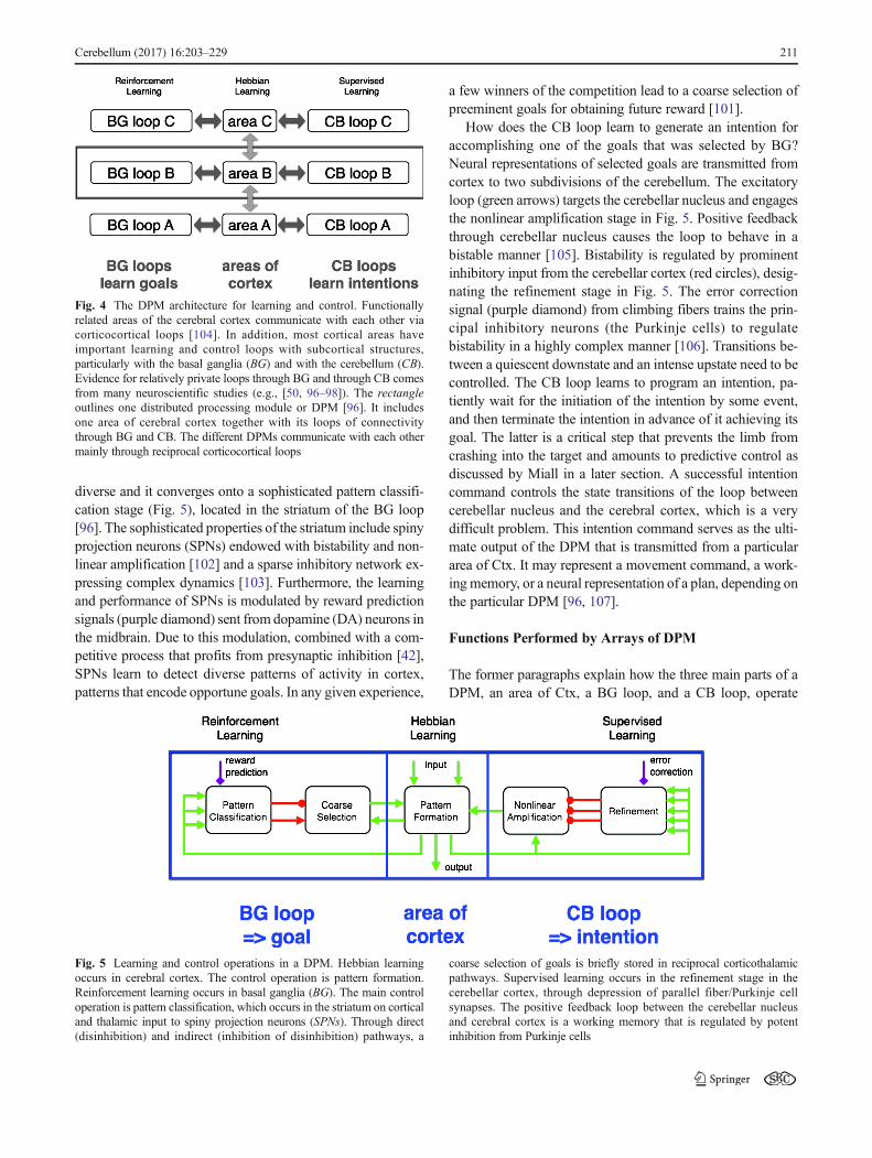
The DPM Architecture for Learning and Control (J.Houk)
DPMs are distributed processing modules [96] based on theanatomy and physiology of the loops that the basal ganglia(BG) and the cerebellum (CB) form with the cerebral cortex(Ctx) (cf. [50, 97, 98]). The DPM architecture illustrated inFig. 4 provides a powerful learning and processing system. Togive a brief overview of its composite functions, BG-Ctxloops discover opportune goals through reinforcement learn-ing [99]. CB-Ctx loops generate intentions capable of achiev-ing these goals through supervised learning [15]. This subcor-tical knowledge is expressed by outputs from BG and CB thatloop back to the same area of Ctx that sent the main input tothe loops. These signals not only force the Ctx to performsubcortically learned actions but also promote learning inCtx based on practice [100]. As a consequence, knowledgethat is gleaned in subcortical structures is progressivelyexported from BG and CB to the cerebral cortex [101].
Operations in the Subcortical Loops
Learning and control functions are defined by the operationsperformed in different DPM stages of any given subcorticalloop (Fig. 5). Pattern formation in cerebral cortex operatesmuch like the classical idea of an unsupervised neural net-work. It learns from practice (Hebbian learning) how to com-bine inputs in order to form useful outputs. The loops throughBG and CB tell it what to practice and thus tutor the cortex toperform what has been learned in the subcortical loops. Asmentioned above, the BG loop learns opportune goals, and theCB loop generates and refines intentions, such that they arecapable of accomplishing the goals. On a slower time scale,the cerebral cortex learns through practice how to encode thisknowledge into cortical habits. Cortical habits are stimulus–response operations that can be performed without subcorticalhelp.
How does the BG loop learn appropriate sets of goalsamong which to choose? The input to BG from cortex is
210 Cerebellum (2017) 16:203–229
diverse and it converges onto a sophisticated pattern classifi-cation stage (Fig. 5), located in the striatum of the BG loop[96]. The sophisticated properties of the striatum include spinyprojection neurons (SPNs) endowed with bistability and non-linear amplification [102] and a sparse inhibitory network ex-pressing complex dynamics [103]. Furthermore, the learningand performance of SPNs is modulated by reward predictionsignals (purple diamond) sent from dopamine (DA) neurons inthe midbrain. Due to this modulation, combined with a com-petitive process that profits from presynaptic inhibition [42],SPNs learn to detect diverse patterns of activity in cortex,patterns that encode opportune goals. In any given experience,
a few winners of the competition lead to a coarse selection ofpreeminent goals for obtaining future reward [101].
How does the CB loop learn to generate an intention foraccomplishing one of the goals that was selected by BG?Neural representations of selected goals are transmitted fromcortex to two subdivisions of the cerebellum. The excitatoryloop (green arrows) targets the cerebellar nucleus and engagesthe nonlinear amplification stage in Fig. 5. Positive feedbackthrough cerebellar nucleus causes the loop to behave in abistable manner [105]. Bistability is regulated by prominentinhibitory input from the cerebellar cortex (red circles), desig-nating the refinement stage in Fig. 5. The error correctionsignal (purple diamond) from climbing fibers trains the prin-cipal inhibitory neurons (the Purkinje cells) to regulatebistability in a highly complex manner [106]. Transitions be-tween a quiescent downstate and an intense upstate need to becontrolled. The CB loop learns to program an intention, pa-tiently wait for the initiation of the intention by some event,and then terminate the intention in advance of it achieving itsgoal. The latter is a critical step that prevents the limb fromcrashing into the target and amounts to predictive control asdiscussed by Miall in a later section. A successful intentioncommand controls the state transitions of the loop betweencerebellar nucleus and the cerebral cortex, which is a verydifficult problem. This intention command serves as the ulti-mate output of the DPM that is transmitted from a particulararea of Ctx. It may represent a movement command, a work-ingmemory, or a neural representation of a plan, depending onthe particular DPM [96, 107].
Functions Performed by Arrays of DPM
The former paragraphs explain how the three main parts of aDPM, an area of Ctx, a BG loop, and a CB loop, operate
Fig. 4 The DPM architecture for learning and control. Functionallyrelated areas of the cerebral cortex communicate with each other viacorticocortical loops [104]. In addition, most cortical areas haveimportant learning and control loops with subcortical structures,particularly with the basal ganglia (BG) and with the cerebellum (CB).Evidence for relatively private loops through BG and through CB comesfrom many neuroscientific studies (e.g., [50, 96–98]). The rectangleoutlines one distributed processing module or DPM [96]. It includesone area of cerebral cortex together with its loops of connectivitythrough BG and CB. The different DPMs communicate with each othermainly through reciprocal corticocortical loops
Fig. 5 Learning and control operations in a DPM. Hebbian learningoccurs in cerebral cortex. The control operation is pattern formation.Reinforcement learning occurs in basal ganglia (BG). The main controloperation is pattern classification, which occurs in the striatum on corticaland thalamic input to spiny projection neurons (SPNs). Through direct(disinhibition) and indirect (inhibition of disinhibition) pathways, a
coarse selection of goals is briefly stored in reciprocal corticothalamicpathways. Supervised learning occurs in the refinement stage in thecerebellar cortex, through depression of parallel fiber/Purkinje cellsynapses. The positive feedback loop between the cerebellar nucleusand cerebral cortex is a working memory that is regulated by potentinhibition from Purkinje cells
Cerebellum (2017) 16:203–229 211

individually. Now, I would like to discuss how arrays ofDPMs function in combination. The work done on mirrorneurons [108] and on imitation learning [109] provides excel-lent topics. Mirror neurons are active when a subject performsa goal-directed action and also when the subject observesanother individual performing the same action. This specialcorrespondence between observation and action has beencalled congruence. If the DPM theory is generally applicable,it has to account for the emergence of congruence duringontogenesis. As a consequence of developmental plasticity,the infant must learn not only how to reach and grasp a spe-cific target but also higher level actions such as bringing amorsel of food to the mouth and eating it. The infant mustalso learn to recognize when another individual performs thesame actions. Hierarchically diverse levels of goal are learnedin BG loops guided by reinforcement learning. SPNs mustlearn not only to fire when a subject is presented a goal forits own action but also when it observes another individualperforming the same action. Meanwhile in CB loops, the in-fant must learn parameterizable intentions for generating out-comes capable of fulfilling the different goals. Often it is as-sumed that the theory of reinforcement learning specifies howbasal ganglia learn primitive actions (say, muscle commands)and ensembles of them (say whole movements or longercourses of actions), but this is not biologically realistic.Muscle commands, and other kinds of intention, are clearlylearned and generated in CB loops to satisfy goals that arelearned and selected by BG loops. Normally, the two kindsof loop work together to select and generate appropriate ac-tions. These learning and control operations need to be ex-plored in simulations of DPM arrays with different parametersets.
An early stage of imitation learning can be traced to thesuperior temporal sulcus (STS) using arguments that focusonly on the cortical areas that are involved in the mirror neu-ron network [109]. Most models do not invoke the subcorticalloops that learn to control each cortical node in the mirrorneuron network. I agree with Caligiore and colleagues [4] thatthis focus of the mirror neuron field is inadequate. We nowhave a good understanding of how loops through BG discoveropportune goals and how loops through CB adapt to generateand refine intentions that accomplish these goals. The focus ofmirror neuron research needs to shift towards exploring howthese goal/intention congruences are learned by arrays ofDPMs.Within the hierarchy of the mirror neuron system, eachDPM evokes a different meaning. In the course of develop-mental practice, these different kinds of knowledge areexported to the different cortical nodes for more rapid auto-matic execution. The latter are examples of cortical habits,which clearly may include sequential chunks of action.
What is so special about subcortical learning as opposedto learning in the cerebral cortex? Briefly stated, learning inBG and CB profits from training information as summarized
in Table 1. Hebbian learning in Ctx depends only on coin-cidence detection with no timed-pulse training signals, justthe coincidence of input and output that occurs during prac-tice in the presence of tonic permissive factors that gatebroad time intervals when learning can occur. In the BGloop, the timed training signals are brief dopamine pulsesthat signal predictions of when future reward may occur[99]. In the CB loop, parallel fiber input and Purkinje celldepolarization signal coincidence detection in a particularspine, and training information is conveyed by climbingfibers that detect precisely when error corrections occur[107, 110]. Thus, BG and CB receive well-timed rewardand error signals as training information. Together with co-incidence detection, these reward and error signals consoli-date learning. When all three learning algorithms are simu-lated in combination, task performance can be quite remark-able [41, 111]. Testing DPM learning hypotheses is nowfacilitated by a new functional magnetic reso-nance imaging (fMRI) method for assessing the corre-spondence between a learning rule and the area of the brainthat performs that type of learning [112].
Cerebellum and Basal Ganglia Work Togetherfor Model-Based Actions (K. Doya)
Based on the anatomical and physiological evidence availableby the end of the last century, I proposed a theoretical frame-work that the cerebellum, the basal ganglia, and the cerebralcortex are specialized for supervised, reinforcement, and un-supervised learning, respectively [41, 111]. This view has re-ceived some support and raised some debate among theoreti-cians and experimentalists.
An important combination of supervised learning and rein-forcement learning is model-based action selection and learn-ing. Supervised learning in the cerebellum, including the cer-ebellar cortex and nuclei, can create a Bforward model^ thatpredicts the results of executed actions [64]. Reinforcementlearning in the basal ganglia can provide a Bstate valuefunction^ that evaluates the goodness of states. Because thedominant view at that time was that the outputs of the cere-bellum and the basal ganglia form separate channels in thethalamus [6], I hypothesized that model-based action selectioncan be implemented by a cortico-cerebellar loop predicting theresult of an imaginary action, the cortico-basal ganglia loopfor evaluating its goodness, and an activation of dopamineneurons for triggering execution of the imagined action (Fig.7 of [111], reproduced here as Fig. 6). A more specific pro-posal was that the striosome (or patch) compartment, whichprojects to dopamine neurons, learns state value functionwhile the matrix compartment, which projects to the pallidum,learns state-action value functions [41, 99].
212 Cerebellum (2017) 16:203–229

In the last decade, there have been significant new obser-vations regarding the interaction between the cerebellum andthe basal ganglia. The cerebellar output is disynaptically con-nected to the basal ganglia through dentate nucleus–centrolateral thalamus–striatum pathway [12]. It was recentlydemonstrated that stimulation of dentate neurons evokesshort-latency (about 10 ms) responses in about half of thestriatal neurons [52]. It was further revealed that the basalganglia output is also disynaptically connected to the cerebel-lum through subthalamic nucleus–pontine nucleus–cerebellarcortex pathway [13]. Thus, the existence of the short-cut con-nections between the cerebellum and the basal ganglia is be-coming a new anatomical consensus [3], but what are theircomputational roles?
A possible role of the dentato-thalamo-striatal pathway ismodel-based evaluation of action candidates. The forwardmodel learned in the lateral cerebellum predicts the resultingsensory state for a candidate action and its output is sentthrough the thalamus to the striatum, which estimates the val-ue of the predicted state. An interesting observation [52] is thatoptogenetic stimulation of the dentate-thalamo-striatal path-way reverts cortico-striatal long-term depression (LTD) to
LTP, which had been reported to happen with dopamine stim-ulation [114]. Previous self-stimulation experiments showedthat the striosome compartment is more effective in inducingself-stimulation than the matrix compartment [115]. It is notknown whether the dentato-thalamo-striatal pathway has anypreference projections, but a possible scenario is that the cer-ebellar input activates the striosome neurons that activate do-pamine neurons.
The function of the subthalamo-ponto-cerebellar pathwayis harder to interpret. The subthalamic nucleus is a part of theBindirect pathway^ of the basal ganglia that is implicated inaction inhibition and aversive learning [116, 117]. Thus, apossible function of the pathway is to provide a BStop!^ signalto the cerebellum for withholding ongoing movements.Another possibility, though highly speculative, is to signalBoff-line^ status to the cerebellum, notifying that currentlythe basal ganglia have withheld execution of motor programs(though inhibition of the thalamus and midbrain motor nucleivia globus pallidus and substantia nigra reticulata, respective-ly) so that the cerebellar internal models can be safely used foroff-line mental simulation.
Here, I presented some views based on the hypothesis thatthe cerebellum provides internal models of the body and theenvironmental dynamics for model-based motor control anddecisions [41, 111]. There are, however, proposals and accu-mulating evidence suggesting that the frontal and parietal cor-tices as well as the hippocampus play important roles inmodel-based planning and decisions [118–120]. A possibledifference of the internal models provided by the cerebellumand the cortex may be that the cerebellum learns deterministicinput–output mapping with its mostly feed-forward circuit,while the cortex learns joint or conditional probability modelsthrough iterative dynamics using its heavily recurrent circuit.Probabilistic models are more general, as it includes determin-istic models as extreme cases, but deterministic models havethe virtue of simplicity. The hippocampus may enable reuseand editing of episodic memories for planning about the fu-ture. These ideas are all speculative, but recent optogenetictools for pathway-specific circuit manipulations can make itpossible to test these hypotheses. The big challenge for ustheoreticians is to provide hypotheses worthy of laborioustesting.
Table 1 Different learning rulesin cerebral cortex, basal ganglia,and cerebellum
Brain site Cerebral cortex Basal ganglia Cerebellar cortex
Cellular site Excitatoryafferentsonto pyramidalcells
Striatum cortical afferentsonto spiny neurons
Parallel fibers ontoPurkinje cells
Learning Rule &operation
Hebbian &coincidence
Reinforcement & coincidence ·reward
Supervised & coincidence· error
Permissive factors Cholinergic + Dopaminergic + Noradrenergic +
This table illustrates key features of three learning rules in the brain (adapted with permission from Houk [113])
state: x(t)
value: V
model: F
state: x*(t+1) action: u(t)
value: Vreward: R
- + +
action: u*(t)
x
environment
δ *: DA
lateral cerebellum
motor cortex
striosome
sensory cortexpremotor
cortex
striosome
Fig. 6 A possible implementation of model-based action selection bycombining forward models in the cerebellum and reward predictors inthe basal ganglia. From [111] with permission
Cerebellum (2017) 16:203–229 213

A Systems-Level View of Cerebellar Motorand Cognitive Function (R.C. Miall)
A Uniform Structure and Specific Connections
The cytoarchitecture of the cerebellar cortex is strikingly con-sistent, both across its span and across species. This very ex-tensive neural sheet, estimated to be equal in area of onehemisphere of the human cerebral cortex [121], is heavilyinterconnected with cerebral cortical areas, and recently evi-dence of bidirectional connections between cerebellum andbasal ganglia has emerged [6, 13]. The consistent cerebellarstructure and circuitry, reciprocally connected to many differ-ent extra-cerebellar structures, suggest strongly that a commonneural operation is performed within each micro-complex. Incontrast, cerebral cortical areas are uniquely organized, witheach cortical area dedicated to a specific function or functions.Function follows form.
A Singular Processing Hypothesis
Three critical questions arise from this view (none of which isnew and all have been raised before). The first question is whatmight be this proposed single operation or transformation per-formed by the cerebellar cortex. This is analogous to the algo-rithmic level of analysis that DavidMarr proposed 40 years ago[122]. There are still multiple alternative answers, ranging froma timing device, or associative learning network, to my pre-ferred hypothesis, as a short-term predictor—specifically thatthe cerebellum contributes a state estimation towards forwardmodelling, predicting the causal chain from motor commandsto changes in state of the modelled system [123]. We haverecent evidence that the same may hold true for prediction inlanguage processing [124].
The second question is how the cerebellar neural circuitrysupports whatever singular operation it achieves (Marr’s phys-ical level of analysis). A full answer will depend on a deeperunderstanding than we currently have of the processing ofeach cell type, the interactions between the interneurons with-in the cerebellum, and the consequences for information pro-cessing of the interconnections between mossy fiber inputsand Purkinje cell outputs [125]. This must include not onlyinteractions between cells in the cerebellar cortex but also theshort-range connections between cortex and deep cerebellarnuclei and between cerebellum and inferior olive. This latterconnection is probably one of the most critical to understand,as the evidence for climbing fiber-driven LTD at the Purkinjecell inputs is overwhelming, and evidence for more subtlechanges at other synapses within the cortical sheet is strong.So understanding what the inferior olivary teaching signalrepresents will determine what cortical process is sculptedand refined by these synaptic events [126].
The third question is analogous to Marr’s computationallevel of analysis. What is the function of the interconnectednetwork running, for example, from parietal and sensorimotorcortical areas through the cerebellum and its output to thethalamus, and projecting back to the same sensorimotor cor-tical areas? Here, the answer must be given in terms of thenetwork and cannot be answered by reference to cerebellar orcortical processes in isolation.
Cerebro-Cerebellar Loops
Evidence for cerebellar interactions with cortical sensorimotorareas is found at all levels—from tracer studies in non-humanprimates, human diffusion tensor imaging and functional con-nectivity measures, lesion studies, and electrical and magneticstimulation studies. What these cannot tell us is what signalsare carried or the functional role of the cerebro-cerebellar loop.Our experimental approach has been to use TMS to disruptcerebellar operations and test consequences on behavior [124,127]; in both studies, we showed change in behavior consis-tent with a loss of predictions generated by the cerebellum—the former study tested linguistic prediction, the latter reachingbehavior. We are using functional imaging to understand theco-activation of cerebral and cerebellar areas [129] and morerecently to explore how their connectivity changes as a con-sequence of learning [130]. We have also used tDCS to ma-nipulate either cerebellar or cerebral excitability, and this ishighlighting a secondary modulatory level of cerebellar-cerebral interaction [131–133]. In detail, we have shown cor-responding changes in cognitive tasks when we depress cere-bellar activity with cathodal tDCS or activate frontal cortexwith anodal tDCS.We believe the former effect is mediated bycerebellar disinhibition of cerebral cortex; both Lago-Rodriguez and Galea and Popa and Kishore discuss thismech-anism in more detail below.
However, two results are of particular mention. We firstreported that the functional connectivity within the cerebellumand a separate fronto-parietal network was enhanced shortlyafter a period of sensory motor learning [130]. The implicationis that cerebellar processes may contribute to the consolidationof a motor skill, probably leading to long-term change in bothcerebellar and cerebral sites. Second, after a similar period oflearning in the serial reaction time task (SRTT, a sequencelearning paradigm) [155], we again saw short-term changeswithin 10 min in cerebellar, frontal, and occipital areas that ledto later changes, measured at 6 h after learning, within senso-rimotor cortex and medio-temporal areas, perhaps reflectinglong-term memory store. One group, learning under explicitconditions, showed early increased connectivity withincerebello-frontal and visual cortical areas and later increasedconnectivity in sensorimotor cortex. The other group learningunder implicit conditions showed only the sensorimotor cor-tical network early after learning, shifting to a medial temporal
214 Cerebellum (2017) 16:203–229
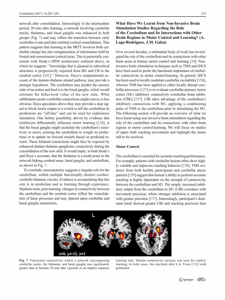
network after consolidation. Interestingly in the intermediateperiod, 30 min after learning, a network involving cerebellarnuclei, thalamus, and basal ganglia was enhanced in bothgroups (Fig. 7) and may reflect the transition between earlycerebellar events and later cerebral cortical consolidation. Thispattern suggests that learning in the SRTT involves both cer-ebellar change but also reorganization of information held byfrontal and sensorimotor cortical areas. This is potentially con-sistent with Houk’s DPM architecture outlined above, inwhich he suggests ‘^knowledge that is gleaned in subcorticalstructures is progressively exported from BG and CB to thecerebral cortex [101].^ However, Doya’s computational ac-count of the dentato-thalamo-striatal pathway may provide astronger hypothesis. The cerebellum may predict the sensorystate of an action and feed it to the basal ganglia, which wouldestimate the behavioral value of the new state. Whatsubthalamo-ponto-cerebellar connections might convey is lessobvious; Doya speculates above they may provide a stop sig-nal to block motor output or a switch to tell the cerebellum itspredictions are Boff-line^ and can be used for exploratorysimulation. One further possibility, driven by evidence thatreinforcers differentially influence motor learning [128], isthat the basal ganglia might modulate the cerebellum’s sensi-tivity to errors, priming the cerebellum to weight its predic-tions or to update its forward models based on predicted re-ward. These bilateral connections might thus be exposed byenhanced dentato-thalamo-ganglionic connectivity during theconsolidation of the new skill. It would imply, in both Houk’sand Doya’s accounts, that the thalamus is a nodal point in thenetwork linking cerebral areas, basal ganglia, and cerebellum,as shown in Fig. 7.
To conclude, neuroanatomy suggests a singular role for thecerebellum, within multiple functionally distinct cerebro-cerebello-thalamic circuits. Evidence is accumulating that thisrole is in prediction and in learning through experience.Medium-term, post-learning, changes in connectivity betweenthe cerebellum and the cerebral cortex reflect the consolida-tion of these processes and may depend upon cerebellar andbasal ganglia interactions.
What Have We Learnt from Non-Invasive BrainStimulation Studies Regarding the Roleof the Cerebellum and Its Interactions with OtherBrain Regions in Motor Control and Learning? (A.Lago-Rodriguez, J.M. Galea)
Over several decades, a substantial body of work has investi-gated the role of the cerebellum and its connections with otherbrain areas in human motor control and learning [34]. Non-invasive brain stimulation techniques such as TMS and tDCShave been used to probe the functional importance of cerebel-lar connectivity in motor control/learning. In general, tDCShas been used to locally modulate cerebellar excitability [156],whereas TMS has been applied to either locally disrupt cere-bellar processes [127] or to evaluate cerebellar-primary motorcortex (M1) inhibitory connectivity (cerebellar brain inhibi-tion (CBI)) [157]. CBI takes advantage of the cerebellum’sinhibitory connections with M1, applying a conditioningpulse of TMS to the cerebellum prior to stimulating the M1.The following section will provide an overview of what wehave learnt using non-invasive brain stimulation regarding therole of the cerebellum and its connections with other brainregions in motor control/learning. We will focus on studiesof upper limb reaching movements and highlight the issuesstill to be resolved.
Motor Control
The cerebellum is essential for accurate reaching performance.For example, patients with cerebellar lesions often show high-ly variable and imprecise reaching behavior [158]. TMS evi-dence from both healthy participants and cerebellar ataxicpatients [159] suggest that human’s ability to perform accuratereaching is highly dependent on the strength of connectivitybetween the cerebellum and M1. Put simply, increased inhib-itory output from the cerebellum to M1 (CBI) correlates withmovement precision, where stronger inhibition is associatedwith greater precision [157]. Interestingly, participant’s dom-inant hand showed greater CBI and reaching precision than
Fig. 7 Functional connectivity within a network encompassingcerebellar nuclei, the thalamus, and basal ganglia was significantlygreater than at baseline 30 min after exposure to an implicit sequence
learning task. Similar connectivity increase was seen for explicitlearning; in both cases, this declined after 6 h. From [155] withpermission
Cerebellum (2017) 16:203–229 215

their non-dominant hand. It is tempting to suggest that stron-ger inhibition by the cerebellum may underlie the preferencefor dominant hand movements. However, it is important topoint out that the observed correlations between CBI and be-havioral precision do not speak to causality. Therefore, it isalso possible that hand preference leads to a practice effectwhich enhance cerebellar connectivity [157]. What role couldthe connectivity between the cerebellum and M1 play inreaching accuracy? Accurate state estimation (e.g., estimatingcurrent limb position) is vital for the precise control ofreaching movements. It is believed that a cerebellar forwardmodel, which predicts the sensory consequences of motorcommands, is crucial for accurate and up-to-date state estima-tion [127]. The correlation between behavioral precision andhigher CBI would be consistent with this framework, particu-larly if onemakes the assertion that increased CBI is indicativeof a stronger forward model [157].
Motor Adaptation
In addition to motor control, the cerebellum plays a pivotalrole in error-based motor learning, often referred to as motoradaptation [31]. This process is crucial for maintaining accu-rate reaching behavior in response to novel environments thatcause errors in performance. Modulation of cerebellar activityby tDCS leads to polarity-dependent changes in motor adap-tation rates to visuomotor [31], locomotor [160], and dynamic[161] novel perturbations. Whereas anodal tDCS leads tofaster motor adaptation, cathodal stimulation decreases adap-tation rates [31, 160, 161]. This clearly identifies a prominentrole of the cerebellum in motor adaptation; however, howimportant are its connections to other brain areas? Previouswork has shown that the cerebellum decreases its inhibitionover M1 (decreased CBI) when movement corrections (motoradaptation) are required to respond to environmental changes[162, 163]. These results are in line with a reduction of cere-bellar Purkinje cells activity in response to error signals andsupport the notion that mechanisms similar to LTD are themost important neurophysiological processes underlying thecerebellar contribution to motor adaptation [164].
Outstanding Issues
Posterior Parietal Cortex (PPC)
Current theories of motor control suggest that the anatomicalconnection between the cerebellum and PPC plays a criticalrole in maintaining accurate voluntary movements [34].Despite this, no brain stimulation work has investigated thefunctional importance of cerebellar-PPC connectivity to mo-tor control and adaptation. One reason for this is the complex-ity involved in such a study. For example, this issue could beinvestigated by comparing the influence of concurrent
cerebellar tDCS on cerebellar-M1 and PPC-M1 paired-puleTMS. This complex brain stimulation design would then haveto be coupled with a motor task.
Basal Ganglia/Frontal Cortex
There is now increasing evidence that error-based motor learn-ing occurs through multiple processes that are not all cerebellar-dependent (reinforcement learning/use-dependent learning/explicit strategies) [165]. Although discussion of these processesis beyond this current review, it is important to mention that theycan often take place concurrently or even in a competingmanner[166]. However, very little is known regarding the neural basisof the interaction between cerebellar-dependent motor adapta-tion and possible frontal/basal ganglia-dependent learning (ex-plicit strategies/reinforcement learning). Although we know thatthe cerebellum has connections to both the basal ganglia and thefrontal cortex [37], the functional relevance of these connectionsto the interaction between different learning processes is current-ly unknown. Once again, the complexity involved in studyingthese connections with brain stimulation is significant. One pos-sible solutionwould be to examine the interaction of thesemotorlearning processes during concurrent brain stimulation (TMS/tDCS) and fMRI.
Cerebellar Modulation of Cortical Plasticity in BasalGanglia-Related Movement Disorders (T. Popa, A.Kishore)
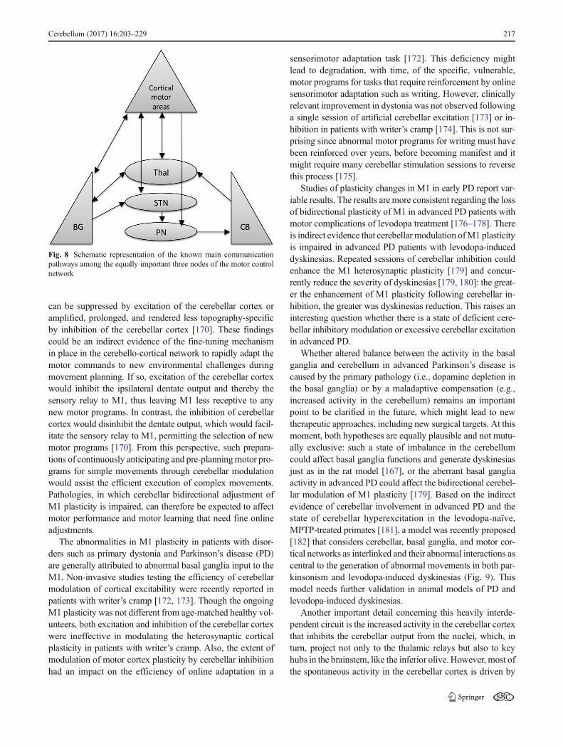
Cerebellum, basal ganglia, and motor areas in the frontal cor-tex are the major nodes in the motor network, and they haveunique cytoarchitectural properties, reciprocal connectivity,and synaptic plasticity mechanisms that allow efficient com-munication among them (Fig. 8). The discovery of reciprocal,topography-specific, subcortical connections between cere-bellum and basal ganglia in primates [12] indicates that thebasal ganglia and cerebellar motor networks are not indepen-dent information processing systems but can exchange infor-mation between them in real-time to provide appropriate in-puts to the frontal cortices. In rats, under physiological condi-tions, the cerebellum modulates cortico-striatal plasticitythrough these connections [52], and the same pathway canalso transmit aberrant cerebellar activity to basal ganglia, gen-erating dystonia and dyskinesias in animals. Cerebellum alsomodulates cortical sensorimotor integration [167, 168] and isimportant for acquiring and maintaining [169] new cortico-spinal integrations. In young healthy humans, non-invasivestimulation studies demonstrated that cerebellum exerts a bi-directional modulation only on heterosynaptic motor cortex(M1) plasticity [170], which is dependent on peripheral sen-sory information reaching the M1 through complex polysyn-aptic pathways [171]. The cortical heterosynaptic plasticity
216 Cerebellum (2017) 16:203–229
can be suppressed by excitation of the cerebellar cortex oramplified, prolonged, and rendered less topography-specificby inhibition of the cerebellar cortex [170]. These findingscould be an indirect evidence of the fine-tuning mechanismin place in the cerebello-cortical network to rapidly adapt themotor commands to new environmental challenges duringmovement planning. If so, excitation of the cerebellar cortexwould inhibit the ipsilateral dentate output and thereby thesensory relay to M1, thus leaving M1 less receptive to anynew motor programs. In contrast, the inhibition of cerebellarcortex would disinhibit the dentate output, which would facil-itate the sensory relay to M1, permitting the selection of newmotor programs [170]. From this perspective, such prepara-tions of continuously anticipating and pre-planningmotor pro-grams for simple movements through cerebellar modulationwould assist the efficient execution of complex movements.Pathologies, in which cerebellar bidirectional adjustment ofM1 plasticity is impaired, can therefore be expected to affectmotor performance and motor learning that need fine onlineadjustments.
The abnormalities in M1 plasticity in patients with disor-ders such as primary dystonia and Parkinson’s disease (PD)are generally attributed to abnormal basal ganglia input to theM1. Non-invasive studies testing the efficiency of cerebellarmodulation of cortical excitability were recently reported inpatients with writer’s cramp [172, 173]. Though the ongoingM1 plasticity was not different from age-matched healthy vol-unteers, both excitation and inhibition of the cerebellar cortexwere ineffective in modulating the heterosynaptic corticalplasticity in patients with writer’s cramp. Also, the extent ofmodulation of motor cortex plasticity by cerebellar inhibitionhad an impact on the efficiency of online adaptation in a
sensorimotor adaptation task [172]. This deficiency mightlead to degradation, with time, of the specific, vulnerable,motor programs for tasks that require reinforcement by onlinesensorimotor adaptation such as writing. However, clinicallyrelevant improvement in dystonia was not observed followinga single session of artificial cerebellar excitation [173] or in-hibition in patients with writer’s cramp [174]. This is not sur-prising since abnormal motor programs for writing must havebeen reinforced over years, before becoming manifest and itmight require many cerebellar stimulation sessions to reversethis process [175].
Studies of plasticity changes in M1 in early PD report var-iable results. The results are more consistent regarding the lossof bidirectional plasticity of M1 in advanced PD patients withmotor complications of levodopa treatment [176–178]. Thereis indirect evidence that cerebellar modulation ofM1 plasticityis impaired in advanced PD patients with levodopa-induceddyskinesias. Repeated sessions of cerebellar inhibition couldenhance the M1 heterosynaptic plasticity [179] and concur-rently reduce the severity of dyskinesias [179, 180]: the great-er the enhancement of M1 plasticity following cerebellar in-hibition, the greater was dyskinesias reduction. This raises aninteresting question whether there is a state of deficient cere-bellar inhibitory modulation or excessive cerebellar excitationin advanced PD.
Whether altered balance between the activity in the basalganglia and cerebellum in advanced Parkinson’s disease iscaused by the primary pathology (i.e., dopamine depletion inthe basal ganglia) or by a maladaptive compensation (e.g.,increased activity in the cerebellum) remains an importantpoint to be clarified in the future, which might lead to newtherapeutic approaches, including new surgical targets. At thismoment, both hypotheses are equally plausible and not mutu-ally exclusive: such a state of imbalance in the cerebellumcould affect basal ganglia functions and generate dyskinesiasjust as in the rat model [167], or the aberrant basal gangliaactivity in advanced PD could affect the bidirectional cerebel-lar modulation of M1 plasticity [179]. Based on the indirectevidence of cerebellar involvement in advanced PD and thestate of cerebellar hyperexcitation in the levodopa-naïve,MPTP-treated primates [181], a model was recently proposed[182] that considers cerebellar, basal ganglia, and motor cor-tical networks as interlinked and their abnormal interactions ascentral to the generation of abnormal movements in both par-kinsonism and levodopa-induced dyskinesias (Fig. 9). Thismodel needs further validation in animal models of PD andlevodopa-induced dyskinesias.
Another important detail concerning this heavily interde-pendent circuit is the increased activity in the cerebellar cortexthat inhibits the cerebellar output from the nuclei, which, inturn, project not only to the thalamic relays but also to keyhubs in the brainstem, like the inferior olive. However, most ofthe spontaneous activity in the cerebellar cortex is driven by
Fig. 8 Schematic representation of the known main communicationpathways among the equally important three nodes of the motor controlnetwork
Cerebellum (2017) 16:203–229 217

intrinsic (non-synaptic) mechanisms and the level of sponta-neous activity in Purkinje cells is reciprocally regulated by theactivity of the inferior olive (through the nucleo-olivary
inhibition). Any regulation of the strength of this negative linkremotely from other brain structures or artificially (pharmaco-logically or with invasive-non-invasive stimulation) could be
a
b
c
Fig. 9 Schematic representation of the basal ganglia-thalamo-corticalloop, the cerebello-thalamo-cortical loop, and the interaction betweenthe two in health (a), in non-dyskinetic Parkinson’s disease, afterlevodopa withdrawal (OFF) and after regular dose of levodopa (ON)(b), and in advanced Parkinson’s disease with levodopa-induceddyskinesia (c). Red arrows represent glutamatergic projections; bluearrows represent GABA-ergic projections; green arrows representdopaminergic projections; dark green arrows in panels b and crepresent the exogenous dopamine from levodopa. The shades of theblocks represent the activity of the respective network nodes. The STNis overactive because of cortical glutamatergic over activity duringdyskinesias and from loss of GPe inhibition in OFF. The STN over
activity locks cerebellar cortex in a persistent hyperactive state andinterferes with its sensory processing function. The behavior of thecortico-ponto-cerebellar projections in non-dyskinetic PD in ON is notreported so far and is predicted by this model to be close to normal (CBctx cerebellar cortex, CM centromedian thalamic nucleus, D1/D2dopamine receptor types of the striatal medium spiny neurons, DNdentate nucleus, GPe globus pallidus externus, GPi globus pallidusinternus, M1 primary motor cortex, PF parafascicular nucleus, PMCpremotor cortex, PN pontine nuclei, SMA supplementary motor area,SNc substantia pars compacta, SNr substantia pars reticulata, STNsubthalamic nucleus, VL ventrolateral thalamic nucleus, VLPo ventro-latero-posterior thalamic nucleus pars oralis, VTA ventral tegmental area)
218 Cerebellum (2017) 16:203–229
a means to adjust the level of spontaneous activity in thecerebellar cortex and subsequently in the whole network.
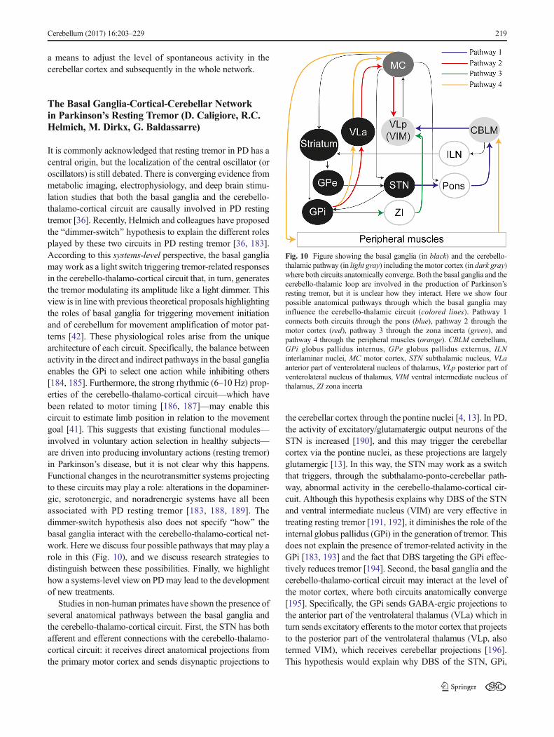
The Basal Ganglia-Cortical-Cerebellar Networkin Parkinson’s Resting Tremor (D. Caligiore, R.C.Helmich, M. Dirkx, G. Baldassarre)
It is commonly acknowledged that resting tremor in PD has acentral origin, but the localization of the central oscillator (oroscillators) is still debated. There is converging evidence frommetabolic imaging, electrophysiology, and deep brain stimu-lation studies that both the basal ganglia and the cerebello-thalamo-cortical circuit are causally involved in PD restingtremor [36]. Recently, Helmich and colleagues have proposedthe Bdimmer-switch^ hypothesis to explain the different rolesplayed by these two circuits in PD resting tremor [36, 183].According to this systems-level perspective, the basal gangliamay work as a light switch triggering tremor-related responsesin the cerebello-thalamo-cortical circuit that, in turn, generatesthe tremor modulating its amplitude like a light dimmer. Thisview is in line with previous theoretical proposals highlightingthe roles of basal ganglia for triggering movement initiationand of cerebellum for movement amplification of motor pat-terns [42]. These physiological roles arise from the uniquearchitecture of each circuit. Specifically, the balance betweenactivity in the direct and indirect pathways in the basal gangliaenables the GPi to select one action while inhibiting others[184, 185]. Furthermore, the strong rhythmic (6–10 Hz) prop-erties of the cerebello-thalamo-cortical circuit—which havebeen related to motor timing [186, 187]—may enable thiscircuit to estimate limb position in relation to the movementgoal [41]. This suggests that existing functional modules—involved in voluntary action selection in healthy subjects—are driven into producing involuntary actions (resting tremor)in Parkinson’s disease, but it is not clear why this happens.Functional changes in the neurotransmitter systems projectingto these circuits may play a role: alterations in the dopaminer-gic, serotonergic, and noradrenergic systems have all beenassociated with PD resting tremor [183, 188, 189]. Thedimmer-switch hypothesis also does not specify Bhow^ thebasal ganglia interact with the cerebello-thalamo-cortical net-work. Here we discuss four possible pathways that may play arole in this (Fig. 10), and we discuss research strategies todistinguish between these possibilities. Finally, we highlighthow a systems-level view on PD may lead to the developmentof new treatments.
Studies in non-human primates have shown the presence ofseveral anatomical pathways between the basal ganglia andthe cerebello-thalamo-cortical circuit. First, the STN has bothafferent and efferent connections with the cerebello-thalamo-cortical circuit: it receives direct anatomical projections fromthe primary motor cortex and sends disynaptic projections to
the cerebellar cortex through the pontine nuclei [4, 13]. In PD,the activity of excitatory/glutamatergic output neurons of theSTN is increased [190], and this may trigger the cerebellarcortex via the pontine nuclei, as these projections are largelyglutamergic [13]. In this way, the STN may work as a switchthat triggers, through the subthalamo-ponto-cerebellar path-way, abnormal activity in the cerebello-thalamo-cortical cir-cuit. Although this hypothesis explains why DBS of the STNand ventral intermediate nucleus (VIM) are very effective intreating resting tremor [191, 192], it diminishes the role of theinternal globus pallidus (GPi) in the generation of tremor. Thisdoes not explain the presence of tremor-related activity in theGPi [183, 193] and the fact that DBS targeting the GPi effec-tively reduces tremor [194]. Second, the basal ganglia and thecerebello-thalamo-cortical circuit may interact at the level ofthe motor cortex, where both circuits anatomically converge[195]. Specifically, the GPi sends GABA-ergic projections tothe anterior part of the ventrolateral thalamus (VLa) which inturn sends excitatory efferents to the motor cortex that projectsto the posterior part of the ventrolateral thalamus (VLp, alsotermed VIM), which receives cerebellar projections [196].This hypothesis would explain why DBS of the STN, GPi,
Fig. 10 Figure showing the basal ganglia (in black) and the cerebello-thalamic pathway (in light gray) including the motor cortex (in dark gray)where both circuits anatomically converge. Both the basal ganglia and thecerebello-thalamic loop are involved in the production of Parkinson’sresting tremor, but it is unclear how they interact. Here we show fourpossible anatomical pathways through which the basal ganglia mayinfluence the cerebello-thalamic circuit (colored lines). Pathway 1connects both circuits through the pons (blue), pathway 2 through themotor cortex (red), pathway 3 through the zona incerta (green), andpathway 4 through the peripheral muscles (orange). CBLM cerebellum,GPi globus pallidus internus, GPe globus pallidus externus, ILNinterlaminar nuclei, MC motor cortex, STN subthalamic nucleus, VLaanterior part of venterolateral nucleus of thalamus, VLp posterior part ofventerolateral nucleus of thalamus, VIM ventral intermediate nucleus ofthalamus, ZI zona incerta
Cerebellum (2017) 16:203–229 219
and VIM are all effective in treating tremor [191, 192, 194].Third, the basal ganglia may engage the cerebello-thalamo-cortical circuit also through the zona incerta, which receivesprojections from the GPi and sends GABA-ergic projectionsto the VLp [197]. Finally, the basal ganglia may interact withthe cerebello-thalamo-cortical circuit through the peripheralnervous system. That is, after tremor initiation by the basalganglia, tremor-related somatosensory afferents transmitted tothe cerebellum may engage the cerebello-thalamo-cortical cir-cuit [198]. However, this hypothesis does not fit with datashowing that peripheral deafferentation, peripheral anesthesiaof tremulous muscles, andmechanical perturbations have littleeffect on PD tremor (for a review, see [36]). To conclude, thereis currently no definite evidence which of the pathwaysconnecting the basal ganglia with the cerebello-thalamo-cortical circuit contribute to PD tremor.
Given the complex involvement of different neural circuits,a systems-level perspective on PD tremor is required to an-swer this question. Systems-level questions require systems-level outcome measures, and the development of new analysistools has made this possible. For example, rather than testingfor altered activity in a set of brain regions, or altered func-tional connectivity between two different brain regions, dy-namic causal modelling (DCM) allows one to statisticallycompare different network configurations based on fMRI da-ta. A second approach could rely on computational modelling[39, 199] that has the advantage to allow one to directly mea-sure the effects of adding or removing specific pathways onthe oscillatory output of the system. Finally, animal models oftremulous PD may be helpful to test whether the resection ofspecific pathways interferes with tremor. For example, thisapproach has been applied to a mouse model of dystoniawhere resection of the anatomical pathway between cerebel-lum and basal ganglia was shown to reduce dystonic symp-toms [200].
A systems-level view of tremormay improve existing treat-ments, for example by tailoring GPi-DBS to ongoing activityin the motor cortex [201]. It may also facilitate the develop-ment of new treatments. For example, computational modelsinformed by functional imaging may allow us to test new anti-tremor treatments Bin silico^ before transferring them to ani-mals or patients [199].
Summary and Conclusions
The nine manuscripts collected in this consensus paper in-volved 18 scholars whose expertise covers different scientificdisciplines including, among others, neurophysiology, theo-retical neuroscience, and neuropsychology. A first notable factthat emerges from the paper is that, although starting fromsuch different backgrounds, all the contributors agreed onthe importance of studying the cerebellar function from an
integrative perspective that takes into account the interplaybetween cerebellar, cortical, and basal ganglia networks.Bostan and Strick underline how the reciprocal anatomicalloops between the cerebellum and the cerebral cortex involveboth motor and non-motor cortical areas. Verschure et al. fur-ther support and expand the analysis of Bostan and Strick byunderlining the role of the nucleo-olivary pathway to managethe contribution of feedback and feed-forward aspects of mo-tor control. Similarly, Jörntell underlines how the spinal inputsinto the cerebellum allow it to be informed about the excitato-ry drive on low level motor functions, so also adding thespinal cord to the systems-level view on cerebellum.Furthermore, Bostan and Strick emphasize their recent find-ings on the connectivity between the cerebellum and the basalganglia. While most of the authors propose possible systems-level effects of these bidirectional cerebellar-basal ganglia an-atomical pathways, Houk decided to not explicitly includethem in his entry. Houk claims that they are too sparse andthere are not enough data to properly address the questionabout which role they might play in the cerebello-cortical-basal ganglia system. Houk only proposes here that such col-lateral synapses might grow in strength the gain of the positivefeedback managed by the cerebellar-cortical loops discussedin his entry and, as a consequence, such loops would becomeextremely difficult to control. Aside from this, there is unan-imous agreement between all the authors of the consensuspaper that future experimental and computational work isneeded to understand the function of cerebellar-basal gangliacircuitry in both motor and non-motor functions. In this re-spect, Doya proposes novel untested theories regarding therole of such cerebellar-basal ganglia loop.
The cross-disciplinary discussion between the contributorsallows the identification of some points of agreement anddisagreement about specific mechanisms through which thecerebellum expresses its functions in synergy with cortexand basal ganglia according to a systems-level view. Below,we summarize the result of such discussion by consideringthree main topics addressed by the authors: (i) cerebellarsystems-level mechanisms for learning and adaptation; (ii)cerebellar systems-level mechanisms underlying dystoniaand Parkinson’s disease; and (iii) techniques for systems-level analysis of cerebellar functions.
Cerebellar Systems-Level Mechanisms for Learningand Adaptation
Houk reviews his previous proposal claiming that cerebellum,cortex, and basal ganglia form DPMs where basal ganglia-thalamo-cortical loops discover goals through reinforcementlearning processes; cerebello-thalamo-cortical loops learn togenerate intentions through supervised learning. According tothis proposal, knowledge that is gleaned in basal ganglia andcerebellum is progressively transferred to the cerebral cortex.
220 Cerebellum (2017) 16:203–229
The basal ganglia-cortex and cerebellum-cortex loops tutorthe cortex to learn what has been learned in the subcorticalloops. Verschure et al. agree with this view and underline howthe learning processes that Houk proposes for cerebellum,basal ganglia, and cortex are compatible with Doya’s attribu-tion of supervised, unsupervised, and reward-based learningto, respectively, cerebellum, cortex, and basal ganglia.Interestingly, Verschure et al. also suggest that the regulationof the cerebellar feedback inhibition to the olive discussed intheir entry may be the mechanism by which the cerebellarcontribution to each DPM gradually adjusts to contextual re-quirements. Indeed, the DPM systems-level architecture maybenefit of a mechanism allowing a flexible reconfiguration (orweighting) of its basic layout. The network could entertainmodes of behavior that are more Bcerebello-centric^ (for in-stance to adapt to a new change in a sensorimotor contingen-cy) to Bcortex-centric^ behaviors. Similarly, Miall agrees withthe general ideas proposed byHouk, even if hemainly stressesthe role of the cerebellum as a predictor.
Doya elaborates his previous theory of motor control andlearning according to which the cerebellum, the basal ganglia,and the cerebral cortex are specialized for supervised, rein-forcement, and unsupervised learning, respectively, in orderto account for the recent findings on the circuitry between thecerebellum and the basal ganglia reviewed by Bostan andStrick. In particular, the author suggests that a possible roleof the dentato-thalamo-striatal pathway may be a model-basedevaluation of action candidates during the reinforcementlearning processes. On the other side, the function of thesubthalamo-ponto-cerebellar pathway may be to provide aBStop!^ signal to the cerebellum to activate a motor programfor withholding ongoing movements. Another possibility maybe that the subthalamo-ponto-cerebellar circuit may signal anBoff-line^ status to the cerebellum, notifying that currently thebasal ganglia have withheld execution of motor programs sothat the cerebellar internal models can be safely used for off-line mental simulation. Kishore and Popa consider this latterhypothesis highly speculative. They do not completely agreewith the first hypothesis, observing that while it might be truefor gaze control, axial muscle control, and background spinalcircuits’ modulation, it might be not true for complex motorcontrol of the limbs as well as for motor plan discrimination.This is therefore one example of topic where consensus cannotbe reached yet and a topic that is critical for future research toexplore.
Lago-Rodriguez and Galea agree with the Doya’s perspec-tive and extend it by specifying the importance of the anatom-ical connections between cerebellum and posterior parietalcortex to motor control and adaptation. The authors reviewevidence suggesting that error-based motor learning occursthrough multiple processes that are not all cerebellar-dependent (reinforcement learning/use-dependent learning/explicit strategies) and that require the involvement of a
cortical-subcortical network including cerebellum, frontal cor-tex, and basal ganglia. This view is also in agreement with theevidence reviewed by Bostan and Strick and with the theoret-ical proposal of Houk. In addition, Lago-Rodriguez and Galeadiscuss recent data suggesting that cerebellar inhibition is im-portant for implementing motor control, while the alleviationof cerebellar inhibition is associated with motor adaptation(see the paragraph below on BTechniques for systems-levelanalysis of cerebellar functions^ for more details on thenon-invasive brain stimulation techniques used by Lago-Rodriguez and Galea to study this effect). Caligiore et al.support this dual role of the cerebellum underlying how theincreased motor adaptation due to an alleviation of cerebellarinhibition raises the question whether the cerebellum has anintrinsic role in motor adaptation or whether the cerebellumBopens^M1 to the influence of other networks (such as trans-cortical or fronto-striatal systems).
Miall presents neuroimaging evidence for the involvementof cerebro-cerebellar and cerebellar-basal ganglia intercon-nections in learning and long-term memory consolidation.On this basis, Miall supports the idea that the cerebellum em-ploys a set of operating/learning principles that are replicatedmany times across the cerebellar cortex. Each replication has aspecific function because it connects with specific brain re-gions.Miall gives three possible operating/learning principles,namely as a timing device, an associative learning network,and a short-term predictor, stressing the importance of thelatter with respect to the other two. Not all the contributorsagreed with this preference for the role of the cerebellum as ashort-term predictor. For example, Houk supports the idea thatall the three singular operating/learning principles are equallyimplemented by the cerebellum working in concert with cor-tical and basal ganglia areas. However, Miall also suggeststhat studies of learning from experience indicate that knowl-edge is consolidated in several cerebral sites outside the cere-bellum after a time delay. Houk agrees with this view andsuggests a possible mechanism underlying it which is basedon the cerebello-cortical systems-level learning processes de-scribed in his entry. In particular, Houk describes an exporta-tion mechanism whereby knowledge can be transferred fromthe cerebellum to the cerebral cortex through rehearsal orpractice. This mechanism, which relies on Hebbian learningin cortex, tutored by input from the cerebellum, may be at thebasis of the consolidation processes discussed by Miall. Thishypothesis—which has parallels in the way other brain areassuch as the hippocampus might Btrain^ the cerebral cortex off-line [202, 203]—deserves future investigations.
Cerebellar Systems-Level Mechanisms UnderlyingDystonia and Parkinson’s Disease
Popa and Kishore describe how the altered interaction be-tween cerebellar and fronto-striatal systems may play a role
Cerebellum (2017) 16:203–229 221
in movement disorders such as dystonia and Parkinson’s dis-ease. In the same line, Caligiore et al. discuss how pathologyin the interconnections between the cerebellum and the cere-bral cortex and between the cerebellum and the basal gangliareviewed by Bostan and Strick may contribute to the manifes-tation of resting tremor in Parkinson’s disease. There was alarge agreement between the authors of the consensus paperabout these systems-level perspectives to study dystonia andParkinson’s disease. Caligiore et al. underline an interestingquestion raised by the paper of Popa and Kishore about towhat extent the altered balance between activity in the basalganglia and cerebellum in advanced Parkinson’s disease iscaused by the primary pathology (i.e., dopamine depletion inthe basal ganglia) or by a maladaptive compensation (e.g.,increased activity in the cerebellum). Verschure et al. stresshow the contribution of Popa and Kishore is very important asit spells out functional hypotheses on the systems-level inter-action between cerebellum, basal ganglia, and cortex that canbe translated into clinical applications. Verschure et al. pro-pose that the nucleo-olivary inhibition discussed in their entrymay have a critical role in explaining the hypotheses put for-ward by Popa and Kishore: the over-activation of the cerebel-lar cortex discussed by Popa and Kishore may result fromexcessive nucleo-olivary inhibition that, by silencing the infe-rior olive, increases the tonic level of activity in Purkinje cells[79]. This points to this specific pathway as a possible targetfor studies with animal models of Parkinson’s disease. It isalso interesting to note a further link between the contributionof Popa and Kishore and the contributions of Lago-Rodríguezand Galea and of Miall: they all describe what appear to becongruent first- and second-order effects of the same basicprinciple of cerebellar control of the motor cortex. Namely,that cerebellar activity has an initial short-term effect on thecontrol of neuronal activity in the motor cortex and a secondlong-term effect in controlling neuronal activity in motor cor-tex through plasticity.
Houk appreciates the Bdimmer-switch^ hypothesis pro-posed in [36, 183], further elaborated by Caligiore et al.,underlining how it fits well with the DPM model of brainarchitecture outlined in his contribution. In addition, Houkproposes a possible systems-level mechanism within theDPM model to account for resting tremor. Using the DPMmodel, Houk predicts that in Parkinson’s disease, the loopthrough basal ganglia suffers from depleted dopamine andtherefore learns goals poorly. As a consequence, it initiatesactions pathologically in the positive feedback loop betweenthe motor cortex and the cerebellar nucleus: instead of a nor-mal motor command, what is initiated is an oscillation at thetremor frequency, and this oscillation is sustained and ampli-fied by positive feedback. Purkinje cells in the cerebellar cor-tex have difficulty to refine this tentative action, so they occa-sionally just increase their discharge to force the bistable loopinto its down state. This terminates the tremor completely.
Then, after a while, the basal ganglia try again to initiate anaction, and again, this results in an oscillation rather than in aproper action. This hypothesis suggests that tremor initiationmay be a failed initiation of a voluntary action, which emergesas a tremor oscillation in the dopamine-depleted basal ganglialoop. This suggestion is at first surprising, given that the clas-sical Parkinson’s tremor occurs at rest and disappears whenpatients make a voluntary action [204]. On the other hand,there is empirical support for such a mechanism: Hallett andcolleagues [205] have shown that the pattern of alternatingactivity in agonist and antagonist muscles seen duringParkinson’s disease resting tremor strongly resembles the ac-tivity seen during voluntary flexion of the arm. They alsofound that in many patients with Parkinson’s disease, a singleBbeat of tremor^ preceded voluntary movements, even whenthere was no clinically noticeable tremor. This suggests that(the initiation of) resting tremor and voluntary actions arisefrom similar oscillations in the motor cortex, which may ex-plain why they do not occur simultaneously [36].
Techniques for Systems-Level Analysis of CerebellarFunctions
The contributions of Popa and Kishore and of Lago-Rodríguez and Galea touch a field that is fundamental to thedebate of this consensus paper: the study of systems-levelinteractions using non-invasive perturbation methods. Thecontribution by Popa and Kishore describes the first test ofprotocols of TMS applied to the cerebellum aimed at the alle-viation of dyskinesia in PD patients. The success of such pro-tocols is explained based on the modulation of motor cortexplasticity by the cerebellum. In the same line, Lago-Rodríguezand Galea review the effect of TMS and tDCS perturbations ofthe cerebello-cortical interactions both in control and learningduring reaching tasks. The premise is that TMS in the cere-bellum has an inhibitory effect on the M1—the so-called CBI.Thus, the main result reviewed is that CBI is lateralized stron-ger for the dominant hemisphere. Additionally, it is alsohighlighted that CBI decreases during motor adaptation.Regarding the interactions between cerebellum and basal gan-glia, the authors sketch the methods that would allow the testof those interactions with non-invasive techniques.Interestingly, the data discussed by Miall in his contributionfurther support the analysis of the method discussed by Lago-Rodríguez and Galea. Moreover, the data discussed by Lago-Rodríguez and Galea, related to the critical contribution ofcerebellar inhibition in motor control and adaptation, and thehypothesis supported by Verschure et al. about the gradualrecruitment of the cerebellum through the regulation of thenucleo-olivary inhibition, reinforce reciprocally. Verschure etal. propose a mechanism (idiosyncratic of the olivo-cerebellarsystem) through which the amplitude of the modulations ofthe cerebellar output could be adjusted following contextual
222 Cerebellum (2017) 16:203–229

changes. Verschure et al. suggest that the sudden introductionof a perturbation in an adaptation protocol may cause a loss ofcertainty that would induce the cerebellum to decrease itsanticipatory/predictive contribution to ongoing behavior.Then, a tempting hypothesis is that CBI can be a read-out ofthat contribution and its decrease a fingerprint of the ensuingmodulation.
Most contributors of this paper agree that the involvementof trans-cortical and fronto-striatal systems to support the dualrole of the cerebellum for motor control/adaptation (Lago-Rodriguez and Galea) and the interaction between thecerebello-fronto-striatal pathways (Popa and Kishore) and be-tween subthalamo-ponto-cerebellar and cerebello-thalamo-cortical circuits (Caligiore et al.) is an important objectivefor further research and will likely require new network-based monitoring/therapeutic methods and systems-levelcomputational models.
Cortico-Subcortical Interactions Support HigherCognitive Functions
Another emerging theme across several contributions of thisconsensus paper is that the systems-level view pursued herecould be important to re-think the traditional hypotheses onthe motor and especially the cognitive functions of the cere-bellum in the light of the most recent evidence ([1, 2, 133]; seethe recent consensus paper of Koziol and colleagues [51]). Inthis respect, Bostan and Strick highlight how the reciprocalanatomical loops between the cerebellum and the cerebralcortex involve both motor and non-motor cortical areas. Inthe same line, Miall underlines how the cortical-cerebellarnetwork can be important for managing predictive mecha-nisms during language processing [124]. This claim is sup-ported by empirical data showing that the right lateral cerebel-lum is connected with cortical language and higher cognitiveareas such as the dorsolateral prefrontal cortex and the Broca’sarea [1, 124].
This perspective suggests that higher level cognitive pro-cesses are not segregated within Bhigher-level cortical areas,^but they are realized in the same cortical-subcortical loops thatsupport actions. In more details, cortical areas implement cog-nitive functions through mechanisms involving cerebellumand basal ganglia, and they depend on subcortical processesin order to execute many of their functions efficiently. In thisway, higher-level functions of the cortex are expressedthrough its interactions with Blower level^ systems which, inturn, are critical in modulating cortical functions [134]. Thisview contrasts the traditional distinction between brain areaswhich support higher cognition (mainly cortex) and brainareas which support action (mainly basal ganglia/cerebellum)and perception [135, 136]. This perspective is also supportedby the embodied-grounded theories of cognition that are in-creasingly challenging traditional views of cognition by
arguing that cognitive representations forming high-levelknowledge are grounded in sensory and motor experiencesrather than being processed in an amodal/abstract conceptualsystem [137–140]. An increasing amount of empirical evi-dence supports embodied cognition views. Examples includechanges in motor behavior or perceptual experience as a resultof semantic processing [140–150], or changes in categoriza-tion [151, 152] and mental manipulations of objects [153,154], that reflect sensory and motor experiences (see [141]for a recent review).
Given the established empirical foundation, and the rela-tively underspecified theories to date, many researchers areextremely interested in embodied-grounded cognition butare clamoring for more specification about how the cortical-subcortical brain areas may be involved [139]. Embodied-cognition views are also supported by conceptual and compu-tationalmodels proposing specific computational mechanismson how high-level cognition processes might strongly dependon processes implemented in brain sensorimotor areas. Forexample, an intriguing hypothesis is that one such cortico-cerebellar loop might permit the control of internal thoughtprocesses, reusing the same internal models and mechanismsthat permit the cortico-cerebellar control of overt actions[142]. A related hypothesis is that the architecture of highercognition and executive function is a sophistication of—andlargely reuses—existing sensorimotor circuits and associatedinternal models, rather than relying on segregated neuronalcircuits, but in an off-line (detached) mode [143]. In this per-spective, the off-line re-enactment of action, perception, andemotional processes, which is based on internally generated(not stimulus-dependent) brain dynamics [198], realizeshigher cognitive functions such as planning and long-termdecisions and also entrains the online sensorimotor processesthat ultimately realize the planned courses of actions [9, 144,145]. Supporting this idea is the fact that the neuronal systemsfor mental imagery and motor preparation are closely related[20, 146–148, 153, 154]. The conceptual and computationalmodels summarized here only grasp the surface of thecortical-subcortical processes that support higher cognition.The systems-level view of cerebellar functions presented inthis article might be the basis of further theoretical, empirical,and modelling investigations addressing the problem of whichbrain structures and mechanisms underlie embodied-grounded cognition and emotional/motivational processes.
Concluding Remarks
As this paper and especially the final discussion has highlight-ed, the integrative dynamics of the cerebello-basal ganglia-thalamo-cortical system is a vibrant and relatively new theme,which is already generating a variety of empirical studies,computational explorations, and theoretical hypotheses.While a definite consensus statement on how the cerebellum
Cerebellum (2017) 16:203–229 223
works in synergy with basal ganglia and cortical areas has notyet been reached, the contributions presented in this paper allrecognize the central importance of studying cerebellar func-tion from a wider, systems-level perspective that fully ac-knowledges its close interplay with different brain areas. Thepapers cover a broad spectrum of themes, some of which arerelatively more consolidated, while others are more novel andeven speculative, thus deserving future investigations. Thecombination and elaboration of the ideas presented here rep-resent a first step towards a new consensus on the function ofthe cerebellum within larger brain networks and hold thepromise to understand the cerebellar, basal ganglia, and corti-cal functional and learning processes in radically differentways.
Acknowledgments The present research was supported by theEuropean Commission 7th Framework Programme (FP7/2007-2013),BChallenge 2—Cognitive Systems, Interaction, Robotics,^ grant agree-ment No. ICT IP-231722, project BIM-CLeVeR—Intrinsically MotivatedCumulative Learning Versatile Robots^ to D. Caligiore; by the HumanFrontier Science Program (HFSP), award number RGY0088/2014 to G.Pezzulo; in parts by funds from the Office of Research and Development,Medical Research Service, Department of Veterans Affairs and NationalInstitutes of Health Grants R01 NS24328, R01 MH56661, P40OD010996, and P30 NS076405 to P. L. Strick; by the EuropeanResearch Council project MotMotLearn (637488) to J. Galea; by theMRC (grant R/J012610/1) to R.C. Miall and Wellcome Trust, grantWT087554 to R.C. Miall and A. Lago-Rodriguez; by the EuropeanResearch Council under the European Union’s Seventh FrameworkProgramme (FP7/2007-2013)/ERC grant agreement no. 341196 to P. F.M. J. Verschure; and by the European Commission’s Horizon 2020socSMC (under agreement number socSMC-641321H2020-FETPROACT-2014) to I. Herreros. We thank the two anonymous re-viewers and ProfessorMichael A. Arbib who have contributed to enhancethe quality of this consensus paper with precious comments.
Conflict of Interest The authors declare that they have no competinginterests.
Open Access This article is distributed under the terms of theCreative Commons Attribution 4.0 International License (http://creativecommons.org/licenses/by/4.0/), which permits unrestricted use,distribution, and reproduction in any medium, provided you give appro-priate credit to the original author(s) and the source, provide a link to theCreative Commons license, and indicate if changes were made.
References
1. Strick PL, Dum RP, Fiez JA. Cerebellum and nonmotor function.Annu Rev Neurosci. 2009;32:413–34.
2. Koziol LF, Budding DE, Chidekel D. Frommovement to thought:executive function, embodied cognition, and the cerebellum.Cerebellum. 2012;11(2):505–25.
3. Bostan AC, Dum RP, Strick PL. Cerebellar networks with thecerebral cortex and basal ganglia. Trends Cogn Sci. 2013;17(5):241–54.
4. Caligiore D, Pezzulo G, Miall RC, Baldassarre G. The contribu-tion of brain sub-cortical loops in the expression and acquisition ofaction understanding abilities. Neurosci Biobehav R.2013;37(10):2504–15.
5. Alexander GE, DeLong MR, Strick PL. Parallel organization offunctionally segregated circuits linking basal ganglia and cortex.Annu Rev Neurosci. 1986;9(1):357–81.
6. Middleton FA, Strick PL. Basal ganglia and cerebellar loops: mo-tor and cognitive circuits. Brain Res Rev. 2000;31(2):236–50.
7. Graybiel AM. The basal ganglia: learning new tricks and loving it.Curr Opin Neurobiol. 2005;15(6):638–44.
8. Yin HH, Knowlton BJ. The role of the basal ganglia in habitformation. Nat Rev Neurosci. 2006;7(6):464–76.
9. Pezzulo G, Rigoli F, Friston K. Active Inference, homeostaticregulation and adaptive behavioural control. Prog Neurobiol.2015. doi:10.1016/j.pneurobio.2015.09.001.
10. Baldassarre G, Caligiore D, Mannella F. The hierarchical organi-sation of cortical and basal-ganglia systems: a computationally-informed review and integrated hypothesis. In: Baldassarre G,Mirolli M, editors. Computational and robotic models of the hier-archical organization of behavior. Berlin: Springer; 2013. p. 237–70.
11. Ichinohe N, Mori F, Shoumura K. A di-synaptic projection fromthe lateral cerebellar nucleus to the laterodorsal part of the striatumvia the central lateral nucleus of the thalamus in the rat. Brain Res.2000;880(1):191–7.
12. Hoshi E, Tremblay L, Féger J, Carras PL, Strick PL. The cerebel-lum communicates with the basal ganglia. Nat Neurosci.2005;8(11):1491–3.
13. Bostan AC, Dum RP, Strick PL. The basal ganglia communicatewith the cerebellum. Proc Natl Acad Sci U S A. 2010;107(18):8452–6.
14. Pelzer EA, Hintzen A, Goldau M, Cramon DY, Timmermann L,Tittgemeyer M. Cerebellar networks with basal ganglia: feasibilityfor tracking cerebello-pallidal and subthalamo-cerebellar projec-tions in the human brain. Eur J Neurosci. 2013;38(8):3106–14.
15. Houk JC, Buckingham JT, Barto AG. Models of the cerebellumand motor learning. Behav Brain Sci. 1996;19(3):368–83.
16. Kawato M, Kuroda S, Schweighofer N. Cerebellar supervisedlearning revisited: biophysical modeling and degrees-of-freedomcontrol. Cur Opin Neurobiol. 2011;21(5):791–800.
17. Rasmussen A, Jirenhed DA, Zucca R, Johansson F, Svensson P,Hesslow G. Number of spikes in climbing fibers determines thedirection of cerebellar learning. J Neurosci. 2013;33(33):13436–40.
18. Dayan P, Abbott LF. Theoretical neuroscience: computational andmathematical modeling of neural systems. J Cognitive Neurosci.2013;15(1):154–5.
19. Caligiore D, Borghi AM, Parisi D, Baldassarre G. Affordancesand compatibility effects: a neural-network computational model.In: Mayor J, Ruh N, Plunkett K editors. Connectionist models ofbehaviour and cognition II: Proceedings of the 11th NeuralComputation and Psychology Workshop. Singapore: WorldScientific; 2008. p. 15–26
20. Caligiore D, Borghi AM, Parisi D, Baldassarre G. TRoPICALS: acomputational embodied neuroscience model of compatibility ef-fects. Psychol Rev. 2010;117(4):1188.
21. Hikosaka O, Nakamura K, Sakai K, Nakahara H. Central mecha-nisms of motor skill learning. Cur Opin Neurobiol. 2002;12(2):217–22.
22. Redgrave P, Gurney K. The short-latency dopamine signal: a rolein discovering novel actions? Nat Rev Neurosci. 2006;7(12):967–75.
23. Hua SE, Houk JC. Cerebellar guidance of premotor network de-velopment and sensorimotor learning. Learn Memory. 1997;4(1):63–76.
24. Arbib MA, Schweighofer N, Thach WT. Modeling the cerebel-lum: from adaptation to coordination. In: Glencross DJ, Piek JP,editors. Motor control and sensory-motor integration: issues and
224 Cerebellum (2017) 16:203–229

directions. Amsterdam: North-Holland Elsevier Science; 1995. p.11–36.
25. Schweighofer N, Arbib MA, Dominey PF. A model of the cere-bellum in adaptive control of saccadic gain. I. The model and itsbiological substrate. Biol Cybern. 1996a;75:19–28.
26. Schweighofer N, Arbib MA, Dominey PF. A model of the cere-bellum in adaptive control of saccadic gain. II. Simulation results.Biol Cybern. 1996b;75:29–36.
27. Penhune VB, Doyon J. Cerebellum and M1 interaction duringearly learning of timed motor sequences. Neuroimage.2005;26(3):801–12.
28. Thoma P, Bellebaum C, Koch B, Schwarz M, Daum I. The cere-bellum is involved in reward-based reversal learning. Cerebellum.2008;7(3):433–43.
29. Izawa J, Rane T, Donchin O, Shadmehr R. Motor adaptation as aprocess of reoptimization. J Neurosci. 2008;28(11):2883–91.
30. Izawa J, Shadmehr R. Learning from sensory and reward predic-tion errors during motor adaptation. PLoS Comput Biol.2011;7(3):e1002012.
31. Galea JM, Vazquez A, Pasricha N, de Xivry JJO, Celnik P.Dissociating the roles of the cerebellum and motor cortex duringadaptive learning: the motor cortex retains what the cerebellumlearns. Cereb Cortex. 2011;21(8):1761–70.
32. Scott SH. The computational and neural basis of voluntary motorcontrol and planning. Trends Cogn Sci. 2012;16(11):541–9.
33. Penhune VB, Steele CJ. Parallel contributions of cerebellar,striatal and M1 mechanisms to motor sequence learning. BehavBrain Res. 2012;226(2):579–91.
34. Shadmehr R, Krakauer JW. A computational neuroanatomy formotor control. Exp Brain Res. 2008;185(3):359–81.
35. Kishore A, Popa T, Meunier S. Cerebellar influence on motorcortex plasticity: behavioral implications for Parkinson’s disease.Front Neurol. 2014;5:68. doi:10.3389/fneur.2014.00068.
36. Helmich RC, Hallett M, Deuschl G, Toni I, Bloem B. Cerebralcauses and consequences of parkinsonian resting tremor: a tale oftwo circuits? Brain. 2012;135:3206–26.
37. Wu T, Hallett M. The cerebellum in Parkinson’s disease. Brain.2013;136:696–709.
38. Jahanshahi M et al. Self-initiated versus externally triggeredmovements. Brain. 1995;118:913–33.
39. Caligiore D, Borghi AM, Parisi D, Ellis R, Cangelosi A,Baldassarre G. How affordances associated with a distractor ob-ject affect compatibility effects: a study with the computationalmodel TRoPICALS. Psychol Res. 2013;77:7–19.
40. Conte A, Khan N, Defazio G, Rothwell JC, Berardelli A.Pathophysiology of somatosensory abnormalities in Parkinson’sdisease. Nat Rev Neurol. 2013;9:687–97.
41. Doya K. Complementary roles of basal ganglia and cerebellum inlearning and motor control. Cur Opin Neurobiol. 2000;10(6):732–9.
42. Houk JC, Bastianen C, Fansler D, Fishbach A, Fraser D, Reber PJ,et al. Action selection and refinement in subcortical loops throughbasal ganglia and cerebellum. Philos Trans R Soc Lond BBiol Sci.2007;362(1485):1573–83.
43. Allen GI, Tsukahara N. Cerebrocerebellar communication sys-tems. Physiol Rev. 1974;54:957–1006.
44. Percheron G, François C, Talbi B, Yelnik J, Fénelon G. The pri-mate motor thalamus. Brain Res Rev. 1996;22:93–181.
45. Dum RP, Strick PL. An unfolded map of the cerebellar dentatenucleus and its projections to the cerebral cortex. J Neurophysiol.2003;89:634–9.
46. Shinoda Y, Sugiuchi Y, Futami T, Izawa R. Axon collaterals ofmossy fibers from the pontine nucleus in the cerebellar dentatenucleus. J Neurophysiol. 1992;67(3):547–60.
47. Holdefer RN, Houk JC, Miller LE. Movement-related dischargesin the cerebellar nuclei persist after local injections of GABAaantagonists. J Neurophysiol. 2005;93(1):35–43.
48. Glickstein M, May JG, Mercier BE. Corticopontine projection inthe macaque: the distribution of labeled cortical cells after largeinjections of horseradish peroxidase in the pontine nuclei. J CompNeurol. 1985;235:343–59.
49. Schmahmann JD, Pandya DN. Preluneate, occipitotemporal, andparahippocampal projections to the basis points in rhesus monkey.J Comp Neurol. 1993;337:94–112.
50. Kelly RM, Strick PL. Cerebellar loops with motor cortex andprefrontal cortex of a nonhuman primate. J Neurosci. 2003;23:8432–44.
51. Koziol LF, Budding D, Andreasen N, D’Arrigo S, Bulgheroni S,Imamizu H, et al. Consensus paper: the cerebellum’s role in move-ment and cognition. Cerebellum. 2014;13:151–77.
52. Chen CH, Fremont R, Arteaga-Bracho EE, Khodakhah K. Shortlatency cerebellar modulation of the basal ganglia. Nat Neurosci.2014;17:1767–75.
53. Verschure PFMJ, Voegtlin T, Douglas RJ. Environmentally medi-ated synergy between perception and behaviour in mobile robots.Nature. 2003;425(6958):620–4.
54. Verschure PFMJ, Pennartz CM, Pezzulo G. Thewhy, what, where,when and how of goal-directed choice: neuronal and computation-al principles. Phil Trans R Soc B. 2014;369(1655):20130483.
55. Thompson RF, Steinmetz JE. The role of the cerebellum in clas-sical conditioning of discrete behavioral responses. Neuroscience.2009;162(3):732–55.
56. Medina JF, Repa JC, Mauk MD, LeDoux JE. Parallels betweencerebellum-and amygdala-dependent conditioning. Nat RevNeurosci. 2002;3(2):122–31.
57. Jirenhed DA, Hesslow G. Are Purkinje cell pauses drivers of clas-sically conditioned blink responses? Cerebellum 2015:1–9.
58. Manto M et al. Consensus paper: roles of the cerebellum in motorcontrol—the diversity of ideas on cerebellar involvement inmove-ment. Cerebellum. 2012;11(2):457–87.
59. Carrel AJ, Zbarska S, Zenitsky GD, Bracha V. A trigeminal con-ditioned stimulus yields fast acquisition of cerebellum-dependentconditioned eyeblinks. Behav Brain Sci. 2012;226(1):189–96.
60. Troncoso J, Múnera A, Delgado-García JM. Classical condition-ing of eyelid and mystacial vibrissae responses in conscious mice.Learn Memory. 2004;11(6):724–6.
61. Green JT, Steinmetz JE. Purkinje cell activity in the cerebellaranterior lobe after rabbit eyeblink conditioning. Learn Memory.2005;12(3):260–9.
62. Hesslow G, Yeo CH. The functional anatomy of skeletal condi-tioning. In: Moore JW, editor. A neuroscientist’s guide to classicalconditioning. New York: Springer; 2002. p. 86–146.
63. McCormick DA, Thompson RF. Cerebellum: essential involve-ment in the classically conditioned eyelid response. Science.1984;223(4633):296–9.
64. Wolpert DM, Miall RC, Kawato M. Internal models in the cere-bellum. Trends Cogn Sci. 1998;2(9):338–47.
65. Herreros I, Verschure PFMJ. Nucleo-olivary inhibition balancesthe interaction between the reactive and adaptive layers in motorcontrol. Neural Netw. 2013;47:64–71.
66. Mauk MD, Ruiz BP. Learning-dependent timing of Pavlovianeyelid responses: differential conditioning using multiple inter-stimulus intervals. Behav Neurosci. 1992;106:1–16.
67. Gormezano I, Kehoe EJ, Marshall BS. Twenty years of classicalconditioning with the rabbit. Prog Psychobiol Physiol Psychol.1983;10:197–275.
68. Heiney SA, Wohl MP, Chettih SN, Ruffolo LI, Medina JF.Cerebellar-dependent expression of motor learning during eye-blink conditioning in head-fixed mice. J Neurosci. 2014;34(45):14845–53.
Cerebellum (2017) 16:203–229 225

69. Bracha V, Zbarska S, Parker K, Carrel A, Zenitsky G, Bloedel JR.The cerebellum and eye-blink conditioning: learning versus net-work performance hypotheses. Neuroscience. 2009;162(3):787–96.
70. Gruart A, Schreurs BG, del Toro ED, Delgado-García J. Kineticand frequency-domain properties of reflex and conditioned eyelidresponses in the rabbit. J Neurophysiol. 2000;83(2):836–52.
71. Verschure PFMJ, Mintz M. A real-time model of the cerebellarcircuitry underlying classical conditioning: a combined simulationand robotics study. Neurocomputing. 2001;38:1019–24.
72. Hesslow G, Ivarsson M. Inhibition of the inferior olive duringconditioned responses in the decerebrate ferret. Exp Brain Res.1996;110(1):36–46.
73. Herreros I, Giovannucci A, Taub AH, Hogri R,Magal A, BamfordS, et al. A cerebellar neuroprosthetic system: computational archi-tecture and in vivo test. Front Bioeng Biotechnol. 2014;2:14. doi:10.3389/fbioe.2014.00014.
74. Hofstötter C, Mintz M, Verschure PFMJ. The cerebellum in ac-tion: a simulation and robotics study. Eur J Neurosci. 2002;16(7):1361–76.
75. Emken JL, Benitez R, Sideris A, Bobrow JE, Reinkensmeyer DJ.Motor adaptation as a greedy optimization of error and effort. JNeurophysiol. 2007;97(6):3997–4006.
76. Brandi S, Herreros I, Verschure PFMJ. Optimization of the antic-ipatory reflexes of a computational model of the cerebellum. In:Lepora NF, Mura A, Krapp HG, Verschure PFMJ, Prescott TJ,editors. Biomimetic and biohybrid systems. Berlin: Springer;2014. p. 11–22.
77. De Zeeuw CI et al. Ultrastructural study of the GABAergic, cere-bellar, and mesodiencephalic innervation of the cat medial acces-sory olive: anterograde tracing combined with immunocytochem-istry. J Comp Neurol. 1989;284(1):12–35.
78. Ohmae S, Medina JF. Climbing fibers encode a temporal-difference prediction error during cerebellar learning in mice.Nature Neurosc. 2015;18(12):1798–803.
79. Bengtsson F, Hesslow G. Cerebellar control of the inferior olive.Cerebellum. 2006;5(1):7–14.
80. Oscarsson O. Functional organization of spinocerebellar paths. In:Iggo A, editor. Somatosensory system. Berlin: Springer; 1973. p.339–80.
81. Shrestha SS et al. Excitatory inputs to four types of spinocerebellartract neurons in the cat and the rat thoraco-lumbar spinal cord. JPhysiol. 2012;590(Pt 7):1737–55.
82. Spanne A, Jörntell H. Processing of multi-dimensional sensorimo-tor information in the spinal and cerebellar neuronal circuitry: anew hypothesis. PLoS Comput Biol. 2013;9(3):e1002979.
83. Soderblom C et al. 3D imaging of axons in transparent spinalcords from rodents and nonhuman primates. Eneuro. 2015. doi:10.1523/ENEURO.0001-15.2015.
84. Bortoff GA, Strick PL. Corticospinal terminations in two new-world primates: further evidence that corticomotoneuronal con-nections provide part of the neural substrate for manual dexterity.J Neurosci. 1993;13(12):5105–18.
85. Geborek P et al. A survey of spinal collateral actions of feline ventralspinocerebellar tract neurons. Eur J Neurosci. 2013;37(3):380–92.
86. Azim E et al. Skilled reaching relies on a V2a propriospinal inter-nal copy circuit. Nature. 2014;508(7496):357–63.
87. Alstermark B et al. Integration in descending motor pathways con-trolling the forelimb in the cat. 8. Ascending projection to the lateralreticular nucleus from C3-C4 propriospinal also projecting to fore-limb motoneurones. Exp Brain Res. 1981;42(3-4):282–98.
88. Santello M, Baud-Bovy G, Jörntell H. Neural bases of hand syn-ergies. Front Comput Neurosci. 2013;7:23.
89. Bengtsson F, Jörntell H. Specific relationship between excitatoryinputs and climbing fiber receptive fields in deep cerebellar nucle-ar neurons. PLoS One. 2014;9(1):e84616.
90. Alstermark B, Isa T. Circuits for skilled reaching and grasping.Annu Rev Neurosci. 2012;35:559–78.
91. Dean P et al. The cerebellar microcircuit as an adaptive filter:experimental and computational evidence. Nat Rev Neurosci.2010;11(1):30–43.
92. Jörntell H, Ekerot CF. Reciprocal bidirectional plasticity of paral-lel fiber receptive fields in cerebellar Purkinje cells and their af-ferent interneurons. Neuron. 2002;34(5):797–806.
93. Geborek P et al. Cerebellar cortical neuron responses evoked fromthe spinal border cell tract. Front Neural Circuits. 2013;7:157.
94. Spanne A, Jörntell H. Questioning the role of sparse coding in thebrain. Trends Neurosci. 2015;38:417–27.
95. Jörntell H. Cerebellar synaptic plasticity and the credit assignmentproblem. Cerebellum. 2014. doi:10.1007/s12311-014-0623-y.
96. Houk JC. Agents of the mind. Biol Cybern. 2005;92:427–37.97. Houk JC,Wise SP. Distributedmodular architectures linking basal
ganglia, cerebellum, and cerebral cortex: their role in planning andcontrolling action. Cereb Cortex. 1995;5:95–110.
98. Kelly RM, Strick PL. Macro-architecture of basal ganglia loopswith the cerebral cortex: use of rabies virus to reveal multisynapticcircuits. Prog Brain Res. 2004;143:449–59.
99. Houk JC, Adams JL, Barto AG. A model of how the basal gangliagenerates and uses neural signals that predict reinforcement. In:Houk JC, Davis JL, Beiser DG, editors. Models of informationprocessing in the basal ganglia. Cambridge, MA: MIT Press;1995. p. 249–74.
100. Merzenich MM, Sameshima K. Cortical plasticity and memory.Cur Opin Neurobiol. 1993;3:187–96.
101. Houk JC. Action selection and refinement in subcortical loopsthrough basal ganglia and cerebellum. In: Seth AK, Prescott TJ,Bryson JJ, editors. Modelling natural action selection. Cambridge:Cambridge University Press; 2012. p. 176–207.
102. Gruber AJ, Solla SA, Surmeier DJ, Houk JC. Modulation ofstriatal single units by expected reward: a spiny neuron modeldisplaying dopamine-induced bistability. J Neurophysiol.2003;90:1095–114.
103. Ponzi A, Wickens JR. Optimal balance of the striatal mediumspiny neuron network. PLoS Comput Biol. 2013;9:e1002954.
104. Goldman-Rakic PS. Topography of cognition: parallel distributednetworks in primate association cortex. Annu Rev Neurosci.1988;11:137–56.
105. Wang J, Dam G, Yildirim S, Rand W, Wilensky U, Houk JC.Reciprocity between the cerebellum and the cerebral cortex: nonlin-ear dynamics in microscopic modules. Complexity. 2008;14:29–45.
106. Berthier NE, Singh SP, Barto AG, Houk JC. Distributed represen-tation of limb motor programs in arrays of adjustable pattern gen-erators. J Cogn Neurosci. 1993;5:56–78.
107. Houk J, Mugnaini E. Cerebellum. In: Squire LR, Bloom FE,Roberts JL, McConnell SK, editors. Fundamental neuroscience.San Diego: Academic; 2003. p. 841–72.
108. Rizzolatti G, Craighero L. The mirror-neuron system. Annu RevNeurosci. 2004;27:169–92.
109. Iacoboni M. Neural mechanisms of imitation. Curr OpinNeurobiol. 2005;15:632–7.
110. Barto AG, Fagg AH, Sitkoff N, Houk JC. A cerebellar model oftiming and prediction in the control of reaching. Neural Comput.1999;11:565–94.
111. Doya K. What are the computations of the cerebellum, the basalganglia and the cerebral cortex? Neural Netw. 1999;12:961–74.
112. Scheidt RA et al. Remembering forward: neural correlates ofmemory and prediction in human motor adaptation.Neuroimage. 2012;59:582–600.
113. Houk JC. Voluntary movement: control, learning and memory. In:Koob GF, Le Moal M, Thompson RF, editors. Encyclopedia ofbehavioral neuroscience. Oxford: Academic; 2010. p. 455–8.
226 Cerebellum (2017) 16:203–229
114. Reynolds JNJ, Wickens JR. Dopamine-dependent plasticity ofcorticostriatal synapses. Neural Netw. 2002;15:507–21.
115. White NM, Hiroi N. Preferential localization of self-stimulationsites in striosomes/patches in the rat striatum. Proc Natl Acad SciU S A. 1998;95:6486–91.
116. Frank MJ, Seeberger LC, O’Reilly RC. By carrot or by stick:cognitive reinforcement learning in parkinsonism. Science.2004;306:1940–3. doi:10.1126/science.1102941.
117. Hikosaka O, Isoda M. Switching from automatic to controlledbehavior: cortico-basal ganglia mechanisms. Trends Cogn Sci.2010;14:154–61. doi:10.1016/j.tics.2010.01.006.
118. DawND, Niv Y, Dayan P. Uncertainty-based competition betweenprefrontal and dorsolateral striatal systems for behavioral control.Nat Neurosci. 2005;8:1704–11. doi:10.1038/nn1560.
119. Glascher J, DawN, Dayan P, O’Doherty JP. States versus rewards:dissociable neural prediction error signals underlyingmodel-basedand model-free reinforcement learning. Neuron. 2010;66:585–95.doi:10.1016/j.neuron.2010.04.016.
120. Pfeiffer BE, Foster DJ. Hippocampal place-cell sequences depictfuture paths to remembered goals. Nature. 2013;497:74–9. doi:10.1038/nature12112.
121. Sereno MI, Diedrichsen J, Tachrount M, Silva G, De Zeeuw CI.Reconstruction and unfolding of the human cerebellar cortex fromhigh-resolution post-mortem MRI. Abst Soc Neurosci. 2014;Abstract No. 733.01, Washington D.C.
122. Marr D, Poggio T. From understanding computation to under-standing neural circuitry. Artificial Intelligence Laboratory. A.I.Memo 1976; Massachusetts Institute of Technology. AIM-357.
123. Hardwick R, Daglioglou M, Miall RC. State estimation and thecerebellum. In: Manto M, Gruol D, Schmahmann J, Koibuchi N,Rossi F, editors. Handbook of cerebellum and cerebellar disorders.Dordrecht: Springer; 2013. p. 1297–313.
124. Lesage E, Morgan BE, Olson AC, Meyer AS, Miall RC.Cerebellar rTMS disrupts predictive language processing. CurBiol. 2012;22:R794.
125. D’Angelo E, Mazzarello P, Prestori F, Mapelli J, Solinas S,Lombardo P, et al. The cerebellar network: from structure to func-tion and dynamics. Brain Res Rev. 2011;66:5–15.
126. Ito M. Error detection and representation in the olivo-cerebellarsystem. Front Neural Circuits. 2013;7:1.
127. Miall RC, Christensen LOD, Cain O, Stanley J. Disruption of stateestimation in the human lateral cerebellum. PLoS Biol. 2007;5:2733–44.
128. Galea JM, Mallia E, Rothwell J, Diedrichsen J. The dissociableeffects of punishment and reward on motor learning. NatNeurosci. 2015;18:597–602.
129. Miall RC, Reckess GZ, Imamizu H. The cerebellum coordinateseye and hand tracking movements. Nat Neurosci. 2001;4:638–44.
130. Albert NB, Robertson EM, Miall RC. The resting human brainand motor learning. Cur Biol. 2009;19:1023–7.
131. Pope P, Miall RC. Task-specific facilitation of cognition by cath-odal transcranial direct current stimulation of the cerebellum.Brain Stimul. 2012;5:84–94.
132. Pope P, Brenton J,Miall RC. Task-specific facilitation of cognitionby anodal transcranial direct current stimulation of the prefrontalcortex. Cereb Cortex. 2015.
133. Schmahmann JD. From movement to thought: anatomic sub-strates of the cerebellar contribution to cognitive processing.Hum Brain Mapp. 1996;4:174–98.
134. Koziol LF, Budding DE. Subcortical structures and cognition:implications for neuropsychological assessment. SpringerScience & Business Media 2009.
135. Clark A. Magic words: how language augments human computa-tion. In: Carruthers P, Boucher J, editors. Language and thought:interdisciplinary themes. Cambridge: Cambridge UniversityPress; 1998. p. 162–83.
136. Lakoff G, Johnson M. Philosophy in the flesh. New York: BasicBooks; 1999.
137. Barsalou LW. Perceptual symbol systems. Behav Brain Sci.1999;22(4):577–609.
138. Pezzulo G, Barsalou LW, Cangelosi A, Fischer MH, McRae K,Spivey MJ. The mechanics of embodiment: a dialog on embodi-ment and computational modeling. Front Psychol. 2011;2:5.
139. Borghi AM, Scorolli C, Caligiore D, Baldassarre G, Tummolini L.The embodied mind extended: using words as social tools. FrontPsychol. 2013;4:214.
140. Caligiore D, Fischer MH. Vision, action and language unifiedthrough embodiment. Psychol Res. 2013;77(1):1–6.
141. Barsalou LW.Grounded cognition. AnnuRev Psychol. 2008;59:617–45.
142. Ito M. Control of mental activities by internal models in the cere-bellum. Nat Rev Neurosci. 2008;9:304–13.
143. Pezzulo G, Castelfranchi C. Thinking as the control of imagina-tion: a conceptual framework for goal-directed systems. PsycholRes. 2009;73:559–77.
144. Pezzulo G. An active inference view of cognitive control. FrontPsychol. 2012;3:478.
145. Jeannerod M. Motor cognition. Oxford: University Press; 2006.146. Cisek P, Kalaska JF. Neural correlates of mental rehearsal in dorsal
premotor cortex. Nature. 2004;431:993–96.147. Cisek P, Kalaska JF. Neural mechanisms for interacting with a world
full of action choices. Annu Rev Neurosci. 2010;33:269–98.148. Bonaiuto J, Arbib MA. Extending the mirror neuron system mod-
el, II: what did I just do? A new role for mirror neurons. BiolCybern. 2010;102:341–59.
149. Boulenger V, Roy AC, Paulignan Y, Deprez V, Jeannerod M,Nazir TA. Cross-talk between language processes and overt motorbehavior in the first 200 ms of processing. J Cogn Neurosci.2006;18:1607–15.
150. Meteyard L, Bahrami B, Vigliocco G. Motion detection and mo-tion verbs language affects low-level visual perception. PsycholSci. 2007;18(11):1007–13.
151. Smith LB. Action alters shape categories. Cogn Sci. 2005;29:665–79.
152. Ross BH, Wang RF, Kramer AF, Simons DJ, Crowell JA. Actioninformation from classification learning. Psychon Bull Rev.2007;14:500–4.
153. Wexler M, Kosslyn SM, Berthoz A. Motor processes in mentalrotation. Cognition. 1998;68:77–94.
154. Seepanomwan K, Caligiore D, Cangelosi A, Baldassarre G.Generalisation, decision making, and embodiment effects in men-tal rotation: a neurorobotic architecture tested with a humanoidrobot. Neural Netw. 2015;72:31–47.
155. Sami S, Robertson EM,Miall RC. The time course of task-specificmemory consolidation effects in resting state networks. JNeurosci. 2014;34:3982–92.
156. Galea JM, Jayaram G, Ajagbe L, Celnik P. Modulation of cerebel-lar excitability by polarity-specific noninvasive direct current stim-ulation. J Neurosci. 2009;29:9115–22.
157. Schlerf JE, Galea JM, Spampinato D, Celnik PA. Laterality differ-ences in cerebellar-motor cortex connectivity. Cereb Cortex.2015;25(7):1827–34.
158. Bhanpuri NH, Okamura AM, Bastian AJ. Predicting and correctingataxia using amodel of cerebellar function. Brain. 2014;137:1931–44.
159. Ugawa Y, Genba-Shimizu K, Rothwell JC, Iwata M, Kanazawa I.Suppression of motor cortical excitability by electrical stimulationover the cerebellum in ataxia. Ann Neurol. 1994;36:90–6.
160. Jayaram G et al. Modulating locomotor adaptation with cerebellarstimulation. J Neurophysiol. 2012;107:2950–7.
161. Herzfeld DJ et al. Contributions of the cerebellum and the motorcortex to acquisition and retention of motor memories.NeuroImage. 2014;98:147–58.
Cerebellum (2017) 16:203–229 227

162. Schlerf JE, Galea JM, Bastian AJ, Celnik PA. Dynamic modula-tion of cerebellar excitability for abrupt, but not gradual,visuomotor adaptation. J Neurosci. 2012;32:11610–7.
163. Jayaram G, Galea JM, Bastian AJ, Celnik P. Human locomotoradaptive learning is proportional to depression of cerebellar excit-ability. Cereb Cortex. 2011;21:1901–9.
164. Medina JF, Lisberger SG. Links from complex spikes to localplasticity and motor learning in the cerebellum of awake-behaving monkeys. Nat Neurosci. 2008;11:1185–92.
165. Haith AM, Krakauer JW. Model-based and model-free mechanismsof human motor learning. Adv Exp Med Biol. 2013;782:1–21.
166. Taylor JA, Ivry RB. Cerebellar and prefrontal cortex contributionsto adaptation, strategies, and reinforcement learning. Prog BrainRes. 2014;210:217–53.
167. Popa D, Spolidoro M, Proville R, Guyon N, Belliveau L, Léna C.Functional role of the cerebellum in gamma-band synchronizationof the sensory and motor cortices. J Neurosci. 2013;33:6552–6.
168. Proville R, SpolidoroM, GuyonN, Dugué G, Selimi F, Isope P, et al.Cerebellum involvement in cortical sensorimotor circuits for the con-trol of voluntary movements. Nat Neurosci. 2014;17:1233–9.
169. Wolpaw JR, Chen XY. The cerebellum in maintenance of a motorskill: a hierarchy of brain and spinal cord plasticity underlies H-reflex conditioning. Learn Mem. 2006;13:208–15.
170. Popa T, Velayudhan B, Hubsch C, Pradeep S, Roze E, VidailhetM, et al. Cerebellar processing of sensory inputs primes motorcortex plasticity. Cereb Cortex. 2013;23:305–14.
171. HamadaM, Galea J, Lazzaro V, Mazzone P, Ziemann U, RothwellJ. Two distinct interneuron circuits in human motor cortex arelinked to different subsets of physiological and behavioral plastic-ity. J Neurosci. 2014;34:12837–49.
172. Hubsch C, Roze E, Popa T, RussoM, Balachandran A, Pradeep S,et al. Defective cerebellar control of cortical plasticity in writer’scramp. Brain. 2013;136:2050–62.
173. Sadnicka A, Hamada M, Bhatia KP, Rothwell JC, Edwards MJ.Cerebellar stimulation fails to modulate motor cortex plasticity inwriting dystonia. Mov Disord. 2014;29:1304–7.
174. Linssen MW, van Gaalen J, Munneke MA, Hoffland BS, HulstijnW, van de Warrenburg BP. A single session of cerebellar thetaburst stimulation does not alter writing performance in writer’scramp. Brain. 2015;138(Pt 6):e355. doi:10.1093/brain/awu321.
175. Meunier S, Popa T, Hubsch C, Roze E, Kishore A. Reply: a singlesession of cerebellar theta burst stimulation does not alter writingperformance in writer’s cramp. Brain. 2015;138(Pt 6):e355. doi:10.1093/brain/awu321.
176. Huang Y-Z, Rothwell JC, Lu C-S, Chuang W-L, Chen R-S.Abnormal bidirectional plasticity-like effects in Parkinson’s dis-ease. Brain. 2011;134:2312–20.
177. Morgante F, Espay A, Gunraj C, Lang A, Chen R. Motor cortexplasticity in Parkinson’s disease and levodopa-induced dyskine-sias. Brain. 2006;129:1059–69.
178. Kishore A, Popa T, Velayudhan B, Joseph T, Balachandran A,Meunier S. Acute dopamine boost has a negative effect on plas-ticity of the primarymotor cortex in advanced Parkinson’s disease.Brain. 2012;135:2074–88.
179. Kishore A, Popa T, Balachandran A, Chandran S, Pradeep S,Backer F, et al. Cerebellar sensory processing alterations impactmotor cortical plasticity in Parkinson’s disease: clues from dyski-netic patients. Cereb Cortex. 2014;24:2055–67.
180. Koch G, Brusa L, Carrillo F, Lo GE, Torriero S, Oliveri M, et al.Cerebellar magnetic stimulation decreases levodopa-induced dys-kinesias in Parkinson disease. Neurology. 2009;73:113–9.
181. Heman P, Barcia C, Gómez A, Ros CM, Ros-Bernal F, Yuste JE, etal. Nigral degeneration correlates with persistent activation of cer-ebellar Purkinje cells in MPTP-treated monkeys. HistolHistopathol. 2012;27:89–94.
182. Kishore A, Popa T. Cerebellum in levodopa-induced dyskinesias: theunusual suspect in the motor network. Front Neurol. 2014;5:157.
183. Helmich RC, Janssen MJ, Oyen WJ, Bloem BR, Toni I. Pallidaldysfunction drives a cerebellothalamic circuit into Parkinson trem-or. Ann Neurol. 2011;69:269–81.
184. Mink JW. The basal ganglia: focused selection and inhibition ofcompeting motor programs. Prog Neurobiol. 1996;50:381–425.
185. Frank MJ. Computational models of motivated action selection incorticostriatal circuits. Curr Opin Neurobiol. 2011;21:381–6.
186. Marsden JF, Ashby P, Limousin-Dowsey P, Rothwell JC, BrownP. Coherence between cerebellar thalamus, cortex and muscle inman. Brain. 2000;123:1459–70.
187. Groß J, Kujala J, Hämäläinen M, Timmermann L, Schnitzler A,Salmelin R. Dynamic imaging of coherent sources: studying neu-ral interactions in the human brain. PNAS. 2001;98:694–9.
188. Qamhawi Z, Towey D, Shah B, Pagano G, Seibyl J, Marek K, etal. Clinical correlates of raphe serotonergic dysfunction in earlyParkinson’s disease. Brain 2015;138(Pt 10):2964–73. doi:10.1093/brain/awv215.
189. Isaias IU, Marzegan A, Pezzoli G, Marotta G, Canesi M, BiellaGE, et al. A role for locus coeruleus in Parkinson tremor. FrontHum Neurosci. 2011;5:179. doi:10.3389/fnhum.2011.00179.
190. Parent A, Hazrati LN. Functional anatomy of the basal ganglia. I.The cortico-basal ganglia-thalamo-cortical loop. Brain Res Rev.1995;20:91–127.
191. Krack P, Batir A, Van Blercom N, Chabardes S, Fraix V, ArdouinC, et al. Five-year follow-up of bilateral stimulation of the subtha-lamic nucleus in advanced Parkinson’s disease. N Engl J Med.2003;349:1925–34.
192. Benabid AL, Pollak P, Hoffmann D, Gervason C, Hommel M,Perret JE, et al. Long-term suppression of tremor by chronic stim-ulation of the ventral intermediate thalamic nucleus. Lancet.1991;337:403–6.
193. Hurtado JM, Gray CM, Tamas LB, Sigvardt KA. Dynamicsof tremor-related oscillations in the human globus pallidus:a single case study. Proc Natl Acad Sci U S A. 1999;96:1674–9.
194. Lozano AM, Lang AE, Galvez-Jimenez N, Miyasaki J, Duff J,HutchisonWD, et al. Effect of GPi pallidotomy onmotor functionin Parkinson’s disease. Lancet. 1995;346:1383–7.
195. Hoover JE, Strick PL. The organization of cerebellar and basalganglia outputs to primary motor cortex as revealed by retrogradetransneuronal transport of herpes simplex virus type 1. J Neurosci.1999;19:1446–63.
196. Kultas-Ilinsky K, Sivan-Loukianova E, Ilinsky IA. Reevaluationof the primary motor cortex connections with the thalamus inprimates. J Comp Neurol. 2003;457:133–58.
197. Plaha P, Khan S, Gill SS. Bilateral stimulation of the caudal zonaincerta nucleus for tremor control. J Neurol Neurosurg Psychiatr.2008;79:504–13.
198. Volkmann J, Joliot M, Mogilner A, Ioannides AA, Lado F, FazziniE, et al. Central motor loop oscillations in parkinsonian restingtremor revealed by magnetoencephalography. Neurology.1996;46:1359–70.
199. Caligiore D, Helmich R, Hallett M, Timmermann L, Toni I,Baldassarre G (in preparation). Abnormal interactions in thebasal ganglia-cortex-cerebellum system produce Parkinsonsymptoms.
200. Calderon DP, Fremont R, Kraenzlin F, Khodakhah K. The neuralsubstrates of rapid-onset dystonia-parkinsonism. Nat Neurosci.2011;14:357–65.
201. Rosin B, Slovik M, Mitelman R, Rivlin-Etzion M, Haber SN,Israel Z, et al. Closed-loop deep brain stimulation is superior inameliorating parkinsonism. Neuron. 2011;72:370–84.
228 Cerebellum (2017) 16:203–229

202. Foster DJ, Wilson MA. Reverse replay of behavioural sequencesin hippocampal place cells during the awake state. Nature.2006;440:680–3.
203. Pezzulo G, van der Meer MA, Lansink CS, Pennartz C. Internallygenerated sequences in learning and executing goal-directed be-havior. Trends Cogn Sci. 2014;18(12):647–57.
204. Papengut F, Raethjen J, Binder A, Deuschl G. Rest tremor sup-pression may separate essential from parkinsonian rest tremor.Parkinsonism Relat Disord. 2013;19(7):693–7.
205. Hallett M, Shahani BT, Young RR. Analysis of stereotyped vol-untary movements at the elbow in patients with Parkinson’s dis-ease. J Neurol Neurosurg Psychiatry. 1977;40:1129–35.
Cerebellum (2017) 16:203–229 229