contributions of the rhogef activity of p210 bcr/abl to
TRANSCRIPT

Contributions of the RhoGEF activity ofp210 BCR/ABL to disease progression
The Harvard community has made thisarticle openly available. Please share howthis access benefits you. Your story matters
Citation Tala, Ilona, Ru Chen, Tinghui Hu, Ethan R Fitzpatrick, David AWilliams, and Ian P Whitehead. 2014. “Contributions of the RhoGEFactivity of p210 BCR/ABL to disease progression.” Leukemia 27 (5):1080-1089. doi:10.1038/leu.2012.351. http://dx.doi.org/10.1038/leu.2012.351.
Published Version doi:10.1038/leu.2012.351
Citable link http://nrs.harvard.edu/urn-3:HUL.InstRepos:11879884
Terms of Use This article was downloaded from Harvard University’s DASHrepository, and is made available under the terms and conditionsapplicable to Other Posted Material, as set forth at http://nrs.harvard.edu/urn-3:HUL.InstRepos:dash.current.terms-of-use#LAA

Contributions of the RhoGEF activity of p210 BCR/ABL todisease progression
Ilona Tala, BS1, Ru Chen, PhD1, Tinghui Hu, PhD1, Ethan R Fitzpatrick, PhD1, David AWilliams, MD2, and Ian P Whitehead, PhD1
1New Jersey Medical School-University Hospital Cancer Center, University of Medicine andDentistry of New Jersey, Newark, NJ 07101, USA2Hematology/Oncology Division, Children's Hospital Boston and Dana-Farber Cancer Institute,Harvard Medical School, 300 Longwood Ave, Boston, MA 02115
AbstractWe have previously identified a tyrosine kinase-independent, guanine nucleotide exchange factor(GEF) activity that is contained within the region of p210 BCR/ABL that distinguishes it fromp190 BCR/ABL. In the current study we have compared the transforming activity of p190 BCR/ABL, p210 BCR/ABL, and a mutant that lacks GEF activity (p210 BCR/ABL(S509A)). In cell-based, ex vivo, and murine bone marrow transplantation assays (BMT) the transforming activity ofp210 BCR/ABL(S509A) mimics p190 BCR/ABL, and is distinct from p210 BCR/ABL. Thus, inthe BMT assay, the p190 BCR/ABL and p210 BCR/ABL(S509A) transplanted mice exhibit amore rapid onset of disease than mice transplanted with p210 BCR/ABL. The reduced diseaselatency is associated with erythroid hyperplasia in the absence of anemia, and expansion of theMEP, CMP and GMP populations, producing a phenotype that is similar to acute myeloidleukemia (AML-M6). The disease phenotype is readily transplantable into secondary recipients.This is consistent with ex vivo clonogenicity assays where p210 BCR/ABL preferentially supportsthe growth of CFU-GM, while p190 BCR/ABL and the mutant preferentially support the growthof BFU-E. These results suggest that the GEF activity that distinguishes p210 BCR/ABL fromp190 BCR/ABL actively regulates disease progression.
KeywordsChronic myelogenous leukemia; p210 BCR/ABL; RhoGEF domain; Rho GTPase
IntroductionThe Philadelphia chromosome results from a translocation that joins sequences from theBCR gene located on chromosome 22, with sequences from the ABL gene located onchromosome 9 (1, 2). Depending upon the location of the breakpoint within BCR, threedifferent forms of the BCR/ABL fusion protein can be produced: p190 BCR/ABL, p210BCR/ABL, and p230 BCR/ABL. Although all three fusion products are associated withleukemia, the clinical outcomes are discrete (1). p210 BCR/ABL is the most commonvariant and is causally associated with virtually all cases of CML; p190 BCR/ABL is found
Users may view, print, copy, download and text and data- mine the content in such documents, for the purposes of academic research,subject always to the full Conditions of use: http://www.nature.com/authors/editorial_policies/license.html#terms
Correspondence: Dr IP Whitehead, Cancer Center H level, New Jersey Medical School, 205 South Orange Avenue, Newark, NJ07101, Phone: 973-972-5215, FAX: 973-972-2668, [email protected].
The authors declare no conflict of interest.
NIH Public AccessAuthor ManuscriptLeukemia. Author manuscript; available in PMC 2014 February 21.
Published in final edited form as:Leukemia. 2013 April ; 27(5): 1080–1089. doi:10.1038/leu.2012.351.
NIH
-PA Author Manuscript
NIH
-PA Author Manuscript
NIH
-PA Author Manuscript

in approximately 25% of ALL; and p230 BCR/ABL has been observed in a subset of thechronic neutrophilic leukemias (3). In all cases the leukemia is dependent upon aconstitutive tyrosine kinase activity that resides within the ABL sequences. Since thedifference in the three BCR/ABL variants lies within the contributions from BCR, it seemslikely that activities that reside within BCR may also contribute to disease progression.Consistent with this, cell- and animal-based studies have identified BCR sequences that areeither required to support the tyrosine kinase activity (4–6), or are substrates for this activity(7). However, since all these activities lie within regions of BCR that are retained in all thefusion variants, they cannot account for the variant-specific disease outcome.
Residues 426-927 of BCR are of particular interest since they are contained within p210BCR/ABL, but not p190 BCR/ABL. Whether or not the strong association of p190, andp210 BCR/ABL with ALL and CML, respectively, reflects the influence of these residueson leukemogenic activity, or simply reflects the innate tendency of hematopoeitic lineages toform specific rearrangements, remains unsettled. It is possible that these sequences mayrepresent an inert spacer that regulates ABL-encoded tyrosine kinase activity by modulatingthe interaction between amino-terminal sequences of BCR and regulatory sequences withinABL(4, 8). It has been reported that p190 BCR/ABL has greater intrinsic tyrosine kinaseactivity than p210 BCR/ABL, although several studies suggest that this difference may bemodest (9–11). Contained within residues 426-927 of BCR are tandem Dbl homology (DH)and pleckstrin homology (PH) domains that are characteristic of RhoGEF family members(12). Two recent studies indicate that this domain is active, and utilizes RhoA as a substratein the context of p210 BCR/ABL (13, 14). p210 BCR/ABL can activate RhoA in cell-basedassays, while p190 BCR/ABL cannot (15), and the introduction of mutations into the DHdomain of p210 BCR/ABL that are predicted to eliminate the RhoGEF activity inhibit itsability to activate RhoA (13, 14). Whether or not this domain contributes to diseaseprogression has not yet been determined.
The murine BMT model for CML provides an experimental system that allows directcomparisons of the disease phenotype that arises from specifically mutated versions of p210BCR/ABL. Although p190 BCR/ABL and p210 BCR/ABL are able to induce amyeloproliferative-like disorder in this model, p190-BCR/ABL does so with a shorterlatency (9, 10, 16). In the current study we compare the transforming activity of p190 BCR/ABL, p210 BCR/ABL, and a p210 BCR/ABL mutant that lacks RhoGEF activity. In bothcell- and animal-based models, p190 BCR/ABL and the RhoGEF mutant of p210 BCR/ABLhave virtually identical transforming activity, which is distinct from p210 BCR/ABL. Thus,whereas p210 BCR/ABL predominantly induces myeloid leukemias in the murine BMTmodel, p190 BCR/ABL and the mutant induce mixed myeloid/erythroid leukemias whichare associated with a reduced disease latency. These results support a model in which theGEF activity that distinguishes p210 BCR/ABL from p190 BCR/ABL actively regulatesdisease progression by influencing lineage-specific leukemic expansion.
Materials and methodsMolecular constructs and cell culture
The MSCV-IRES-gfp (MIG) retroviral vector has been previously described (Addgene,Cambridge, MA) (17). MSCV-bcr-abl/p190-IRES-gfp, MSCV-bcr-abl/p210-IRES-gfp andMSCV-bcr-abl/p210(S509A)-IRES-gfp contain full-length p190 BCR/ABL, p210 BCR/ABL, and a p210 BCR/ABL RhoGEF mutant respectively (14). Phoenix-Ecotropic cells(ATCC, Manhassas, VA) were maintained in DMEM supplemented with 10% FBS(Gemini, Woodland, CA). Ba/F3 cells were cultured in RPMI supplemented with 10% FBSand IL-3-conditioned WEHI media. High titer retrovirus was generated using Phoenix-Ecotropic packaging cells as previously described (14).
Tala et al. Page 2
Leukemia. Author manuscript; available in PMC 2014 February 21.
NIH
-PA Author Manuscript
NIH
-PA Author Manuscript
NIH
-PA Author Manuscript

Protein expressionWestern blot analysis was performed as previously described (18). Antibodies used include:anti-Bcr (N20), anti-RhoA (26C4), anti-MLC2 (Fl-172) (Santa Cruz, Santa Cruz, CA); anti-phospho-MLC2(Ser19) (Cell Signaling, Danvers, MA). Affinity purification assays tomeasure the levels of endogenous GTP-bound RhoA were performed using Rhotekin-RBDProtein GST beads (Cytoskeleton, Denver, CO) as previously described (19).
Cell proliferation and apoptosis assaysBa/F3 cells were infected by retroviral particles that encode MSCV-bcr-abl/p210-IRES-gfp,MSCV-bcr-abl/p210(S509A)-IRES-gfp, MSCV-bcr-abl/p190-IRES-gfp, or cognate vector.At 48 hr post-infection cells that express GFP were sorted by FACS, seeded in media withand without IL-3, and then grown for 24, 48 and 72 hr time points. For proliferation assayscells were counted using a hemacytometer. For cell cycle analysis cells were washed withPBS, fixed with ethanol for 20 minutes, then resuspended in PI-RNase solution (50μg/ml PI+ 100μg/ml RNase A in PBS), incubated for another 20 minutes, and analyzed by flowcytometry. Quantification of apoptotic cells was performed using the Annexin V-BiotinApoptosis Detection Kit according to the manufacturer's instructions (Calbiochem, La Jolla,CA). All experiments were performed on a minimum of three independent sorts.
AnimalsBALB/c mice used in these studies were housed under pathogen-free conditions. All animalcare, housing, and experimentation was conducted in accordance with protocols approved bythe Institutional Animal Care and Use Committee of UMDNJ-New Jersey Medical School.
Bone marrow transductionRetroviral transduction of bone marrow was done as previously described (20). Briefly,bone marrow cells were harvested and an enriched stem cell/mononuclear cell fraction waspurified via Ficoll separation (Histopaque-1083; Sigma, St. Louis, MO). If cells were to beused for transplantations the mice were injected with 3.0 mg 5-Fluorouracil 6 days prior toharvesting. Prior to retroviral transduction, cells (2 × 10⁶/ml) were prestimulated for 48hours in IDMEM containing 10% Premium FBS (Atlanta Biologicals, Lawrenceville, GA),2% Penicillin/Streptomycin, and 100 ng/ml each of recombinant murine TPO, G-CSF, andSCF (Peprotech, Rocky Hill, NJ). For primary transduction, cells were resuspended inappropriate retroviral supernatant containing growth factors and seeded onto fibronectincoated wells ((21); Retronectin; Takara Bio, Otsu, Japan). After 24 hours incubation, cellswere resuspended in fresh retroviral supernatant and incubated for an additional 48 hours.GFP positive cells were then sorted by FACS (FACSDiVA, BD Biosciences, FranklinLakes, NJ).
Ex vivo analysis of murine hematopoietic progenitor cellsGFP sorted primary bone marrow cells were serially diluted in triplicate in 24-well plates inWhitlock-Witte media (RPMI supplemented with 5% FBS, 200 μM L-glutamine, 50 μM 2-mercaptoethanol and penicillin-streptomycin). Cells were cultured for 3 weeks and werescored as positive for transformation when the number of cell exceeded 1×10⁶ cells per mlof medium. MethoCult GF M3434 and M3630 (StemCell Technologies, Vancouver,Canada) was used to detect and quantify mouse hematopoietic progenitors in bone marrowfollowing the manufacturer's instructions. To confirm the identity of colonies, cells weredissociated and stained with Hema 3.
Tala et al. Page 3
Leukemia. Author manuscript; available in PMC 2014 February 21.
NIH
-PA Author Manuscript
NIH
-PA Author Manuscript
NIH
-PA Author Manuscript

Bone marrow transplantationFor bone marrow transplantations both GFP positive and negative cells were sorted byFACS (FACSDiVA, BD Biosciences, Franklin Lakes, NJ). Transplantations were performedby injecting 5.0 × 10⁴ GFP positive cells and 3.5 × 10⁵ GFP negative cells into the tail veinof recipient BALB/c mice that had been lethally irradiated with a split dose of 900 rads(Mark I irradiator (model 68A), J.L. Shepherd and Associates). For secondarytransplantations, a total of 1×10⁶ cells (5×10⁵ bone marrow cells and 5×10⁵ spleen cells)from diseased mice were injected into the tail veins of sublethally (450rad) irradiatedBALB/c mice.
Evaluation of disease progression by flow cytometryMice were euthanized through CO2 inhalation, and hematopoietic organs were harvested.Antibodies used for staining were CD11b(Mac-1)-PE, CD45R/B220-APC, CD3e-PerCP-Cy5.5, CD71-PE, Ter119-APC and CD117-PerCP-Cy5.5 (BD Biosciences, Franklin Lakes,NJ). Flow cytometry analysis was performed on a FACSCalibur using CellQuest software(BD Biosciences, Franklin Lakes, NJ).
HistopathologyPeripheral blood smears were made with blood obtained from tail-vein puncture, andstaining was performed using Hema 3 solutions (Fisher Scientific, Pittsburgh, PA). Solidorgans were harvested and fixed in 10% neutral buffered formalin (Sigma, St. Louis, MI)prior to paraffin sectioning. Five micron sections were then stained with hematoxylin andeosin. Complete blood cell counts were performed on heparinized blood obtained throughcardiac puncture. Manual counts were conducted using a hemacytometer and automatedcounts were conducted by Antech Diagnostics (Lake Success, NY).
Progenitor analysis by flow cytometryBone marrow cells were harvested at death and stained with biotinylated antibodies specificfor the following lineage markers: CD45R (B220), Cd11b, CD8a, CD4, CD3e, Ter-119,Gr-1 and IL-7Rα (eBioscience, San Diego, CA), incubated for 30 min, then washed andstained with the following additional markers: PE-Texas Red-Streptavidin, APC-Cy7-Sca-1,Brilliant Violet 421-CD117, (BioLegend, San Diego, CA), PE-CD34 and PE-Cy7-CD16/CD32 (BD Biosciences, Franklin Lakes, NJ). FACS analysis was performed using the BDLSR II flow cytometer (BD Biosciences, Franklin Lakes, NJ).
StatisticsStatistical analyses were performed using GraphPad Prism version 5.0 (GraphPad Software).Data sets in Figure 1, 2 and 4 were analyzed using one-way ANOVA, followed by Student'st-test. Once the statistical analysis was complete for Figure 2, the data was converted to foldactivations to facilitate ease of comparison. Kaplan-Meier curves were analyzed using aLog-rank (Mantel-Cox) test.
ResultsThe RhoGEF activity of p210 BCR/ABL targets RhoA signaling in hematopoietic cells
We have previously shown that the RhoGEF activity of p210 BCR/ABL utilizes RhoA as asubstrate in 293T cells (14). To confirm that it also utilizes RhoA as a substrate inhematopoietic cells we infected Ba/F3 cells with bicistronic retroviral vectors that encodep190 BCR/ABL, p210 BCR/ABL, or p210 BCR/ABL(S509A), along with GFP. GFP+ cellswere sorted and then Western blots were performed to confirm equal expression of theproteins (Figure 1A). Affinity precipitation assays for activated RhoA were then performed
Tala et al. Page 4
Leukemia. Author manuscript; available in PMC 2014 February 21.
NIH
-PA Author Manuscript
NIH
-PA Author Manuscript
NIH
-PA Author Manuscript

(Figure 1B). Although the level of total RhoA is equivalent in all cell lines, the level ofactivated RhoA is significantly higher in cells expressing p210 BCR/ABL compared tovector controls. A small, but significant increase in activated RhoA is also observed in cellsthat express p210 BCR/ABL(S509A) but not p190 BCR/ABL. These observations suggestthat although the p210 BCR/ABL(S509A) mutant is substantially impaired in its ability toactivate RhoA, it may retain some residual RhoGEF activity.
Myosin light chain 2 (MLC2) is a component of myosin that is phosphorylated on Ser 19 byROCK, the preferred effector molecule for RhoA. To confirm activation of RhoA-mediatedsignaling in the transduced cell lines, lysates were examined for levels of phosphorylatedMLC2 (p-MLC2(Ser19)). When compared to vector transduced cells, we observesignificantly elevated levels of p-MLC(Ser19) in cells that express p210 BCR/ABL, but notin those transduced with p190 BCR/ABL or p210 BCR/ABL(S509A) (Figure 1C). This isconsistent with the RhoA activity assays, and suggests that the mutant is impaired in RhoAsignaling in hematopoietic cells.
The RhoGEF activity of p210 BCR/ABL influences interleukin 3 (IL-3)-dependent and -independent growth in hematopoietic cells
The Ba/F3 cells that were used to analyze Rho activity were then compared for proliferationin the presence or absence of IL-3. In the presence of IL-3, cells that express p210 BCR/ABL grow significantly slower than cells that express cognate vector (Figure 1D). Incontrast, cells that express p190 BCR/ABL or p210 BCR/ABL(S509A) show an equivalent2-fold increase in growth relative to vector. In the absence of IL-3 the majority of vectortransduced cells are dead by 72h (Figure 1E). As expected, the p210 BCR/ABL transducedcells are able to proliferate in the absence of IL-3 and achieve a cell density at 72h that issimilar to their density in the presence of IL-3 (5 × 10⁵ /ml vs 4 × 10⁵/ml) (Figure 1E).Although both the p190 BCR/ABL and p210 BCR/ABL(S509A) transduced cells alsoproliferate in the absence of IL-3, they achieve a cell density at 72h that is significantlylower than the p210 BCR/ABL transduced cells. Thus, although all three constructs wereable to confer IL-3 independent growth, p190 BCR/ABL and p210 BCR/ABL(S509A) didso to a significantly lesser degree.
Next we determined whether the difference in growth rates of the p210 BCR/ABL and p210BCR/ABL(S509A) transduced cells in the absence of IL-3 could be attributed to a differencein their resistance to apoptosis. For this analysis we plated 5-fold more cells than theproliferation assays in order to more accurately measure apoptosis in the vector transducedcells. When compared to vector transduced cells, cells that express p210 BCR/ABL aresignificantly more resistant to apoptosis at 72 h (45% vs 70% annexin positive at 72h)(Figure 1F). In contrast cells that express p190 BCR/ABL and p210 BCR/ABL(S509A)undergo apoptosis at a similar frequency than cells that express vector. Thus, although bothconstructs can induce a higher rate of proliferation, they do not provide the survivaladvantage provided by p210 BCR/ABL. To confirm these observations the cells were thenstained with PI and a cell cycle analysis was performed (Figure 1G). This analysisconfirmed the increased proportion of cells in sub-G1 for the p190 BCR/ABL and p210BCR/ABL(S509A) expressing cell lines, and a reduced number of cycling cells. Thisdifference is apparent by 24 hr post-infection.
RhoGEF activity influences transformation in murine bone marrow ex vivo assaysTo explore the role of the RhoGEF domain in the transformation of murine hematopieticprogenitor cells, bone marrow was collected from BALB/C mice and infected withretrovirus that express p190 BCR/ABL, p210 BCR/ABL, p210 BCR/ABL(S509A), orcognate vector. Cells were then sorted for GFP expression and plated in Whitlock-Witte
Tala et al. Page 5
Leukemia. Author manuscript; available in PMC 2014 February 21.
NIH
-PA Author Manuscript
NIH
-PA Author Manuscript
NIH
-PA Author Manuscript

media at various dilutions in triplicate. p210 BCR/ABL induced rapid outgrowth of culturesseeded with as few as 1×10⁴ cells while vector expressing cells were transformationdeficient (Figure 2A). The outgrowth of p190 BCR/ABL and p210 BCR/ABL(S509A)infected cells was observed only at densities of 3×10⁴ or higher, and the number of daysrequired to reach saturation at this concentration was significantly higher. To determinewhether the constructs also differ in their ability to induce outgrowth of specific lineagesbone marrow colony formation was assessed on media that supports growth of granulocyte-macrophage progenitors (CFU-GM) and erythroid progenitors (BFU-E) (M3434), and onmedia that supports growth of B cell progenitor cells (CFU-preB) (M3630). When wecompared p210 BCR/ABL to vector on the M3434 media, we observed an equivalentnumber of BFU-E colonies (Figure 2B), but a significant increase in CFU-GM colonies onthe p210 BCR/ABL plates (Figure 2C). In contrast, when we compared p190 BCR/ABL, orthe p210 BCR/ABL(S509A) mutant, to vector on the same media, we observed significantlyfewer CFU-GM colonies (Figure 2C) and significantly more BFU-E colonies (Figure 2B).Thus, when grown on media that supports the growth of both BFU-E and CFU-GM, p210BCR/ABL favors CFU-GM expansion, while p190 BCR/ABL and the mutant favor BFU-Eexpansion. In the CFU-preB cell assay all three constructs exhibited a similar, high level oftransformation (Figure 3D). To confirm the identity of the colonies induced by theconstructs, cells were dissociated from randomly chosen colonies and stained with Hema 3.As expected, the BFU-E colonies are comprised of reticulocytes and nucleated red bloodcells, the CFU-GM colonies are comprised of myeloid precursors, and the CFU-pre-Bcolonies are comprised of lymphoid precursors (Figure 2B–D, right panels).
RhoGEF activity contributes to disease progression in a bone marrow transplantation(BMT) model for CML
To examine the role of the RhoGEF domain in vivo we determined whether it contributes todisease progression in the murine bone marrow transplantation model for CML. Consistentwith previous studies (10, 17, 22), all p210 BCR/ABL transplanted mice (3 independentexperiments, n = 20 total) become moribund within 23–38 days of transplantation displayingcachexia, a decrease in activity and increased respirations (Figure 3A, Table 1). WBCcounts taken at death are elevated (Table 1). An examination of peripheral blood smearsprepared when the mice become moribund reveal polychromasia and extensive leukocytosiswith a predominant expansion of myeloblasts and neutrophils (Figure 3B, Table 1). Incontrast, mice transplanted with p190 BCR/ABL or p210 BCR/ABL(S509A) begin toexhibit signs of overt illness earlier than those transplanted with p210 BCR/ABL (within 12days of transplantation) and the average lifespan and the range of ages at which the micebecome moribund are significantly shorter (Figure 3A, Table 1). WBC counts are elevated,and outside the normal range, but 3 fold lower than p210 BCR/ABL transplanted mice atdeath. The peripheral blood smears prepared when the mice become moribund also revealpolychromasia, but a more limited expansion of the myeloid lineage than p210 BCR/ABLtransplanted mice. In addition, large numbers of nucleated red blood cells are observed(nRBCs; Figure 3B).
All mice were subject to histologic examination. All mice had splenomegaly (Figure 3B andTable 1) at death and showed disruption of both the white and red pulp by infiltratingmyeloid cells and foci of extramedullary hematopoeisis (Figure 3C). In the liver, myeloidcells infiltrated both sinusoids and portal tracts. As previously seen in other studies (10, 17,22), large numbers of myeloid cells were present in pulmonary capillaries, together withextensive focal hemorrhage and consolidated regions. Despite the similar pathologyassociated with the three constructs, the organ architecture was consistently better preservedat death in mice transplanted with p190 BCR/ABL and p210 BCR/ABL(S509A), and thespleen size was consistently smaller.
Tala et al. Page 6
Leukemia. Author manuscript; available in PMC 2014 February 21.
NIH
-PA Author Manuscript
NIH
-PA Author Manuscript
NIH
-PA Author Manuscript

Five animals for each condition that were sacrificed due to becoming moribund were alsosubject to immunophenotyping (Table 2). Mononuclear cells were collected from peripheralblood, spleen and bone marrow, and examined for the expression of GFP, myeloid (CD11b),T cell (CD3), and B cell (B220) markers. At death approximately 86% of WBCs from micetransplanted with p210 BCR/ABL are GFP+ compared to 53% for p190 BCR/ABL and 49%for p210 BCR/ABL(S509A) (Table 1). When this is considered along with the lower WBCcounts at death (Table 2), we conclude that the shortened lifespan of the p190 BCR/ABLand p210 BCR/ABL(S509A) transplanted mice cannot be simply attributed to acceleratedmyeloid expansion.
Staining of the mononuclear cells with myeloid and lymphoid specific markers revealsmodest qualitative differences in disease phenotype. For the p210 BCR/ABL transplantedmice all animals have myeloid leukemias. The p190 BCR/ABL and p210 BCR/ABL(S509A) transplanted mice also predominantly show myeloid expansion but also,elevated levels of B and T cells are observed in the marrow and spleen. Overall the diseasephenotype of p190 BCR/ABL and p210 BCR/ABL(S509A) mice are indistinguishable.
To examine the expansion of the erythroid lineage, peripheral blood was collected at deathfrom additional mice, and CBCs were performed to determine the number of nRBCs,reticulocytes, and erythrocytes (Table 3). At death the p210 BCR/ABL transplanted micehave normal total RBC counts, but show a 3 fold increase in reticulocytes relative to vectortransplanted mice, and a small increase in nRBCs. Hemoglobin and hematocrit levels arerelatively normal. The p190 BCR/ABL and p210 BCR/ABL(S509A) transplanted mice alsodo not exhibit anemia, and have normal hemoglobin and hematocrit levels. However, theyhave even higher reticulocyte counts (6 fold increase), and abnormally high numbers ofcirculating nRBCs. To confirm the expansion of the erythroid lineage we stained theperipheral blood, bone marrow, and spleen of the mice for erythroid markers (Table 3,CD71+/Ter119+). For this analysis mature RBCs were removed with Ficoll, rather than lysisbuffer, to preserve fragile nRBCs. Although all three constructs show elevated numbers ofCD71+/Ter119+ cells relative to vector, a consistently higher number of cells is observed inthe p190 BCR/ABL and p210 BCR/ABL(S509A) transplanted mice (Table 3).
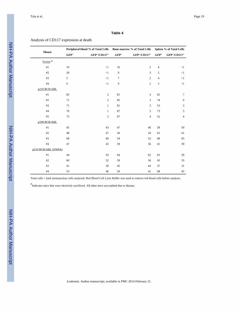
The elevated numbers of blast cells observed in the blood smears suggests that the p190BCR/ABL and p210 BCR/ABL(S509A) transplanted mice may be succumbing to moreacute leukemia's than the p210 transplanted mice. To confirm this, peripheral blood, bonemarrow and spleen was collected from randomly selected mice and cells were stained forCD117 (Table 4), a marker for AML. In all three tissues examined the number of GFP+/CD117+ cells was slightly increased in the p210 BCR/ABL transplanted mice relative to thevector transplanted mice. In contrast, the number of GFP+/CD117+ cells was dramaticallyincreased in the p190 BCR/ABL and p210 BCR/ABL(S509A) transplanted mice suggestinga more acute phenotype. In order to determine whether a particular progenitor populationwas selectively amplified in the p190 BCR/ABL and p210 BCR/ABL(S509A) transplantedmice, progenitor flow was performed on the bone marrow cells. Our analysis showed thatGMPs, CMPs and MEPs are significantly increased in the p190 BCR/ABL and p210 BCR/ABL(S509A) transplanted mice relative to the p210 BCR/ABL transplanted mice, but thegreatest increase is observed for the MEPs (Figure 4A and 4B).
In order to confirm that the erythroid expansion observed in the p190 and mutanttransplanted mice is an element of the malignancy, and not a reactive process, secondarytransplantations were performed. Of the five primary tumors examined from the p210 BCR/ABL transplanted mice two transmitted a myeloproliferative disease with a longer latencybut similar phenotype (Figure 4C). The remaining three developed a B-ALL phenotype withlonger disease latency. For all of the primary tumors examined for the p190 BCR/ABL and
Tala et al. Page 7
Leukemia. Author manuscript; available in PMC 2014 February 21.
NIH
-PA Author Manuscript
NIH
-PA Author Manuscript
NIH
-PA Author Manuscript

p210 BCR/ABL(S509A) transplanted mice it was possible to transmit the erythroleukemiato the secondary recipients. Thus, in all cases the disease in the secondary recipients closelyresembled the primary erythroleukemic disease, both in phenotype and latency. Collectivelythese observations suggest that the p190 BCR/ABL and p210 BCR/ABL(S509A)transplanted mice succumb to acute erythroid leukemia's similar to M6a in humans. Theyfurther suggest that either the MEPs have acquired self-renewal ability in the p190 BCR/ABL or mutant transplanted mice, or that the leukemia's are originating from a commonprogenitor population that is capable of differentiating via myeloid and erythroid lineages.
DiscussionTo directly assess the contribution of RhoGEF activity to p210 BCR/ABL transformation,we have introduced a single amino acid substitution into the DH domain of p210 BCR/ABLthat impairs RhoGEF activity, without affecting tyrosine kinase activity. When tested in cell,ex vivo, and animal-based models of transformation this mutant induces a disease phenotypethat is discrete from p210 BCR/ABL, but indistinguishable from p190 BCR/ABL. Whenviewed in its entirety, our data suggests that the RhoGEF activity of p210 BCR/ABLdirectly contributes to transforming activity, and may account for the difference in diseaseoutcome associated with p190 BCR/ABL and p210 BCR/ABL.
A comparison of transforming activity in cultured cells reveals that the RhoGEF activitymay have differential effects on both proliferation and survival. Thus, in the presence ofIL-3, cells that express vector, p190 BCR/ABL or the p210 BCR/ABL(S509A) mutantproliferate more rapidly than those that express p210 BCR/ABL, suggesting that theRhoGEF activity can be limiting with respect to proliferation. Interestingly, the p190 BCR/ABL and mutant expressing Ba/F3 cells do not enjoy a substantive survival advantage whenIL-3 is withdrawn, which leads to a disproportionate reduction in proliferative potentialrelative to the p210 BCR/ABL expressing cells. Thus, the RhoGEF activity limitsproliferation, but supports survival. The ability of p210 BCR/ABL to provide survivalsignaling has been well documented (23). Although survival is thought to be predominantlymediated by tyrosine kinase-dependent activation of the PI3K/AKT pathway, themechanism of activation remains unsettled (23). Current evidence suggests the pathway canbe activated by the GRB2/GAB2 complex which is recruited to Y177 in the BCR sequences(24), or alternatively, by recruitment and activation of CRKL and CBL (25). p210 BCR/ABL has also been shown to activate STAT5 which regulates the expression of anti-apoptotic genes such as MCL-1 and BCL-xL (26, 27). Our current study also implicatesRhoA mediated signaling pathways in p210 BCR/ABL survival signaling. RhoA signalinghas been shown to promote survival in a variety of tissue types (28–31), and was recentlyshown to be required for the B cell activating factor-mediated survival response (32).
In the absence of Il-3 there does appear to be a limited population of p190 BCR/ABL andmutant expressing cells that can evade apoptosis and retain a high proliferative capacity.This possibility is supported by a previous study which showed that p190 BCR/ABLtransformed BA/F3 cells proliferate more rapidly than p210 BCR/ABL transformed cells inthe absence of IL-3 (10). In this case, the authors used cell lines that had been stably selectedwith neomycin, and thus would have negatively selected against cells undergoing apoptosis,and positively selected for those that retain their high proliferative potential.
Our observation that both p190 BCR/ABL and the p210 BCR/ABL(S509A) mutant are stillable to drive myeloproliferation in the bone marrow transplantation model is consistent withprevious studies which showed that p190 BCR/ABL induces myeloid leukemia's withreduced latency if the donor mice are pre-treated with 5-FU (9, 10). However, we observedthat myeloid expansion and organ disruption at death was much more limited than in mice
Tala et al. Page 8
Leukemia. Author manuscript; available in PMC 2014 February 21.
NIH
-PA Author Manuscript
NIH
-PA Author Manuscript
NIH
-PA Author Manuscript

transplanted with p210 BCR/ABL. Instead we observed leukocytosis, coupled withexpansion of nRBCs, and reticulocytosis, suggesting that the leukemic expansion isqualitatively different. An examination of lineage specific markers and progenitor analysisconfirmed the presence of elevated numbers of GMPs, CMPs and MEPs consistent withacute erythroleukemia (AML-M6) and is likely to account for the reduced disease latencyassociated with the transplanted mice. These tumors are readily transplantable intosecondary recipients suggesting that they arise from a common progenitor population that iscapable of differentiating via myeloid and erythroid lineages. Although previous studieshave not noted the erythroid expansion (9, 10), these studies did not indicate that CBCs wereperformed, or that erythroid markers were examined. Thus, the phenotype may have beenpresent, but not noticed.
The enhanced ability of p190 BCR/ABL and p210 BCR/ABL(S509A) to drive expansion ofthe erythroid lineage was further confirmed by their ability to induce BFU-Es in colonyforming assays. It has been shown previously that when grown in media that allowsdetection of CFU-GM alone, both p190 BCR/ABL and p210 BCR/ABL support the growthof CFU-GM colonies (9). However, when we cultured bone marrow cells in the presence ofmedia that allows detection of both BFU-E and CFU-GM, we observed that p210 BCR/ABLpreferentially supports the growth of CFU-GM, while p190 BCR/ABL and the mutantpreferentially support the growth of BFU-E. This suggests that the RhoGEF domain bothactively supports myeloid expansion, and limits erythroid expansion. The former possibilityis supported by a recent study in which an inhibitor of the RhoA/ROCK complex was shownto oppose the proliferation of human CML CD34+ cells (33).
BCR/ABL rearrangements are found in 1–3% of cases of AML (including AML-M6) (34–37), and cases of erythroleukemic blast crisis have been described in CML (38). Sincetransition to blast crisis is generally thought to be triggered by the acquisition of secondarymutations, our results suggest that loss of Rho signaling in the leukemic stem cell thatoccurs secondary to p210 BCR/ABL transformation, may be sufficient to switch the fate ofthese cells from myeloid expansion to erythroid expansion, and account for theseerythroleukemic blasts.
AcknowledgmentsThis work was supported by Public Health Service grants CA097066 (IPW) and DK62757 (DAW).
References1. Melo JV. The diversity of BCR-ABL fusion proteins and their relationship to leukemia phenotype.
Blood. 1996; 88:2375–2384. [PubMed: 8839828]
2. Sawyers CL. Chronic myeloid leukemia. The New England Journal of Medicine. 1999; 340:1330–1340. [PubMed: 10219069]
3. Pane F, Frigeri F, Sindona M, Luciano L, Ferrara F, Cimino R, et al. Neutrophilic-chronic myeloidleukemia: a distinct disease with a specific molecular marker (BCR/ABL with C3/A2 junction).Blood. 1996; 88:2410–2414. [PubMed: 8839830]
4. Muller AJ, Young JC, Pendergast AM, Pondel M, Landau NR, Littman DR, et al. BCR first exonsequences specifically activate the BCR/ABL tyrosine kinase oncogene of Philadelphiachromosome-positive human leukemias. Mol Cell Biol. 1991; 11:1785–1792. [PubMed: 2005881]
5. McWhirter JR, Galasso DL, Wang JY. A coiled-coil oligomerization domain of Bcr is essential forthe transforming function of Bcr-Abl oncoproteins. Mol Cell Biol. 1993; 13:7587–7595. [PubMed:8246975]
6. Maru Y, Afar DE, Witte ON, Shibuya M. The dimerization property of glutathione S-transferasepartially reactivates Bcr-Abl lacking the oligomerization domain. J Biol Chem. 1996; 271:15353–15357. [PubMed: 8663064]
Tala et al. Page 9
Leukemia. Author manuscript; available in PMC 2014 February 21.
NIH
-PA Author Manuscript
NIH
-PA Author Manuscript
NIH
-PA Author Manuscript

7. Pendergast AM, Quilliam LA, Cripe LD, Bassing CH, Dai Z, Li N, et al. BCR-ABL-inducedoncogenesis is mediated by direct interaction with the SH2 domain of the GRB-2 adaptor protein.Cell. 1993; 75:175–185. [PubMed: 8402896]
8. Pendergast AM, Muller AJ, Havlik MH, Maru Y, Witte ON. BCR sequences essential fortransformation by the BCR-ABL oncogene bind to the ABL SH2 regulatory domain in anonphosphotyrosine-dependent manner. Cell. 1991; 66:161–171. [PubMed: 1712671]
9. Demehri S, O'Hare T, Eide CA, Smith CA, Tyner JW, Druker BJ, et al. The function of thepleckstrin homology domain in BCR-ABL-mediated leukemogenesis. Leukemia. 2009; 24:226–229. [PubMed: 19759561]
10. Li S, Ilaria RL Jr. Million RP, Daley GQ, Van Etten RA. The P190, P210, and P230 forms of theBCR/ABL oncogene induce a similar chronic myeloid leukemia-like syndrome in mice but havedifferent lymphoid leukemogenic activity. The Journal of Experimental Medicine. 1999;189:1399–1412. [PubMed: 10224280]
11. Lugo TG, Pendergast AM, Muller AJ, Witte ON. Tyrosine kinase activity and transformationpotency of bcr-abl oncogene products. Science. 1990; 247:1079–1082. [PubMed: 2408149]
12. Whitehead IP, Campbell S, Rossman KL, Der CJ. Dbl family proteins. Biochim Biophys Acta.1997; 1332:F1–23. [PubMed: 9061011]
13. Daubon T, Chasseriau J, Ali AE, Rivet J, Kitzis A, et al. Differential motility of p190bcrabl- andp210bcr-abl-expressing cells: respective roles of Vav and Bcr-Abl GEFs. Oncogene. 2008;27:2673–2685. [PubMed: 18059343]
14. Sahay S, Pannucci NL, Mahon GM, Rodriguez PL, Megjugorac NJ, et al. The RhoGEF domain ofp210 Bcr-Abl activates RhoA and is required for transformation. Oncogene. 2008; 27:2064–2071.[PubMed: 17922031]
15. Harnois T, Constantin B, Rioux A, Grenioux E, Kitzis A, et al. Differential interaction andactivation of Rho family GTPases by p210bcr-abl and p190bcr-abl. Oncogene. 2003; 25(22):6445–6454. [PubMed: 14508524]
16. Kelliher M, Knott A, McLaughlin J, Witte ON, Rosenberg N. Differences in oncogenic potencybut not target cell specificity distinguish the two forms of the BCR/ABL oncogene. Mol Cell Biol.1991; 11:4710–4716. [PubMed: 1875948]
17. Pear WS, Miller JP, Xu L, Pui JC, Soffer B, et al. Efficient and rapid induction of a chronicmyelogenous leukemia-like myeloproliferative disease in mice receiving P210 bcr/abltransducedbone marrow. Blood. 1998; 92:3780–3792. [PubMed: 9808572]
18. Olabisi OO, Mahon GM, Kostenko EV, Liu Z, Ozer HL, et al. Bcr interacts with components ofthe endosomal sorting complex required for transport-I and is required for epidermal growth factorreceptor turnover. Cancer Res. 2006; 66:6250–6257. [PubMed: 16778200]
19. Korus M, Mahon GM, Cheng L, Whitehead IP. p38 MAPK-mediated activation of NF-kappaB bythe RhoGEF domain of Bcr. Oncogene. 2002; 21:4601–4612. [PubMed: 12096337]
20. Thomas EK, Cancelas JA, Chae HD, Cox AD, Keller PJ, et al. Rac guanosine triphosphatasesrepresent integrating molecular therapeutic targets for BCR-ABL-induced myeloproliferativedisease. Cancer Cell. 2007; 12:467–478. [PubMed: 17996650]
21. Hanenberg H, Xiao XL, Dilloo D, Hashino K, Kato I, et al. Colocalization of retrovirus and targetcells on specific fibronectin fragments increases genetic transduction of mammalian cells. Naturemedicine. 1996; 2:876–882.
22. Zhang X, Ren R. Bcr-Abl efficiently induces a myeloproliferative disease and production of excessinterleukin-3 and granulocyte-macrophage colony-stimulating factor in mice: a novel model forchronic myelogenous leukemia. Blood. 1998; 92:3829–3840. [PubMed: 9808576]
23. Hazlehurst LA, Bewry NN, Nair RR, Pinilla-Ibarz J. Signaling networks associated with BCR-ABL-dependent transformation. Cancer Control. 2009; 16:100–107. [PubMed: 19337196]
24. Sattler M, Mohi MG, Pride YB, Quinnan LR, Malouf NA, et al. Critical role for Gab2 intransformation by BCR/ABL. Cancer Cell. 2002; 1:479–492. [PubMed: 12124177]
25. Sattler M, Salgia R, Okuda K, Uemura N, Durstin MA, et al. The proto-oncogene productp120CBL and the adaptor proteins CRKL and c-CRK link c-ABL, p190BCR/ABL and p210BCR/ABL to the phosphatidylinositol-3' kinase pathway. Oncogene. 1996; 12:839–846. [PubMed:8632906]
Tala et al. Page 10
Leukemia. Author manuscript; available in PMC 2014 February 21.
NIH
-PA Author Manuscript
NIH
-PA Author Manuscript
NIH
-PA Author Manuscript

26. Gesbert F, Griffin JD. Bcr/Abl activates transcription of the Bcl-X gene through STAT5. Blood.2000; 96:2269–2276. [PubMed: 10979976]
27. Sillaber C, Gesbert F, Frank DA, Sattler M, Griffin JD. STAT5 activation contributes to growthand viability in Bcr/Abl-transformed cells. Blood. 2000; 95:2118–2125. [PubMed: 10706883]
28. Gallagher ED, Gutowski S, Sternweis PC, Cobb MH. RhoA binds to the amino terminus ofMEKK1 and regulates its kinase activity. J Biol Chem. 2004; 279:1872–1877. [PubMed:14581471]
29. Kobayashi K, Takahashi M, Matsushita N, Miyazaki J, Koike M, et al. Survival of developingmotor neurons mediated by Rho GTPase signaling pathway through Rho-kinase. J Neurosci. 2004;24:3480–3488. [PubMed: 15071095]
30. Yoshida T, Clark MF, Stern PH. The small GTPase RhoA is crucial for MC3T3-E1 osteoblasticcell survival. Journal of Cellular Biochemistry. 2009; 106:896–902. [PubMed: 19184980]
31. Zhu S, Korzh V, Gong Z, Low BC. RhoA prevents apoptosis during zebrafish embryogenesisthrough activation of Mek/Erk pathway. Oncogene. 2008; 27:1580–1589. [PubMed: 17873909]
32. Zhang S, Zhou X, Lang RA, Guo F. RhoA of the Rho family small GTPases is essential for Blymphocyte development. PloS one. 7:e33773. [PubMed: 22438996]
33. Burthem J, Rees-Unwin K, Mottram R, Adams J, Lucas GS, et al. The rho-kinase inhibitorsY-27632 and fasudil act synergistically with imatinib to inhibit the expansion of ex vivo CD34(+)CML progenitor cells. Leukemia. 2007; 21:1708–1714. [PubMed: 17554385]
34. Pompetti F, Spadano A, Sau A, Mennucci A, Russo R, et al. Long-term remission in BCR/ABL-positive AML-M6 patient treated with Imatinib Mesylate. Leukemia research. 2007; 31:563–567.[PubMed: 16916543]
35. Soupir CP, Vergilio JA, Dal Cin P, Muzikansky A, Kantarjian H, et al. Philadelphia chromosome-positive acute myeloid leukemia: a rare aggressive leukemia with clinicopathologic featuresdistinct from chronic myeloid leukemia in myeloid blast crisis. American Journal of ClinicalPathology. 2007; 127:642–650. [PubMed: 17369142]
36. Morgan GJ, Wiedemann LM, Chan LC, Price CM, Kanfer EJ, et al. A case of M-BCR-rearranged,Philadelphia-positive AML that relapsed as chronic phase CML. Blood. 1990; 75:317–318.[PubMed: 2403819]
37. Price CM, Rassool F, Shivji MK, Gow J, Tew CJ, et al. Rearrangement of the breakpoint clusterregion and expression of P210 BCR-ABL in a "masked" Philadelphia chromosome-positive acutemyeloid leukemia. Blood. 1988; 72:1829–1832. [PubMed: 3179449]
38. Sharma P, Dhingra KK, Roy S, Singh T. An acute myeloid leukemia M6b blast crisis with giantproerythroblasts in chronic myeloid leukemia. Journal of Pediatric Hematology/Oncology. 2009;31:220–221. [PubMed: 19262253]
Tala et al. Page 11
Leukemia. Author manuscript; available in PMC 2014 February 21.
NIH
-PA Author Manuscript
NIH
-PA Author Manuscript
NIH
-PA Author Manuscript

Figure 1.The RhoGEF activity of p210 BCR/ABL regulates RhoA and influences IL-3 dependent andindependent growth in hematopoietic cells. Ba/F3 cells were infected with retroviralparticles that encode MSCV-bcr-abl/p210-IRES-gfp, MSCV-bcr-abl/p210(S509A)-IRES-gfp, MSCV-bcr-abl/p190-IRES-gfp, or cognate vector. (A–C) GFP-positive cells weresorted, and then plated for 48 hr. (A) Lysates were collected and examined by Western blotfor expression of the Bcr-Abl constructs. (B) Lysates were also examined by affinityprecipitation assays to measure levels of total (RhoA) and activated (GTP-RhoA) RhoA, and(C) by Western blot for levels of total (MLC2) and phosphorylated (p-MLC2(Ser19))myosin light chain 2. (A–C) All Western blots were performed in triplicate and quantitatedusing Quantity One software (BioRad). Quantitated data shown are derived from threeindependent sorts, and show standard deviations, and statistical significance relative to MIGvector controls (B and C). (D–G) GFP-positive cells from the same sorts were immediatelyplated in the presence (D) or absence (E, F & G) of IL-3. (D&E) Cells were counted at theindicated time points. (F) The percentage of apoptotic cells was determined at the indicatedtime points. (G) The percentage of cells in Sub-G1, G1, S and G2/M cell cycle phases wasdetermined at indicated time points. (A–G) Data shown are an average of three independentexperiments performed on three independent sorts and show standard deviations, andstatistical significance as indicated (* p < 0.05, ** p < 0.01, *** p < 0.001).
Tala et al. Page 12
Leukemia. Author manuscript; available in PMC 2014 February 21.
NIH
-PA Author Manuscript
NIH
-PA Author Manuscript
NIH
-PA Author Manuscript

Figure 2.Loss of RhoGEF activity alters the transforming potential of p210 BCR/ABL in murine exvivo assays. Bone marrow was collected from BALB/c mice and infected with retroviralparticles that encode MSCV-bcr-abl/p210-IRES-gfp, MSCV-bcr-abl/p210(S509A)-IRES-gfp, MSCV-bcr-abl/p190-IRES-gfp, or cognate vector. (A) GFP positive cells werecollected and plated at the indicated cell numbers per well and in the absence of cytokines.Assays were performed in triplicate. A well was scored positive when the number of viablecells reached 106/well. Cell growth was monitored for 21 days. (B) GFP positive cells werecollected and plated in MethoCult media that supports the growth of both BFU-E and CFU-GM (B & C), or CFU-pre-B (D). Colonies were counted and expressed as fold changerelative to p210 BCR/ABL. Data shown is the average of three independent experiments andshows standard deviations, and statistical significance relative to p210 (* p < 0.05, ** p <0.01, *** p < 0.001). Cells were dissociated from random colonies and stained with Hema 3to confirm the identity of the colonies. Right panels show a representative colony and thestaining of the dissociated cells. Images of colonies were visualized using an AdvancedMicroscopy Group microscope; EVOS (Bothell, WA, USA) at 4×PH magnification usingMicron(EVOS) 2.0 software. Images of cells from colonies were visualized under oil using aCarl Zeiss microscope; Zeiss Axio Imager A1 (Thornword, NY, USA) equipped withACHROPLAN, 100x, 1.25 numerical aperture, and acquired using AxioCam MRC cameraand Axiovision 4.7.1 software.
Tala et al. Page 13
Leukemia. Author manuscript; available in PMC 2014 February 21.
NIH
-PA Author Manuscript
NIH
-PA Author Manuscript
NIH
-PA Author Manuscript

Figure 3.Altered disease progression in mice transplanted with the p210 BCR/ABL(S509A) mutant.Bone marrow was collected from BALB/c mice and infected with retroviral particles thatencode MSCV-bcr-abl/p210-IRES-gfp, MSCV-bcr-abl/p210(S509A)-IRES-gfp, MSCV-bcr-abl/p190-IRES-gfp, or cognate vector. GFP positive cells were collected and used for thebone marrow transplantation assay. (A) Survival curves for recipient mice transplanted withvector, p190 BCR/ABL, p210 BCR/ABL or p210 BCR/ABL(S509A). Kaplan-Meier curveswere generated for three independent experiments. BMT = bone marrow transplantation.When comparing p210 BCR/ABL with p210 BCR/ABL(S509A), Mantel-Cox tests of thethree survival curves yielded values of p = 0.0011 (χ2 = 10.73), p = 0.0013 (χ2 = 10.38) andp < 0.0001, (χ2 = 16.35) respectively. (B) Blood smears were performed weekly on the miceto monitor disease progression. The representative smears, and spleens, shown were takenwhen mice became moribund except p210 (day 14) which was done on day 14 post-BMT.Black arrows indicate nucleated red blood cells. (C) Spleen, liver, and lung tissues were alsoharvested at the time of death, and processed slides were stained with hematoxylin andeosin. Images of blood smears were visualized under oil using a Carl Zeiss microscope;Zeiss Axio Imager A1 (Thornword, NY, USA) equipped with ACHROPLAN, 100x, 1.25numeical aperture, and acquired using AxioCam MRC camera and Axiovision 4.7.1software. Images of solid organs were visualized with the same microscope using Ec Plan-NEOPLUAR 10x, 0.3 numerical aperture.
Tala et al. Page 14
Leukemia. Author manuscript; available in PMC 2014 February 21.
NIH
-PA Author Manuscript
NIH
-PA Author Manuscript
NIH
-PA Author Manuscript

Figure 4.The RhoGEF activity of BCR/ABL limits the self-renewal of myeloid progenitors in theBMT model. Bone marrow cells isolated from diseased mice at death were used forimmunophenotypic analysis of progenitor populations. (A) Representative FACS stainingprofiles of progenitor populations. (B) Percentages of each progenitor populations (GMP,CMP and MEP) relative to total GFP positive cells. Values were derived from 4 mice pergroup, and represented as averages. Data shows statistical significance of total progenitorpopulation relative to p210 (* p < 0.05, ** p < 0.01, *** p < 0.001). (C) Survival curves ofsecondary transplanted mice. A total of 1×10⁶ cells (bone marrow and spleen) isolated fromdiseased mice were injected into sublethally irradiated BALB/c mice. When comparingp210 BCR/ABL with p210 BCR/ABL(S509A) a Mantel-Cox test for the survival curvesyielded a value of p = 0.004 (χ2 = 8.28).
Tala et al. Page 15
Leukemia. Author manuscript; available in PMC 2014 February 21.
NIH
-PA Author Manuscript
NIH
-PA Author Manuscript
NIH
-PA Author Manuscript

NIH
-PA Author Manuscript
NIH
-PA Author Manuscript
NIH
-PA Author Manuscript
Tala et al. Page 16
Table 1
Comparison of WBC counts, spleen weight, blasts and lifespan
Mouse WBC (103/μl) spleen weight (g) % blasts lifespan (day)
Vector1 5.77±0.94 0.096±0.008 0 NA
p210 BCR/ABL 118.5±31.94 0.878±0.098 26.4±5.88 28 (23–38)
p190 BCR/ABL 34.97±33.02 0.438±0.067 83.2±4.35 16 (15–21)
p210 BCR/ABL(S509A) 39.74±25.11 0.737±0.123 82.1±5.73 16 (15–17)
Data shown is for a minimum of 5 mice per group.
For lifespan, the range of ages at death is shown in parentheses.
% blasts = percentage of blasts in peripheral blood as assessed by morphology.
1Indicates mice that were electively sacrificed. All other mice succumbed due to disease.
Leukemia. Author manuscript; available in PMC 2014 February 21.

NIH
-PA Author Manuscript
NIH
-PA Author Manuscript
NIH
-PA Author Manuscript
Tala et al. Page 17
Tabl
e 2
Imm
unop
heno
typi
ng o
f di
seas
e pr
ogre
ssio
n in
a B
MT
mod
el f
or C
ML
Mou
se (
Day
at
deat
h or
sacr
ific
e)
Per
iphe
ral b
lood
% o
f T
otal
Cel
lsB
one
mar
row
% o
f T
otal
Cel
lsSp
leen
% o
f T
otal
Cel
ls
%G
FP
+G
FP
+ /C
D11
b+G
FP
+ /C
D3+
GF
P+ /
B22
0+%
GF
P+
GF
P+ /
CD
11b+
GF
P+ /
CD
3+G
FP
+ /B
220+
%G
FP
+G
FP
+ /C
D11
b+G
FP
+ /C
D3+
GF
P+ /
B22
0+
Vec
tor
1
#1 (
Day
29)
134
<1
<1
179
<1
<1
113
<1
<1
#2 (
Day
30)
2<
1<
1<
14
2<
1<
16
3<
1<
1
#3 (
Day
30)
164
<1
<1
2610
<1
<1
104
<1
<1
#4 (
Day
32)
103
<1
<1
115
<1
<1
52
<1
<1
#5 (
Day
32)
123
<1
<1
113
<1
<1
113
<1
<1
p210
BC
R/A
BL
#1 (
Day
29)
8877
<1
<1
8373
<1
<1
7462
<1
<1
#2 (
Day
32)
8770
<1
<1
7961
<1
<1
6350
<1
<1
#3 (
Day
30)
8078
<1
<1
8275
<1
<1
6548
<1
<1
#4 (
Day
32)
9078
<1
<1
8265
<1
<1
6351
<1
<1
#5 (
Day
30)
8668
<1
<1
7954
<1
<1
6330
<1
<1
p190
BC
R/A
BL
#1 (
Day
16)
7170
1<
156
367
443
1910
7
#2 (
Day
17)
5553
11
5335
55
4716
68
#3 (
Day
17)
4641
32
4527
66
2918
54
#4 (
Day
16)
3332
<1
<1
4025
53
3416
53
#5 (
Day
17)
6257
<1
<1
5033
107
4129
92
p210
BC
R/A
BL
(S5
09A
)
#1 (
Day
16)
5552
<1
<1
5240
102
3621
141
#2 (
Day
15)
5048
<1
<1
4739
2<
141
365
<1
#3 (
Day
16)
4745
1<
137
256
440
1919
2
#4 (
Day
15)
5147
4<
143
3210
135
1915
1
#5 (
Day
15)
4038
2<
137
2610
135
2014
1
1 Vec
tor
mic
e w
ere
elec
tivel
y sa
crif
iced
. All
othe
r m
ice
succ
umbe
d du
e to
dis
ease
.
Leukemia. Author manuscript; available in PMC 2014 February 21.

NIH
-PA Author Manuscript
NIH
-PA Author Manuscript
NIH
-PA Author Manuscript
Tala et al. Page 18
Table 3
Analysis of erythroid dysplasia at death
Mouse nRBC/100 WBC % reticulocytes RBC (106/(μl) Hemoglobin, g/dl % hematocrit CD71+/Ter119+
% of total cells
Vector1 0 3.81±1.9 8.75±0.26 13.92±0.44 43.9±1.57 13.33±1.52
p210 BCR/ABL 2.85±2.85 14.39±4.61 9.65±1.47 16.36±2.37 52.70±8.31 30.33±2.3
p190 BCR/ABL 222.9±154.6 24.71±8.27 8.20±0.69 13.43±1.22 43.97±4.36 71.67±5.77
p210 BCR/ABL (S509A) 145.6±140.1 28.59±6.74 9.93±0.91 14.86±1.37 47.33±4.37 73.67±4.93
Data shown is for a minimum of 5 mice per group.
nRBC = nucleated red blood cells. WBC = white blood cells. RBC = red blood cells. Total cells = total mononuclear cells analyzed.
1Indicates mice that were electively sacrificed. All other mice succumbed due to disease.
Leukemia. Author manuscript; available in PMC 2014 February 21.
NIH
-PA Author Manuscript
NIH
-PA Author Manuscript
NIH
-PA Author Manuscript
Tala et al. Page 19
Table 4
Analysis of CD117 expression at death
MousePeripheral blood % of Total Cells Bone marrow % of Total Cells Spleen % of Total Cells
GFP+ GFP+/CD117+ GFP+ GFP+/CD117+ GFP+ GFP+/CD117+
Vector a
#1 10 <1 10 2 4 <1
#2 20 <1 9 3 5 <1
#3 5 <1 7 2 4 <1
#4 9 <1 9 2 5 <1
p210 BCR/ABL
#1 83 2 81 3 65 7
#2 71 2 85 3 74 6
#3 71 1 81 3 53 3
#4 70 1 87 3 73 5
#5 75 2 87 4 52 4
p190 BCR/ABL
#1 45 43 47 46 58 50
#2 48 47 36 34 63 61
#3 68 66 54 53 68 65
#4 47 43 39 36 41 39
p210 BCR/ABL (S509A)
#1 44 43 64 62 63 59
#2 60 52 39 36 45 35
#3 41 30 45 44 37 31
#4 53 46 50 41 68 45
Total cells = total mononuclear cells analyzed. Red Blood Cell Lysis Buffer was used to remove red blood cells before analysis.
aIndicates mice that were electively sacrificed. All other mice succumbed due to disease.
Leukemia. Author manuscript; available in PMC 2014 February 21.