copyright warning &...
TRANSCRIPT

Copyright Warning & Restrictions
The copyright law of the United States (Title 17, United States Code) governs the making of photocopies or other
reproductions of copyrighted material.
Under certain conditions specified in the law, libraries and archives are authorized to furnish a photocopy or other
reproduction. One of these specified conditions is that the photocopy or reproduction is not to be “used for any
purpose other than private study, scholarship, or research.” If a, user makes a request for, or later uses, a photocopy or reproduction for purposes in excess of “fair use” that user
may be liable for copyright infringement,
This institution reserves the right to refuse to accept a copying order if, in its judgment, fulfillment of the order
would involve violation of copyright law.
Please Note: The author retains the copyright while the New Jersey Institute of Technology reserves the right to
distribute this thesis or dissertation
Printing note: If you do not wish to print this page, then select “Pages from: first page # to: last page #” on the print dialog screen

The Van Houten library has removed some ofthe personal information and all signatures fromthe approval page and biographical sketches oftheses and dissertations in order to protect theidentity of NJIT graduates and faculty.

ABSTRACT
SPINNING OF COLLAGEN FIBERS AND CHARACTERIZING THERMAL,MECHANICAL, TENSILE AND STRUCTURAL PROPERTIES
byGrace Evangelin Tatagiri
Collagen is the most important building block in the entire animal world. It plays an
important role in regeneration of broken bones and wound healing, also helps grow blood
vessels to feed the healing areas. The use of synthetic materials has been extensive, but
these materials have there own limits and drawbacks. Poor biocompatibility of these
materials is the main issue and often result in inflammations. Hence collagen is being
heralded as one of the most appropriate replacement as an implantable material. The
reasons being that it is a naturally occurring material, exhibits high biocompatibility low
antigenicity and host response. This study involes spinning collagen fibers using
dispersion made from bovine tendon, and to modify their properties via cross-linking by
treating the fibers with gluteraldehdye. These fibers are characterized for mechanical
properties, effect of temperature on dimension changes, temperature dependent heat flow,
and temperature dependent weight. The above tests are conducted using TMA, DSC and
TGA.The fiber diameters and surface features are studied using SEM. The results of
these analyses are compared with cross-linking and without cross-linking for each of the
three dispersion percentages. The mechanical behavior of cross linked collagen fibers
was enhanced relative to the non cross linked fibers with higher denaturation temperature
and lower tensile deformation.

SPINNING OF COLLAGEN FIBERS AND CHARACTERIZING THERMAL,MECHANICAL, TENSILE AND STRUCTURAL PROPERTIES
byGrace Evangelin Tatagiri
A ThesisSubmitted to the Faculty of
New Jersey Institute of TechnologyIn Partial Fulfillment of the Requirements for the Degree of
Master of Science in Biomedical Engineering
Department of Biomedical Engineering
January 2004


APPROVAL PAGE
SPINNING OF COLLAGEN FIBERS AND CHARACTERIZING IN TERMS OFTHERMAL, MECHANICAL, TENSILE AND STRUCTURAL PROPERTIES
Grace Evangelin Tatagiri
Dr. Michael Jaffe, Thesis AdvisorResearch Professorbiomedical Engineering, NJIT.
Dr. Treena Arinzeh, Committee MInber Date:Assistant Professor in Biomedical Engineering, NJIT
Dr. GOO Collins, Committee Member hate:Visiting Scientist, Medical Device and Concept Laboratory, NJIT

BIOGRAPHICAL SKETCH
Author: Grace Evangelin Tatagiri
Degree: Master of Science
Date: January 2004
Undergraduate and Graduate Education:
• Master of Science in Biomedical Engineering,New Jersey Institute of Technology, Newark, New Jersey. 2004.
• Bachelor of Engineering Biomedical Engineering,K.L.E. Society's College of Engineering and Technology, India. 2001.
Major: Biomedical Engineering

Dedicated to:
My beloved
Grandparents, Parents, Sisters and Brothers.
"Thy word is a lamp unto my feet, and a light unto my path" Psalms 119:105
v

ACKNOWLEDGEMENT
I would like to express my deepest appreciation to Dr. Michael Jaffe who not only
served as research advisor but also gave encouragement, support and lot of
resources.
I would like to extend my thanks to Dr. Joseph Nichols and Dr.Nels Lauritzen
who helped in providing various resources.
I am very grateful to Dr. George Collins and Joseph Pickton for helping with
various experiments, offering continuous expert advice, reassurance and also for
being on the thesis committee.
Special thanks to Dr. Treena Arinzeh for serving as committee member.
Above all thankful to the Lord Almighty, family and friends for their support and
encouragement.
vi

TABLE OF CONTENTS
Chapter Page
1 INTRODUCTION 1
1.1 General Statement of Problem 1
1.2 Objective 2
2 BACKGROUND INFORMATION 3
2.1 Chemistry of Collagen 3
2.2 Types of Collagen 5
2.3 Background of Collagen Producing Process 9
2.4 Background of Spinning Process 10
3 EXPERIMENTAL DESIGN 11
3.1 Making of Collagen Dispersion 11
3.2 The Experimental Setup for Spinning 15
3.3 Chemical Modification of Collagen to Cross-link the Fibers 19
3.4 Characterization of Collagen Fibers 25
3.5 TGA-Thermal Gravimetric Analysis 27
3.6 DSC-Differential Scanning Calorimetry 27
3.7 TMA-Thermal Mechanical Analysis 29
3.8 Tensile Modulus 29
3.9 SEM-Scanning Electron Microscope 30
3.10 URM-Universal Research Microscope 30
vii

TABLE OF CONTENTS
(Continued)
Chapter Page
4 RESULTS AND DISCUSSION 31
4.1 TMA-Results 33
4.2 DSC-Results 36
4.3 DSC-Results 40
4.4 Tensile Modulus-Results 43
4.5 SCM -Results 46
4.6 URM-Results 49
5 CONCLUSIONS 52
APPENDIX A SAFETY PROCEDURES 53
APPENDIX B TGA, DSC, TMA PLOTS AND SEM FIGURES 54
REFERENCES 81
viii

Table
LIST OF TABLES
Page
3.1 Weights after Grinding the Tendon 12
3.2 Weight after Placing in the Oven 13
3.3 Resultant Weights of Collagen 13
3.4 pH Reading of the Washes 15
4.1 Types of Collagen Fibers 32
4.2 TGA-Temperature and their Corresponding Weight Loss 33
4.3 DSC-Fibers with their Corresponding Tg and Td 37
4.4 TMA-Fibers and the Temperature at which they Break 40
4.5 Tensile Strengths of the Fibers 43
ix

LIST OF FIGURES
Figure Page
2.1 Structure of collagen 3
2.2 Major Amino acids of collagen 4
3.1 The various sections of bovine tendon 11
3.2 Schematic diagram of spinning of collagen fiber 17
3.3 Collagen cross-linked with gluteraldehyde 24
4.1 TGA plot of collagen 1% dispersion 34
4.2 TGA plot of collagen 1% dispersion cross-linked 35
4.3 DSC plot of collagen with 1% dispersion —Ramp 38
4.4 DSC plot of collagen with 1% dispersion - heat/cool/heat 39
4.5 TMA plot of collagen with 1% dispersion 41
4.6 TMA plot of collagen with 1% dispersion cross linked 42
4.7 Tensile Modulus plot 1% dispersion 44
4.8 Tensile Modulus plot with 1% dispersion and cross-links 45
4.9 SEM picture with 1 % dispersion 47
4.10 SEM picture with 1 % dispersion and cross-linked 48
4.11 URM picture with 1% dispersion with and without cross links 49
4.12 URM picture with 1.6% dispersion with and without cross-links 50
4.13 URM picture with 2% dispersion with and without cross-links 51

CHAPTER 1
INTRODUCTION
1.1 General Statement of Problem
Collagen is the most important building block in the entire animal world; it is the tie that
binds the animal kingdom together. Being nature's most abundant protein polymer, more
than a third of the body's protein is collagen making up 75% of the skin. The more
Science learns about the body, the more integral we see collagen to be. Collagen acts as
scaffolding for bodies, controls cell shape and differentiation. It plays an important role
in regeneration of broken bones and wound healing and also helps grow blood vessels to
feed the healing areas. Collagen is the fibrous protein constituent of skin, cartilage, bone,
and other connective tissue.
The use of synthetic materials, such as polyester fiber or polytetrafluoroethylene
as implants designed to replace diseased or damaged body parts has been extensive.
These materials have however, enjoyed limited success. This has been due to the poor
biocompatibility of many of these materials, which among other problems frequently
initiate persistent inflammatory reactions. Additionally, the failure of the body to
integrate these materials, because they do not break down and do not lend themselves to
remodeling by tissue cells that may come into contact with them, causes further problems
(1).
Collagen is being heralded as one of the most useful biomaterials in the realm of drug
delivery applications, for surgical purposes (2), and collagen scaffolds for tissue
regeneration (3). The reasons are very clear as collagen offers many unique and
advantageous properties that make it a versatile material. First and foremost, because
1

collagen is a naturally occurring material, it exhibits an extremely high biocompatibility
and safety, or conversely elicits a low antigenicity and host response. Secondly, collagen
is biodegradable and bioreabsorbable. These properties, in turn, are controlled via
regulating the degree of cross-linking. Lastly, collagen is easily modifiable and can be
combined with several synthetic polymers to produce a variety of medical devices and
drug delivery systems.
1.2 Objective
The main objective of this work is to spin collagen fibers using dispersion made from
bovine tendon, and to modify their properties via cross-linking by treating the fibers with
gluteraldehdye. This work is directed at incorporating the ability to cross link collagen
fibers into the set of capabilities of Medical Device and Concept Laboratory. Fibers are
spun using a gel, which has various percentages of collagen dispersed in ethanol. The
percentages of dispersion under study are 1%, 1.6% and 2%. These fibers are
characterized in terms of mechanical behavior, effect of temperature on dimension
changes, temperature dependent heat flow, and temperature dependent weight. The above
tests are conducted using Thermal Mechanical Analysis (TMA), Differential Scanning
Calorimetry (DSC) and Thermal Gravimetric Analysis (TGA) respectively. The fiber
diameters and surface features are studied using a Scanning Electron Microscope (SEM).
The results of these analyses are compared with cross-linking and without cross-linking
for each of the three dispersion percentages.
2

CHAPTER 2
BACKGROUND INFORMATION
2.1 Chemistry of Collagen
Collagen is characterized by the formation of triple helices in which three polypeptide
chains are wound tightly around one another in a ropelike structure. The triple helix
domains of the collagens consist of repeats of the amino acid sequence Gly-X-Y. A
glycine (the smallest amino acid, with a hydrogen side group is required in every third
position in order for the polypeptide chains to pack together close enough to form the
collagen triple helix.
Figure 2.1 Three-polypeptide chains coil around one another in a characteristic triplehelix structure
3

Proline is frequently found in the X position and hydroxyproline in the Y position,
because of their ring structure, these amino acids stabilize the helical conformations of
the polypeptide chains (4).
4
Figure 2.2 Major Amino Acids of Collagen Proline, Hydroxyproline, Lysine,hydroxylysine.
The primary structure has complete sequence of amino acids along each of three
polypeptide chains as well as the location of interchain cross-links in relation to this
sequence (5). Approximately one-third of the residues are glycine and another quarter or
so are proline or hydroxyproline. The structure of the bifunctional interchain cross-link is
the relatively complex condensation product of a reaction involving lysine and
hydroxylysine residues; this reaction continues as the organism matures, thereby
rendering the collagens of older animals more difficult to extract (6).
The secondary structure is the local configuration of a polypeptide chain that
results from satisfaction of stereochemical angles and hydrogen-bonding potential of

peptide residues. In collagen, the abundance of glycine residues plays a key
configurational role in the triplet Gly-X-Y, where X and Y are frequently proline or
hydroxyproline, respectively, these two amino acids that direct the chain configuration
locally by the rigidity of their ring structures. The absence of a side group in glycine
permits the close approach of polypeptide chain in the collagen triple helix (7). The
tertiary structure refers to the global configuration of the polypeptide chains; it represents
the pattern according to which the secondary structures are packed together within the
complete macromolecule and it constitutes the structural unit that can exists as a
physiochemically stable entity in solution, namely, the triple helical collagen molecule.
The fourth-order quaternary structure denotes the repeating supermolecular unit
structure, comprising several molecules packed in a specific lattice, which constitutes the
basic element of the solid state (microfibril). Collagen molecules are packed in a quasi-
hexagonal lattice at an interchain distance of about 1.3 nm, which shrinks considerably
when the microfibril is dehydrated. Higher levels of order, eventually leading to gross
anatomical features, which can be seen with the naked eye, have been proposed but there
is no agreement on their definition (5, 6, 7).
2.2 Types of Collagen
At this stage 13 kinds of vertebrate collagen are known, plus some smaller molecules
looking like collagen parts suspected to be members of the family and each type has
evolved to serve a distinct purpose. Collagen varies depending on the anatomical region.
From muscle to bone to cartilage to blood vessels to nerves to various parts of the skin,
which itself is the largest organ in the body.
5

The differences among these collagen siblings come at the ends of each collagen
molecule. It's as though nature created specified arms able to share their fingertips with
the tips of different molecules in appropriate cells. That's how they connect. But all
collagen middles are the same. Three strands of repeating amino acids coil themselves,
left-handed, into the unique collagen triple helix. Then these coils weave themselves
right-handed into a cable, like small steel wires braided into the cables of a suspension
bridge. In fact, collagen has a greater tensile strength than steel. Presumably, this
complex structure was devised by nature to be invulnerable to the circulating enzymes
and other materials in the body. Nature accomplished this purpose superbly, which is
why no other enzyme (of the many thousands in the body) but a "collagenase" can break
it into its component parts.
When the body needs to build any new cellular structure as in the healing process,
for example, collagen and/or collagen fragments play a central role. Although the role of
collagen as scaffolding has been known for some time, we now know that collagen
controls cell shape and differentiation, migration, and the synthesis of a number of
proteins.
They are insoluble, stable and have long biological half-life, high tensile strength
and contractibility. They are secreted by connective tissue cells, as well as by a variety of
other cell types. As a major component of skin, bone teeth, tendons, ligaments, cartilage,
and connective tissue. The characteristic feature of a typical collagen molecule is its long,
stiff, triple-stranded helical structure in which three collagen polypeptide chains are
wound around one another in a ropelike super helix. Its properties are diverse and
remarkable. In tendon it has a tensile strength equal to that of light steel wire. In the
6

cornea it is as transparent as water. It accounts for the toughness of leather, the tenacity of
glue and the viscousness of gelatin. So far about 25 distinct collagen chains have been
identified, each encoded by a separate gene. Different combinations of these genes are
expressed in different tissues. Although in principle more than 10,000 types of collagen
molecules could be assembled from various combinations of the 25 or so chains, only
about 15 types of collagen molecules have been found. The main types of collagen found
in connective tissues are types I, H, V, XI-type I being the principal collagen of skin and
bone and by far the most common. These are the fibrillar collagens and have the ropelike
structure. After being secreted into ordered polymers called collagen fibrils, which are
thin (10-300nm in diameter) structures, many hundreds of microns long in mature tissues
and clearly visible in electron micrographs.
The collagen fibrils often aggregate into larger, cable like bundles, which can be
seen in the light microscope as collagen fibers several micrometers in diameter. Types IX
and XII are called fibril-associated collagens as they decorate the surface of collagen
fibrils. They are thought to link these fibrils to one another and to other components in
the extracellular matrix. Types IV and VII are network-forming collagens. Type IV
molecules assemble into a felt like sheet or meshwork that constitutes a major part of
mature laminae while type VII molecules form dimmers that assemble into specialized
structures called anchoring fibrils, which help attach the basal lamina multilayered
epithelia to the underlying connective tissue and therefore are especially abundant in the
skin.
7

Type I. Major collagen found in skin, tendon, bone, dentin; a fetal form also exists:
Mutations in the cleaving of the procollagen I have been shown to be responsible for
Ehrlers-Danlos syndrome type VII, a disorder characterized by extreme joint
hypermobility and congenital bilateral hip dislocations.Other mutations in collagen I
produce osteogenesis imperfecta, a highly variable condition (depending on the place of
the mutation) characterized by deformed bones, short stature, and abnormalities of teeth.
Type II. Specific for cartilage and vitreous humor.
Mutations in collagen II can cause abnormalities of skeletal development and eye
structure.
Type HI. Frequently found with type I in skin, muscles, and blood vessels.
Mutations in collagen III cause Ehlers-Danlos syndrome.
This is a life threatening and debilitating disease characterized by extreme arterial and
capillary fragility. Death is often due to hemorrhage.
Type V. Fetal tissues, placenta, interstitial tissues
Appears to be necessary for extracellular matrix assembly in connective tissues.
Knockouts of this gene in mice have spinal deformities and poor skin and eye
development.
Type XI. Cartilage. Mutations in collagen XI are responsible for the Stickler Syndrome,
characterized by cartilage and joint problems, cleft palate, as well as poor retinal
development.
8

2.3 Background of Collagen Producing Process
Furukawa et al, (8) describes a process for producing regenerated collagen fiber with
excellent water resistance and undergoes substantially no waving on contact with water.
This is suitable as a substitute for human hair, animal hair, or as catgut. The starting
material used in this invention is a solution of solubilized collagen for which the source
was collagen from the raw hide of animals, such as cattle or pigs, fresh or salted, with an
alkali or an enzyme and preparing an acidic aqueous solution. This collagen was
accomplished by treating soluble collagen having metallic salt aqueous solution. At the
end they prepare an aqueous solution with an acid for spinning fiber from it. This acidic
collagen solution is spun through a spinneret of an inorganic salt, such as sodium sulfate,
sodium chloride, to obtain fibrous solubilized collagen.
To get excellent water resistance, researchers cross link these fibers with a water
soluble organic cross linking agents include monoaldehydes, e.g., formaldehyde,
acetaldehyde, methyiglyoxal, and acrolein; dialdehydes, e.g., glyoxal, malondialdehyde,
succindialdehyde, glutaraldehyde, phthalaldehyde, dialdehyde, and starch; epoxy
compounds, e.g., glycol glycidyl ether or a poiyoi glycidyl ether, and a glycidyl ester of a
monocarboxylic acid, dicarboxylic acid or polycarboxylic acid, cromic acid; N-methylol
compounds, e.g., urea, melamine, acrylamide, methacrylamide, and N-methylol
compounds derived from polymers of these compounds; water-soluble polyurethane
obtained by introducing an isocyante group into a polyol or a polycarboxylic acid and
adding sodium hydrogen sulfite; chlorotriazine derivatives, e.g.,monochlorotriazine and
dichlorotriazine; sulfuric ester of oxyethylsulfone or vinylsulfone derivatives, tanning,
and synthetic tannin. These water-soluble organic crosslinking agents may be used either
9

individually or in combination of two or more of them. Among these agents,
formaldehyde and glutaraldehyde are preferred because they are generally used in the
lather industry and are therefore easily available.
The solution is usually adjusted to a pH of 7 to 13 with for example, boric acid,
sodium acetate or sodium hydroxide. If the pH of the solution is less than 7, the cross
linking reaction is retarded and if it exceeds 13, the peptide linkage of the solubilized
collagen is susceptible to hydrolysis. The temperature of the solution is preferably 40
degree C or less.
2.4 Background of Spinning Process
The apparatus relates to a method for forming a collagen fiber. The method includes
directing liquid collagen solution or dispersion into a coagulation bath to form a
continuous collagen fiber. This dehydrated continuous collagen fiber is dried up as it
comes out of the coagulation bath using cold air at room temperature.
The spinneret of this design is particularly adapted to the extrusion of a dispersion
of swollen collagen fibril into an acetone and ammonia-dehydrating bath to form a
collagen monofilament. In the production of collagen monofilament, it is the general
practice to force an aqueous dispersion of collagen through a spinneret, having single or
multiple orifices, into a dehydrating medium, which converts the material into a filament.
As the filament-forming collagen emerges from the orifice the dehydrating medium will
cause the collagen to set, by removing the solvent from it.
10

CHAPTER 3
EXPERIMENTAL DESIGN
3.1 Making of Collagen Dispersion
The process to extract collagen used in this project is developed with the help of Mr. Nels
Lauritzen and Dr. J. Nichols of Prodex Science Inc., located in Princeton, New Jersey,
USA. The process starts with complete Bovine Superficial flexor and deep flexor tendons
as obtained from cattle.
Figure 3.1 The various sections of bovine tendon.
The various sections of bovine tendon are illustrated in Figure 3.1 In this figure
the letters "A" through "D" has arbitrarily designate certain sections of the tendon. The
"A" portions consist of sheaths, which surround the two "C" sections. The "A" portions
are also connected directly to the "B" tendon. The "C" material consists of two small
dense shanks which branch off the larger "D" section. These "C" portions contain a large
11

percentage of reticulin. That section of the single shank identified by the letter "D" in
Figure 3.1 is the preferred portion of the tendon for preparing the collagen dispersion.
Clean the "D" portion of the tendon and freeze tendon in water. The clean tendons are
then sliced to a thickness of about 12-15_millimeters using the NBI Nantsune deli slicer.
Thicker slices swell slowly in aqueous acid solution and are difficult to disperse. Thinner
slices disperse readily but the dispersion when extruded has poor tensile strength. It is
observed that the tendon sections are sliced across the major axis, as lengthwise slicing
seems to result in a slower swelling.
The weight of sliced tendon was 947.1gm. Using electric meat grinder, grind the
sliced tendon; transfer the ground tendon into a beaker.
An adequate sample of the sliced tendon is analyzed at this time for total solids, as
the moisture contained in the tendon received from various suppliers at different times is
not constant. Take out and weight 2.0 ± 0.5 gm of wet ground tendon in to three different
aluminum dry dish. Close the beaker tightly with aluminum foil, so that tendon does not
leave moisture.
12

Place the aluminum dishes with the wet tendon into the oven at 100 °C for a
minimum of 4 hours, later for 3 days in 100 + 20 °C. Take out the aluminum dishes from
the oven and weigh them.
13
Initial dry weight of ground tendon available for purification.
(Wet Weight of ground tendon) x (percent solid) = Dry Weight of tendon to be purified.
The ground tendon is next treated with a buffer solution. Preparation of buffer
solution is, 8.4 liter of KH2P04 (Potassium phosphate monobasic) solution by adding
41.25 gm of KH2PO4 to 8.4 liter of distilled or demineralized water. Then 1.5gm of
sodium hydroxide is added to the solution to get the pH around 6.

Add the ground tendon to the above solution. Warm up to 37°C and add 10gm of
ficin enzyme derived from plant. Dissolve this ground tendon in 300m1 of solution taken
from previously prepared 8.4 litter buffer batch. Immediately, add 300 ml ficin premix
uniformly to the buffer solution. The buffer enzyme treatment is needed to dissolve the
elastin, which encircles and ties together the collagen fibers. By this treatment
substantially all of the elastin is dissolved and can be removed. The tendon-enzyme
mixture is incubated at a temperature of about 37 °C for 15 to 20 hours.
• Wet weight of ground tendon added is 864.0gm.
• Temperature of solution 36 °C (ficin added 10.0gm)
Stir intermittently and keep the solution at 37°C +2 for 1 hour. Now to deactivate
the enzyme and to wash the soluble proteins and lipids, prepare a solution of NH 4N03
(Ammonium Nitrate) and NaC10 2 (Sodium Clorite) in 8.4 liters of distilled water.
Add NaCl2 to solution during last 5 minute of preparation. Wash three (3) times,
15 minutes for each time (wash) with 3 liters of distilled water per wash and also squeeze
out excess water. Monitor the pH at each wash.
14

Table 3.4 pH reading of the Washes
15
Store the squeezed dry collagen in cool and dry place. To prepare the collagen
dispersion, 500 ml of distilled water and 500m1 of methanol is taken, mix them together,
and then 2 gm of cyano acetic acid is added. This mixture is then poured into the beaker
with collagen. This is allowed to sit for couple of minutes.
• Weight of collagen = 21.98gm
• Weight of cyano acetic acid = 2.0gm
This mixture is then poured into a blender for three to five continuous cycles. 10
seconds at low speed, 10 seconds at medium speed, and 10 second at high speed. These
three speeds count as one cycle. These cycles can be repeated if needed. A 5-minute
pause between each cycle is taken to prevent heat due to friction. Centrifuging the
mixture at 4000 rpm for 5 minutes then follows this. A very clean, colorless material is
obtained. This is now ready to be spun.
3.2 The Experimental Setup for Spinning
Shown in Figure 3.2 in a schematic view, is an apparatus for spinning a collagen fiber
monofilament. The term "monofilament" as used here, means a single thread of oriented
collagen fibril as extruded through a single orifice spinneret. Item 1 is syringe pump

made by Harvard Systems Model number 901 on which syringes of 10 cc, 20 cc, 30 cc,
or 50 cc filled with collagen dispersion can be fixed. Item 2 is the needle attached to
syringe, made up of stainless steel with diameter ranges from 16, 18, 20, 22 gauges. The
needle is bent at 90° angle so that its orifice can face right above the conveyer belt item 6.
Needles made of stainless steel are preferable because they have the property to resist
corrosive environment of acetone and ammonia. Items 3A, B, C, D, E, F are nylon rollers
which are 1 inch in diameter. They have bore in the center of about 0.30 inch which make
them look like wheels and their function is to rotate the conveyer belts. In order to resist
the corrosive environment of coagulation bath, the rollers used are made up of nylon.
These nylon rollers are attached to a stainless steel frame, item 16, inside the coagulation
bath item 4. Coagulation solution item 5 which may contain 2 liters of solution originally
made up by adding 1.5 milligrams of NH 3OH (Ammonium Hydroxide) and 60 grams of
water to 2 liters of fresh HPCL grade acetone. The original water content of this HPCL
grade acetone is 4 grams per liter. This will give pH between 9-10 in the coagulation
bath.
If less ammonia is present in the spin bath, the extruded filament is too soft when
formed at the needle opening and if too much ammonia is present in the bath, the
filament is brittle and cannot be stretched to obtain the desired orientation. In the
coagulation bath, acetone takes out the water from collagen fiber and ammonia
neutralizes the acid to give strength to the monofilament.
The water present in the spin bath has the opposite effect, in that too much water
will result in an excessively soft filament, and too little water will give a brittle filament
that cannot be stretched. Thus, the ammonia present in the acetone bath will compensate
16

to some extent for the water present and vice versa. The composition of the spin bath may
be maintained relatively constant by adding ammonia, acetone and water at every 15
minutes and constantly checking pH by using hand held pH meter.
17
Figure 3.2 Schematic diagram of spinning of collagen fiber.
Item 6 is the conveyer belt made up of polyethylene and has thickness of 0.15
cm, width of 3.5 cm and 48 inches in length. Polyethylene is resistant to corrosive
environment of acetone and ammonia, it also does not like collagen, that means after
drying the collagen fiber does not stick to the surface of the conveyer belt, so the fiber
readily comes off the conveyer belt. 7 is the cold air blower, very much similar to hair
dryer, which is aimed on the conveyer belt right near the end of coagulation bath to
increase the amount of time required for drying. Item 8A and B is the clamp made up of

aluminum onto which rollers are attached. Item 9 is the rail made up of iron on which
these clamps are mounted. Items 10 and 11 are two pulleys. Item 10 is attached to roller
Item 3F with 0.25 inch bore size, where as Item 11 is attached to electric motor's Item
14A shaft with 0.5 inch bore size. Item 12 is the timing belt made up of rubber of 4
inches in length, which connect two pulleys. Item 13 is the shaft of electric motor of size
0.5 inches. Items 14A, 14B are two low speed DC motors with speed controllers. The
lowest speed that can be achieved is 2 inches/min, and the fastest speed that can be
achieved is 10 inches/min. Item 15 is the take up roller coated with Teflon or
Polyethylene film. From the trial and error experiment it is observed that to get
uniformity in fiber the identical speed of motor is 3 inch/mm and speed of syringe pump
is 2 inches/min.
Using this apparatus we make collagen monofilament from three different
dispersion concentrations; i.e. 1%, 1.6%, and 2%. It is observed that with 1% dispersion,
it gives us a film like monofilament which has the tape or film like structure instead of
round fiber because the viscosity of 1% dispersion is low and when the fiber comes out
from needle orifice it sets on the conveyer belt throughout the coagulation bath. Since the
viscosity is very low it gets flat to form a tape or film like structure.
With 1.6% dispersion, which has more collagen in it and has comparatively more
viscosity than 1 %, it almost looks like gel and produces round fiber. When 1.6%
dispersion comes out of the needle orifice it does not get flat because of the higher
viscosity. This also happens with 2% dispersion. Fibers with concentration higher than
2% cannot be spun because 2% in itself is very viscous and is found to be difficult to get
through needle with small orifice.
18

There are two drawbacks of this apparatus. One is it does not have any
mechanism to remove air bubbles from the dispersion. Due to this problem, at times leads
to the breakage of the continuous fiber that is spun using the syringe pump. This problem
can be overcome by centrifuging the dispersion at more than 5000 rpm. The other
solution of this problem is filling the syringe with desired concentration of dispersion and
let it sit for one or two days. This will remove the air bubble automatically, if not all, at
least most of them. The second drawback is it does not have any cross-linking bath. This
problem here in this project is overcome by using a separate bath to which cross linking
agent is added. This bath is used in the place of coagulation bath in above design. The
fiber is then passed through this cross-linking bath for cross-linking the fibers.
3.3 Chemical Modification of Collagen to Cross Link the Fibers
The primary structure of collagen is made up of long sequences of some 20 different
amino acids. Since each amino acid has its own chemical identity, there are 20 types of
pendant side groups, each with its own chemical reactivity, attached to the polypeptide
chain backbone. As examples, there are carboxylic side groups (from glutamic acid and
aspartic acid residues), and primary amino groups (lysine, hydroxylysine, and arginine
residues), and hydroxlic groups (tyrosine and hydroxylysine). The collagen molecule is
therefore subject to modification by a large variety of chemical reagents. Because of the
chemical versatility there are many ways for the collagen to be chemically modified. In
addition it is possible to react with only a specific type of amino acid rather than all
amino acid residue types carrying the same functional group, also requires chemical
analysis (9).
19
Historically, the chemical modification of collagen has been practiced in the
leather industry (since about 50 % of the protein content of cowhide is collagen) and in
the photographic gelatin industry. Today, the increasing use of collagen in biomaterials
applications has provided renewed incentive for novel chemical modification, primarily
in two areas. First, implanted collagen is subject to degradable attack by collagenases,
and chemical cross-linking is a well-known means of decelerating the degradation rate.
Second, collagen extracted from an animal source elicits production of antibodies
(immunogenicity). Although it is widely accepted that collagen elicits synthesis of a far
smaller concentration of antibodies than other proteins e.g., albumin, treatment with
specific reagents, including enzymatic treatment is occasionally used to reduce the
immunogenicity of collagen (10).
Collagen-based implants are normally degraded by collagenases, naturally
occurring enzymes, which attack the triple helical molecule at a specific location. Two
characteristic products result, namely, the N-terminal fragment which amounts to about
two thirds of the molecule, and the one—quarter C-terminal fragment. Both of these
fragments become spontaneously transformed (denatured) to gelatin at physiological
temperatures via the helix-coil transition and the gelatinized fragments are then cleaved
to oligopeptides by naturally occurring enzymes, which degrade several other tissue
proteins (non specific proteases) (11).
Collagenases are naturally present in healing wounds and are credited with a
major role in the degradation of collagen fibers at the site of trauma. At about the same
time the degradation of collagen and of the extra cellular matrix components proceeds in
the wound bed, these components are being synthesized by cells in the wound bed (12).
20

Eventually, new architectural arrangements, such as scar tissue form a stable between
adjacent organs, which allows the healed organ to continue functioning at a nearly
physiological level. The combined process of collagen degradation and scar synthesis is
often referred to as remodeling. One of the frequent challenges in the design of collagen
implants is to modify collagen chemically in a way, which either accelerates or slows
down the rate of its degradation at the implantation site to a desired level (12).
An effective method for reducing the degradation rate of collagen by naturally
occurring enzymes is chemical cross-linking (13). A very simple self-cross-linking
procedure, dehydrative cross-linking, is based on the fact that removal of water below
about 1% by weight insolubilizes collagen as well as gelatin by inducing formation of
interchain peptide bonds. The nature of the cross-links formed can be inferred from the
results of studies using chemically modified gelatins. Gelatin which had been modified
either by esterification of the carboxylic groups of aspartyl-gluamyl residues or by
acetylation of the s-amino groups of lysyl residues remained soluble in aqueous solvents
after exposure of the solid protein to high temperature, while unmodified gelatins lost
their solubility. Insolubilization of collagen and gelatin following severe dehydration has
been, accordingly, interpreted as the result of drastic removal of the aqueous product of a
condensation reaction, which led to the formation of interchain amide links. The
proposed mechanism is consistent with results, obtained by titration; showing that the
number of free carboxylic groups and free amino groups in collagen are both
significantly decreased following high temperature treatment (14).
Removal of water to the extent necessary to achieve a density of cross-links in
excess of 10 -5 moles of cross-linking dry gelatin, which corresponds to an average
21

molecular weight between cross-links, M c , of about 70 kDa, can be achieved within hours
by exposure to temperatures in excess of 105 °C under atmospheric pressure. The
possibility that the cross-linking achieved under these conditions is caused by a pyrolytic
reaction has been ruled out (12). Furthermore, chromatographic data have shown that the
amino acid composition of collagen remains intact after exposure to 105 °C for several
days. In fact, it has been observed that gelatin can be cross-linked by exposure to
temperatures as low as 25 °C provided that a sufficiently high vacuum is present to
achieve the drastic moisture removal, which appears to drive the cross-linking reaction
(15).
Exposure of highly hydrated collagen to temperatures in excess of about 37 °C is
known to cause reversible melting of the triple helical structure, as described earlier. The
melting point of the triple helix increases with the collagen-diluent ratio from 37 °C, the
helix-coil transition of the infinitely dilute solution, to about 120 °C for collagen swollen
with as little as 20% wt. diluent and up to about 210 °C, the melting point of anhydrous
collagen. Accordingly, it is possible to cross-link collagen using the drastic dehydration
procedure described earlier without loss of the triple helical structure. It is sufficient to
adjust the moisture content of collagen to a low enough level prior to exposure to the high
temperature levels required for rapid dehydration (16).
Dialdehydes have been long known in the leather industry as effective tanning
agents and in histological laboratories as useful fixatives. Both of these applications are
based on the reaction between the dialdehyde and the I- amino group of lysyl residues in
the protein, which induces formation of interchain cross-links. Glutaraldehyde cross-
linking is a relatively widely used procedure. The nature of the cross-link formed has
22

been the subject of controversy, primarily because of the complex, apparently polymeric,
character of this reagent. Considerable evidence supports the proposed anabilysine
structure, which is derived from two lysine side chains and two molecules of
glutaraldehyde (13, 16).
Evidence for other mechanisms has been presented. Compared with other
aldehydes, glutaraldehyde has shown itself to be a particularly effective cross-linking
agent, as judged, for example, by its ability to increase the cross-link density. The Mc
values provide the experimenter with a series of collagens in which the enzymatic
degradation rate can be studied over a wide range, thereby affording implants, which
effectively disappear from tissue between a few days and several weeks following
implantation. Although the mechanism of the reaction between glutaraldehyde and
collagen at neutral pH is understood in part, the reaction in acidic media has not been
studied extensively (13,16). Evidence that covalent cross-linking is involved comes from
measurements of the equilibrium tensile modulus of films that have been treated to
induce cross-linking and have subsequently been gelatinized by treatment in 1 M NaCI at
70°C. Under such conditions, only films that have been converted into a three-
dimensional network support an equilibrium tensile force; by contrast, uncross-linked
specimens dissolve readily in the hot medium (15).
23

Figure 3.3 Collagen cross-linked with gluteraldehyde (27).
The Figure 3.3 shows how gluteraldehyde reacts with the amino acids of collagen
to form cross-links. There is reaction of amine with aldehydes through condensation. The
immunogenicity of the collagen used in implants is significant and has been studied
assiduously using laboratory preparations. However, the clinical significance of such
immunogenicity has been shown to be very low, and it is often considered to be
negligible. This immense simplification of the immunoligical problem of using collagen
as a biomaterial was recognized a long time ago by manufacturers of collagen-based
sutures. The apparent reason for the low antigenecity of type I collagen stems from the
small species difference among type I collagens (e.g., cows vs. human). Such similarity
is, in turn, probably understandable in terms of the inability of the triple helical
configuration to incorporate the substantial amino acid substitutions, which characterize
species differences with other proteins. The relative constancy of the structure of the
triple helix among the various species is, in fact, the reason why collagen is sometimes
referred to as a "successful" protein in terms of its evolution or, rather, the lack of it (17,
18,10).
24

In order to modify the immunogenicity of collagen, it is useful to consider the
location of its antigenic determinants, i.e., the specific chemical groups that are
recognized as foreign by the immunoligical system of the host animal. The
configurationally (or conformational) determinants of collagen depend on the presence
of the intact triple helix and, consequently, are abolished when collagen is denatured into
gelatin; the latter event occurs spontaneously after the triple helix is cleaved by a
collagenase (10,11). Gelatinization exposes the sequential determinant of collagen over
the short period during which gelatin retains its macromolecule character, before it is
cleared away following attack by one of several non-specific proteases. Controlling the
stability of the triple helix during processing of collagen, therefore, prevents the display
of the sequential determinants (19, 4).
Sequential determinants also exist in the nonhelical end (telopeptide region) of the
collagen molecule and this region has been associated with most of the immunogenicity
of collagen-based implants. Several enzymatic treatments have been devised to cleave the
telopeptide region without destroying the triple helix. Treating collagen with
glutaraldehyde not only reduces its degradation rate by collagenase but also appears to
reduce its antigenecity as well. The mechanism of this effect is not well understood.
3.4 Characterization of Collagen Fibers
The purpose of this work is to determine whether the physical behaviour change of
collagen fiber change as a function of chemical treatment. In all the cases there is a need
to know whether an inflammatory response results in the presentation to the implanted
25

implanted tissue the cellular components that lead to calcification, or whether a change in
the protein structure causes the initiation of the calcification process.
The tissue evaluated here is the bovine tendon collagen. Several different fiber treatment
processes were evaluated to see if changes in protein structure could be identified like
treatment with glutaraldehyde. The analytical methods used for the initial studies were
developed to study physical changes in polymers. They include differential scanning
calorimetry (DSC), thermal mechanical analysis (TMA), thermal gravimetric analysis
(TGA). The diameters and surface features are analyzed using SEM and Universal
Research Microscope.
Materials
1. Collagen gel with 1% viscosity treated with ethanol and spun into fibers dehydratedusing a acetone bath (pH 9-10).
2. Collagen gel with 1% viscosity treated with ethanol and spun into fibers. These fibersare treated with glutaraldehyde during the dehydration process in acetone. This resultsin producing fibers with cross-links.
3. Collagen gel with 1.687% viscosity treated with ethanol and spun into fibersdehydrated using a acetone bath (pH 9-10).
4. Collagen gel with 1.687% viscosity treated with ethanol and spun into fibers. Thesefibers are treated with glutaraldehyde during the dehydration process in acetone. Thisresults in producing fibers with cross-links.
5. Collagen gel with 2% viscosity treated with ethanol and spun into fibers dehydratedusing a acetone bath (pH 9-10).
6. Collagen gel with 2% viscosity treated with ethanol and spun into fibers. These fibersare treated with glutaraldehyde during the dehydration process in acetone. This resultsin producing fibers with cross-links.
26

3.5 Thermal Gravimeteric Analysis
Thermal gravimetric analysis was carried out on Q50 Thermogravimetric Analyzer. Non-
isothermal experiments were performed in the temperature ranges of —50 to 250 degree C
at heating rate of 10 degree C per min on each sample. The average sample size was 3 mg
and the nitrogen flow-rate was 50 cm3 per min.
The thermogravimetric data was analysed using the associated TGA-Q50
software. The loss of mass in terms of percentage loss in weight from the dependence of
the heating rate was the apparent results of this test.
The collagen fiber was cut with a scissors into tiny bits of pieces and weighed.
The pan with the collagen fibers was covered with a porous metal screen to keep the light
fibers from blowing out of the pan. The scan rate was 10 degree Cumin over a range of
room temperature to 250 degrees. These parameters were selected based on the previous
studies conducted on rat-tail tendon and collagen of pericardium.
3.6 DSC- Differential Scanning Calorimetry
Thermal analysis of collagen fibers is used as a diagnostic tool to evaluate the effect of
temperature and stress conditions. The effect of temperature on the collagen fibers has
however has not been adequately studied. This method while easy to apply, does not
provide any insight into structural changes occurring in the fiber. This study is based on
the understanding of collagen structure. Collagen chains consist of three helical
polypeptide chains held together by hydrogen bonds (21). The thermoreversible
transformation of collagen to gel is interpreted as the disintegration of these helical
structures into random coils. Upon cooling, random coils (22) undergo a conformational
27

coil to helix transition during which they attempt to reform the original collagen
structure(23). Depending on the temperature and time at which the random coils are
allowed to cool, they form less organized gel. In this sense gel is prepared by complete
thermal denaturation of collagen (23) followed by partial renaturation through nucleation
and growth of crystalline links (24). The resulting three-dimensional network is
responsible for the strength and integrity of the gel. Only a fraction of the
macromolecules comprises the helical structure. The space between the fibrils is
composed of disordered amorphous polypeptide chains (25) providing the elasticity to the
collagen fiber.
The purpose of this DSC experiments is to gain an understanding of the structural
changes of collagen fiber when exposed to heating and cooling by observing the relative
heat flow using ramp and heat-cool-heat using thermal analysis.
Thermal behavior and melting temperatures are determined with Q100 differential
scanning calorimetry, TA Instruments New Castle, DE. Samples for thermal analysis are
prepared as follows. The collagen fiber was cut with a scissors into tiny bits of pieces and
weighed. The weight was 2-5 mg then hermetically sealed into aluminum DSC pans and
crimped. An empty pan with a cover was also crimped and placed as a reference sample.
The equipment was first operated on Ramp mode and then followed by Heat-Cool-Heat
mode. The scan rate was 10 degree Cummins over a range of —50 to 250 degrees. For heat
cool heat operation mode the scan rate for the cooling cycle was kept at 5 degree C/min
in order to accurately study the thermal events. These parameters were selected based on
the previous studies conducted on rat-tail tendon and collagen of pericardium.
28

3.7 Thermal Mechanical Analysis
This test is conducted using the TMA 2940 Thermo mechanical Analyzer made by TA
Instruments. This test is used to evaluate the effect of temperature on the change in
dimensions. The results are analyzed using the software. It is expected that as the strength
of the collagen fiber increase, the temperature at which it break also increase.
Collagen fiber is cut using a scissors and placed between the clamps used to
mount the sample. The sample is then mounted onto the sample fixture. The initial length
of the fiber is measured automatically by the instrument. It is then subjected to the non-
isothermal temperature ranging from —50 to 250 degrees C. It is heated at the rate of 10
degree C per min. Since an internal cooling system was not available, it was cooled
below room temperature using liquid nitrogen in the furnace.
3.8 Tensile Modulus
This test is conducted using the TMA 2940 Thermo mechanical Analyzer made by TA
Instruments to find the Young's modulus of the collagen fiber at isothermal temperature.
The process of cross-linking after treating the collagen gel with gluteraldehyde is realized
and the slope is listed. It shows that there is cross-linking of terminal amino groups with
treatment of gluteraldehyde. This is demonstrated by the fact that the slope of the plot of
tensile deformation versus applied force decreases when the fiber is cross-linked. The
equipment software plots the changes in the length of the fiber with respect to increase in
force applied. The maximum amount of force applied here 1 Newton.
Collagen fiber is cut using a scissors and placed between the clamps used to mount thesample. The sample is then mounted onto the sample fixture. The initial length
29

Collagen fiber is cut using a scissors and placed between the clamps used to
mount the sample. The sample is then mounted onto the sample fixture. The initial length
of the fiber is measured automatically by the instrument. In this method, the test is
conducted at isothermal temperature. The temperature is kept constant equal to body
temperature i.e 37 degree C.
3.9 Scanning Electron Microscope
The size and surface features of the collagen fibers produced by the different amount of
dispersion and cross-linked and non-cross linked were studied by scanning electron
microscope. Collagen samples about 2-3mm in length were mounted on aluminum stubs.
The instrument used was Leo 1530 VP.
Collagen fibers are placed on the aluminum stubs. Then a double-sided tape, a
piece large enough to place on top of the aluminum stub is cut. The paper on the tape is
peeled off and placed on the stub. Tweezers are used to place the sample onto the tape.
Drop the aluminum stubs into the numbered sample holder and turn screws until tight.
The inner sample holders are placed into the outer sample holder and again the screws are
turned to tightness. The samples were then introduced into the specimen chamber of a
scanning electron microscope and examined for surface structure and diameter.
3.10 Universal Research Microscope
This study was performed on the collagen fibers using a universal research microscope
for obtaining polarized and non-polarized images. About 3-4 mm of the collagen fiber in
length was cut with scissors and mounted on a glass slide. This was then placed URM.
30

CHAPTER 4
RESULTS AND DISCUSSION
The table below shows the different fibers spun in this study to characterize their thermal
stability, mechanical, tensile and structural properties.
31

Table 4.1 Different Types of Collagen Fibers Spun for the Study.

4.1 Thermal Gravimetric Analysis
The thermogravimetric curves for three dispersions with and without cross-linking are
shown below in Figures 4.1a to 4.1f. All the samples showed similar behavior with the
main stage of mass loss. This stage showed at about 250 degree C the loss of adsorbed
water and bound water. These graphs show the weight loss as the temperature increases.
The water content in these fibers is completely evaporated at an average temperature of
245.93, it is seen that there is maximum loss in weight for all the fibers or no further
weight loss irrespective of the amount of percentage dispersion of collagen and cross-
linking at this temperature. It is also observed that there is rapid loss between room
temperature and 100 degree C followed by linear weight loss until 246 degree, which
shows that there is some loosely bound water and some adsorbed water. Figures below
show the plots and the rest in the Appendix B.
33

160 1,40Temperature (CC)
250U universal V3.7 A TA I nostrum m en Bs
Sample. Collagen Film 1%Size. 0 2840 mgMethod: RampComment: with screen
TGAC..... \tga\Grace_TGA\CF1%.002
Operator GraceRun Date: 18-Nov-03 12:52Instrument: TGA Q50 V5 0 Build 164
Figure 4.1 Thermogravimetric analysis (TGA) plot of collagen with 1% dispersion and non cross-linked.

860 260160 150
Temperature (°C)50
100
98 -
96 -
94
,7)
92 -
90 -
88 -24 5.9 4"C87 .23 %
250
ni versa I V3 7 4 74 n stru en t s
Sample: Collagen Film X 1%Size: 0.3200 mgMethod: RampComment: with screen
TGAFile: C....1tg a\Grace_TGA\CFX1%.001Operator GraceRun Date. 18-Nov-03 12:03Instrument: TGA 050 V5.0 Build 164
Figure 4.2 Thermogravimetric analysis (TGA) plots of collagen with 1% dispersion and cross-linked.

4.2 Differential Scanning Calorimetry
During the ramp test, the broad endotherm given by the total heat flow corresponds to the
loss of water content as shown by TGA of the collagen fiber. It is important to remember that
hydrogen bonds that maintain the secondary and tertiary structure of collagen are disrupted
with an increase of temperature inducing the uncoiling of the triple helix in a chains of
random conformation, individually or covalently linked depending on the degree of heating.
The denaturation phenomenon, distinct from degradation implies that the rupture of peptide
bonds leads to the formation of an amorphous polymer, namely gelatin. It can be used as a
test to study the thermal stability of collagen.
This is shown in the graphs at the end of the ramp cycle. Previous calorimetric
measurements on different collagens have shown that the denaturation of the dry protein
occurs in the 180-250° C temperature range as an endothermic peak. This endothermic peak,
which can be assigned, as a first order transition is not reversible on successive heating scans
in the dry state. In this case a peak is observed, which is the denaturation temperature. On
subsequent heat/cool/heat operation the glass transition temperature at an average
temperature of 195degree C is observed.
From the table below we also observe that the denaturation for cross linked fibers
occurs at a higher temperature compared to the collagen fibers without the cross links.
Figures below show the plots and the rest followed in the Appendix B.
36

-7
0.0-
-0 2-
-0.4 -
Td
221 77'C
.......... .-__^. ----
i,..,.I
,,...
..--'- '-'1.--,... 1`Br...,",......
......-''
Sample: Collage film 1%Size:. 3.2000 mgMethod: Ramp
DSCFile: C:... \My Documents\CF1%.001Operator - GraceRun Date: 29-Dec-03 12:03Instrument. DSC 0100 V7 0 Build 244
-0.6 -100
Expo Up
0.2
-50 6 100 200 250Temperature re) Univ ersal V3.7.A. TA I nstru men.
Figure 4.3 DSC plot above shows the heating cycle for 1% dispersed collagen fiber.

Sample: Collage film 1%Size: 32000 mgMethod: Heat/Cool/Heat
DSCDocuments‘CF1%.002f
Operator: GraceRun Date: 29-Deo-03 12:55Instrument: DSC Q100 V7.0 Build 244
m6
0 4-
6 260-50 250-0 6
-100Expo Up
50 100Temperature (°C) Universal V3.7"A TA nostrum men.
0 2 -
o 0-
-0 2 - - ;195.65'C
Tg,...
-04-
Figure 4.4 DSC plot above shows the heat/cool/heat cycle for 1% collagen fiber.

4.3 Thermal Mechanical Analysis
The influence of cross-linking the fibers is studied using thermal mechanical
analysis. It is observed that the and temperature at which a cross-linked fiber breaks
is greater than the fiber without cross-linking. It is also observed that a fiber with
higher percentage of collagen dispersion also breaks at a greater temperature. Table
4.3a shows the different percentages of collagen and also the temperature at which
they break down. As the heating increases there is contraction in the helical
structure. At some point it denatures and looses its mechanical integrity. The
contraction is caused by the rotation of the structural units in the protein
macromolecule, which are oriented. Figures below show the plots and the rest are
given in Appendix B.
40
4000-
0 --
-4000 -
Sample: Collagen fiber -1%Size: 12_8832 mmMethod: Ramp
MA C....0esidop‘,TMA FilesGrace.002Operator GraceRun Date - 15-Oct-03 14:04instrument: 2940 TMA V2-4E
4000
22545C
450Universal V37A TA I nstru rnerr
-4000-50 0
100
150
460
Temperature (DC)
Figure 4.5 TMA plot above shows the contraction and break point of collagen fiber with 1% dispersion,

4000
2000 -
0 -
227 2.3°
100
Temperature (DC)150 200 250
Universal VITA TA I nstru men*
-2000-50 6
Sample: Collagen Fiber (X) 1 %Size: 127850 mmMethod: Ramp
TMAFile: C:--. \Desktop \MA Files Grace_ 011Operator: GraceRun Date: 18-Nov-03 11:34Instrument: 2940 TMA V24E
Figure 4.6 TMA plot above shows the contraction and break point of collagen fiber with 1% dispersion and crosslinked.

4.4 Tensile Modulus
It is a well-known fact that the mechanical properties of tendons and ligaments change
with age. With growth the collagen fibril length and diameter increase which is
associated with increased ultimate tensile strength and moduli and decreased failure
strain. At maturity the tensile strength as well as failure strain appear to decrease which is
associated with the conversion of cross-links to non cross-links. In this study its
determined that the dependence of tensile extension on force applied to the collagen fiber
up to a maximum force of 1 Newton. The inverse slope of plots made in the manner is
related to the Young's modulus. It is observed in the table 4.4 that the slope decreases for
the cross-linked fibers. Using the cross sectional area the estimated modulus is calculated
taking the initial length as 12.6 mm. Figures below show the plots and the rest are
followed in Appendix B.
43

Figure 4.7 This plot above shows the slope of the stress strain curve of collagen film with 1% dispersion.

Figure 4.8 This plot above shows the slope of the stress strain curve of collagen film with 1% dispersion andcross-linked.

4.5 Scanning Electron Microscope
Observation of the appearance of surface and the diameter of these collagen fibers were
carried out using scanning electron microscope. The surface of the collagen is smooth
without pores. The collagen film made with 1% dispersion appears more like a film in
both cross linked and non cross linked fiber. Other fibers made of 1.6% and 2% appeared
cylindrical with diameters ranging from 1 10-24micrometers. Figures below show the SEM
pictures and the rest are followed in the Appendix B.
46

Figure 4.9 The surface of the collagen film with 1% dispersion non cross-linked.

Figure 4.10 The surface of the collagen film with 1% dispersion cross-linked

4.6 Universal Research Microscope
The following figures show the polarized and non polarized images of collagen fibers with
different collagen dispersion percentages and also with cross-linked and non cross-linked
fibers. They exhibit the property of birefringence showing that the fibers are oriented.
49
Figure 4.11 Collagen film with 1% dispersion non-polarized (top) and polarized(bottom).

Figure 4.12 Collagen film with 1.6% dispersion non-polarized (top) and polarized(bottom).
50

51

CHAPTER 5
CONCLUSION
In this study procedures for spinning bovine tendon collagen at three different
dispersion levels (1%, 1.6% and 2%) were developed. Further techniques for
cross-linking these fibers using gluteraldehyde were demonstrated.
Thermal analysis techniques were used to evaluate the physical behavior
of cross-linked and non cross-linked collagen fiber. Based on the results from the
thermo analysis data the mechanical behavior of cross-linked collagen fibers was
enhanced relative to the non cross-linked collagen fibers. This enhancement was
evident in higher denaturation temperature of cross linked collagen fiber
compared to non cross linked collagen fibers as well as a lower tensile
deformation of the cross linked fiber per unit force applied compared to non cross
linked fibers.
Based on these results the ability to cross-link wet spun collagen fiber has
been clearly demonstrated. These fibers with further processing and modification
could be used to make scaffolds for tissue engineering and other surgical
purposes. With the ability to tailor physical behavior via cross-linking these fibers
can be engineered for specific applications.
5.1 Recommendations
The major drawback to spin fibers of longer lengths should be overcome by
finding better techniques to remove the air bubbles that usually block the
continuous flowing collagen gel. Using different concentrations of gluteraldehyde
the degree of cross-linking can be regulated and enhanced to make stronger fibers.
52

APPENDIX A
SAFETY PROCEDURES
The safety procedures of spinning are summarized in this appendix.
• Always wear eye protection, hand protection and protective clothing.
• Handle chemicals used during experimental with care and store in a proper place
• Spinning should always be done inside the hood to avoid the fumes that are highlyinflammable
• Clean syringes, needles, beakers should be used.
• Used solutions should not be disposed in the drains.
• Close the lid of ammonium hydroxide bottle immediately after use.
• Electrical Safety during Spinning:
• Proper care should be taken while connecting the wires
• Keep the wires from crossing the acetone bath
53

APPENDIX B
The figures below show the TGA, DSC, TMA plots and SEM figures of collagen fibersof different percentage dispersions with and without cross-linking.
54

Figure B.1 Thermogravimetric analysis (TGA) plot of collagen with 1.6% dispersion and no cross-linking.

5(31. 200 250Universal V3. TA TA Instrumen
100 150Temperature (DC)
100
98-
96-
94 -
92 -
90-
88 -
24586 .67-ya
86
Sample: Collagen Filber X 1.6%Size: 0.8940 mgMethod: RampComment - with screen
TOGA File: C:.-.Aga‘Grace_TGA‘CFX1-6%0Operator: GraceRun Date 18-Nov-03 13:41Instrument TGA Q50 V5-0 Build 164
Figure B.2 Thermogravimetric analysis (TGA) plots of collagen with 1.6 % dispersion and cross-linked.

Figure B.3 Thermogravimetric analysis (TGA) plot of collagen with 2% dispersion and no cross-linking.

Figure B.4 Thermogravimetric analysis (TGA) plot of collagen with 2% dispersion and with cross-linking.

FigureB.5 DSC plot above shows the heating cycle for 1% collagen fiber with cross links.

Figure B.6 DSC plot above shows the heat/cool/heat cycle for 1% collagen fiber with cross linking.

Figure B.7 DSC plot above shows the ramp heating of collagen fiber with 1.6% dispersion.
0 .0 -
Sample collagen fiber 1.6%Size: 3.5000 mgMethod: Heat/Cool/Heat
DSCFile: C:... \DSC\Cf011Grace\CFF11.6%0Operator: GraceRun Date: 09-Dec-03 11:33Instrument: DSC 0100 V7.0 Build 244
0.2
Tg.182-79°C
0
-0.2 -
-0 4-100
Exo Up
150 100 1 50
Temperature (°C)200
250U Div ersal V3-7A TA I nstru men.
Figure B.8 DSC plot above shows the heat/cool/heat cycle of collagen fiber with 1.6 % dispersion.

-0.8 I I-100 50 100 150 200
E 'AP P Temperature (°C)250
U (live real V3.74 TA 1 nsBru m eaBs
Sample: collagen fiber (X) 16%Size: 35000 mgMethod: Ramp
DSCFile: C. -.\DSC1‘CF1 \CF1,6 (X).001Operator GraceRun Date: 11-Dec-03 09:59Instrument: DISC 0100 V7.0 Build 244
Figure B.9 DSC plot above shows the ramp heating of collagen fiber with 1.6% dispersion and cross linked.

Figure B.10 DSC plot above shows the ramp heating of collagen fiber with 1.6%dispersion with cross links .

Figure B.11 DSC plot above shows the ramp heating of collagen fiber with 2%dispersion,

Figure B.12 DSC plot above shows the heat/cool/heat cycle of collagen fiber with 2 % dispersion.

0.2
731'
0 -
-0.2 -
0U-
-0 4 -
-0 6 -
-0 8-50 5'0 100 150 200
Temperature (CC)E xo U p250
U niersaI I V7.7.A TA. Instruments
Sample- Collagen Fiber (X) 2%Size: 5.2000 mgMethod: Ramp
DSCFife: C.__ \DSC\Cf01‘GraceACFX2-001Operator GraceRun Date: 10--Nov-03 12-33Instrument:. DISC 0100 V7 .0 Build 244
Figure B.13 DSC plot above shows the ramp heating of collagen fiber with 2 % dispersion with cross linking.

-0,6-100 _50 6 100
Exoup Temperature (CC)150 200 250
Universal V3.7 TA I nstru m en Bs
Sample: Collagen Fiber (X) 4%Size: 5.2000 mgMethod: Heat/Cool/Heat
DSCFile: C: \DsckGrace\CFX2.004Operator GraceRun Date: 10-Nov-03 13:46Instrument: DSC Q100 V7.0 Build 244
Figure B.14 DSC plot above shows the heat/cool/heat cycle of collagen fiber with 2 % dispersion and cross-linking.
Sample: Collagen Fiber 1.6%Size: 14.9039 mmMethod: Ramp
TWAFile: C:_,\Desktop\TMA Files\Grace.007Operator. GraceRun Date: 13-Nov-03 17:30Instrument: 2940 MA V2-4E
4000
2000 -
0 -
233 39'C
-4000 -100 50 100 150 200 250
Temperature (°C) U niversa I V3.74, TA I n sBrurn Lents
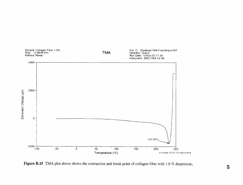
Figure B.15 TMA plot above shows the contraction and break point of collagen fiber with 1.6 % dispersion.
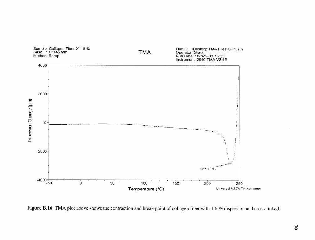
Figure B.16 TMA plot above shows the contraction and break point of collagen fiber with 1.6 % dispersion and cross-linked.
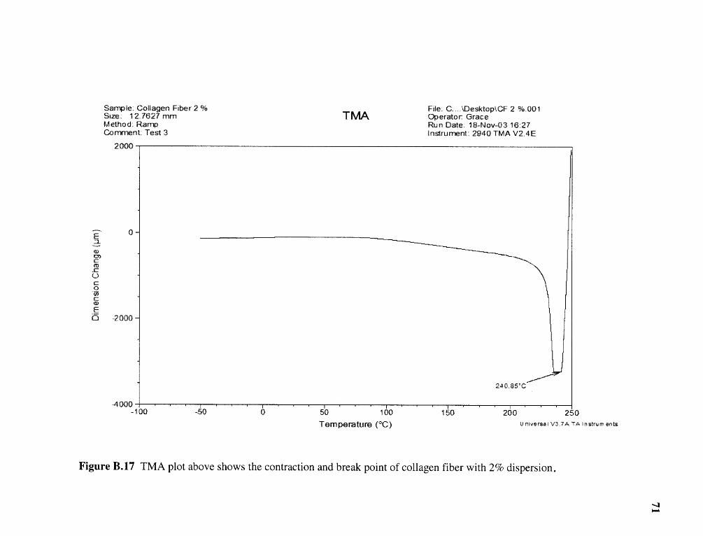
Figure B.17 TMA plot above shows the contraction and break point of collagen fiber with 2% dispersion.

Figure B.18 TMA plot above shows the contraction and break point of collagen fiber with 2% dispersion and cross-linked.

Figure B.19 This plot above shows the slope of the stress strain curve of collagen film with 1% dispersion.

Figure B.20 This plot above shows the slope of the stress strain curve of collagen film with 1.6 % dispersion cross-linked.

Figure B.21 This plot above shows the slope of the stress strain curve of collagen film with 2% dispersion.

Figure B.22 This plot above shows the slope of the stress strain curve of collagen film with 2 % dispersion and cross-linked.

Figure B.23 Collagen fiber with 1.6% dispersion and non cross-linked

Figure B.24 Collagen fiber with 1.6% dispersion and cross links.

Figure B.25 Collagen fiber with 2% dispersion and non cross-linked.

Figure B.26 Collagen fiber with 2% dispersion and cross-linked.

REFERENCES
1. Fofonoff Timothy W. (Dedham, MA); Bell; Eugene (Boston, MA) Apparatusand Method for spinning and processing fiber. US Patent 5,562,946.
2. Steffan; Wolfgang Method of Making of Collagen fibers for surgical use. USPatent 4,404,033.
3. Fredrick H.Silver and David Christiansen. Collagen Scaffolds for TissueRegeneration. UMDNJ-Robert Wood Johnson Medical School, New Jersey.
4. Prockop, D. J, Kivirikko, K. I. (1996). Collagens: Molecular Biology, Diseases,and Potentials for Therapy. Annual Review of Biochemistry, 64, 403-34.
5. Nimni, M.E., Ed (1998). Collagen, vol. III, Biotechnology.
6. Piez, K. A (1985) Collagen. Encyclopedia of Polymer Science and Technology3:699-727.
7. Stenzel, K.H., Miyata, T. and Rubin, A.L. Collagen as a biomaterial, AnnualReviews Inc. Palo Alto, CA, vol3, 231-252.
8. Furukawa; Mitsuru (hyogo, JP); Masahiki (Hyogo, JP); Murata: Shoichi (Hyogo,JP).
9. Sasayama; Atsushi (hyogo, JP) US Patent 5344917. Process for producingregenerated collagen fiber.
10. Stenzel, K. H., Miyata, T., and Rubin, A. L. (1974), Collagen as a biomaterial,Annual Reviews Inc,. Palo Alto, CA, vol. 3, 231-252.
11. Burke, J. F., Yannas, I. V., Quinby, W. C., Jr., Bondoc, C. C., and Jung, W. K.(1981). Successful use of a physiologically acceptable artificial skin in thetreatment of extensive burn injury. Ann. Surg. 194:413-428.
12. Butler, C. E., Compton, C. C., Yannas, I, I. V., and Orgill, D. P. (1995). The effect ofkeratinocyte seeding of collagen-glycosaminoglycan membranes on regenerationskin in a porcine model. 27 th Annual Meeting of the American Burn Association,Albuquerque, NM, April 19-21.
13. Yannas, I. V., Burke, J. F. Orgill, D. P., and Skrabut, E. M. (1982), Wound tissuecan utilize a polymeric template to synthesize a functional extension of skin,Science 215: 174-176.
81

14. Chvapil, M. 1979). Industrial uses of collagen. In Fibrous Proteins: Scientific,Industrial and Medical Aspects, D. A. D. Parry and L. K. Creamer, eds.,Academic Press, London, vol. 1, pp. 247-269.
15. Yannas, I. V., Burke, J. F., Gordon, P. L., and Huang, C., (1977), Multilayermembrane useful as synthetic skin. U. S Patent 4,060.081; Nov 29.
16. Yannas, I.V., Lee, E., Orgill, D. P., Skrabut, E. M., and Murphy, G.F. (G. F.(1989), Synthesis and characterization of model extracellular matrix that inducespartial regeneration of adult mammalian skin, Proc. Natl. Acad. Sci. U.S.A.86:933-937.
17. Yannas, I. V., (1972), Collagen and gelatin in the solid state, Journal of Macro-mol. Sci.-Revs. Macromol. Chem., C7 (1), 49-104.
18. Joseph Nicholas and H. B. Kievens, Collagen Article and the ManufactureThereof. US Patent 2,919,998.
19. Joseph Nicholas and T.L Reissmann, Collagen Article and the ManufactureThereof. US Patent 2,919,999.
20. Lodish, H., Berk, A., Zipursky, S. L., Matsudaira, P., Baltimore, D., & Darnell,J.E. (2000). Molecular Cell Biology. New York: W H Freeman & Co.
21. Prockop, D. J., Kivirikko, K. I (1996). Collagens.
22. Belitz, H. D., 1987. Food Chemistry, Second ed. Springer, Berlin, pp. 426-430.
23. Watenabe, K., Tezuka., Tadahiro, Ishii, 1997. Configuration between re-formedcollagen triple helices and helices and artificially introduced cross-links in gelatingels. Macromolecules 30, 7910-7913.
24. Mackie, A. R., Gunning, A.P., Ridout,M.J.,1998. Gelation of gelatin, observationin the bulk and at the air-water interface. Bioploymers 46, 245-252.
25. Chabala, J. M., Levi-Setti, R., Maternaghan, T.J.,1994. Photographic gelatin layermicrostructures revealed by imaging secondary ion mass spectrometry. J. imagingSci. 39 (3),222-232.
26. Reich,G., 1996. Einflusz der sorbitol-spezifikation and eigenschaftenvonweichgelatinekapselen. Pharm,Ind. 58 (10), 941-946.
27. Flory, P.J., Garrett,R.R., 1958. Phase transition in collagen and gelatin systems. J.Am. Chem. Soc. 80, 4836-4845.
28. Ming-Thau Sheu, Ju-Chun Huang et al., 2000. Characterization of collagen gel
82

solutions and collagen matrices for cell cultures. Biomaterials 22(2001) 1713-1719.
29. V.Samouillan, J. Dandurand et al., 2002. Comparison of chemical treatments onthe chain dynamics and thermal stability of bovine pericardium collagen.
83