coronary ct angiographycirculation.or.kr/workshop/2004fall/file/coronarycta-2004... ·...
TRANSCRIPT
Coronary CT Angiography
Yeon Hyeon Choe, MD
Department of Radiology, Samsung Medical Center,
Sungkyunkwan University

Applications of MDCT: Ischemic Heart Diseases
• Coronary arterial morphology (angiographic display)
• Coronary plaque characterization• Postop. Evaluation (CABG)• Stent patency• Coronary calcium score• Cardiac perfusion• Cardiac function

Techniques of 16-slice CT: Coronary Studies
• Calcium scoring: no contrast, 1.2 mm scan• Postcontrast studies
– Nonionic CM 80-120 ml, 4 ml/s– Delayed scan at 10 min for myocardial imaging
• Heart rate control by preCT medication (beta-blocker) in pt with HR > 60 bpm (metoprolol 50-100 mg p.o., 1 h before)
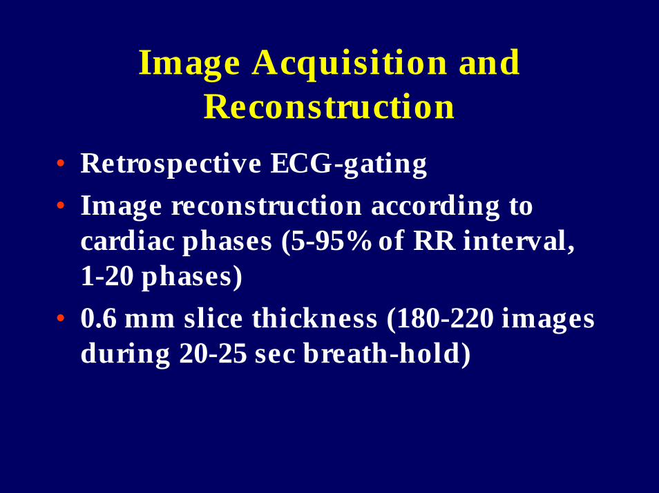
Image Acquisition and Reconstruction
• Retrospective ECG-gating• Image reconstruction according to
cardiac phases (5-95% of RR interval, 1-20 phases)
• 0.6 mm slice thickness (180-220 images during 20-25 sec breath-hold)

3D Techniques andImage Analysis
• Volume rendering • Maximum intensity projection (MIP)
– Vessel analysis– Sub-volume MIP
• Reformation in cardiac short and long axes• Endoscopic view (fly-through)
• Source images
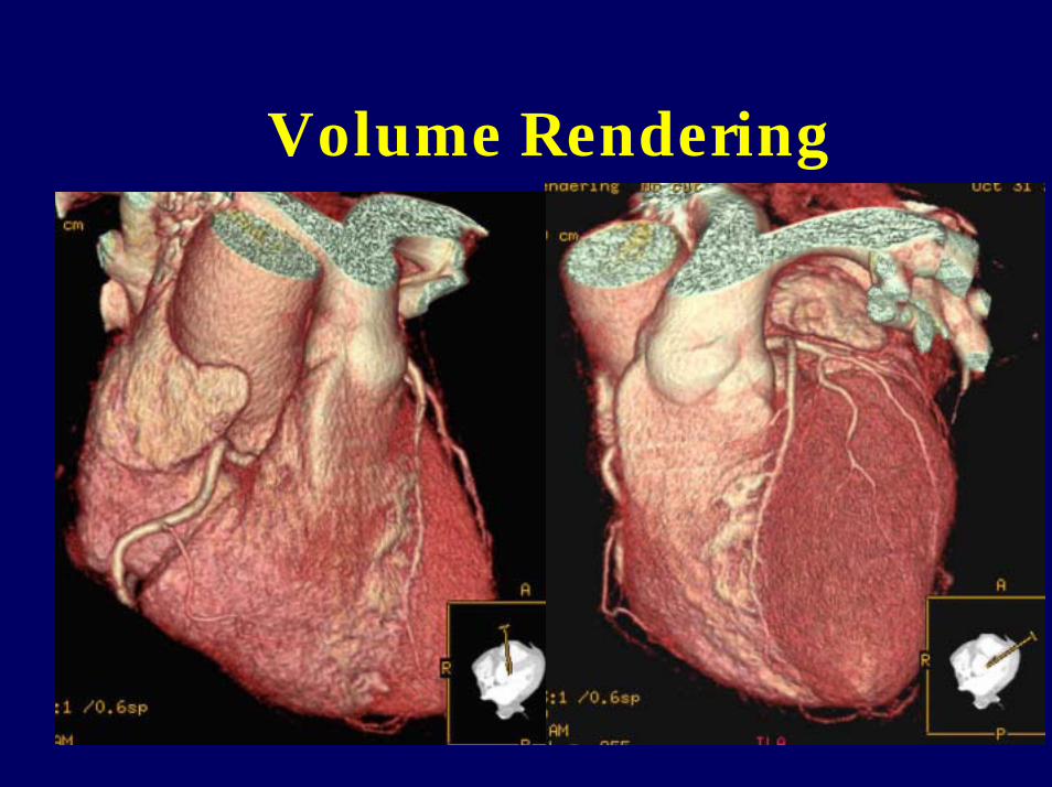
Volume Rendering
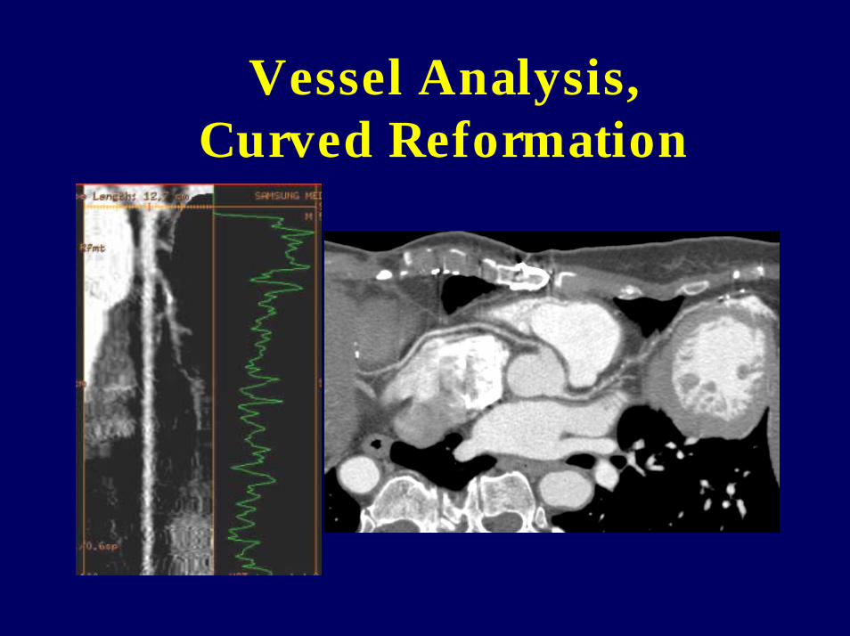
Vessel Analysis, Curved Reformation

Navigation
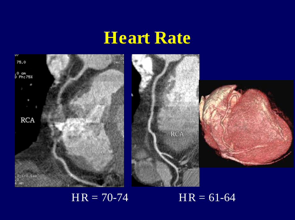
Heart Rate
HR = 70-74 HR = 61-64
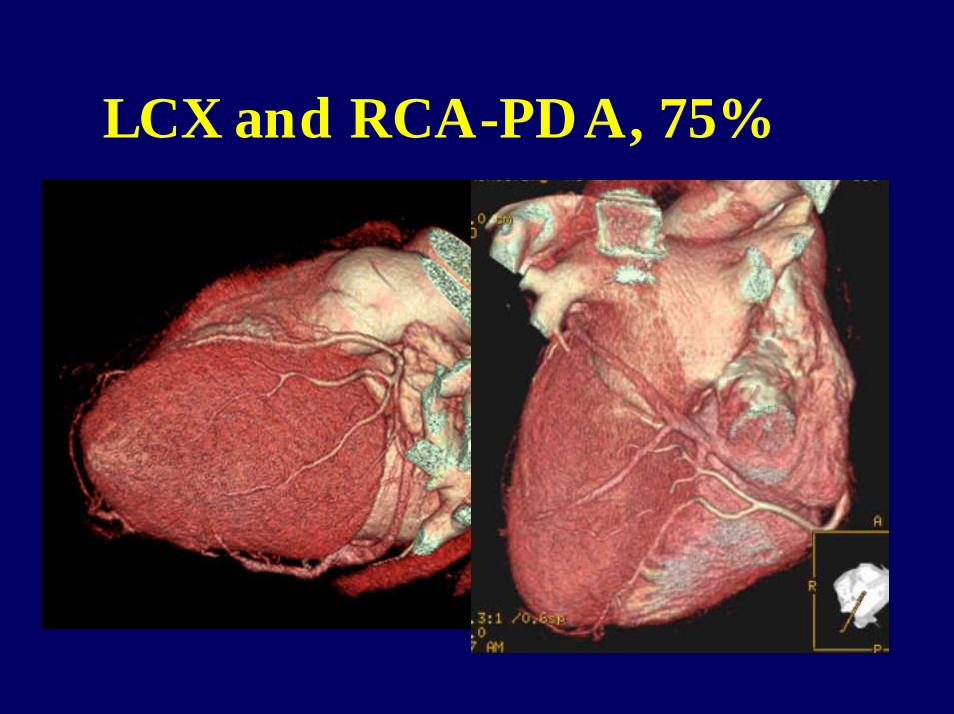
LCX and RCA-PDA, 75%


Stent: Patent vs Occluded
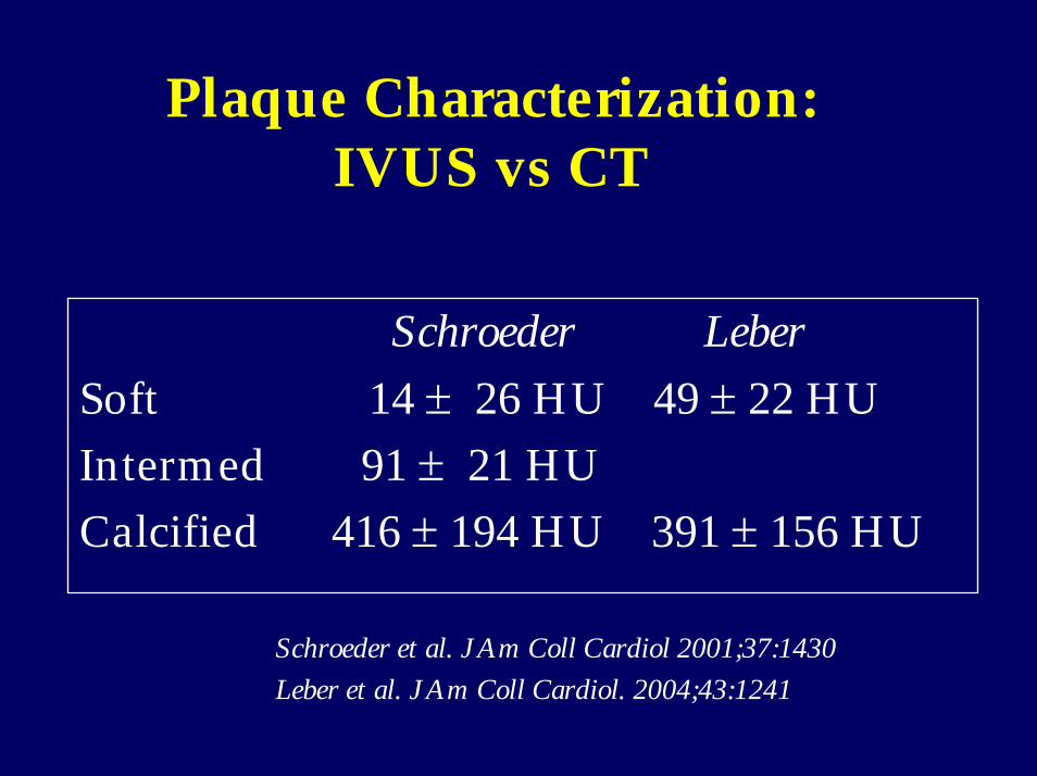
Plaque Characterization: IVUS vs CT
Schroeder LeberSoft 14 ± 26 HU 49 ± 22 HUIntermed 91 ± 21 HUCalcified 416 ± 194 HU 391 ± 156 HU
Schroeder et al. J Am Coll Cardiol 2001;37:1430Leber et al. J Am Coll Cardiol. 2004;43:1241

Fatty Plaque and Stent
LeeYS

16-CT vs IVUS (n = 22)
Noncalcified Plaque
Calcified PlaqueAchenbach et al. Circulation 2004;109:14-17
Sens, 78%;Spec, 87%.
Sens, 94%;Spec, 94%.
For Exclusively Noncalcified Plaque: Sens = 53% (8/15).
Underestimation of plaque volumeby MDCT (24 vs 43 mm3, p <.001)
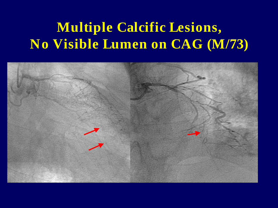
Multiple Calcific Lesions,No Visible Lumen on CAG (M/73)
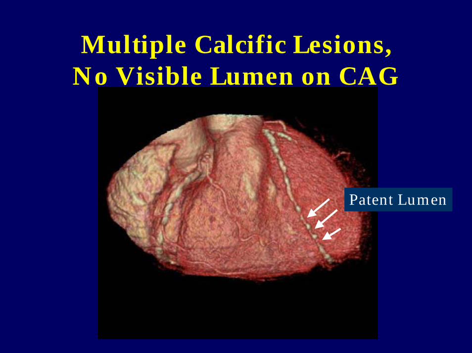
Multiple Calcific Lesions,No Visible Lumen on CAG
Patent Lumen
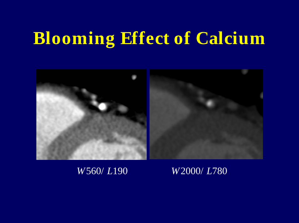
Blooming Effect of Calcium
W560/L190 W2000/L780
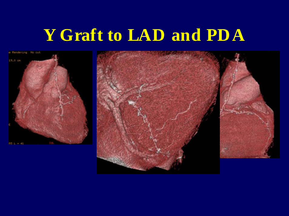
Y Graft to LAD and PDA
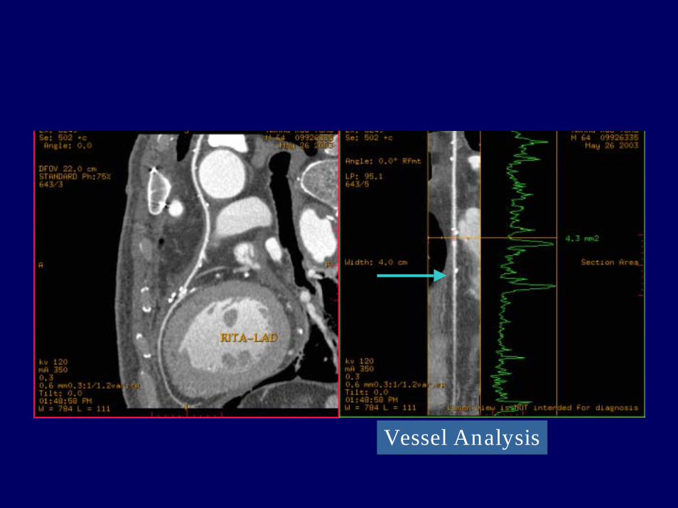
Vessel Analysis

Coronary Calcium Scoring
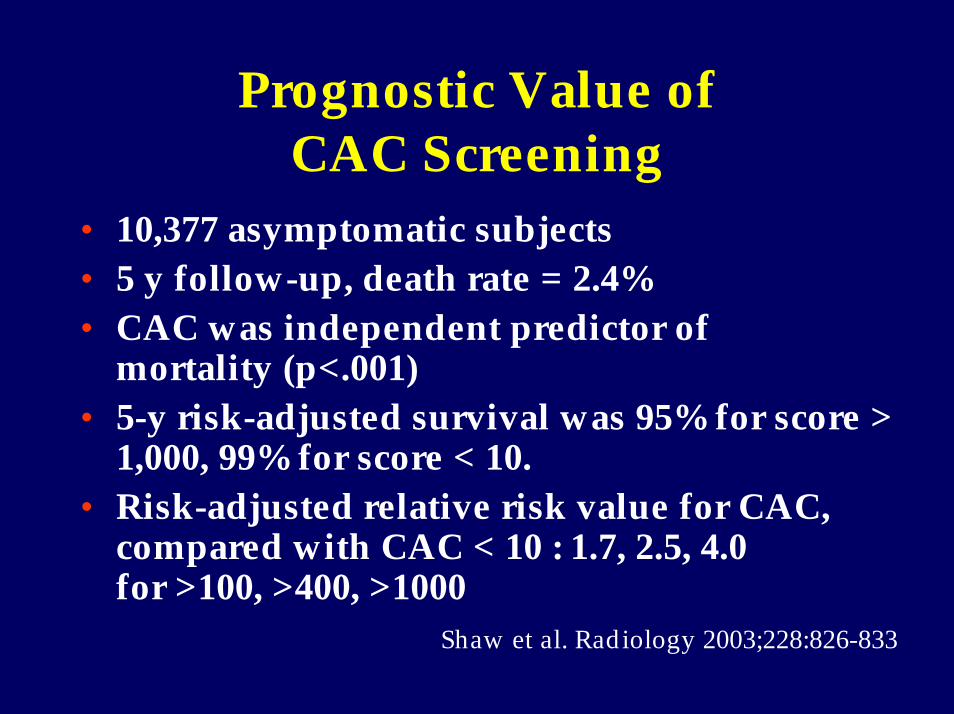
Prognostic Value of CAC Screening
• 10,377 asymptomatic subjects • 5 y follow-up, death rate = 2.4%• CAC was independent predictor of
mortality (p<.001)• 5-y risk-adjusted survival was 95% for score >
1,000, 99% for score < 10.• Risk-adjusted relative risk value for CAC,
compared with CAC < 10 : 1.7, 2.5, 4.0 for >100, >400, >1000
Shaw et al. Radiology 2003;228:826-833

RCA from Lt Coronary Sinus
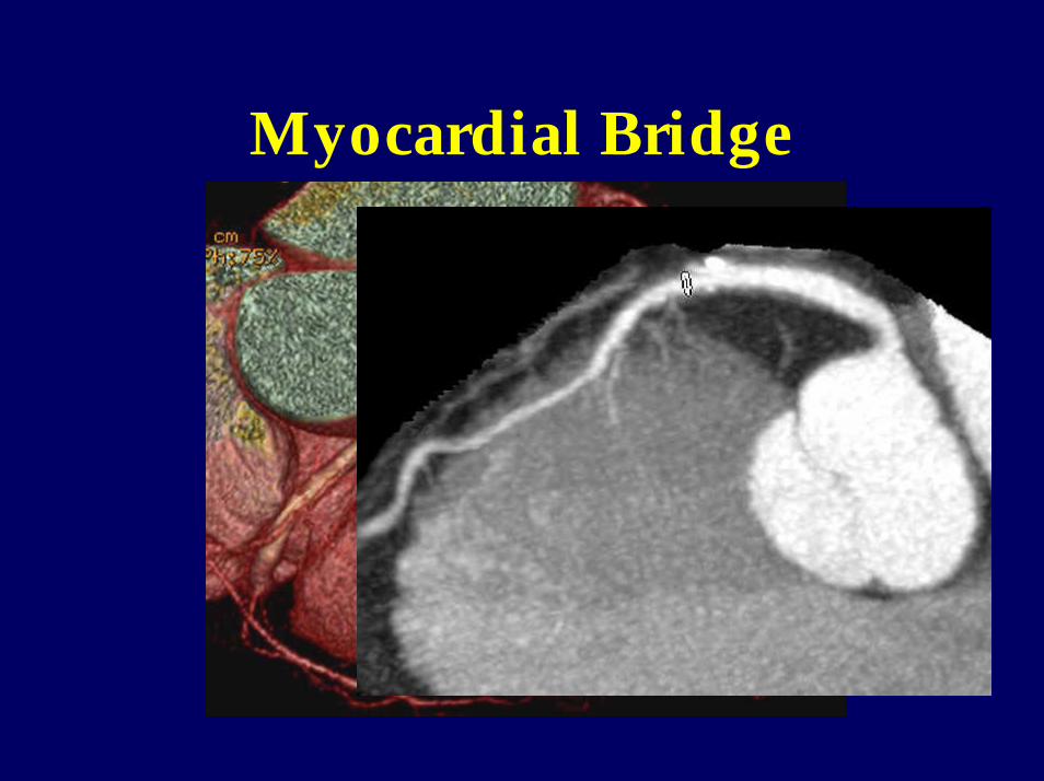
Myocardial Bridge

Kawasaki Disease (M/13y)
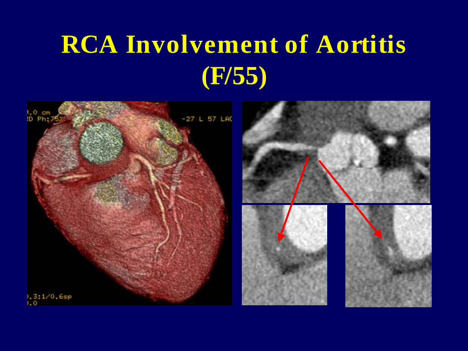
RCA Involvement of Aortitis(F/55)

Collateral Circulation
Vieussen’s Ring: Conal to Conal Collateral
Shin SD, M/73

Patients with Chest PainAtypical
Stress Tests MDCTAcute
Coronary Syndrome
Typical
CAG/InterventionQuitQuit
Stenosis: NoStenosis: Yes

Patient with Acute Chest Pain at ER
• ECG-gated CTA covering whole chest for exclusion of
– Aortic dissection– Pulmonary embolism– Coronary artery disease– Myocardial infarction

MDCT Evaluation of Myocardial Perfusion and
Viability
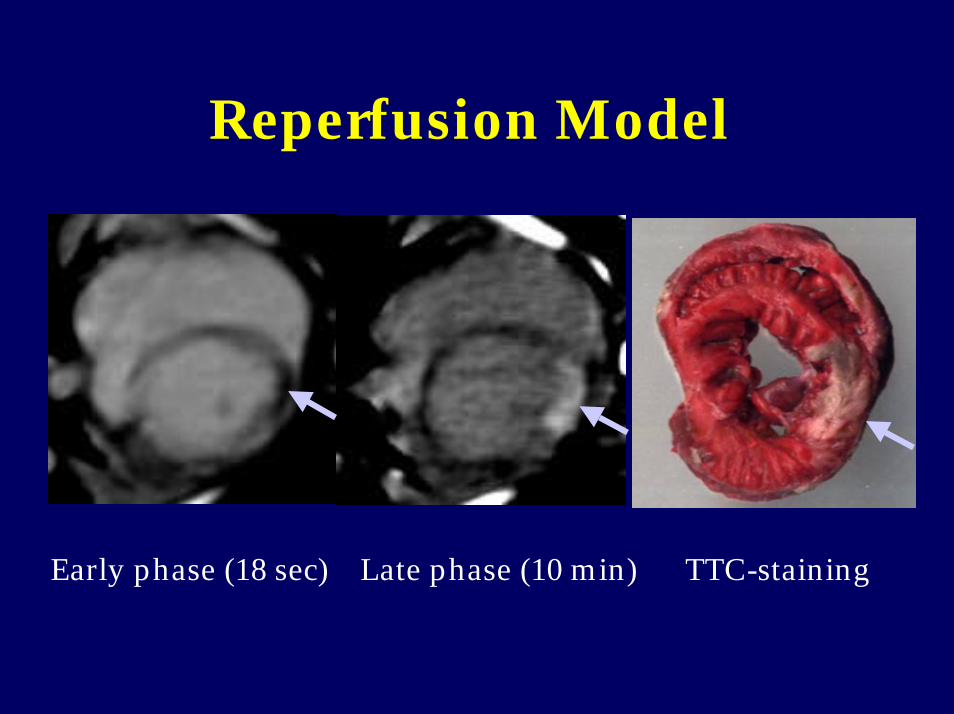
Reperfusion Model
Early phase (18 sec) Late phase (10 min) TTC-staining

Time Course: CT vs MRI in Occlusion Model
Early 5 min 10 min 15 min 23 min

Human Study
• 55 patients underwent MDCT and MRI in the acute stage (within 2 weeks, n = 34) and/or chronic stage (1-36 months, n = 24) of MI

MR Imaging and MDCT• First-pass and 5-min and 15-min delayed
myocardial MR imaging was performed using an 1.5 T scanner (GE Signa CVi) with injection of 0.15 mmol/kg Gd-DTPA
• Within 24 hours after MR imaging, ECG-gated MDCT was performed using GE 4-slice (n = 12) or 16-slice (n = 46) scanner at 25-sec and 10-min delay with injection of 120 ml nonionic contrast at 4 ml/s.

Results
• MDCT showed areas of MI on early phase images in all cases except one (4.1 %) and in all cases on late phase images.
• Myocardial abnormalities on CT correlated well (94.9%) with those on MRI except 3 exam sets without visible lesions on early CT (n = 1) or perfusion MRI (n = 2) in terms of location and depth of the lesions.
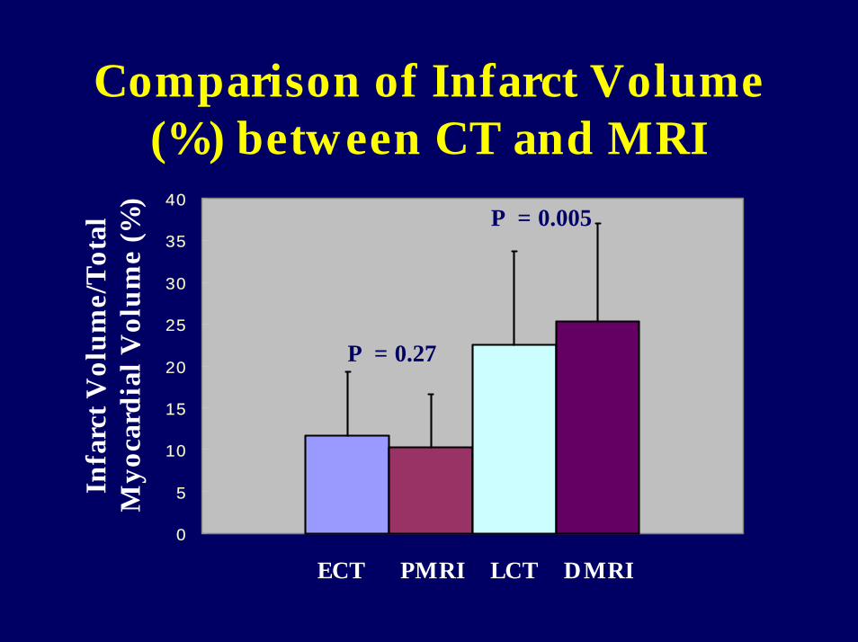
Comparison of Infarct Volume (%) between CT and MRI
0
5
10
15
20
25
30
35
40
Infa
rct V
olum
e/To
tal
Myo
card
ial V
olum
e (%
)
ECT LCTPMRI DMRI
P = 0.27
P = 0.005

Correlation of Infarct Volume (%) between CT and MRI
0
10
20
30
40
50
60
0 10 20 30 40 50 60 70
MRI
MD
CT
ECT vs PMRI
LCT vs DMRI
r = 0.59r = 0.81

AMI, Persistent Perfusion Defect on Late Phase, CT = MRI
CT MRI
Early
Late
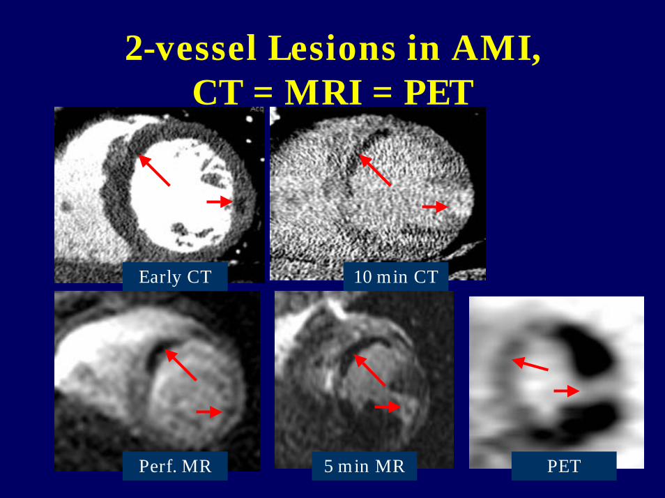
2-vessel Lesions in AMI, CT = MRI = PET
Early CT
5 min MRPerf. MR
10 min CT
PET
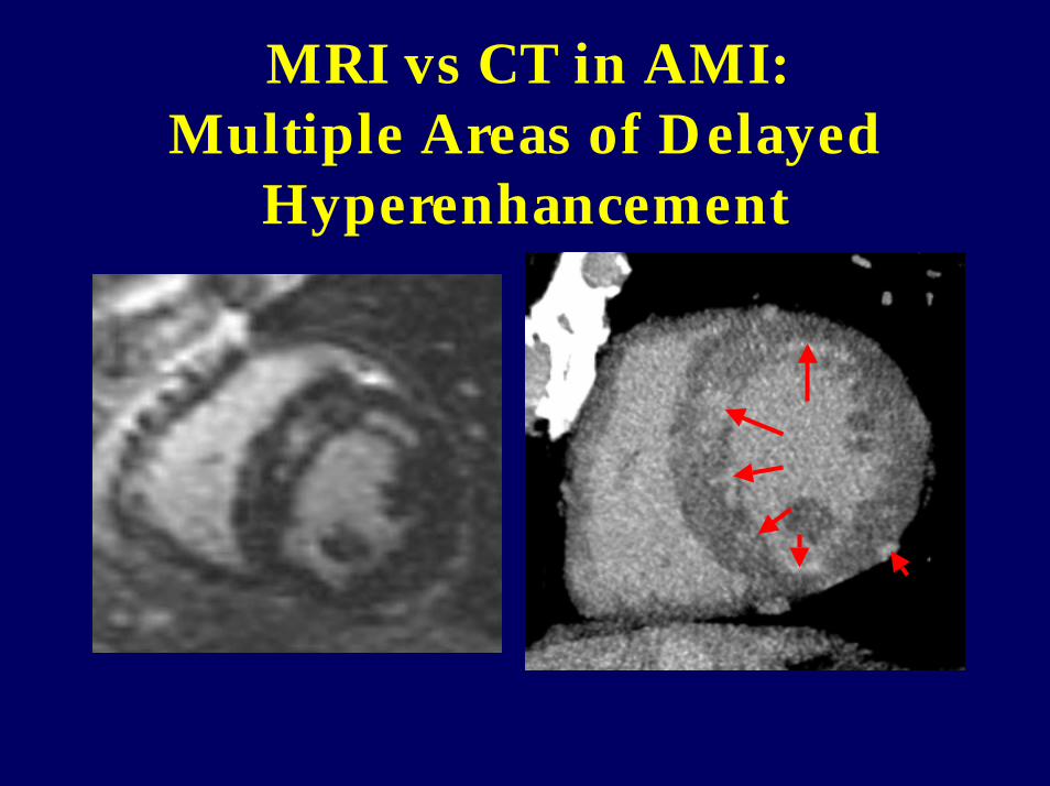
MRI vs CT in AMI: Multiple Areas of Delayed
Hyperenhancement

Color-coded Images with LV Function Assessment in AMI

Stenting Complication

MDCT Assessment of Coronary Artery Stenosis (> 50%)
977993928816Ropers(n = 77)
9780869510016Nieman(n = 58)
98597685684Achenbach(n = 64)
97819781944Knez(n = 44)
NPVPPVSpecSensAssess(%)
Slice(DR)

Limitations of Coronary CTA
• Extensive calcifications• Stents: spatial resolution• Heart rate: temporal resolution • Radiation (3.5-5.9 mSv)• Small branches/septal branches

Cardiac Function Analysis
• Volume ejection fraction• Wall motion analysis• Wall thickness and thickening

New Developments
• 40-slice and 64-slice CT (rotation ~0.33 s, 4-5 s scan)
• Volume CT (flat panel CT)

Summary
• MDCT allows reliable coronary angiography.
• One-stop shop imaging of ischemic heart disease is feasible using MDCT.
Conclusion
• MDCT can exclude or prove significant coronary stenosis.
• Only MDCT can detect early coronary atheroscleosis noninvasively and help start preventive medication in time.

Thank You