cortical ependymoma. case report
TRANSCRIPT
J Bras Neurocirur 29 (4): 667-670, 2018Daher D, Brito A, Diaz D - Cortical ependymoma. Case report
Case Report
Ependymomas are rare central nervous system tumors 1 that arise from differentiated ependymal cells located in the ventricular system, choroid plexus2, and along the central canal of the spinal cord3,4. Representing 2% to 9% of all neuroepithelial tumors, localizations outside the ventricular
system are uncommon, and affect pediatric and adult patients equally 5. Few cases of pure Cortical Ependymomas (CEs) have been reported in the literature 3-8. CEs usually manifest with a size larger than 4cm 6, and symptoms are relatively mild until a later stage 4. Patients tend to present focal neurologic deficits 1, and most frequently seizures. We present a case of a 47-year-old female with a frontoparietal cortical ependymoma manifested as new-onset seizure and left hemiparesis.
Cortical ependymoma. Case report Evolución y Tasa de Recuperación en el Tratamiento Ependimoma Cortical. Relato de caso
Antonio Daher1
Adriana Brito2
Daniela Diaz3
ABSTRACT Ependymomas are rare central nervous system tumors derived from ependymal cells, located in the ventricular system, choroid plexus e central canal of the spinal cord. Ependymomas could appear throughout the craniospinal axis, even outside the ventricular system, although, pure cortical ependymoma are very rare, and only a few cases have been reported in the literature. We hereby present the case of a 46 years-old female, admitted to the hospital, presenting a new-onset seizure. Magnetic Resonance Imaging (MRI) showed a large bilateral frontoparietal tumor, associated to important perilesional edema. Radical resection of the tumor was performed, and the biopsy reported anaplastic ependymoma. Patient underwent complementary radiotherapy.
Keywords: Ependymoma; Cortical ependymoma; Brain tumor
RESUMOEpendimomas são tumores raros do sistema nervoso central derivados de células ependimárias localizadas no sistema ventricular, plexo coroide e canal central da medula espinhal. Os ependimomas podem surgir ao longo do eixo cranioespinhal, mesmo fora do sistema ventricular; entretanto, os casos de ependimomas puramente corticais, são muito raros, com poucos casos reportados na literatura. Apresentamos o caso de uma paciente feminina, 47 anos, admitida no hospital por crise convulsiva. Ressonância magnética (RM) demonstrou volumoso tumor frontoparietal com importante edema perilesional. A ressecção radical do tumor foi realizada, biopsia reportou ependimoma anaplásico e o tratamento foi complementado com radioterapia.
Palavras-chave: Ependimoma; Ependimoma cortical; Tumor cerebral
1 MD, Neurosurgeon, Adjunct Professor of Neurosurgery, Centro Médico Guerra Mendez, Hospital Oncológico Miguel Perez Carreño, Valencia, Venezuela. 2 MD, MR, Medical Residency in Neurosurgery, La Santa Casa Hospital, Centro Médico Guerra Mendez, Hospital Oncológico Miguel Perez Carreño, Valencia, Venezuela.3 MD, Neurosurgeon, Centro Médico Guerra Mendez, Hospital Oncológico Miguel Perez Carreño, Valencia Central Hospital, Valencia, Venezuela.
Received Dec 18, 2019Corrected Dec 19,2019Accepted Dec 20, 2019
INTRODUCTION
J Bras Neurocirur 29 (4): 667-670, 2018Daher D, Brito A, Diaz D - Cortical ependymoma. Case report
Case Report
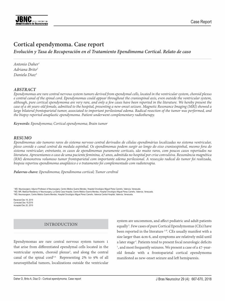
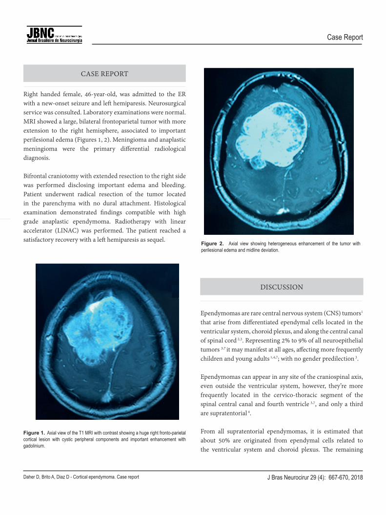
Right handed female, 46-year-old, was admitted to the ER with a new-onset seizure and left hemiparesis. Neurosurgical service was consulted. Laboratory examinations were normal. MRI showed a large, bilateral frontoparietal tumor with more extension to the right hemisphere, associated to important perilesional edema (Figures 1, 2). Meningioma and anaplastic meningioma were the primary differential radiological diagnosis.
Bifrontal craniotomy with extended resection to the right side was performed disclosing important edema and bleeding. Patient underwent radical resection of the tumor located in the parenchyma with no dural attachment. Histological examination demonstrated findings compatible with high grade anaplastic ependymoma. Radiotherapy with linear accelerator (LINAC) was performed. The patient reached a satisfactory recovery with a left hemiparesis as sequel.
Ependymomas are rare central nervous system (CNS) tumors1 that arise from differentiated ependymal cells located in the ventricular system, choroid plexus, and along the central canal of spinal cord 2,3. Representing 2% to 9% of all neuroepithelial tumors 3,7 it may manifest at all ages, affecting more frequently children and young adults 1,4,7; with no gender predilection 3.
Ependymomas can appear in any site of the craniospinal axis, even outside the ventricular system, however, they’re more frequently located in the cervico-thoracic segment of the spinal central canal and fourth ventricle 3,7, and only a third are supratentorial 4.
From all supratentorial ependymomas, it is estimated that about 50% are originated from ependymal cells related to the ventricular system and choroid plexus. The remaining
CASE REPORT
DISCUSSION
Figure 1. Axial view of the T1 MRI with contrast showing a huge right fronto-parietal cortical lesion with cystic peripheral components and important enhancement with gadolinium.
Figure 2. Axial view showing heterogeneous enhancement of the tumor with perilesional edema and midline deviation.
Figure 1. Axial view of the T1 MRI with contrast showing a huge right fronto-parietal
cortical lesion with cystic peripheral components and important enhancement with
gadolinium.
9
Figure 2. Axial view showing heterogeneous enhancement of the tumor with
perilesional edema and midline deviation.
10
J Bras Neurocirur 29 (4): 667-670, 2018Daher D, Brito A, Diaz D - Cortical ependymoma. Case report
Case Report
is distributed throughout brain parenchyma 4. Localization outside the ventricular system is uncommon, and affects pediatric and adult patients equally 9. Few cases of pure Cortical Ependymomas (CEs) have been reported in the literature 3-8, which are located in the cortical ribbon with no identified connection to the ventricular lining 3. In the series of cases presented at the Mayo Clinic in 2011, the most common localization was the frontal lobe, followed by parietal and occipital lobes. A very low frequency of CEs in the temporal lobe has been observed 7.
CEs usually are manifested with a size larger than 4cm 6, and symptoms are relatively mild until a later stage 4. Patients tend to present focal neurologic deficits 1 and most frequently seizures, regardless of their rare localization in the temporal lobe 3,4,7 . CEs have also been reported as incidental during workup for headaches 7.
Even though there is no clear explanation for the histogenesis of CEs, it has been proposed that supratentorial ependymomas could be originated from rests of ependymal cells located in the angle of the ventricle, deep into the adjacent parenchyma5. On the other hand, due to the wide distribution of ependymomas without restriction to the mentioned area, this mechanism seemed doubtful. Progenitor cells hypothesis has been proposed as an explanation of cortical ependymomas pathogenesis 3. In 2005, Hegyi et al. based on the pathologic features of an ectopic retinal ependymoma suggested that the origin of this neoplasm could be explained for Muller cells (with progenitor properties)9 .
CEs had shown a wide spectrum of morphologies, and could be difficult to distinguish from some infiltrative gliomas7, typically showing the characteristic perivascular pseudo-rosettes, and in some cases true ependymal rosettes3,4,7. Less classic features could be found, as tanycitic, epithelial and clear cells, and even rarely spindle cell component and schwannian-like nodules3,7, important for differential diagnosis with Angiocentric Glioma.
According to the World Health Organization (WHO) classification, for CNS tumors, ependymomas have
been traditionally divided into low grade (Grade II), and high grade (Grade III) lesions, reserving Grade I for myxopapillary ependymoma7. A higher proportion of supratentorial ependymomas are high grade when compared with infratentorial ependymomas8. Nevertheless, cortical ependymomas are most commonly low grade with a relatively better clinical course 7 .
Recently in 2016, the WHO CNS tumor was updated, including now molecular parameters in addition to the histological findings, to define many tumor entities10. In this update, a genetically defined ependymoma subtype has been accepted: Ependymoma, RELA fusion–positive, responsible for the majority of supratentorial tumors in children. It is expected that continuing studies of the molecular features will provide a more objective classification to this tumors and, therefore, a major clinical significance to the histological classification 10 .
Magnetic Resonance Imaging (MRI) is the primary modality, for assessment of ependymoma 11 . Intracranial ependymomas conventionally demonstrate hypo-intensity on T1, hyper-intensity on T2, and intermediate-to-high FLAIR signal intensity, relative to gray and white matter 4,7,11. Supratentorial ependymomas tend to demonstrate more heterogeneous T1 and T2 due to a greater tendency toward cyst formation, calcifications and hemorrhage, when compared with infratentorial ependymomas. Commonly, on post gadolinium T1-weighted images, there is avid enhancement of the soft tissue within the tumor, intermixed with poorly or none enhancing areas 11 .
Radical surgical resection is considered to be the best treatment, in light of easier approach for CEs, than intraventricular tumors. Postoperative radiotherapy is used for anaplastic ependymoma, and partially resected tumor 3,4,7, due to the possibility of recurrence, and 30 to 40% chances of leptomeningeal dissemination at recurrence 4,7 . Chemotherapy and prophylactic craniospinal irradiation are not indicated 4 .
CEs appear to have a relatively favorable prognosis when compared to other supratentorial ependymomas 3,4,7.
J Bras Neurocirur 29 (4): 667-670, 2018Daher D, Brito A, Diaz D - Cortical ependymoma. Case report
Case Report
According to Metellus et al. the 5 year survival rate is 57.1% +/- 8.7%; although, histopathological type, localization, extent of resection, age at diagnosis and gender must be considered 3.
1. Koeller KK, Sandberg GD; Armed Forces Institute of Pathology. From the archives of the AFIP. Cerebral intraventricular neoplasms: radiologic-pathologic correlation. Radiographics. 2002;22(6):1473-1505. doi: 10.1148/rg.226025118.
2. Goutelle A, Daher A, Perrin G. Èpendymomes intracrâniens. In Cohadon F. Tumeurs du système nerveux et de ses enveloppes. Paris: Flammarion médecine-sciences; 1989. p. 313-325.
3. Bijwe S, Ansari S, Jadhav V, Palande D. Pure cortical ependymoma: A rare entity. Asian J Neurosurg. 2015;10(2):162-165. doi: 10.4103/1793-5482.152111.
4. Shuangshoti S, Rushing EJ, Mena H, Olsen C, Sandberg GD. Supratentorial extraventricular ependymal neoplasms: a clinicopathologic study of 32 patients. Cancer. 2005;103(12):2598-605. doi: 10.1002/cncr.21111.
5. Van Gompel JJ, Koeller KK, Meyer FB, Marsh WR, Burger PC, Roncaroli F, et al. Cortical ependymoma: an unusual epileptogenic lesion. J Neurosurg. 2011;114(4):1187-1194. doi: 10.3171/2010.12.JNS10846
6. Hegyi L, Peston D, Theodorou M, Moss J, Olver J, Roncaroli F. Primary glial tumor of the retina with features of myxopapillary ependymoma. Am J Surg Pathol. 2005;29(10):1404-1410. doi: 10.1097/01.pas.0000172188.02424.d8.
7. Mohaghegh MR, Chitsaz A, Okhovat AA, Pour EB. Supratentorial cortical ependymoma: An unusual presentation of a rare tumor. Adv Biomed Res. 2015;4:72. doi: 10.4103/2277-9175.153896.
8. Tailor J, Jaunmuktane Z, Brandner S, Sethi H. Supratentorial ependymoma presenting as a cortical cyst with a mural nodule in an adult. J Surg Case Rep. 2015;2015(1):rju124. doi: 10.1093/jscr/rju124.
9. Louis DN, Perry A, Reifenberger G, von Deimling A, Figarella-Branger D, Cavenee WK, et al. The 2016 World Health Organization Classification of Tumors of the Central Nervous System: a summary. Acta Neuropathol. 2016;131(6):803-820. doi: 10.1007/s00401-016-1545-1.
10. Roncaroli F, Consales A, Fioravanti A, Cenacchi G. Supratentorial cortical ependymoma: report of three cases. Neurosurgery. 2005;57(1):E192. doi: 10.1227/01.neu.0000164171.29292.d6.
11. Yuh EL, Barkovich AJ, Gupta N. Imaging of ependymomas: MRI and CT. Childs Nerv Syst. 2009;25(10):1203-13. doi: 10.1007/s00381-009-0878-7.
Antonio Daher, MD, PhD NeurosurgeonAdjunct Professor of NeurosurgeryCentro Médico Guerra MendezHospital Oncológico Miguel Perez CarreñoValencia, Venezuela E-mail: [email protected]
CORRESPONDING AUTHOR
REFERENCES