cortical spreading depression in migraine-time to … · cortical spreading depression in...
TRANSCRIPT
714
DOI: 10.1590/0004-282X20150094
ARTICLEVIEW AND REVIEW
Cortical spreading depression in migraine-time to reconsider?Depressão alastrante cortical na enxaqueca – hora de reconsiderar?Alan J McComas, Adrian R M Upton
Three different mechanisms - vascular, trigeminovascu-lar and cortical hyperexcitability - have been proposed for the pathogenesis of migraine and have recently been the subject of extensive reviews1,2,3,4. In our own assessment5 we present-ed the results of therapeutic transcranial magnetic stimula-tion (tTMS) as evidence that, in at least some patients, vir-tually all migraine symptoms during the headache phase are cortical in origin and that a neuroinflammatory mechanism could not be involved in the initiation and maintenance of the headache; instead, the evidence clearly pointed to corti-cal hyperexcitability6. We now use the results of tTMS once more, this time as part of an argument against one of the cen-tral tenets of migraine pathophysiology, namely, that cortical spreading depression (CSD) is responsible for the aura.
CORTICAL SPREADING DEPRESSION (CSD)
In 1944 Aristides Leão reported the ability to produce tem-porary suppression of electrical activity in the rabbit cortex by any one of several manoeuvres7. The latter included intense electrical stimulation of the brain surface, the application of
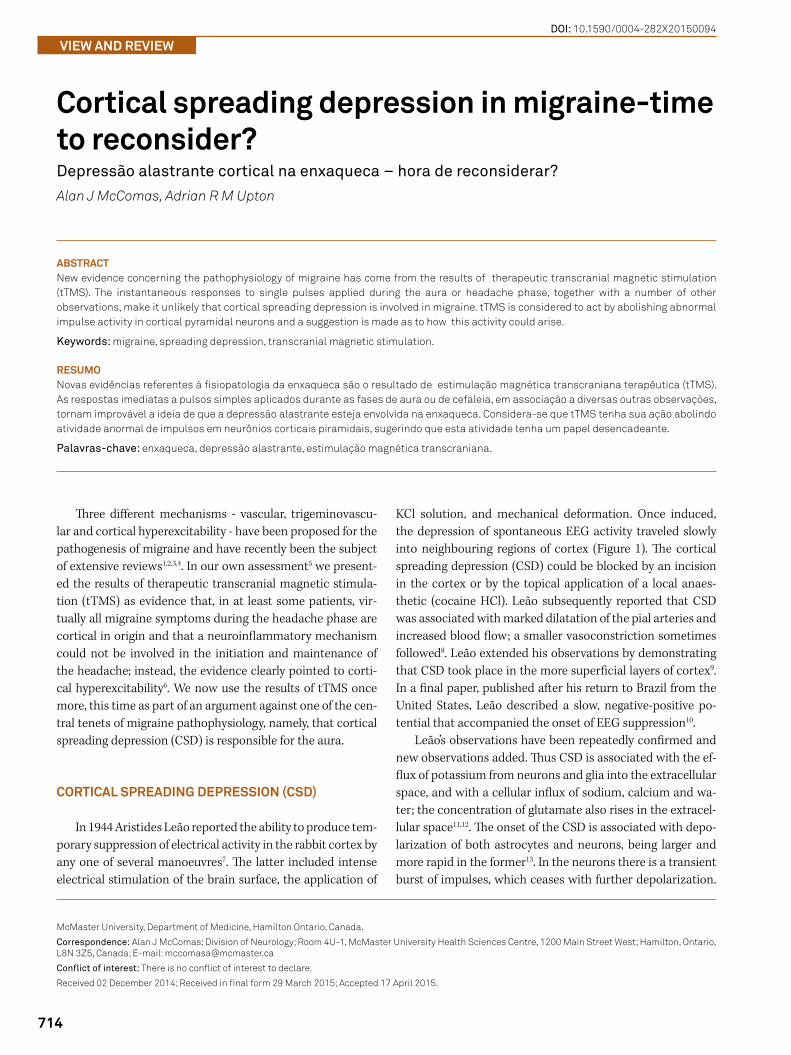
KCl solution, and mechanical deformation. Once induced, the depression of spontaneous EEG activity traveled slowly into neighbouring regions of cortex (Figure 1). The cortical spreading depression (CSD) could be blocked by an incision in the cortex or by the topical application of a local anaes-thetic (cocaine HCl). Leão subsequently reported that CSD was associated with marked dilatation of the pial arteries and increased blood flow; a smaller vasoconstriction sometimes followed8. Leão extended his observations by demonstrating that CSD took place in the more superficial layers of cortex9. In a final paper, published after his return to Brazil from the United States, Leão described a slow, negative-positive po-tential that accompanied the onset of EEG suppression10.
Leão’s observations have been repeatedly confirmed and new observations added. Thus CSD is associated with the ef-flux of potassium from neurons and glia into the extracellular space, and with a cellular influx of sodium, calcium and wa-ter; the concentration of glutamate also rises in the extracel-lular space11,12. The onset of the CSD is associated with depo-larization of both astrocytes and neurons, being larger and more rapid in the former13. In the neurons there is a transient burst of impulses, which ceases with further depolarization.
McMaster University, Department of Medicine, Hamilton Ontario, Canada.
Correspondence: Alan J McComas; Division of Neurology; Room 4U-1, McMaster University Health Sciences Centre, 1200 Main Street West; Hamilton, Ontario, L8N 3Z5, Canada; E-mail: [email protected]
Conflict of interest: There is no conflict of interest to declare.
Received 02 December 2014; Received in final form 29 March 2015; Accepted 17 April 2015.
ABSTRACTNew evidence concerning the pathophysiology of migraine has come from the results of therapeutic transcranial magnetic stimulation (tTMS). The instantaneous responses to single pulses applied during the aura or headache phase, together with a number of other observations, make it unlikely that cortical spreading depression is involved in migraine. tTMS is considered to act by abolishing abnormal impulse activity in cortical pyramidal neurons and a suggestion is made as to how this activity could arise.
Keywords: migraine, spreading depression, transcranial magnetic stimulation.
RESUMONovas evidências referentes à fisiopatologia da enxaqueca são o resultado de estimulação magnética transcraniana terapêutica (tTMS). As respostas imediatas a pulsos simples aplicados durante as fases de aura ou de cefaleia, em associação a diversas outras observações, tornam improvável a ideia de que a depressão alastrante esteja envolvida na enxaqueca. Considera-se que tTMS tenha sua ação abolindo atividade anormal de impulsos em neurônios corticais piramidais, sugerindo que esta atividade tenha um papel desencadeante.
Palavras-chave: enxaqueca, depressão alastrante, estimulação magnética transcraniana.
715Alan J McComas et al. Spreading depression in migraine
It is thought that CSD propagates through the dendritic net-work and/or through gap junctions between astrocytes.
HISTORICAL LINKAGE OF CSD TO MIGRAINE
Leão himself speculated that CSD might be linked in some way to migraine, in part because of its slow passage across the cortex; he was, however, thinking in terms of the dilatation of the pial arteries rather than the change in elec-trical activity. A few years prior to Leão’s first publication, the experimental psychologist, Karl Lashley, gave detailed and lucid descriptions of his own visual auras; the scintillations were interpreted as the result of intense excitation in the vi-sual cortex, while the enlarging scotomas were attributed to inhibition14. Taking the length of the striate cortex to be 67 mm, and allowing 20 minutes for the visual phenomena to reach the margin of the temporal visual field from their central origin, Lashley calculated that the underlying corti-cal process traveled rostrally at approximately 3 mm/minute from the occipital pole. The possible link between CSD and the migraine aura was not pointed out until 1958, in the form of a brief note by Milner15. In the following year Hubel and Wiesel published the results of a microelectrode study of the cat visual cortex, reporting the presence of orientation sensi-tive cells in the primary visual area16. The likelihood that exci-tation of these cells was responsible for the flickering zigzag lines of a visual aura should have been immediately obvious, but instead had to wait 12 years before appearing in print17.
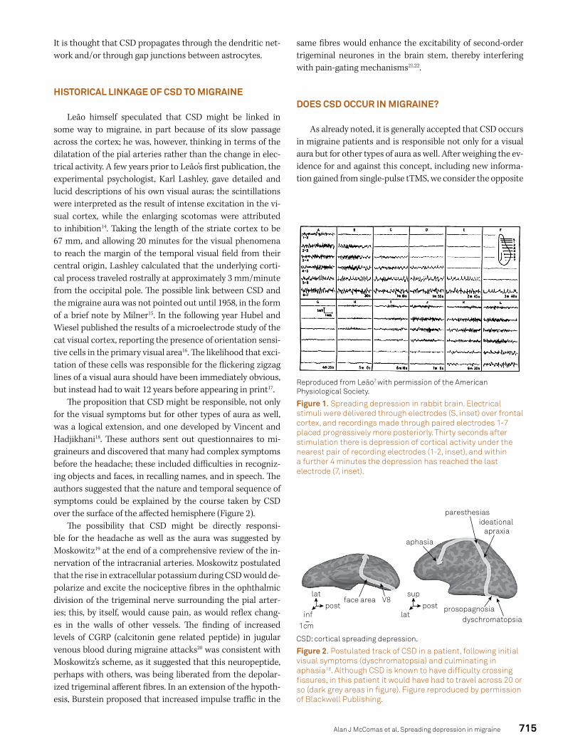
The proposition that CSD might be responsible, not only for the visual symptoms but for other types of aura as well, was a logical extension, and one developed by Vincent and Hadjikhani18. These authors sent out questionnaires to mi-graineurs and discovered that many had complex symptoms before the headache; these included difficulties in recogniz-ing objects and faces, in recalling names, and in speech. The authors suggested that the nature and temporal sequence of symptoms could be explained by the course taken by CSD over the surface of the affected hemisphere (Figure 2).
The possibility that CSD might be directly responsi-ble for the headache as well as the aura was suggested by Moskowitz19 at the end of a comprehensive review of the in-nervation of the intracranial arteries. Moskowitz postulated that the rise in extracellular potassium during CSD would de-polarize and excite the nociceptive fibres in the ophthalmic division of the trigeminal nerve surrounding the pial arter-ies; this, by itself, would cause pain, as would reflex chang-es in the walls of other vessels. The finding of increased levels of CGRP (calcitonin gene related peptide) in jugular venous blood during migraine attacks20 was consistent with Moskowitz’s scheme, as it suggested that this neuropeptide, perhaps with others, was being liberated from the depolar-ized trigeminal afferent fibres. In an extension of the hypoth-esis, Burstein proposed that increased impulse traffic in the
same fibres would enhance the excitability of second-order trigeminal neurones in the brain stem, thereby interfering with pain-gating mechanisms21,22.
DOES CSD OCCUR IN MIGRAINE?
As already noted, it is generally accepted that CSD occurs in migraine patients and is responsible not only for a visual aura but for other types of aura as well. After weighing the ev-idence for and against this concept, including new informa-tion gained from single-pulse tTMS, we consider the opposite
Reproduced from Leão7 with permission of the American Physiological Society.
Figure 1. Spreading depression in rabbit brain. Electrical stimuli were delivered through electrodes (S, inset) over frontal cortex, and recordings made through paired electrodes 1-7 placed progressively more posteriorly. Thirty seconds after stimulation there is depression of cortical activity under the nearest pair of recording electrodes (1-2, inset), and within a further 4 minutes the depression has reached the last electrode (7, inset).
paresthesias
aphasia
1cm
postface area
dyschromatopsiaprosopagnosia
V8lat
infpost
sup
lat
ideational apraxia
CSD: cortical spreading depression.
Figure 2. Postulated track of CSD in a patient, following initial visual symptoms (dyschromatopsia) and culminating in aphasia18. Although CSD is known to have difficulty crossing fissures, in this patient it would have had to travel across 20 or so (dark grey areas in figure). Figure reproduced by permission of Blackwell Publishing.
716 Arq Neuropsiquiatr 2015;73(8):714-721
to be true – that it is very unlikely that CSD has any place in the pathophysiology of migraine.
Evidence in favour of CSD in migraineBlood flow studies
As already noted, Leão reported that the spreading de-pression in exposed rabbit brain was associated with marked dilatation of the pial arteries, with resulting increases in blood flow and in the oxygenation level of the venous blood (‘in the veins, the rate of flow is strikingly increased, and these ves-sels promptly become as scarlet as the arteries8.) The vasodi-latation spread outwards in the affected hemisphere, follow-ing the depression of neural activity; at a given site it reached its peak in 0.5 – 3 minutes and was over in another 1.5 – 3 minutes. The vasodilatation was occasionally followed by a prolonged reduction in arterial caliber. Given Leão’s descrip-tion, it was logical to see whether there was altered cortical blood flow during migraine. The initial studies, undertaken in Copenhagen, were based on the detection of regional brain radioactivity at different times following intracarotid injec-tions of radioactive xenon; the testing was undertaken dur-ing spontaneous or induced migraine auras23. The results were interpreted as evidence of decreased cortical blood flow (oligaemia) during the aura; the reduction was first noted in the posterior part of the brain and it then spread into parietal and temporal areas at a rate of 2-3 mm/min24; rather similar results were subsequently obtained with PET imaging25.
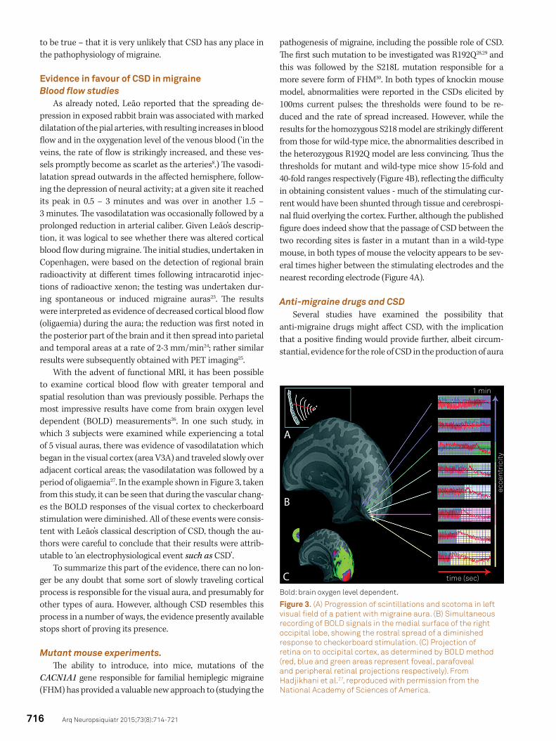
With the advent of functional MRI, it has been possible to examine cortical blood flow with greater temporal and spatial resolution than was previously possible. Perhaps the most impressive results have come from brain oxygen level dependent (BOLD) measurements26. In one such study, in which 3 subjects were examined while experiencing a total of 5 visual auras, there was evidence of vasodilatation which began in the visual cortex (area V3A) and traveled slowly over adjacent cortical areas; the vasodilatation was followed by a period of oligaemia27. In the example shown in Figure 3, taken from this study, it can be seen that during the vascular chang-es the BOLD responses of the visual cortex to checkerboard stimulation were diminished. All of these events were consis-tent with Leão’s classical description of CSD, though the au-thors were careful to conclude that their results were attrib-utable to ‘an electrophysiological event such as CSD’.
To summarize this part of the evidence, there can no lon-ger be any doubt that some sort of slowly traveling cortical process is responsible for the visual aura, and presumably for other types of aura. However, although CSD resembles this process in a number of ways, the evidence presently available stops short of proving its presence.
Mutant mouse experiments.The ability to introduce, into mice, mutations of the
CACN1A1 gene responsible for familial hemiplegic migraine (FHM) has provided a valuable new approach to (studying the
pathogenesis of migraine, including the possible role of CSD. The first such mutation to be investigated was R192Q28,29 and this was followed by the S218L mutation responsible for a more severe form of FHM30. In both types of knockin mouse model, abnormalities were reported in the CSDs elicited by 100ms current pulses; the thresholds were found to be re-duced and the rate of spread increased. However, while the results for the homozygous S218 model are strikingly different from those for wild-type mice, the abnormalities described in the heterozygous R192Q model are less convincing. Thus the thresholds for mutant and wild-type mice show 15-fold and 40-fold ranges respectively (Figure 4B), reflecting the difficulty in obtaining consistent values - much of the stimulating cur-rent would have been shunted through tissue and cerebrospi-nal fluid overlying the cortex. Further, although the published figure does indeed show that the passage of CSD between the two recording sites is faster in a mutant than in a wild-type mouse, in both types of mouse the velocity appears to be sev-eral times higher between the stimulating electrodes and the nearest recording electrode (Figure 4A).
Anti-migraine drugs and CSDSeveral studies have examined the possibility that
anti-migraine drugs might affect CSD, with the implication that a positive finding would provide further, albeit circum-stantial, evidence for the role of CSD in the production of aura
1 min
time (sec)
ecce
ntri
city
A
B
CBold: brain oxygen level dependent.
Figure 3. (A) Progression of scintillations and scotoma in left visual field of a patient with migraine aura. (B) Simultaneous recording of BOLD signals in the medial surface of the right occipital lobe, showing the rostral spread of a diminished response to checkerboard stimulation. (C) Projection of retina on to occipital cortex, as determined by BOLD method (red, blue and green areas represent foveal, parafoveal and peripheral retinal projections respectively). From Hadjikhani et al.27, reproduced with permission from the National Academy of Sciences of America.

717Alan J McComas et al. Spreading depression in migraine
and headache31. The results have been variable; for example, topiramate suppresses CSD effectively inrats32 but not in all cats33, and in both species the CSD suppression is unaccom-panied by changes in bloodflow. In contrast, sumatriptan, a very effective anti-migraine drug, has no effect on CSD34.
Earlier evidence against CSD in migraine: brain recordings and neural function(i). Difficulty in inducing CSD in human subjects
In his first paper Leão7 reported that it was more difficult to evoke CSD in the cat than in the rabbit, and this distinc-tion between brains with convoluted and smooth surfaces has been confirmed35. In the human brain, which has many more gyri than the cat, McLachlan and Girvin36 were unable to ini-tiate CSD in the exposed cortices of 23 patients about to un-dergo resections for intractable epilepsy. There are three points that deserve emphasis. First, using the same techniques (KCl application, electrical and mechanical stimulation), the au-thors had no difficulty in inducing CSD in rats. Second, the re-sults could not be explained by anaesthesia since the patients were only receiving pain medication with fentanyl and droper-idol; though both drugs are known to affect the EEG37, neither appears to interfere with CSD generation38,39. Indeed, the fact that droperidol may terminate migraine attacks40 and yet not affect CSD constitutes another argument against a role for the latter in the pathogenesis of the headache. Third, four of the patients had suffered from migraine, though without auras. Although there have since been reports of CSD detection in human subjects, using electrocorticography sometimes com-bined with scalp EEG, it is relevant that the CSD was only asso-ciated with major brain lesions, either trauma41, subarachnoid haemorrhage or stroke42.
EEG recordings during migraine auraAlthough it is technically possible to detect CSD in hu-
man subjects with scalp electrodes (see above), any depres-sion of EEG activity during either the aura or the headache phase of migraine is mild or absent; there may be some in-crease in slow wave activity but, in a proportion of patients, the EEG may be quite normal43.
Preservation of neural function during the auraIt is well known that sensory function may be lost during
an aura, the best example being the scotoma that replaces the scintillations as the latter progress across the visual field. However, the association is not invariable, for in a minority of subjects the fortification spectra may be superimposed on a normal visual image. Similarly, subjects who describe numb-ness and tingling can still detect touch. Such preservation of function is incompatible with a process, such as CSD, that, at least in animal models, abolishes all neural activity.
New evidence against CSD in migraine: therapeutic transcranial magnetic stimulation
Transcranial magnetic stimulation (TMS) has been em-ployed as a means of exploring CNS function for almost 30 years, its great advantage over electrical stimulation being that it is painless and penetrates the skull more readily; nevertheless the magnetic pulse is converted to an electri-cal one on intersecting suitably disposed biological con-ductors (axons and dendrites). Depending on the choice of target and stimulus parameters, TMS can be employed to either excite or block function in a neural circuit44. The pos-sibility of TMS being a useful treatment for migraine was raised in 2004 when it was found that a course of repetitive
Cum
ulat
ive
frac
tion
of a
nim
als
CSD threshold (µC)
R192Q KI
R192Q KI
wt
wt
stim1
1
2
1
2
50 sec25 m
V
2
0.0
0.2
0.4
0.6
0.8
1.0
1 10 100
A B
CSD: cortical spreading depression.
Figure 4. (A) Slow waves, associated with spreading depressions, in wild-type (wt) and R192Q K1 mutant mice. Positions of stimulating and recording electrodes shown at top. In the wild type mouse the wave takes approximately 60 s to travel the 2.1 mm separating the recording electrodes, for a velocity of 2.1 mm/min. However, the same wave required only 20 s or so to travel the 4.8 mm separating the stimulating electrodes from the nearest recording electrode, corresponding to a velocity of 14.4 mm/min. The extra distances incurred because of the curvature of the hemispheres have been ignored. (B) Cumulative distributions of thresholds for eliciting CSD in mutant and wild-type mice. From van den Maagdenberg et al.30; reproduced with permission of Elsevier.
718 Arq Neuropsiquiatr 2015;73(8):714-721
TMS, when applied to the left dorsolateral prefrontal cor-tex on alternate days, reduced both the frequency and the intensity of migraine attacks; moreover, the benefit persist-ed for at least one month45. Similar, but unexpected, find-ings were subsequently reported in two migraineurs treat-ed in a similar way for depression46. Our own interest in tTMS for migraine arose from observations made on one of the authors, a migraine sufferer himself, and on patient volunteers47. Unlike the aforementioned studies, TMS was applied at the onset of the headache and given as a single pulse, sometimes repeated once, to the back of the head; the headache was found to diminish and sometimes to dis-appear altogether. The working hypothesis - a mistaken one (see below) - was that stimulation of the cortex in advance of a CSD was halting the progress of the latter, rather as the creation of a firebreak may stop a forest fire. Any CSD pres-ent during the attack was presumed to have commenced in the occipital cortex, since visual symptoms constitute the most common aura. Since that time there have been several controlled trials of single or double pulse TMS administered to the occipital cortex during the migraine attack, and in all cases there have been positive outcomes5,48,49,50,51. In the latter two trials, the patients were able to deliver the mag-netic pulses themselves, using a portable hand-held device designed specifically for that purpose.
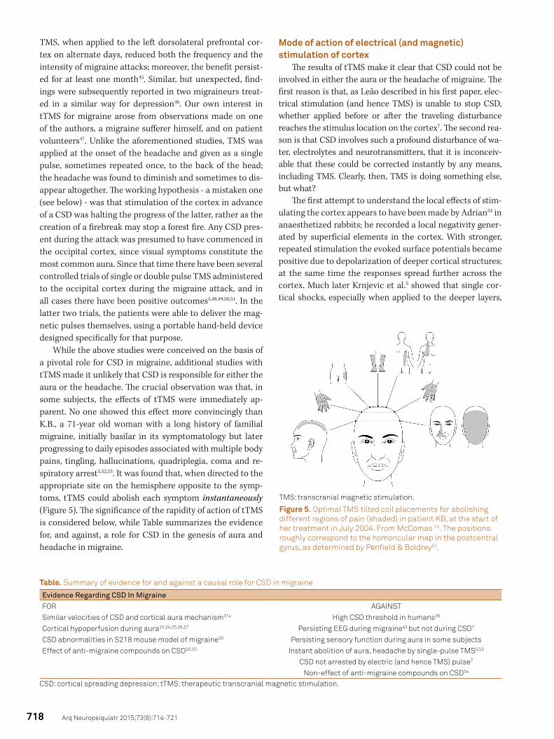
While the above studies were conceived on the basis of a pivotal role for CSD in migraine, additional studies with tTMS made it unlikely that CSD is responsible for either the aura or the headache. The crucial observation was that, in some subjects, the effects of tTMS were immediately ap-parent. No one showed this effect more convincingly than K.B., a 71-year old woman with a long history of familial migraine, initially basilar in its symptomatology but later progressing to daily episodes associated with multiple body pains, tingling, hallucinations, quadriplegia, coma and re-spiratory arrest5,52,53. It was found that, when directed to the appropriate site on the hemisphere opposite to the symp-toms, tTMS could abolish each symptom instantaneously (Figure 5). The significance of the rapidity of action of tTMS is considered below, while Table summarizes the evidence for, and against, a role for CSD in the genesis of aura and headache in migraine.
Mode of action of electrical (and magnetic) stimulation of cortex
The results of tTMS make it clear that CSD could not be involved in either the aura or the headache of migraine. The first reason is that, as Leão described in his first paper, elec-trical stimulation (and hence TMS) is unable to stop CSD, whether applied before or after the traveling disturbance reaches the stimulus location on the cortex7. The second rea-son is that CSD involves such a profound disturbance of wa-ter, electrolytes and neurotransmitters, that it is inconceiv-able that these could be corrected instantly by any means, including TMS. Clearly, then, TMS is doing something else, but what?
The first attempt to understand the local effects of stim-ulating the cortex appears to have been made by Adrian54 in anaesthetized rabbits; he recorded a local negativity gener-ated by superficial elements in the cortex. With stronger, repeated stimulation the evoked surface potentials became positive due to depolarization of deeper cortical structures; at the same time the responses spread further across the cortex. Much later Krnjevic et al.5 showed that single cor-tical shocks, especially when applied to the deeper layers,
TMS: transcranial magnetic stimulation.
Figure 5. Optimal TMS tilted coil placements for abolishing different regions of pain (shaded) in patient KB, at the start of her treatment in July 2004. From McComas 53. The positions roughly correspond to the homoncular map in the postcentral gyrus, as determined by Penfield & Boldrey62.
Table. Summary of evidence for and against a causal role for CSD in migraine
Evidence Regarding CSD In MigraineFOR AGAINSTSimilar velocities of CSD and cortical aura mechanism7,14 High CSD threshold in humans36
Cortical hypoperfusion during aura23,24,25,26,27 Persisting EEG during migraine43 but not during CSD7
CSD abnormalities in S218 mouse model of migraine30 Persisting sensory function during aura in some subjectsEffect of anti-migraine compounds on CSD32,33 Instant abolition of aura, headache by single-pulse TMS5,52
CSD not arrested by electric (and hence TMS) pulse7
Non-effect of anti-migraine compounds on CSD34
CSD: cortical spreading depression; tTMS: therapeutic transcranial magnetic stimulation.
719Alan J McComas et al. Spreading depression in migraine
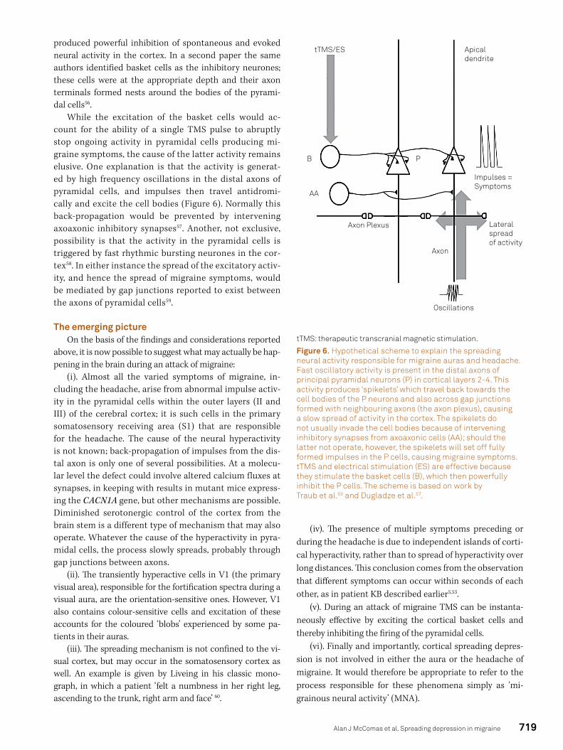
produced powerful inhibition of spontaneous and evoked neural activity in the cortex. In a second paper the same authors identified basket cells as the inhibitory neurones; these cells were at the appropriate depth and their axon terminals formed nests around the bodies of the pyrami-dal cells56.
While the excitation of the basket cells would ac-count for the ability of a single TMS pulse to abruptly stop ongoing activity in pyramidal cells producing mi-graine symptoms, the cause of the latter activity remains elusive. One explanation is that the activity is generat-ed by high frequency oscillations in the distal axons of pyramidal cells, and impulses then travel antidromi-cally and excite the cell bodies (Figure 6). Normally this back-propagation would be prevented by intervening axoaxonic inhibitory synapses57. Another, not exclusive, possibility is that the activity in the pyramidal cells is triggered by fast rhythmic bursting neurones in the cor-tex58. In either instance the spread of the excitatory activ-ity, and hence the spread of migraine symptoms, would be mediated by gap junctions reported to exist between the axons of pyramidal cells59.
The emerging pictureOn the basis of the findings and considerations reported
above, it is now possible to suggest what may actually be hap-pening in the brain during an attack of migraine:
(i). Almost all the varied symptoms of migraine, in-cluding the headache, arise from abnormal impulse activ-ity in the pyramidal cells within the outer layers (II and III) of the cerebral cortex; it is such cells in the primary somatosensory receiving area (S1) that are responsible for the headache. The cause of the neural hyperactivity is not known; back-propagation of impulses from the dis-tal axon is only one of several possibilities. At a molecu-lar level the defect could involve altered calcium fluxes at synapses, in keeping with results in mutant mice express-ing the CACN1A gene, but other mechanisms are possible. Diminished serotonergic control of the cortex from the brain stem is a different type of mechanism that may also operate. Whatever the cause of the hyperactivity in pyra-midal cells, the process slowly spreads, probably through gap junctions between axons.
(ii). The transiently hyperactive cells in V1 (the primary visual area), responsible for the fortification spectra during a visual aura, are the orientation-sensitive ones. However, V1 also contains colour-sensitive cells and excitation of these accounts for the coloured ‘blobs’ experienced by some pa-tients in their auras.
(iii). The spreading mechanism is not confined to the vi-sual cortex, but may occur in the somatosensory cortex as well. An example is given by Liveing in his classic mono-graph, in which a patient ‘felt a numbness in her right leg, ascending to the trunk, right arm and face’ 60.
(iv). The presence of multiple symptoms preceding or during the headache is due to independent islands of corti-cal hyperactivity, rather than to spread of hyperactivity over long distances. This conclusion comes from the observation that different symptoms can occur within seconds of each other, as in patient KB described earlier5,53.
(v). During an attack of migraine TMS can be instanta-neously effective by exciting the cortical basket cells and thereby inhibiting the firing of the pyramidal cells.
(vi). Finally and importantly, cortical spreading depres-sion is not involved in either the aura or the headache of migraine. It would therefore be appropriate to refer to the process responsible for these phenomena simply as ‘mi-grainous neural activity’ (MNA).
Oscillations
Axon
AA
B P
Axon Plexus
tTMS/ES
Impulses =Symptoms
Apical dendrite
Lateral spread of activity
tTMS: therapeutic transcranial magnetic stimulation.
Figure 6. Hypothetical scheme to explain the spreading neural activity responsible for migraine auras and headache. Fast oscillatory activity is present in the distal axons of principal pyramidal neurons (P) in cortical layers 2-4. This activity produces ‘spikelets’ which travel back towards the cell bodies of the P neurons and also across gap junctions formed with neighbouring axons (the axon plexus), causing a slow spread of activity in the cortex. The spikelets do not usually invade the cell bodies because of intervening inhibitory synapses from axoaxonic cells (AA); should the latter not operate, however, the spikelets will set off fully formed impulses in the P cells, causing migraine symptoms. tTMS and electrical stimulation (ES) are effective because they stimulate the basket cells (B), which then powerfully inhibit the P cells. The scheme is based on work by Traub et al.59 and Dugladze et al.57.
720 Arq Neuropsiquiatr 2015;73(8):714-721
UNANSWERED QUESTIONS
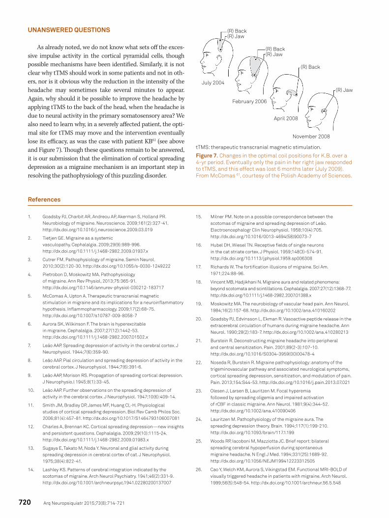
As already noted, we do not know what sets off the exces-sive impulse activity in the cortical pyramidal cells, though possible mechanisms have been identified. Similarly, it is not clear why tTMS should work in some patients and not in oth-ers, nor is it obvious why the reduction in the intensity of the headache may sometimes take several minutes to appear. Again, why should it be possible to improve the headache by applying tTMS to the back of the head, when the headache is due to neural activity in the primary somatosensory area? We also need to learn why, in a severely affected patient, the opti-mal site for tTMS may move and the intervention eventually lose its efficacy, as was the case with patient KB61 (see above and Figure 7). Though these questions remain to be answered, it is our submission that the elimination of cortical spreading depression as a migraine mechanism is an important step in resolving the pathophysiology of this puzzling disorder.
References
1. Goadsby PJ, Charbit AR, Andreou AP, Akerman S, Holland PR. Neurobiology of migraine. Neuroscience. 2009;161(2):327-41. http://dx.doi.org/10.1016/j.neuroscience.2009.03.019
2. Tietjen GE. Migraine as a systemic vasculopathy. Cephalalgia. 2009;29(9):989-996. http://dx.doi.org/10.1111/j.1468-2982.2009.01937.x
3. Cutrer FM. Pathophysiology of migraine. Semin Neurol. 2010;30(2):120-30. http://dx.doi.org/10.1055/s-0030-1249222
4. Pietrobon D, Moskowitz MA. Pathophysiology of migraine. Ann Rev Physiol, 2013;75:365-91. http://dx.doi.org/10.1146/annurev-physiol-030212-183717
5. McComas A, Upton A. Therapeutic transcranial magnetic stimulation in migraine and its implications for a neuroinflammatory hypothesis. Inflammopharmacology. 2009;17(2):68-75. http://dx.doi.org/10.1007/s10787-009-8058-7
6. Aurora SK, Wilkinson F. The brain is hyperexcitable in migraine. Cephalalgia. 2007;27(12):1442-53. http://dx.doi.org/10.1111/j.1468-2982.2007.01502.x
7. Leão AAP. Spreading depression of activity in the cerebral cortex. J Neurophysiol. 1944;7(6):359-90.
8. Leão AAP. Pial circulation and spreading depression of activity in the cerebral cortex. J Neurophysiol. 1944;7(6):391-6.
9. Leão AAP, Morison RS. Propagation of spreading cortical depression. J Neurophysio.l 1945;8(1):33-45.
10. Leão AAP. Further observations on the spreading depression of activity in the cerebral cortex. J Neurophysiol. 1947;10(6):409-14.
11. Smith JM, Bradley DP, James MF, Huang CL-H. Physiological studies of cortical spreading depression. Biol Rev Camb Philos Soc. 2006;81(4):457-81. http://dx.doi.org/10.1017/S1464793106007081
12. Charles A, Brennan KC. Cortical spreading depression—new insights and persistent questions. Cephalalgia. 2009;29(10):1115-24. http://dx.doi.org/10.1111/j.1468-2982.2009.01983.x
13. Sugaya E, Takato M, Noda Y. Neuronal and glial activity during spreading depression in cerebral cortex of cat. J Neurophysiol. 1975;38(4):822-41.
14. Lashley KS. Patterns of cerebral integration indicated by the scotomas of migraine. Arch Neurol Psychiatry. 1941;46(2):331-9. http://dx.doi.org/10.1001/archneurpsyc.1941.02280200137007
15. Milner PM. Note on a possible correspondence between the scotomas of migraine and spreading depression of Leão. Electroencephalogr Clin Neurophysiol. 1958;10(4):705. http://dx.doi.org/10.1016/0013-4694(58)90073-7
16. Hubel DH, Wiesel TN. Receptive fields of single neurons in the cat striate cortex. J Physiol. 1959;148(3)-574-91. http://dx.doi.org/10.1113/jphysiol.1959.sp006308
17. Richards W. The fortification illusions of migraine. Sci Am. 1971;224:88-96.
18. Vincent MB, Hadjikhani N. Migraine aura and related phenomena: beyond scotomata and scintillations. Cephalalgia. 2007:27(12):1368-77. http://dx.doi.org/10.1111/j.1468-2982.2007.01388.x
19. Moskowitz MA. The neurobiology of vascular head pain. Ann Neurol. 1984;16(2):157-68. http://dx.doi.org/10.1002/ana.410160202
20. Goadsby PJ, Edvinsson L, Ekman R. Vasoactive peptide release in the extracerebral circulation of humans during migraine headache. Ann Neurol. 1990;28(2):183-7. http://dx.doi.org/10.1002/ana.410280213
21. Burstein R. Deconstructing migraine headache into peripheral and central sensitization. Pain. 2001;89(2-3):107-10. http://dx.doi.org/10.1016/S0304-3959(00)00478-4
22. Noseda R, Burstein R. Migraine pathophysiology: anatomy of the trigeminovascular pathway and associated neurological symptoms, cortical spreading depression, sensitization, and modulation of pain. Pain. 2013;154:S44-53. http://dx.doi.org/10.1016/j.pain.2013.07.021
23. Olesen J, Larsen B, Lauritzen M. Focal hyperemia followed by spreading oligemia and impaired activation of rCBF in classic migraine. Ann Neurol. 1981;9(4):344-52. http://dx.doi.org/10.1002/ana.410090406
24. Lauritzen M. Pathophysiology of the migraine aura. The spreading depression theory. Brain. 1994;117(1):199-210. http://dx.doi.org/10.1093/brain/117.1.199
25. Woods RP, Iacoboni M, Mazziotta JC. Brief report: bilateral spreading cerebral hypoperfusion during spontaneous migraine headache. N Engl J Med. 1994;331(25):1689-92. http://dx.doi.org/10.1056/NEJM199412223312505
26. Cao Y, Welch KM, Aurora S, Vikingstad EM. Functional MRI-BOLD of visually triggered headache in patients with migraine. Arch Neurol. 1999;56(5):548-54. http://dx.doi.org/10.1001/archneur.56.5.548
November 2008
April 2008
February 2006
July 2004
(R) Back(R) Jaw
(R) Back(R) Jaw
(R) Back
(R) Jaw
tTMS: therapeutic transcranial magnetic stimulation.
Figure 7. Changes in the optimal coil positions for K.B. over a 4-yr period. Eventually only the pain in her right jaw responded to tTMS, and this effect was lost 6 months later (July 2009). From McComas 61, courtesy of the Polish Academy of Sciences.

721Alan J McComas et al. Spreading depression in migraine
27. Hadjikhani N, Sanchez del Rio M, Wu O, Schwartz D, Bakker D, Fischl B et al. Mechanisms of migraine aura revealed by functional MRI in human visual cortex. Proc Natl Acad Sci USA. 2001;98(8):4687-92. http://dx.doi.org/10.1073/pnas.071582498
28. Maagdenberg AM, Pietrobon D, Pizzorusso T, Kaja S, Broos LA, Cesetti T et al. A Cacna1a knockin migraine mouse model with increased susceptibility to cortical spreading depression. Neuron. 2004;41(5):701-10. http://dx.doi.org/10.1016/S0896-6273(04)00085-6
29. Cao YQ, Piedras-Rentería ES, Smith GB, Chen G, Harata NC, Tsien RW. Presynaptic Ca2+ channels compete for channel type-preferring slots in altered neurotransmission arising from Ca2+ channelopathy. Neuron. 2004;43(3):387-400. http://dx.doi.org/10.1016/j.neuron.2004.07.014
30. Maagenberg AM, Pizzorusso T, Kaja S, Repolilli N, Shapovalova M, Hoebeek FE et al. High cortical spreading depression susceptibility and migraine-associated symptoms in Cav2.1 S218L mice. Ann Neurol. 210;67(1):85-98. http://dx.doi.org/10.1002/ana.21815
31. Costa C, Tozzi A, Rainero I, Cupini LM, Calabresi P, Ayata C et al. Cortical spreading depression as a target for anti-migraine agents. J Headache Pain. 2013;14(1):62. http://dx.doi.org/10.1186/1129-2377-14-62
32. Unekawa M, Tomita Y, Toriumi H, Suzuki N. Suppressive effect of chronic peroral topiramate on potassium-induced cortical spreading depression in rats. Cephalalgia. 2012;32(7):518-27. http://dx.doi.org/10.1177/0333102412444015
33. Akerman S, Goadsby PJ. Topiramate inhibits cortical spreading depression in rat and cat: impact in migraine aura. Neuroreport. 2005;16(2):1383-7. http://dx.doi.org/10.1097/01.wnr.0000175250.33159.a9
34. Read SJ, Hirst WD, Upton N, Parsons AA. Cortical spreading depression produces increased cGMP levels in cortex and brain stem that is inhibited by tonabersat (SB-220453) but not sumatriptan. Brain Res. 2001;89(1-2):69-77. http://dx.doi.org/10.1016/S0006-8993(00)03191-7
35. Marshall WH, Essig CF. Relation of air exposure of cortex to spreading depression of Leão. J Neurophysiol. 1951;14(4):265-73.
36. McLachlan RS, Girvin JP. Spreading depression of Leão in rodent and human cortex. Brain Res. 1994;666(1):133-6. http://dx.doi.org/10.1016/0006-8993(94)90295-X
37. Kushikata T, Araki I, Sato T, Hashimoto Y, Ishihara H, Matsuki A. [EEG pattern during total intravenous anesthesia with droperidol, fentanyl and ketamine]. Matsui. 1993;42(8):1194-9. Japanese.
38. Hunfeld M, Pope KJ, Fitzgibbon SP, Willoughby JO, Broberg M. Effects of anesthetic agents on seizure-induction with intra-cortical injection of convulsants. Epilepsy Res. 2013;105(1-2):52-61. http://dx.doi.org/10.1016/j.eplepsyres.2012.12.009
39. Hertle DN, Dreier JP, Woitzik J, Hartings JA, Bullock R, Okonkwo DO et al. Effect of analgesics and sedatives on the occurrence of spreading depolarizations accompanying acute brain injury. Brain. 2012;135(8):2390-8. http://dx.doi.org/10.1093/brain/aws152
40. Nerenberg RH, Friedman BW. Migraine: an evidence-based update. Emerg Med. 2014;46(7):294-316.
41. Fabricius M, Fuhr S, Bhatia R, Boutelle M, Hashemi P, Strong AJ et al. Cortical spreading depression and peri-infarct depolarization in acutely injured human cerebral cortex. Brain. 2006;129(3):778-90. http://dx.doi.org/10.1093/brain/awh716
42. Drenckhahn C, Winkler MKL, Major S, Scheel M, Kang EJ, Pinczolits A et al. Correlates of spreading depolarization in human scalp electroencephalography. Brain. 2012;135(3):853-68. http://dx.doi.org/10.1093/brain/aws010
43. Sand T. EEG in migraine: a review of the literature. Funct Neurol. 1991;6(1):7-22.
44. Hallett M. Transcranial magnetic stimulation and the human brain. Nature. 2000;406(6792):147-50. http://dx.doi.org/10.1038/35018000
45. Brighina F, Piazza A, Vitello G, Aloisio A, Palermo A, Daniele O et al. rTMS of the prefrontal cortex in the treatment of chronic migraine:a pilot study. J Neurol Sci. 2004;227(1):67-71. http://dx.doi.org/10.1016/j.jns.2004.08.008
46. O’Reardon JP, Fontecha JF, Cristancho MA, Newman S. Unexpected reduction in migraine and psychogenic headaches following rTMS treatment for major depression: a report of two cases. CNS Spectr. 2007;12(12):921-5. http://dx.doi.org/10.1017/S1092852900015716
47. Fisher MJ. Goodbye headaches: a gun that shoots magnetic pulses puts and end to an age-old scourge. Discover. 2004;25(8):28-9.
48. Clarke BM, Upton ARKamath MV, Al-Harbi T, Castellanos CM. Transcranial magnetic stimulation for migraine:clinical effects. J Headache Pain. 2006;7(5):341-6. http://dx.doi.org/10.1007/s10194-006-0329-8
49. Mohammed YM, Hughes G, Kothari R, Nkrumah M, Fischell S, Robert F et al. Self-administered transcranial magnetic stimulation (TMS) during the aura phase improves and aborts headaches. Headache. 2006;46:839.
50. Lipton RB, Dodick DW, Silberstein SD, Saper JR, Aurora SK, Pearlman SH et al. Single-pulse transcranial magnetic stimulation for acute treatment of migraine with aura: a randomised, double-blind, parallel-group, sham-controlled trial. Lancet Neurol. 2010;9(4):373-80. http://dx.doi.org/10.1016/S1474-4422(10)70054-5
51. Lipton RB, Pearlman SH. Transcranial magnetic stimulation in the treatment of migraine. Neurotherapeutics. 2010;7(2):204-12. http://dx.doi.org/10.1016/j.nurt.2010.03.002
52. Upton A, McComas A. Abolition of migraine by transcranial magnetic simulation. Abstracts of 40th Meeting of the Canadian Congress of Neurological Sciences; 2005 June 14-18; Otawwa. CalgarY: Canadian Congress of Neurological Sciences; 2005. (Can J Neurol Sci. 2005;32(2 suppl 1):S71.
53. McComas AJ. The artful chameleon: an exploration of migraine and medicine. West Flamborough: Alkat Neuroscience; 2006.
54. Adrian ED. The spread of activity in the cerebral cortex. J Physiol. 1937;88(2):127-61. http://dx.doi.org/10.1113/jphysiol.1936.sp003427
55. Krnjević K, Randić M, Straughan DW. An inhibitory process in the cerebral cortex. J Physiol. 1966;184(1):16-48. http://dx.doi.org/10.1113/jphysiol.1966.sp007902
56. Krnjević K, Randić M, Straughan DW. Nature of a cortical inhibitory process. J Physiol. 1966;184(1):49-77. http://dx.doi.org/10.1113/jphysiol.1966.sp007903
57. Dugladze T, Schmitz D, Whittington MA, Vida I, Gloveli T. Segregation of axonal and somatic activity during fast network oscillations. Science. 2012;336(6087):1458-61. http://dx.doi.org/10.1126/science.1222017
58. Cunningham MO, Whittington MA, Bibbig A, Roopun A, LeBeau FE, Vogt A et al. A role for fast rhythmic bursting neurons in cortical gamma oscillations in vitro. Proc Natl Acad Sci USA. 2004;101(18):7152-7.
59. Traub R, Bibbig A, LeBeau FE, Cunningham MO, Whittington MA. Persistent gamma oscillations in superficial layers of rat auditory neocortex: experiment and model. J Physiol. 2005;562(1):3-8. http://dx.doi.org/10.1113/jphysiol.2004.074641
60. Liveing E. On megrim, sick-headache and some allied disorders: a contribution to the pathology of nerve-storms. London: Churchill; 1873.
61. McComas AJ. TMS in a severe case of complex migraine. Lecture Notes of the ICB Seminar. 2009;83:164-9.
62. Penfield W, Boldrey E. Somatic motor and sensory representation in the cerebral cortex of man as studied by electrical stimulation. Brain. 1937;60(4):389-443. http://dx.doi.org/10.1093/brain/60.4.389