covid echocardiography analysis as predictor of mortality
TRANSCRIPT
Human vs AI-Based Echocardiography Analysis as
Predictor of mortality in Acute COVID-19 Patients:
WASE-COVID Study
Federico M Asch, MD, FASE, FACCDirector, CV Core labs and Cardiac Imaging ResearchMedStar Health Research InstituteWashington, DC - USA
COVID
ACC Scientific Sessions, May 2021
American Society of Echocardiography Foundation,Alliance Partners and Global Collaborators
Federico Asch MD FASE, PIRoberto Lang MD FASE, PI
COVID
DisclosuresWASE- COVID has been funded by
American Society of Echocardiography FoundationMedStar HealthUniversity of Chicago
with in-kind donations by
Institutional Research Grants: Ultromics, TOMTEC, Caption Health, GE, Butterfly.Scientific Advisory Board: Ultromics
Background
• Transthoracic echocardiography (TTE) has emerged as the leading cardiac imaging modality for patients admitted with COVID-19 infection
• Myocardial injury has been linked with poor outcomes, therefore an echocardiogram at admission may prove to be a powerful tool to predict death.
• The role of AI in cardiovascular imaging and specifically echocardiography is expanding, to facilitate image acquisition and analysis
• With reader-dependent technologies such as echocardiography, fully automated, AI-based analysis should result in lower variability of results than those obtained from human reads.
• With increased interpretation consistency, it is foreseeable that the use of automated measurements could improve the capacity to predict outcomes.
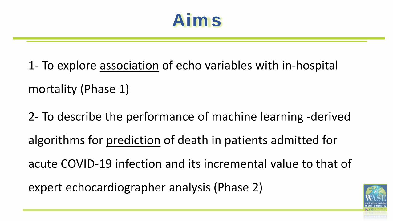
Aims
1- To explore association of echo variables with in-hospital
mortality (Phase 1)
2- To describe the performance of machine learning -derived
algorithms for prediction of death in patients admitted for
acute COVID-19 infection and its incremental value to that of
expert echocardiographer analysis (Phase 2)
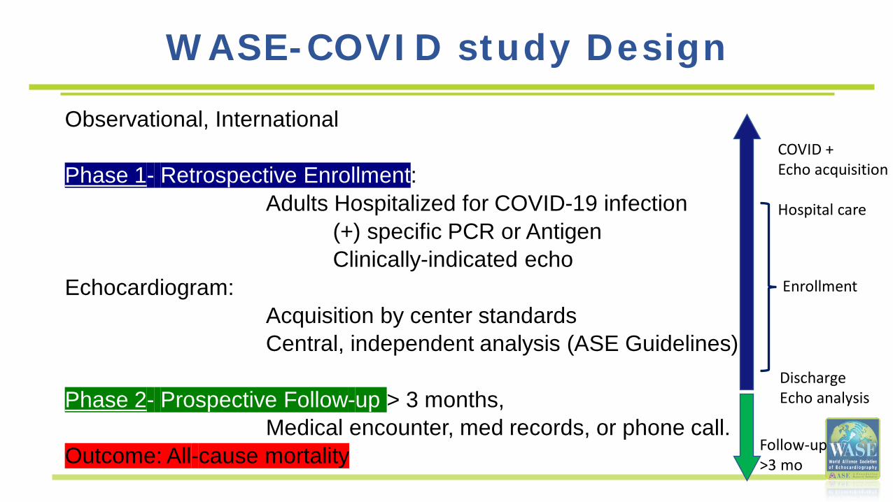
WASE-COVID study Design
Observational, International
Phase 1- Retrospective Enrollment:Adults Hospitalized for COVID-19 infection
(+) specific PCR or AntigenClinically-indicated echo
Echocardiogram: Acquisition by center standardsCentral, independent analysis (ASE Guidelines)
Phase 2- Prospective Follow-up > 3 months,Medical encounter, med records, or phone call.
Outcome: All-cause mortality
COVID +Echo acquisition
Hospital care
DischargeEcho analysis
Follow-up >3 mo
Enrollment
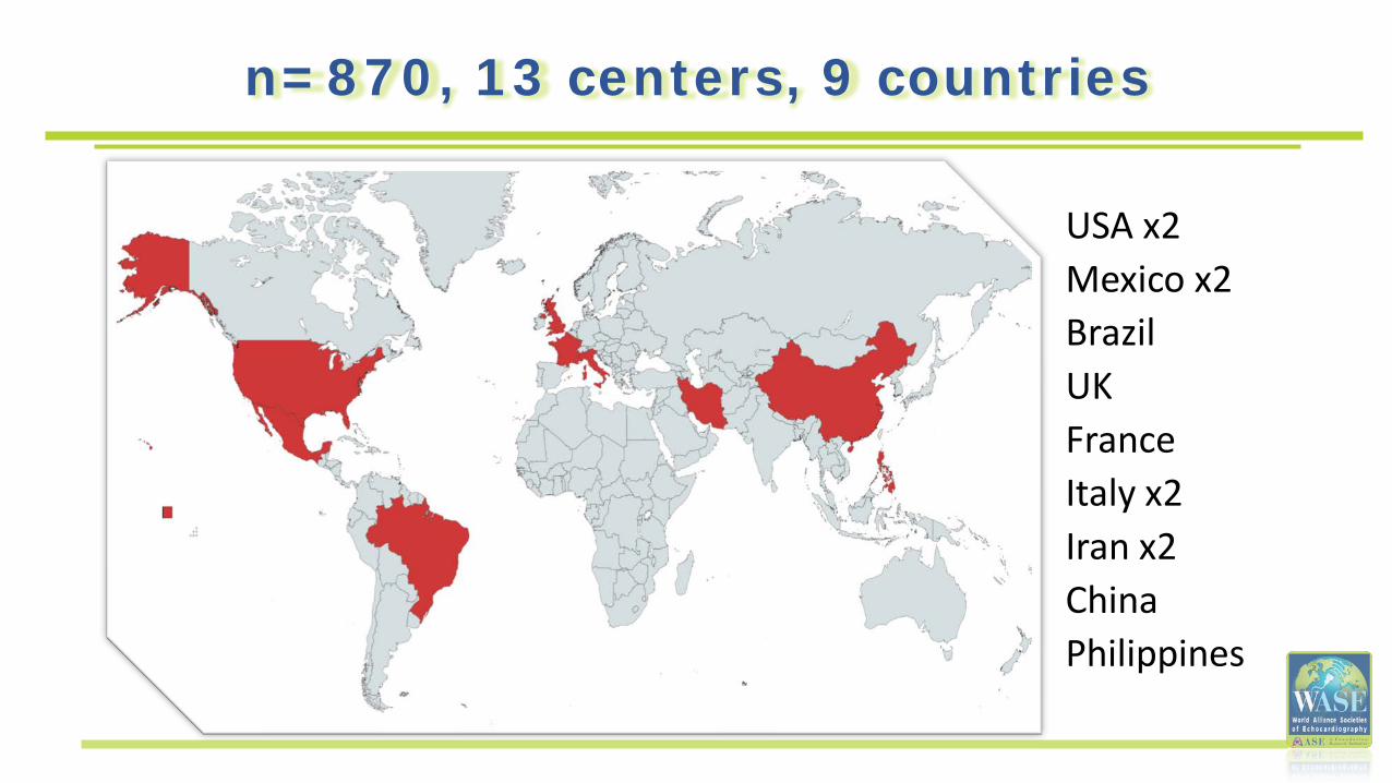
n=870, 13 centers, 9 countries
USA x2Mexico x2BrazilUKFrance Italy x2Iran x2ChinaPhilippines
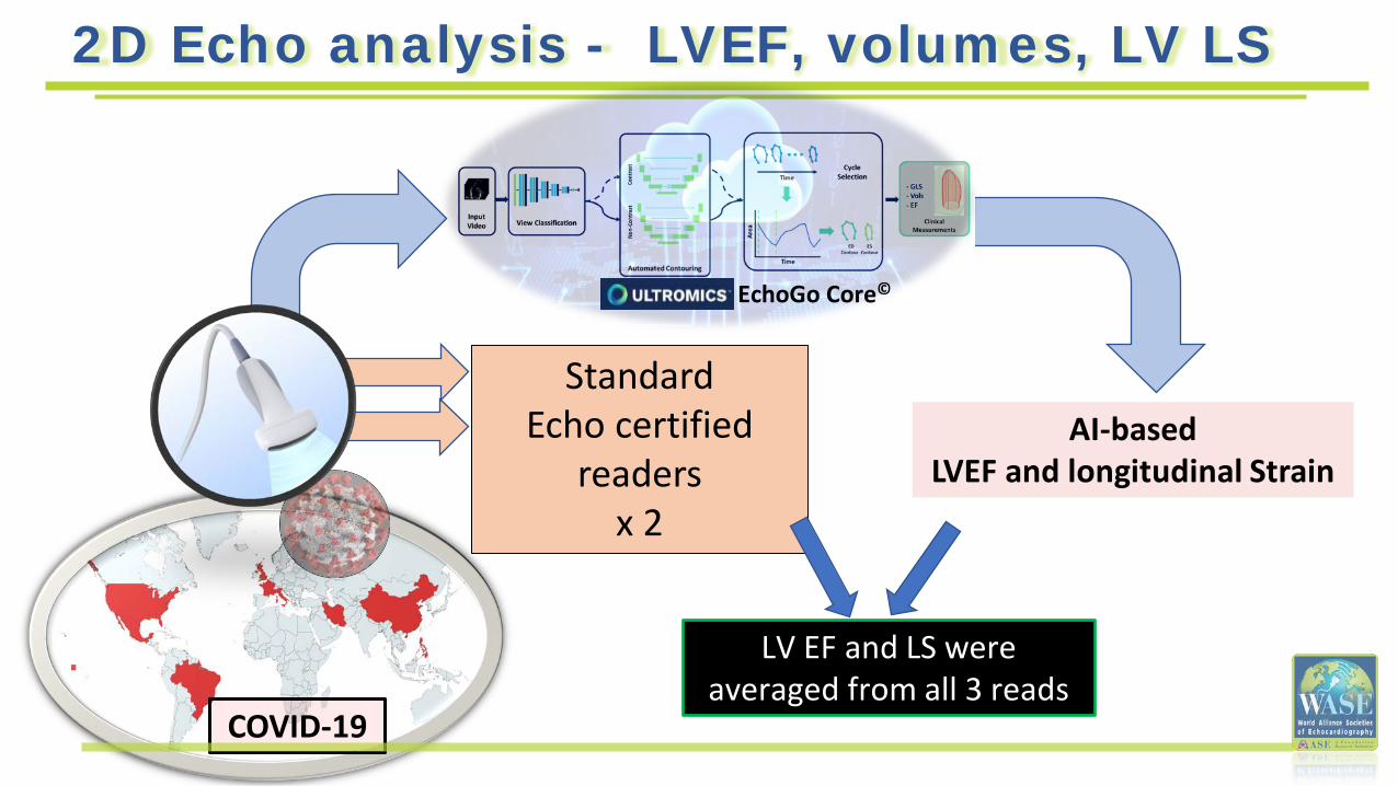
Standard Echo certified
readers x 2
COVID-19
AI-based LVEF and longitudinal Strain
LV EF and LS were averaged from all 3 reads
2D Echo analysis - LVEF, volumes, LV LS
EchoGo Core©
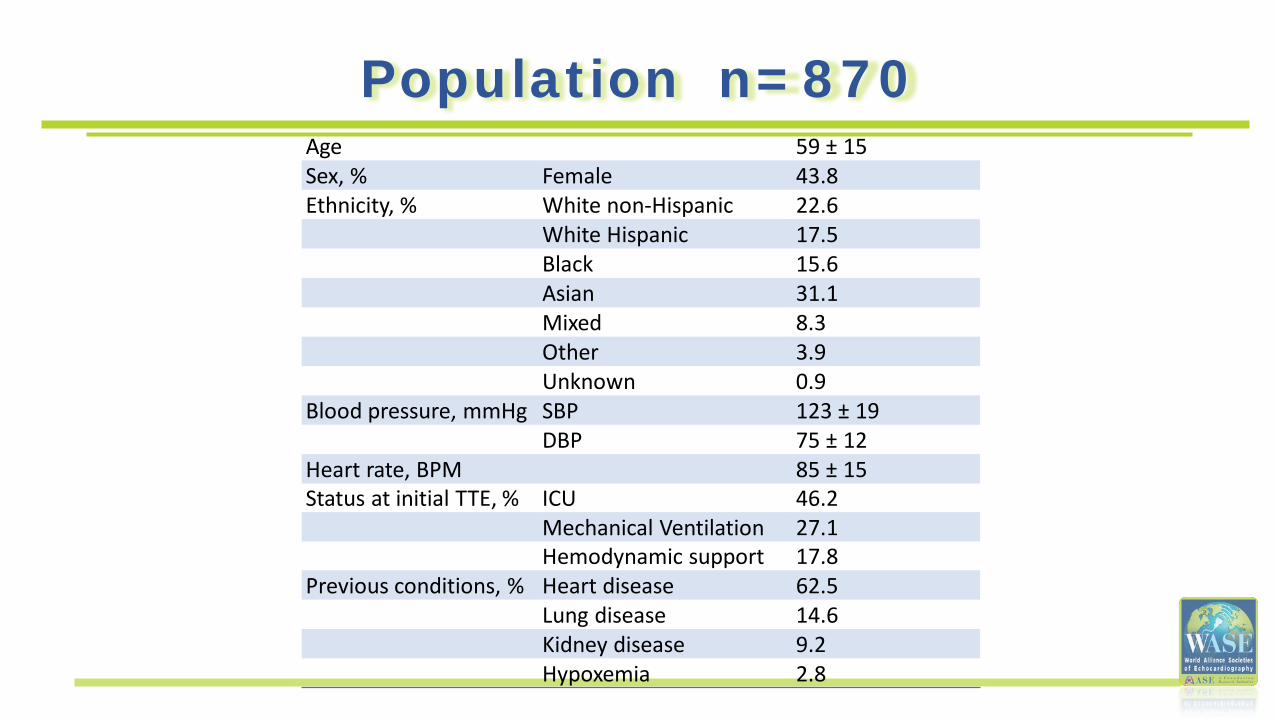
Population n=870 Age 59 ± 15 Sex, % Female 43.8Ethnicity, % White non-Hispanic 22.6
White Hispanic 17.5Black 15.6Asian 31.1Mixed 8.3Other 3.9Unknown 0.9
Blood pressure, mmHg SBP 123 ± 19 DBP 75 ± 12
Heart rate, BPM 85 ± 15 Status at initial TTE, % ICU 46.2
Mechanical Ventilation 27.1Hemodynamic support 17.8
Previous conditions, % Heart disease 62.5Lung disease 14.6Kidney disease 9.2Hypoxemia 2.8
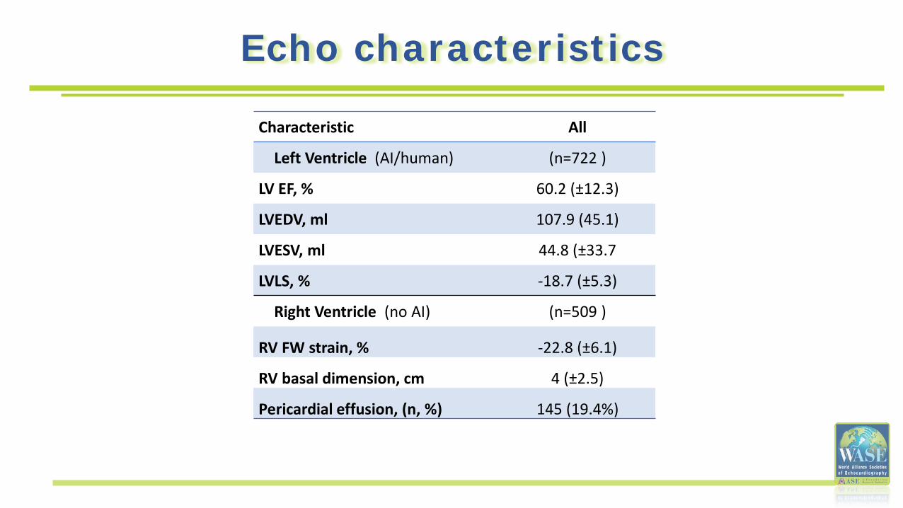
Echo characteristics
Characteristic All
Left Ventricle (AI/human) (n=722 )
LV EF, % 60.2 (±12.3)
LVEDV, ml 107.9 (45.1)
LVESV, ml 44.8 (±33.7
LVLS, % -18.7 (±5.3)
Right Ventricle (no AI) (n=509 )
RV FW strain, % -22.8 (±6.1)
RV basal dimension, cm 4 (±2.5)
Pericardial effusion, (n, %) 145 (19.4%)
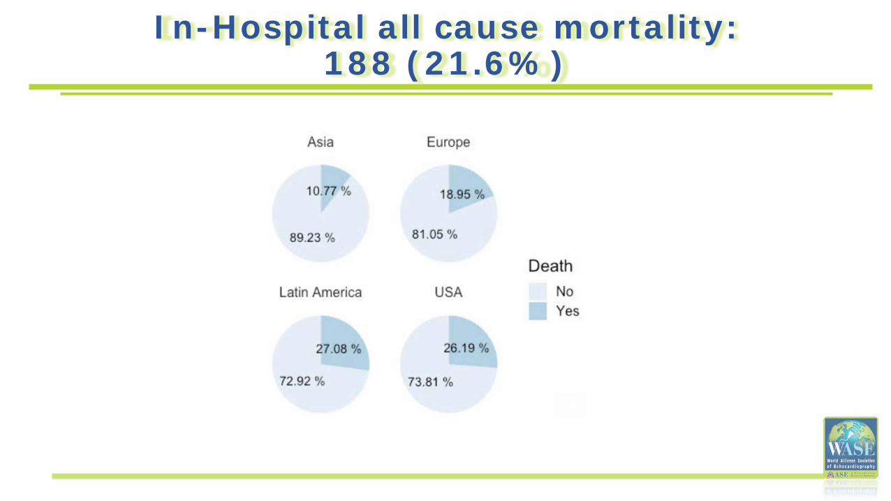
In-Hospital all cause mortality: 188 (21.6%)
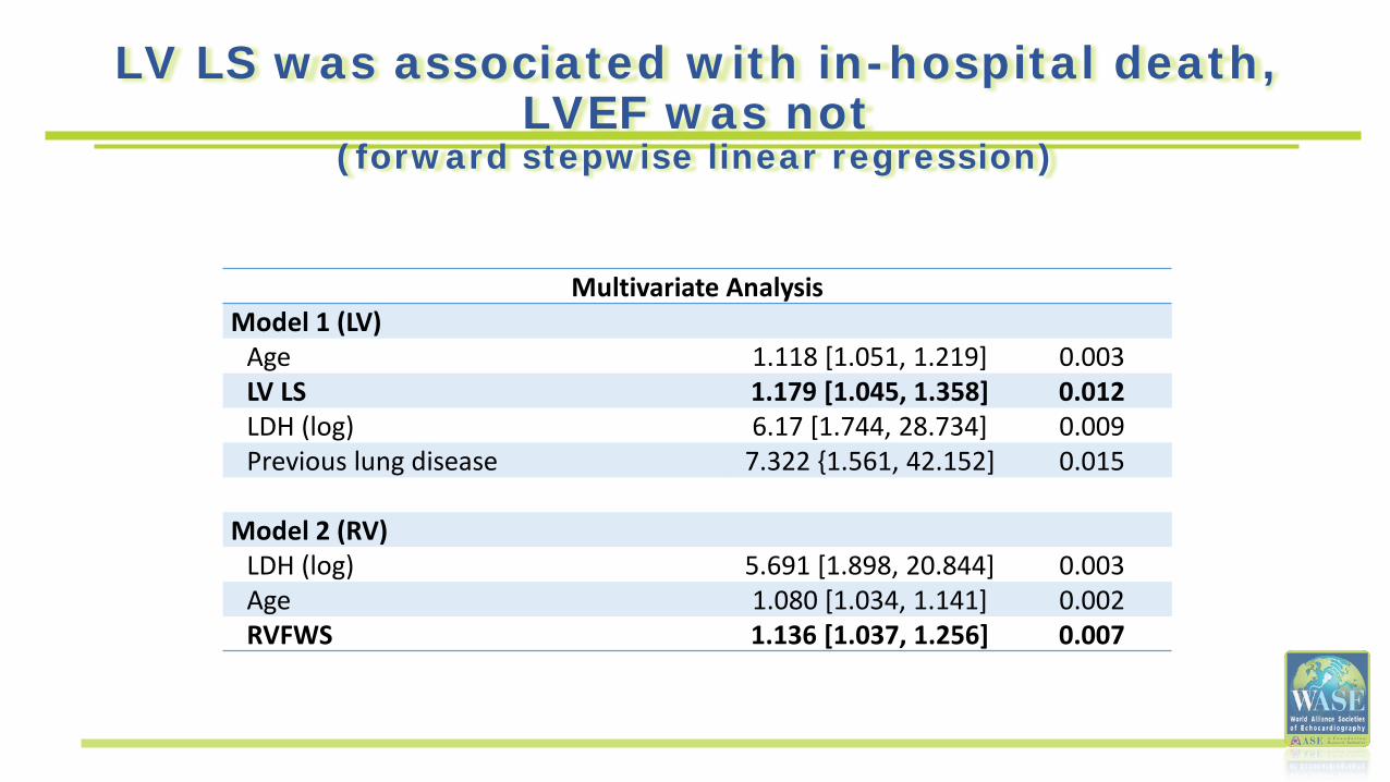
LV LS was associated with in-hospital death, LVEF was not
(forward stepwise linear regression)
Multivariate AnalysisModel 1 (LV)
Age 1.118 [1.051, 1.219] 0.003LV LS 1.179 [1.045, 1.358] 0.012LDH (log) 6.17 [1.744, 28.734] 0.009Previous lung disease 7.322 {1.561, 42.152] 0.015
Model 2 (RV)LDH (log) 5.691 [1.898, 20.844] 0.003Age 1.080 [1.034, 1.141] 0.002RVFWS 1.136 [1.037, 1.256] 0.007
Conclusions – Phase 1
• When measurements were averaged, LV LS, RVFWS, in addition to age, LDH, and previous lung disease were independently associated with in-hospital mortality, while LVEF was not.
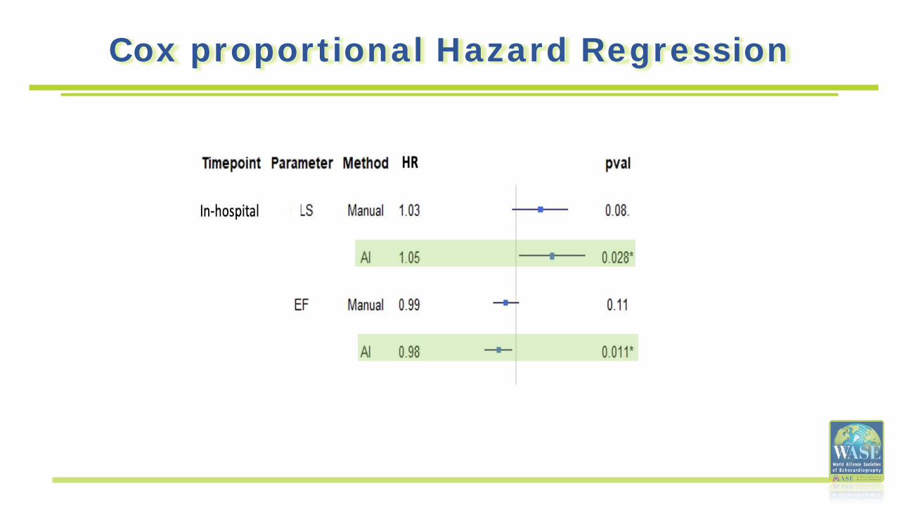
Cox proportional Hazard Regression
Hypothesis (Phase 2)
LVEF and LV LS obtained using AI-derived algorithms
will have less inter-reader variability and
will result in a better predictor of mortality than expert
readers.
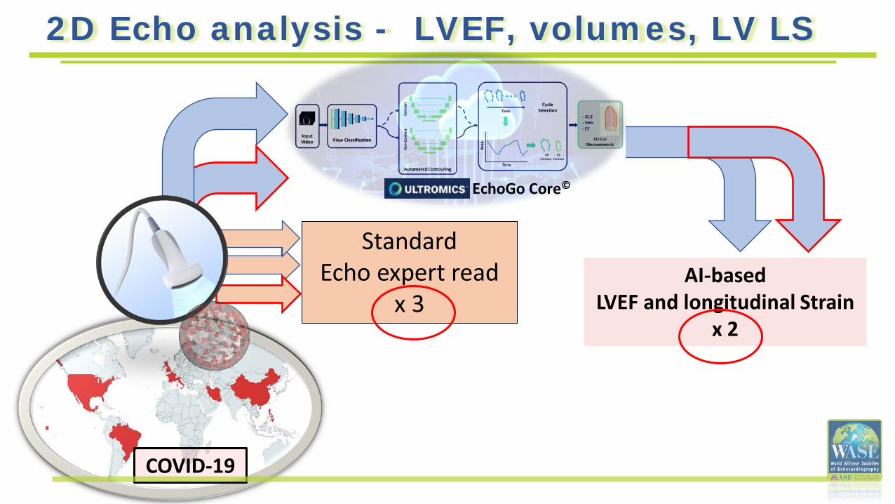
COVID-19
AI-based LVEF and longitudinal Strain
x 2
2D Echo analysis - LVEF, volumes, LV LS
Standard Echo expert read
x 3
EchoGo Core©
Prospective Follow-up
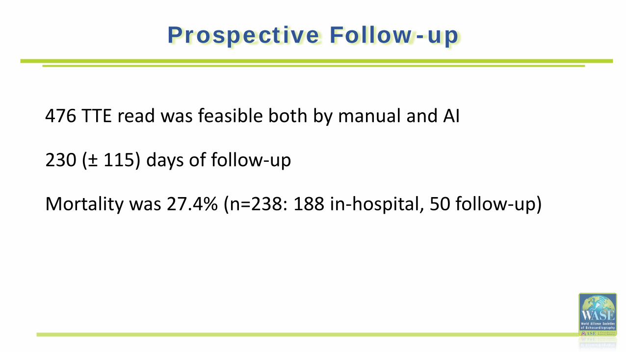
476 TTE read was feasible both by manual and AI
230 (± 115) days of follow-up
Mortality was 27.4% (n=238: 188 in-hospital, 50 follow-up)
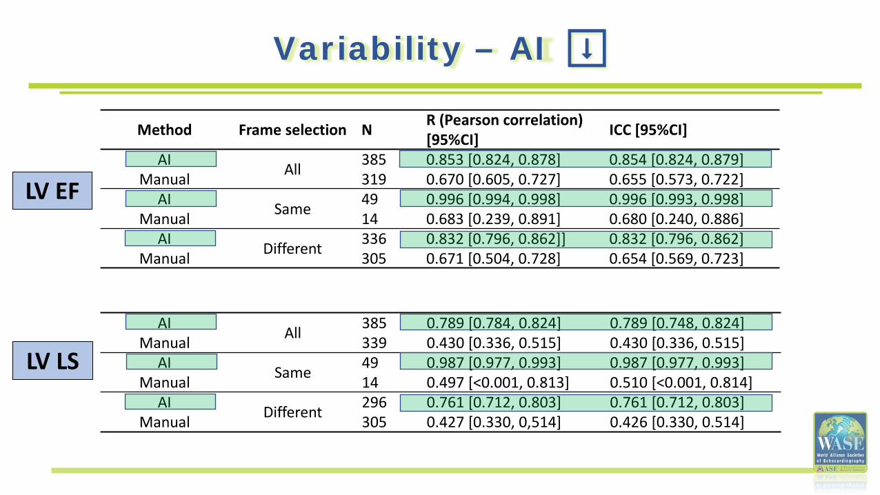
Variability – AI ⬇
Method Frame selection N R (Pearson correlation) [95%CI] ICC [95%CI]
AI All 385 0.853 [0.824, 0.878] 0.854 [0.824, 0.879]Manual 319 0.670 [0.605, 0.727] 0.655 [0.573, 0.722]
AI Same 49 0.996 [0.994, 0.998] 0.996 [0.993, 0.998]Manual 14 0.683 [0.239, 0.891] 0.680 [0.240, 0.886]
AI Different 336 0.832 [0.796, 0.862]] 0.832 [0.796, 0.862]Manual 305 0.671 [0.504, 0.728] 0.654 [0.569, 0.723]
AI All 385 0.789 [0.784, 0.824] 0.789 [0.748, 0.824]Manual 339 0.430 [0.336, 0.515] 0.430 [0.336, 0.515]
AI Same 49 0.987 [0.977, 0.993] 0.987 [0.977, 0.993]Manual 14 0.497 [<0.001, 0.813] 0.510 [<0.001, 0.814]
AI Different 296 0.761 [0.712, 0.803] 0.761 [0.712, 0.803]Manual 305 0.427 [0.330, 0,514] 0.426 [0.330, 0.514]
LV EF
LV LS
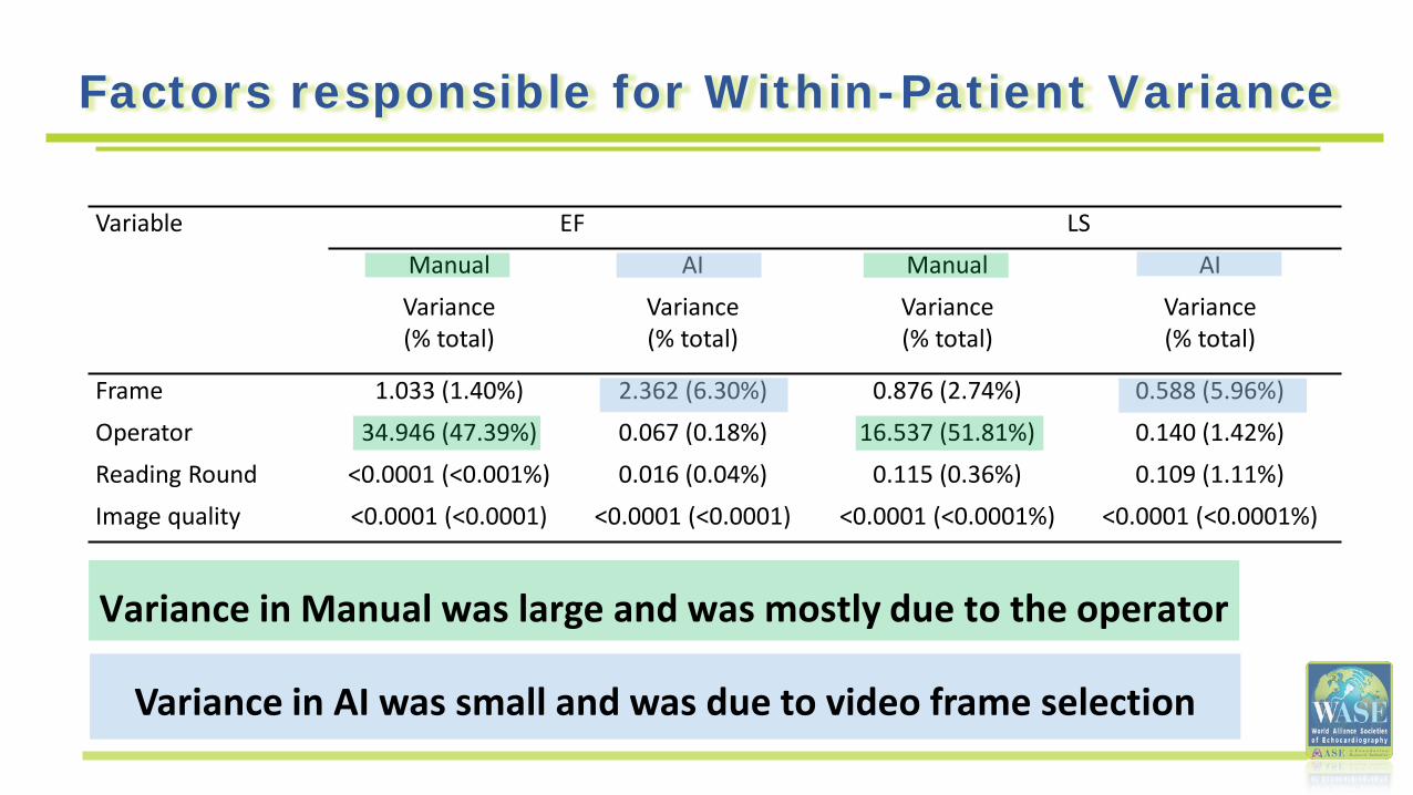
Factors responsible for Within-Patient Variance
Variance in AI was small and was due to video frame selection
Variable EF LS
Manual AI Manual AI
Variance(% total)
Variance(% total)
Variance(% total)
Variance(% total)
Frame 1.033 (1.40%) 2.362 (6.30%) 0.876 (2.74%) 0.588 (5.96%)
Operator 34.946 (47.39%) 0.067 (0.18%) 16.537 (51.81%) 0.140 (1.42%)
Reading Round <0.0001 (<0.001%) 0.016 (0.04%) 0.115 (0.36%) 0.109 (1.11%)
Image quality <0.0001 (<0.0001) <0.0001 (<0.0001) <0.0001 (<0.0001%) <0.0001 (<0.0001%)
Variance in Manual was large and was mostly due to the operator
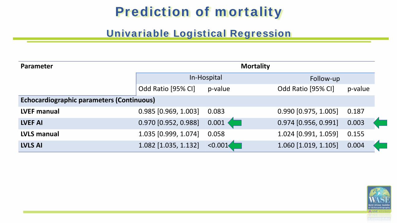
Prediction of mortalityUnivariable Logistical Regression
Parameter MortalityIn-Hospital Follow-up
Odd Ratio [95% CI] p-value Odd Ratio [95% CI] p-valueEchocardiographic parameters (Continuous)LVEF manual 0.985 [0.969, 1.003] 0.083 0.990 [0.975, 1.005] 0.187LVEF AI 0.970 [0.952, 0.988] 0.001 0.974 [0.956, 0.991] 0.003LVLS manual 1.035 [0.999, 1.074] 0.058 1.024 [0.991, 1.059] 0.155LVLS AI 1.082 [1.035, 1.132] <0.001 1.060 [1.019, 1.105] 0.004
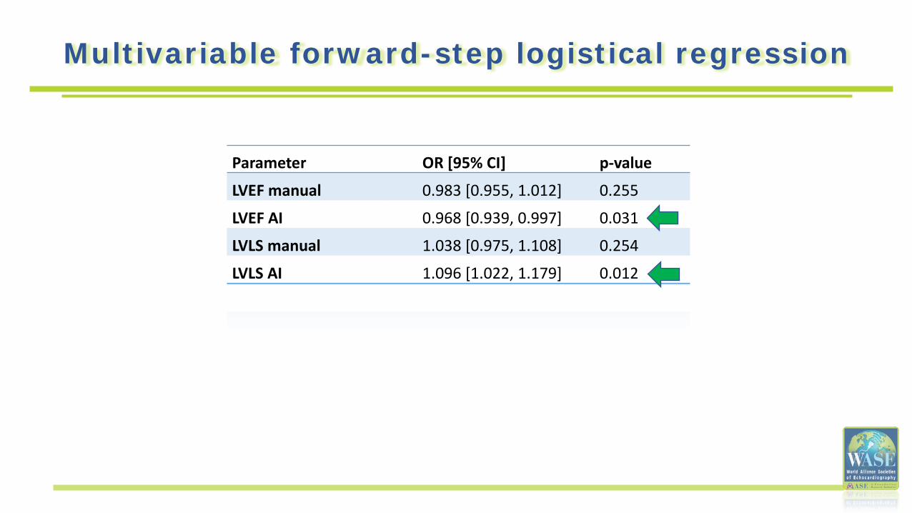
Parameter OR [95% CI] p-value
LVEF manual 0.983 [0.955, 1.012] 0.255
LVEF AI 0.968 [0.939, 0.997] 0.031
LVLS manual 1.038 [0.975, 1.108] 0.254
LVLS AI 1.096 [1.022, 1.179] 0.012
Multivariable forward-step logistical regression
Limitations
• Patients were enrolled in a retrospective manner• Not all echocardiograms could be quantified• Echocardiograms did not include sufficient information to assess the left atrium,
diastolic function and pulmonary pressures• Findings may be applicable to patients with COVID-19, not necessarily to other
patients• However, if broadened to a wider patient population with better image quality,
it is conceivable that AI contouring could be feasible in a much higher proportion of patients and therefore have more power
Conclusions (Phase 2)
• Automated quantification of LVEF and LVLS using AI minimized variability
• AI-based LV analyses, but not manual, were significant predictors of in-hospital and follow-up mortality.
• AI analysis of echoes could increase statistical power to predict outcomes, possibly requiring smaller sample sizes in clinical trials
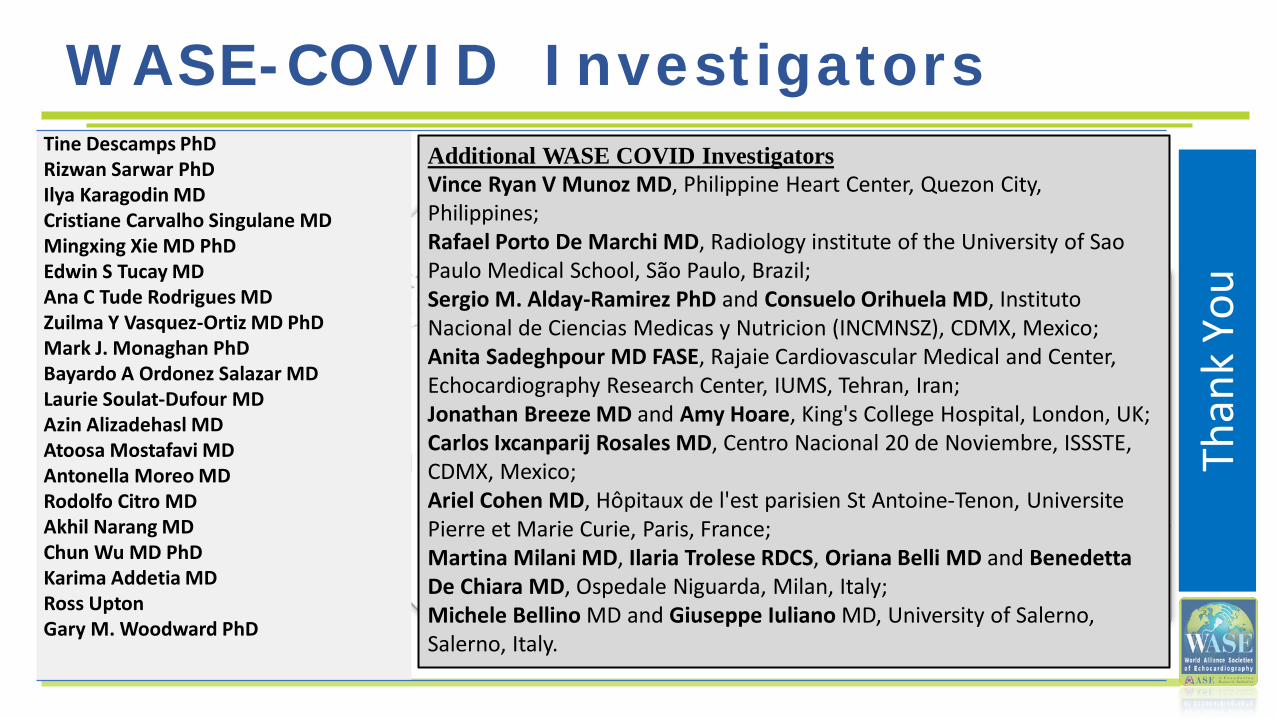
WASE-COVID Investigators
Than
k Yo
u
Tine Descamps PhDRizwan Sarwar PhDIlya Karagodin MD Cristiane Carvalho Singulane MD Mingxing Xie MD PhDEdwin S Tucay MDAna C Tude Rodrigues MDZuilma Y Vasquez-Ortiz MD PhDMark J. Monaghan PhD Bayardo A Ordonez Salazar MDLaurie Soulat-Dufour MDAzin Alizadehasl MDAtoosa Mostafavi MD Antonella Moreo MD Rodolfo Citro MD Akhil Narang MD Chun Wu MD PhDKarima Addetia MD Ross UptonGary M. Woodward PhD
Ultromics Ltd, Oxford, UKRadcliffe Department of Medicine, University of Oxford, UKUniversity of Chicago, Chicago ILUniversity of Chicago, Chicago ILUnion Hospital, Tongji Medical College of HUST, Wuhan, P.R. of ChinaPhilippine Heart Center, Quezon City, PhilippinesRadiology institute of the University of Sao Paulo Medical School, BrazilInstituto Nacional de Ciencias Medicas y Nutricion Salvador Zubiran, CDMX, MexicoKing's College Hospital, London, UKCentro Medico Nacional 20 de Noviembre, ISSSTE, CDMX, MexicoSt Antoine and Tenon Hospital and Sorbonne Université, Paris, FRRajaie CV Medical and Echocardiography Research Center, IUMS, Tehran, IranBaharloo Hospital, Tehran University of Medical Sciences, Tehran, IranDe Gasperis Cardio Center, Niguarda Hospital, Milan, ItalyUniversity of Salerno, Salerno, ItalyNorthwestern University, Chicago ILUnion Hospital, Tongji Medical College of HUST, Wuhan, P.R. of ChinaUniversity of Chicago, Chicago ILUltromics Ltd, Oxford, UKUltromics Ltd, Oxford, UK
Additional WASE COVID InvestigatorsVince Ryan V Munoz MD, Philippine Heart Center, Quezon City, Philippines; Rafael Porto De Marchi MD, Radiology institute of the University of Sao Paulo Medical School, São Paulo, Brazil; Sergio M. Alday-Ramirez PhD and Consuelo Orihuela MD, Instituto Nacional de Ciencias Medicas y Nutricion (INCMNSZ), CDMX, Mexico;Anita Sadeghpour MD FASE, Rajaie Cardiovascular Medical and Center, Echocardiography Research Center, IUMS, Tehran, Iran; Jonathan Breeze MD and Amy Hoare, King's College Hospital, London, UK;Carlos Ixcanparij Rosales MD, Centro Nacional 20 de Noviembre, ISSSTE, CDMX, Mexico; Ariel Cohen MD, Hôpitaux de l'est parisien St Antoine-Tenon, UniversitePierre et Marie Curie, Paris, France; Martina Milani MD, Ilaria Trolese RDCS, Oriana Belli MD and BenedettaDe Chiara MD, Ospedale Niguarda, Milan, Italy; Michele Bellino MD and Giuseppe Iuliano MD, University of Salerno, Salerno, Italy.