crizotinib synergizes with chemotherapy in preclinical...
TRANSCRIPT
Cancer Therapy: Preclinical
Crizotinib Synergizes with Chemotherapy inPreclinical Models of NeuroblastomaKateryna Krytska1, Hannah T. Ryles1, Renata Sano1, Pichai Raman2, Nicole R. Infarinato1,Theodore D. Hansel1, Monish R. Makena3, Michael M. Song3, C. Patrick Reynolds3, andYael P. Moss�e1
Abstract
Purpose: The presence of an ALK aberration correlates withinferior survival for patients with high-risk neuroblastoma. Theemergence of ALK inhibitors such as crizotinib has providednovel treatment opportunities. However, certain ALK muta-tions result in de novo crizotinib resistance, and a phase I trialof crizotinib showed a lack of response in patients harboringthose ALK mutations. Thus, understanding mechanisms ofresistance and defining circumvention strategies for the clinicis critical.
Experimental Design: The sensitivity of human neuroblasto-ma–derived cell lines, cell line–derived, and patient-derivedxenograft (PDX) models with varying ALK statuses to crizotinibcombined with topotecan and cyclophosphamide (topo/cyclo)was examined. Cultured cells and xenografts were evaluated foreffects of these drugs on proliferation, signaling, and cell death,and assessment of synergy.
Results: In neuroblastoma murine xenografts harboring themost common ALK mutations, including those mutations asso-ciated with resistance to crizotinib (but not in those with wild-type ALK), crizotinib combined with topo/cyclo enhanced tumorresponses andmouse event-free survival. Crizotinibþ topo/cycloshowed synergistic cytotoxicity and higher caspase-dependentapoptosis than crizotinib or topo/cyclo alone in neuroblastomacell lines with ALK aberrations (mutation or amplification).
Conclusions: Combining crizotinib with chemotherapeuticagents commonly used in treating newly diagnosed patients withhigh-risk neuroblastoma restores sensitivity in preclinical modelsharboring both sensitive ALK aberrations and de novo–resistantALK mutations. These data support clinical testing of crizotiniband conventional chemotherapy with the goal of integrating ALKinhibition into multiagent therapy for ALK-aberrant neuroblas-toma patients. Clin Cancer Res; 22(4); 948–60. �2015 AACR.
IntroductionNeuroblastomas are embryonal tumors that arise from the
malignant transformation of neural crest-derived cells and typi-cally present in early childhood (1). Despitemajor enhancementsin treatment approaches over the past several decades, the curerate for patients with high-risk neuroblastoma lags significantlybehind that of other common childhood cancers, and this diseasecontributes substantialmorbidity andmortality in patients (2). Inthis era of more rational therapies, new treatment strategiestargeting key oncogenic drivers are urgently needed. Thecomplexities of signaling networks that modify therapeutic
vulnerability in cancer cells mandate the study of drug combina-tions to prevent or reverse tumor drug resistance. The discoverythat somatically acquired activatingmutations and amplificationsof the Anaplastic Lymphoma Kinase (ALK) oncogene oftendrive the malignant process in a subset of neuroblastomas posi-tions ALK inhibition strategies as a promising therapeuticapproach (3–6).
ALK is an orphan receptor tyrosine kinase (RTK) normallyexpressed only in the developing and neonatal brain with apostulated role in the regulation of neuronal differentiation(7, 8). Dysregulation of ALK signaling has been associated withthe development of various cancers (9), most notably anaplasticlarge-cell lymphoma (ALCL) in a translocated form fused to theN-terminal of nucleophosmin (NPM; ref. 10), and fused to theechinodermmicrotubule-associated protein-like 4 (EML4) in 3%to 5% non–small cell lung cancers (NSCLC; ref. 11). ALK'soncogenic targets exert their growth advantage and antiapoptoticeffects through activation of numerous downstream pathwayssuch as PI3K/Akt, MAPK, and STAT3 (12). Pharmacologic ALKinhibition in ALCL models is associated with decreased levels ofphosphorylatedALK and its downstream effectors, and ultimatelyculminates with cell-cycle arrest and apoptosis (13). Early-phaseclinical studies of crizotinib, a dual ALK/MET small-moleculetyrosine kinase inhibitor, in pretreated patients with advancedrelapsed/refractory NSCLC harboring ALK rearrangementsyielded dramatic response rates (14, 15). This validated ALK asa therapeutic target and led to expedited FDA approval of crizo-tinib in patients with ALK-translocated lung cancer.
1Division of Oncology and Center for Childhood Cancer Research,TheChildren's Hospital of Philadelphia, Philadelphia, Pennsylvania. 2Divi-sion of Oncology, The Center for Biomedical Informatics (CBMi), TheChildren's Hospital of Philadelphia Research Institute, Philadelphia,Pennsylvania. 3Cancer Center, Texas Tech University Health SciencesCenter School of Medicine, Lubbock, Texas.
Note: Supplementary data for this article are available at Clinical CancerResearch Online (http://clincancerres.aacrjournals.org/).
K. Krytska and H.T. Ryles contributed equally to this article.
Corresponding Author: Yael P. Moss�e, Children's Hospital of Philadelphia,University of Pennsylvania Perelman School of Medicine, 3501 Civic CenterBoulevard, CTRB 3056, Philadelphia, PA 19104. Phone: 215-590-0965; Fax:267-426-0685; E-mail: [email protected]
doi: 10.1158/1078-0432.CCR-15-0379
�2015 American Association for Cancer Research.
ClinicalCancerResearch
Clin Cancer Res; 22(4) February 15, 2016948
on May 26, 2018. © 2016 American Association for Cancer Research. clincancerres.aacrjournals.org Downloaded from
Published OnlineFirst October 5, 2015; DOI: 10.1158/1078-0432.CCR-15-0379

ALKmutations are observed in 8% of neuroblastoma tumorsand span the entire spectrum of patients, ranging fromcongenital cases to adolescents and young adults. Within thehigh-risk subset of patients, the frequency of ALK aberrations is14% (10% point mutation and 4% amplification) and isindependently predictive of inferior outcome, supporting theusefulness of defining ALK status at diagnosis for prognosticand therapeutic stratification of these high-risk patients (16).Somatic ALK mutations at three sites (R1275, F1174, andF1245) occur most frequently in neuroblastoma, located inkey regulatory regions of the ALK kinase domain. Although celllines harboring the R1275Q mutation are initially sensitive todirect ALK kinase inhibition, cells harboring the F1174- andF1245 residue mutation are relatively resistant (16). Biochem-ical studies have shown that the reduced sensitivity of theF1174L mutation to crizotinib, and other ATP-competitive ALKinhibitors, is due in part to an increased ATP-binding affinitythat must be compensated for by higher doses of ALK inhibitorsor alternative therapeutic modalities (16).
A now completed pediatric phase I trial and ongoing phase IItrial of crizotinib (17), the most studied ALK inhibitor in neuro-blastoma, have shown activity in a subset of neuroblastomapatients, but the frequency and duration of responses are mar-ginal. Combining crizotinib with conventional genotoxic agentsmay provide superior antitumor activity than either approachused alone. The TP53 tumor suppressor gene is a key regulator ofapoptosis, senescence, cell-cycle arrest, and DNA repair and,unlike in many adult cancers, retains wild-type activity in neu-roblastoma (18, 19). Neuroblastomas can acquire a sustainedhigh-level drug resistance during chemotherapy, which can beattributed to acquired p53mutations and/or loss of p53 function(20, 21). Harnessing this central pathway and developing strat-egies to induce p53 functional activation may provide an oppor-tunity to enhance therapeutic efficacy. In the present study, wesought to provide the preclinical data that support integration ofcrizotinib into multiagent chemotherapy for patients with high-risk neuroblastoma.
Materials and MethodsMouse xenograft studies
Female CB17 SCID mice were subcutaneously implanted withhuman neuroblastoma NB-1643 (ALK-R1275Q mutation, TP53WT), SH-SY5Y (ALK-F1174Lmutation, TP53WT),NB-EBc1 (ALK-WT, expresses robust phosphorylated ALK, TP53 WT), SK-N-AS(ALK-WT, TP53-H168R mutation), NB-SD (ALK-WT, TP53-C176F mutation), and NB-1691 (ALK-WT, TP53 WT, MDM2amplified) xenografts and the Felix-PDX (ALK-F1245C, TP53WT)patient-derived xenograft (PDX) established post-mortem fromthe blood of a patient who died of a progressive MYCN-non-amplified high-risk neuroblastoma. Mice with engrafted tumorsthat reached 200mm3 in size were randomized into groups of 10per condition. Tumors were measured using a digital caliper atthe initiation of the study and one to two times per week duringthe treatment period. For all experiments, mice were weighedtwice weekly. The studies consisted of the following groups: (i)vehicle control; (ii) crizotinib; (iii) topotecan plus cyclophospha-mide (topo/cyclo); and (iv) crizotinib þ topo/cyclo. Mice con-tinuously received either vehicle (solvent medium of 0.5%meth-yl-cellulose, 0.5% Tween-80 in water, QD, PO), crizotinib (100mg/kg QD, PO), topo/cyclo (0.05 mg/kg and 20 mg/kg, respec-tively, IP), or crizotinib plus topo/cyclo treatment. The treatmentschedule for the chemotherapeutic drugs was as follows: i.p. 5days on treatment, 16days off treatment, cycle resumedonday21.The event resulting in mouse euthanasia was disease progression,defined as the tumor volume either reaching four times the initialvolume or 3 cm3. In the biology studies with SH-SY5Y and Felix-PDX tumors, mice were enrolled at 300 mm3, and randomizedinto the same four treatment groups as described above.Miceweretreated for 3 days, euthanized, and their tumors were collected 4hours after last treatment. All animal work was conducted accord-ing to relevant national and international guidelines and stepswere taken to minimize tumor burden and drug-related sideeffects. All animal studies were approved by The Children'sHospital of Philadelphia (Philadelphia, PA) IACUC (protocol# 2012-5-643).
Cell lines and reagentsThe neuroblastoma cell lines NB-1643 (ALK-R1275Q, TP53
WT), SH-SY5Y (ALK-F1174L, TP53 WT), NB-EBc1 (ALK WT,expresses phosphorylated ALK, TP53 WT), LAN-5 (ALK-R1275Q, TP53WT), LAN-6 (ALK-D1091N, TP53WT), NB-1 (ALKamplified, TP53WT), NB-SD (ALK-F1174L, TP53-C176F), KELLY(ALK-F1174L, TP53-P177T), NB-1691 (ALK WT, TP53 WT,MDM2 amplified), SK-N-AS (ALK WT, TP53-H168R), SK-N-BE(2)C (ALK WT, TP53-C135F), and NBL-S (ALK WT, TP53 WT),were obtained from the Children's Hospital of Philadelphia cellbank and were cultured in RPMI-1640 media supplemented with10% FBS, 1% L-glutamine, 1% penicillin/streptomycin, and 0.2%gentamycin. The cell lines COG-N-295 (ALK-F1174L, TP53 WT),COG-N-415 (ALK-F1174L TP53 WT), COG-N-247 (ALK WT,TP53 WT), CHLA-122 (ALK WT, TP53 WT), and COG-N-297(ALK WT, TP53 mutated) were established in the laboratory ofDr. C. Patrick Reynolds and obtained through the Children'sOncology Group Cell Culture and Xenograft Repository (www.cogcell.org). COG designated cell lines were cultured in IscovesModified Dulbecco Medium supplemented with 20% FBS, 1% L-glutamine, and 1% ITS (Sigma Aldrich). All cells were cultured inan atmosphere of 37�C and 5% CO2. Annual genotyping
Translational Relevance
Despite intensive, multimodal therapies, patients withhigh-risk neuroblastoma still face unacceptably highmortalityrates. We have previously shown that ALK activation is presentin 14%of patients with high-risk disease and that the presenceof an ALK aberration (point mutation or amplification) cor-relates with inferior survival in these patients. Importantly, theF1174L and F1245C amino acid substitutions, which com-prise two of the hot spot mutations, confer intrinsic resistanceto crizotinib and present a significant obstacle in the clinic. Inan effort to develop novel and more effective ALK inhibitionstrategies, we evaluated the in vitro and in vivo response of ALKaberrant neuroblastoma models to the ALK inhibitor, crizo-tinib, in combination with conventional chemotherapyagents, topotecan and cyclophosphamide. We provide thepreclinical rationale for clinical testing of crizotinib withconventional chemotherapy with the goal of integrating ALKinhibition into multiagent therapy for patients with high-riskALK aberrant neuroblastoma.
Crizotinib and Chemotherapy Synergize in Neuroblastoma
www.aacrjournals.org Clin Cancer Res; 22(4) February 15, 2016 949
on May 26, 2018. © 2016 American Association for Cancer Research. clincancerres.aacrjournals.org Downloaded from
Published OnlineFirst October 5, 2015; DOI: 10.1158/1078-0432.CCR-15-0379
(AmpFLSTR Identifiler Kit) of these lines and a single nucleotidepolymorphism array analysis (IlluminaH550)were performed toensure maintenance of cell identity using methods as previouslydescribed (22). Reagents for the in vitro studies included 4-hydro-peroxycyclophosphamide (4-HC; Niomech), topotecan (Sigma-Aldrich), and crizotinib (Pfizer, Inc). Identification of cell linesand xenografts were confirmed at time of experimentation byshort tandem repeat (STR) analysis validated against the Chil-dren's Hospital of Philadelphia database.
In vitro cytotoxicity assaysFor Cell Titer Glo (CTG; Promega) drug combination experi-
ments, in vitro cell viability was assessed by measuring levels ofATP using the luminescent detection assay measured on theGloMax-Multi Microplate Multimode Reader (Promega). Cellswere plated at their predetermined seeding densities on day 1,treated 24 hours later on day 2, and then read 72 hours later onday 5. Cells were first tested with single agents and Dm (absoluteIC50) values were established for each drug in each cell line. Thecells were then plated and treated with single agent and thecombination, and the results were analyzed.
Drug combination analysis was also conducted using theDIMSCAN assay, a semiautomatic fluorescence-based digitalimage microscopy system that quantifies viable cells in tissueculture multiwell plates on the basis of their selective accumula-tion and cleavage of fluorescein diacetate to fluorescein (23).DIMSCAN is capable of measuring cytotoxicity over a 4-logdynamic range by quantifying total fluorescence per well, whichis proportional to the number of viable, clonogenic cells aftereliminating background fluorescence with digital thresholdingand eosin Y quenching. Briefly, cell lines were seeded into 96-wellplates in 150 mL of complete medium (3,000–14,000 cells perwell) and incubated overnight. Crizotinib, topotecan, and 4-HC(the active metabolite of cyclophosphamide) in 50 mL of com-pletemediumwere added to the wells (12 replicate wells per eachconcentration of drugs). Cell lines were incubated in the presenceof these single agents or in combination for 96 hours, after whichfluorescein diacetate in 50 mL of 0.5% eosin Y (final concentrationof fluorescein diacetate 60 mg/mL) was added to eachwell and thecells were incubated for an additional 25 minutes at 37�C. Totalfluorescence was then measured with DIMSCAN and the resultswere expressed as surviving fractions of treated cells comparedwith control cells that were exposed to vehicle.
Determination of synergyResults were analyzed for synergistic, additive, or antagonistic
effects using the combination index (CI) method developed byChou and Talalay (24). The quantified CI values obtained by thismethod are defined as synergistic if CI<1, additive if CI ¼ 1, orantagonistic if CI>1. A confidence interval of<0.1 is represented asþþþþþ and indicates strong synergism. The isobologrammeth-od evaluates the interactions at a chosen effect level and istherefore useful in inspecting the drug interactions at the corre-sponding concentrations, often the median effect concentrations.Levels of cell viability and CI were obtained by software analysisand subsequently graphed in CI versus fraction affected (Fa) plots(Fig. 2A–E). Using both CalcuSyn v.2.0 (Biosoft Inc.), and Com-puSynv.1.0 (ComboSyn, Inc.),wedetermined the concentrationsof crizotinib and topotecan required to produce a defined single-agent effect (e.g., IC50), and placed the values on the x- and y-axesin a two-coordinate plot. Second, the concentrations of the two
drugs used in combination to provide the same effect were placedin the same plot. Synergy, additivity, or antagonism are indicatedwhen the concentrations of crizotinib and topotecan are locatedbelow, on, or above the line, respectively.
Synergy byDIMSCANwas determined using CalcuSyn software(24).
To further evaluate the dose-dependent drug interaction ofcrizotinib and topotecan, isobolograms at effect levels of 50%and 75% cell growth inhibition were created. Because the singleagents alone or in combination usually reach 50% inhibition, theisobologram at 50% provides an actual comparison for singledrug versus combination. The 75% isobologram illustrates theutility of combining these drugs at higher effect levels, withpractical implications in the clinic. Data points above or belowthe line of additivity indicate antagonism or synergy, respectively.
Western blot analysisAll cell lines were plated at their predetermined seeding
density and treated 24 hours later. Cells were harvested 8 hoursposttreatment and lysed according to a previously describedprotocol (25). Protein concentration was calculated using theBCA method and 30 to 50 mg of protein were separated on 4%to 12% Bis-Tris gradient gels, transferred to PVDF membranes(Millipore) overnight at 4�C, and immunoblotted againstpALKY1604, total ALK, cleaved caspase-3, cleaved PARP, phos-pho-p53S15, p21, PUMA, GAPDH, and b-actin (Cell SignalingTechnology) and MDM2 (Abcam) and total p53 (DO-1; SantaCruz Biotechnology), overnight at 4�C. All primary antibodieswere diluted 1:1,000, except GAPDH and b-actin, which werediluted 1:5,000. Membranes were washed and probed withsecondary antibodies at a dilution of 1:10,000 (Santa CruzBiotechnology). When probed for multiple proteins, mem-branes were stripped using 0.1 mol/L glycine, 0.5% Tween20 (at pH 2.5) twice for 30 minutes.
Statistical analysisFor in vivo statistical analysis, mixed-effects models were used
to assess tumor volume over time between treated samples andvehicle controls. Event-free survival (EFS) was defined as thepercentage of mice that survived while on therapy, wheresurvival was defined as the lack of an "event." An event wasdefined as a tumor volume reaching above 3 cm3 or 4� theinitial tumor volume, at which point the mice were sacrificedand taken off study. EFS percentages were estimated using theKaplan–Meier method and survival curves were compared byusing the log-rank test. Statistical analyses were performedusing the "nlme", "survival", and "multcomp" packages in theR statistical programming language (26–28). Statistical signif-icance was defined as P values < 0.05.
For in vitro densitometric analysis, ImageJ software (1.47v;NIH) was used to quantify intensity of the bands. Results wereconsidered statistically significant when P values were <0.05.Protein levels were estimated by quantification of the immuno-blot bands. The in vitro results were analyzed by performing alinear regression on the data, with the vehicle set as the Y intercept.The values from each treatment group were averaged and thencompared between groups. For the in vivo Western blot quanti-fication, because each lane represents one mouse, and there wereseveral mice ran per group, the groups were analyzed with eachmouse serving as a replicate. An ANOVA was performed on these
Krytska et al.
Clin Cancer Res; 22(4) February 15, 2016 Clinical Cancer Research950
on May 26, 2018. © 2016 American Association for Cancer Research. clincancerres.aacrjournals.org Downloaded from
Published OnlineFirst October 5, 2015; DOI: 10.1158/1078-0432.CCR-15-0379
samples, and a Tukey significant difference (HSD) post-hoc testwas done to test the significance between the groups. Significanceof results was designated as follows: �,P<0.05; ��,P<0.01; ���,P<0.001. The error reported is the SEM.
shRNA p53 Knockdown and functionality testingNB-1643 and SH-SY5Y cells were stably transfected with ret-
rovirus targeting p53 (Origene TL320558A and TL3200558C) orcopGFP control (Origene TR30021). Infected cells were selectedfor and kept in a medium containing 0.5 mg/mL puromycin.Knockdown efficiency was measured by qRT-PCR, with TP53(probe from Applied Biosystems; Hs00153349_m1) expressionlevels normalized to the geometric mean of two housekeepinggenes, IPO8 (Applied Biosystems; Hs00183533_m1) and UBC(Applied Biosystems, HS00824723_m1). All qRT-PCR reactionswere performed in triplicate. The DDCt method was used toanalyze fold change, and percentage knockdown was calculatedcompared with GFP control.
P53 functionality was tested by treating cells with 25 mmol/Lmelphalan (L-PAM) for 16 hours and assaying induction of totalp53, MDM2, and p21. Densitometric analysis was conducted onthe p21 protein bands that were normalized to the loadingcontrol. Functional p53 was defined as having a greater than 2-fold increase of p21 upon L-PAM treatment compared with anontreated control.
ResultsCrizotinib combined with standard cytotoxic agents sensitizedALK-mutant neuroblastoma xenografts with wild-type TP53
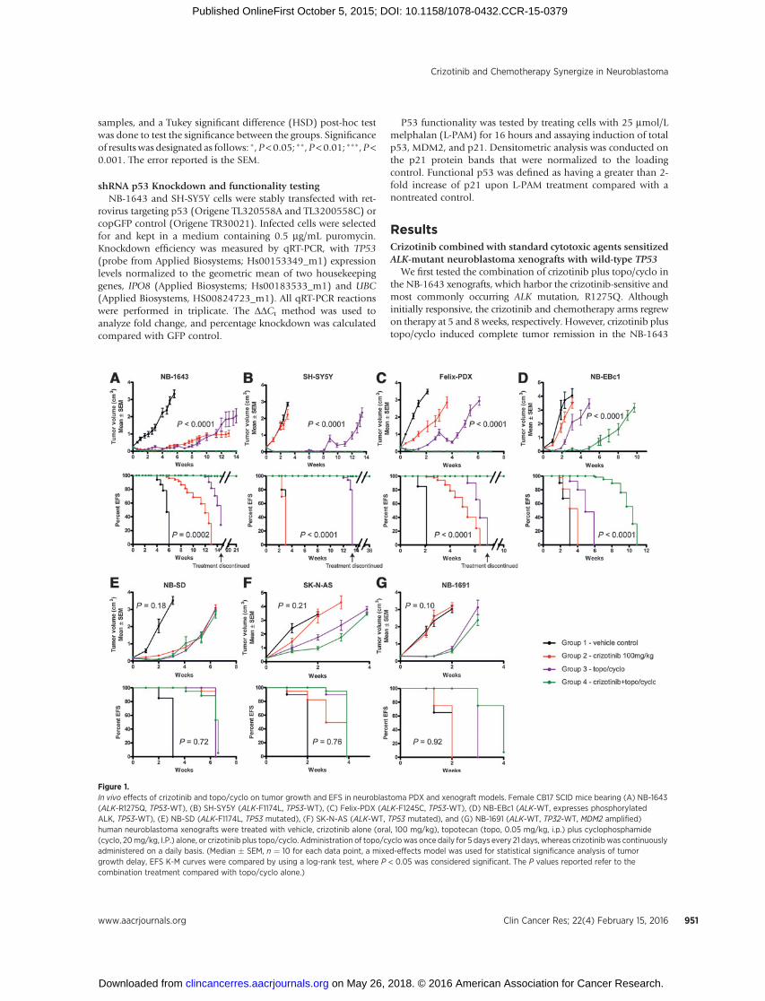
We first tested the combination of crizotinib plus topo/cyclo inthe NB-1643 xenografts, which harbor the crizotinib-sensitive andmost commonly occurring ALK mutation, R1275Q. Althoughinitially responsive, the crizotinib and chemotherapy arms regrewon therapy at 5 and 8 weeks, respectively. However, crizotinib plustopo/cyclo induced complete tumor remission in the NB-1643
Figure 1.In vivo effects of crizotinib and topo/cyclo on tumor growth and EFS in neuroblastoma PDX and xenograft models. Female CB17 SCID mice bearing (A) NB-1643(ALK-R1275Q, TP53-WT), (B) SH-SY5Y (ALK-F1174L, TP53-WT), (C) Felix-PDX (ALK-F1245C, TP53-WT), (D) NB-EBc1 (ALK-WT, expresses phosphorylatedALK, TP53-WT), (E) NB-SD (ALK-F1174L, TP53 mutated), (F) SK-N-AS (ALK-WT, TP53 mutated), and (G) NB-1691 (ALK-WT, TP32-WT, MDM2 amplified)human neuroblastoma xenografts were treated with vehicle, crizotinib alone (oral, 100 mg/kg), topotecan (topo, 0.05 mg/kg, i.p.) plus cyclophosphamide(cyclo, 20mg/kg, I.P.) alone, or crizotinib plus topo/cyclo. Administration of topo/cyclo was once daily for 5 days every 21 days, whereas crizotinib was continuouslyadministered on a daily basis. (Median � SEM, n ¼ 10 for each data point, a mixed-effects model was used for statistical significance analysis of tumorgrowth delay, EFS K-M curves were compared by using a log-rank test, where P < 0.05 was considered significant. The P values reported refer to thecombination treatment compared with topo/cyclo alone.)
Crizotinib and Chemotherapy Synergize in Neuroblastoma
www.aacrjournals.org Clin Cancer Res; 22(4) February 15, 2016 951
on May 26, 2018. © 2016 American Association for Cancer Research. clincancerres.aacrjournals.org Downloaded from
Published OnlineFirst October 5, 2015; DOI: 10.1158/1078-0432.CCR-15-0379
Krytska et al.
Clin Cancer Res; 22(4) February 15, 2016 Clinical Cancer Research952
on May 26, 2018. © 2016 American Association for Cancer Research. clincancerres.aacrjournals.org Downloaded from
Published OnlineFirst October 5, 2015; DOI: 10.1158/1078-0432.CCR-15-0379
model for 14 weeks of treatment (Fig. 1A, top), with tumorsregrowing on average, 6.6 weeks after therapy had ended. Survivalin the combination group was superior to survival in all the otherconditions (Fig. 1A, bottomand Supplementary Table S1).Wenextevaluated this combination therapy in SH-SY5Y xenografts, whichharbor the second most frequent ALK mutation (F1174L), previ-ously shown tobe resistant to crizotinib (16,29). In agreementwithprevious data, crizotinib alone lacked antitumor activity in thismodel, with treated tumors growing as fast as the untreated cohort.Although initially effective, topo/cyclo did not demonstrate asustained response, with tumors regrowing on therapy at 8 weeks(Fig. 1B, top and Supplementary Table S1). However, combiningcrizotinib with topo/cyclo achieved rapid and sustained completetumor regressions for the duration of treatment. Mice treated withtopo/cyclo þ crizotinib showed significantly improved EFS anddecreased tumor growth rates compared with all groups, andmaintained complete responses for 24 weeks after cessation oftreatment (Fig. 1B, bottom and Supplementary Table S1).
We next assessed the combination in Felix-PDX PDXs, whichharbor the third most common ALK mutation (R1245C) inneuroblastoma. Similarly to SH-SY5Y, these xenografts have beenshown to display de novo resistance to crizotinib (21). Althoughtreatmentwith crizotinib or the topo/cyclo alone displayed amildtumor growth delay, Felix-PDX treated with the topo/cyclo þcrizotinib achieved and maintained complete responses (Fig. 1C,top andSupplementary Table S1), and a significant increase inEFS(Fig. 1C, bottom and Supplementary Table S1). Combinationtherapy led to complete regression for the duration of treatment inFelix-PDX mice, with regrowth of tumors occurring 3 weeks aftercessation of therapy.
The combination was then tested in NB-EBc1, an ALK WTxenograft that displays robust constitutive ALK activation andhas been previously shown to be dependent on ALK signaling forgrowth (6). The combination therapy in this model was signif-icantly more effective than vehicle, crizotinib alone, and chemo-therapy alone, delaying tumor growth for 4 weeks before on-treatment progression (Fig. 1D, top andSupplementary Table S1).Although crizotinib and topo/cyclo alone resulted in significantlydecreased tumor volumes compared with vehicle, neither therapyincreased survival rates over control, in contrast with the pro-longed survival seen with topo/cyclo þ crizotinib (Fig. 1D,bottom and Supplementary Table S1). All treatments were welltolerated by the mice, with no signs of systemic toxicity or weightloss (Supplementary Fig. S1).
Crizotinib þ topo/cyclo was not efficacious in xenograftsharboring TP53 mutations
We next investigated the activity of crizotinib in combinationwith cytotoxic agents against crizotinib-resistant xenograftmodels harboring various ALK and TP53 mutations. Althoughthe growth of NB-SD xenografts harboring ALK F1174L and TP53mutations was significantly delayed by all treatment conditions
compared with vehicle, and while the combination treated armshowed evidence of pALK abrogation (Supplementary Fig. S4), allxenografts experienced rapid tumor progression while on therapy(Fig. 1E and Supplementary Table S1). Importantly, combiningcrizotinib with topo/cyclo did not increase antitumor activitycompared with single-agent crizotinib or topo/cyclo. Finally, wesought to test the efficacy of the combination inmodels withwild-type ALK and TP53 mutations using the xenografts SK-N-AS andNB-1691. As expected, crizotinib alone did not affect tumorgrowth in either xenograft model (Fig. 1F, top and 1G top andSupplementary Table S1). Both the cytotoxic agents and thecombination treatments resulted in a mild tumor growth delay,with tumors rapidly progressing on therapy. The combinationshowed no greater efficacy in these xenografts than therapy withtopo/cyclo alone. In agreement with the tumor growth rates, EFSanalysis demonstrated a similar pattern in both xenograft models(Fig. 1E–G, bottom).
The combination of crizotinib and chemotherapy was activeagainst patient-derived ALK-mutant neuroblastoma cell lineswith functional p53
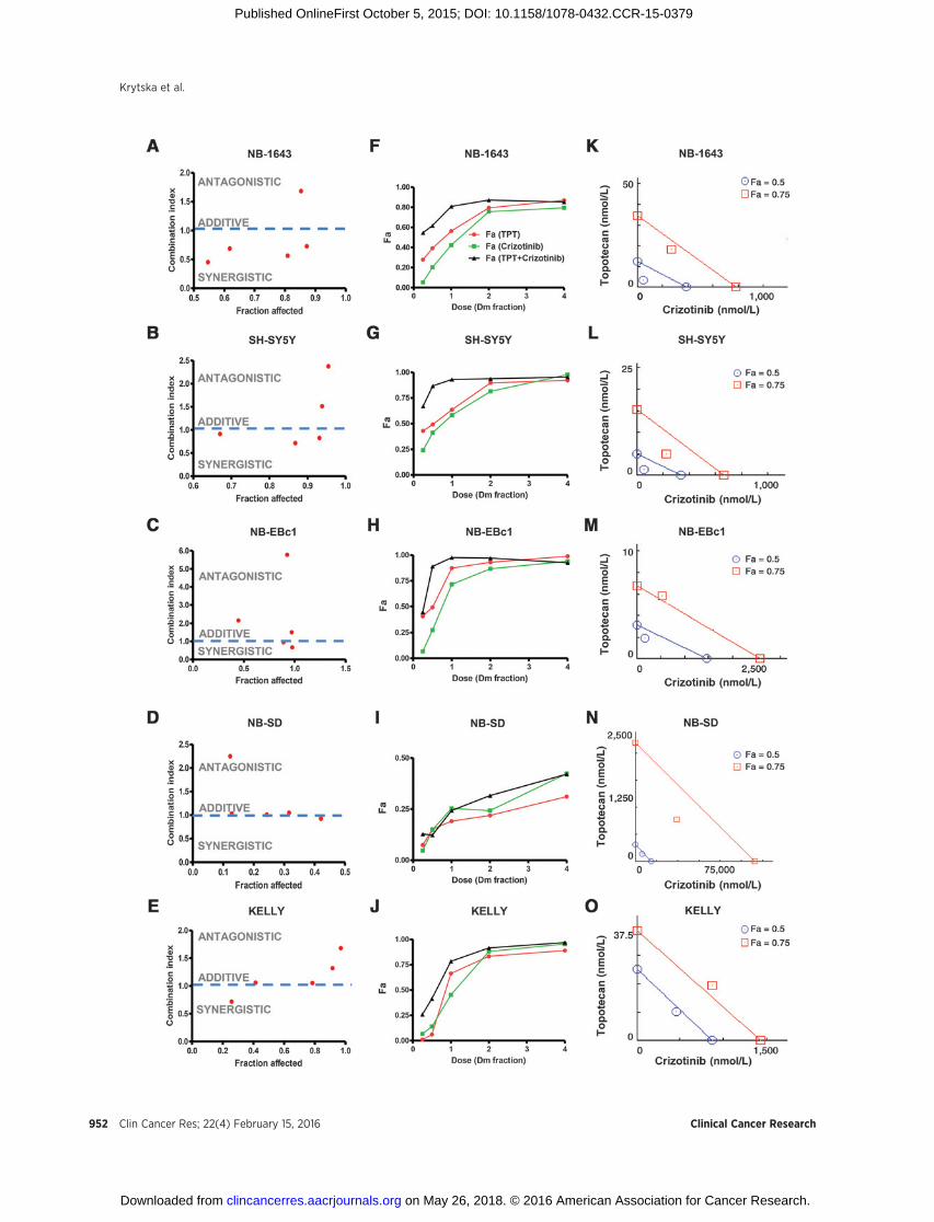
To assay for synergy between crizotinib and cytotoxic agents invitro, we used two methods of analysis: the median-effect CI andthe isobologram.We first tested crizotinib and topotecan as singleagents and determined the median-effect dose (Dm) values(equivalent to the absolute IC50), in a panel offive neuroblastomacell lines differing in their genotypic profiles (Fig. 2 and Supple-mentary Table S3). Crizotinib and topotecan were the leasteffective as single agents and in combination in NB-SD (Fig.2D) and KELLY (Fig. 2E), both cell lines that harbor a TP53mutation. In the p53 functional cell lines, the combinationtherapy resulted in a greater inhibition of cell viability comparedwith single agent treatments, and demonstrated a high level ofinhibition (high Fa). The CI analysis for NB-1643 (Fig. 2A) andSH-SY5Y (Fig. 2B), both ALK-mutated cell lines, revealed severalvalues less than 1.0, indicating a synergy that was stronger atlower-dose combinations. The data were also presented in dose-response curves (Fig. 2F–J), where the dose of drug, as a fraction ofthe cell lines' respective Dm values, was plotted against thefraction of cells affected by treatment (Fa). In all cell lines exceptfor NB-SD, the combination treatment resulted in a greater Fa atlower doses compared with single agent crizotinib or topotecan.NB-EBc1 showed mild synergy at two doses (Fig. 3C), with thehigher doses reflecting antagonism. The dose-reduction indices(DRI) at Dm ranged from 2 to 7 for crizotinib and from 2 to 4 fortopotecan in cell lines exhibiting synergy (Supplementary TableS3), suggesting that such synergistic interactions between crizo-tinib and topotecan provide the opportunity to reduce the con-centrations of individual drugs and thereby potentially reducetheir associated toxicities. Indeed, the maximal activities of thesingle agents could be extended to over 90% cell growth inhibi-tion when applied in combination.
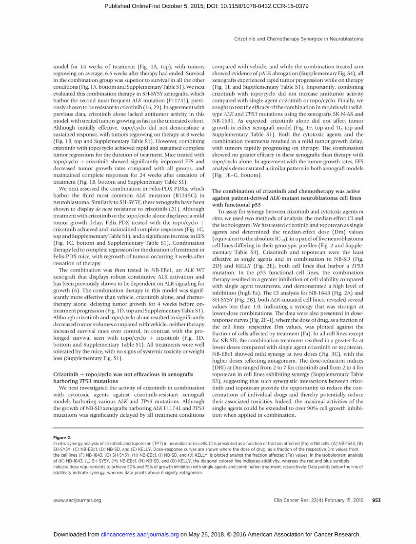
Figure 2.In vitro synergy analysis of crizotinib and topotecan (TPT) in neuroblastoma cells. CI is presented as a function of fraction affected (Fa) in NB cells: (A) NB-1643, (B)SH-SY5Y, (C) NB-EBc1, (D) NB-SD, and (E) KELLY. Dose–response curves are shown where the dose of drug, as a fraction of the respective Dm values fromthe cell lines (F) NB-1643, (G) SH-SY5Y, (H) NB-EBc1, (I) NB-SD, and (J) KELLY, is plotted against the fraction affected (Fa) values. In the isobologram analysisof (K) NB-1643, (L) SH-SY5Y, (M) NB-EBc1, (N) NB-SD, and (O) KELLY, the diagonal colored line indicates additivity, whereas the red and blue symbolsindicate dose requirements to achieve 50% and 75% of growth inhibition with single agents and combination treatment, respectively. Data points below the line ofadditivity indicate synergy, whereas data points above it signify antagonism.
Crizotinib and Chemotherapy Synergize in Neuroblastoma
www.aacrjournals.org Clin Cancer Res; 22(4) February 15, 2016 953
on May 26, 2018. © 2016 American Association for Cancer Research. clincancerres.aacrjournals.org Downloaded from
Published OnlineFirst October 5, 2015; DOI: 10.1158/1078-0432.CCR-15-0379
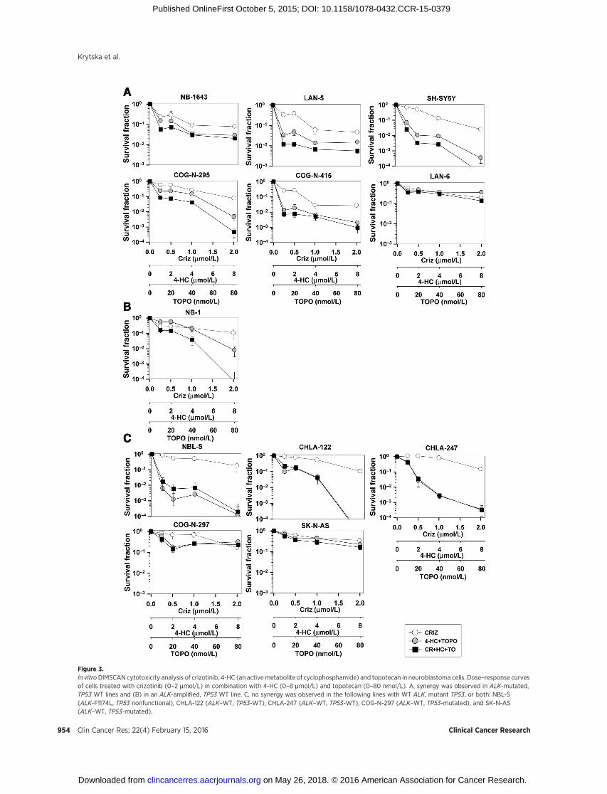
Figure 3.In vitroDIMSCAN cytotoxicity analysis of crizotinib, 4-HC (an activemetabolite of cyclophosphamide) and topotecan in neuroblastoma cells. Dose–response curvesof cells treated with crizotinib (0–2 mmol/L) in combination with 4-HC (0–8 mmol/L) and topotecan (0–80 nmol/L). A, synergy was observed in ALK-mutated,TP53 WT lines and (B) in an ALK-amplified, TP53 WT line. C, no synergy was observed in the following lines with WT ALK, mutant TP53, or both: NBL-S(ALK-F1174L, TP53 nonfunctional), CHLA-122 (ALK–WT, TP53-WT), CHLA-247 (ALK–WT, TP53-WT), COG-N-297 (ALK–WT, TP53-mutated), and SK-N-AS(ALK–WT, TP53-mutated).
Krytska et al.
Clin Cancer Res; 22(4) February 15, 2016 Clinical Cancer Research954
on May 26, 2018. © 2016 American Association for Cancer Research. clincancerres.aacrjournals.org Downloaded from
Published OnlineFirst October 5, 2015; DOI: 10.1158/1078-0432.CCR-15-0379
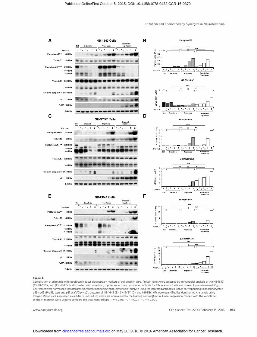
Figure 4.Combination of crizotinib with topotecan induces downstream markers of cell death in vitro. Protein levels were assessed by immunoblot analysis of (A) NB-1643,(C) SH-SY5Y, and (E) NB-EBc1 cells treated with crizotinib, topotecan, or the combination of both for 8 hours with fractional doses of predetermined IC50s.Cell lysateswere normalized for total protein content and subjected to immunoblot analysis using the indicated antibodies. Bands corresponding to phosphorylated-p53-ser15 (P-p53, top) and p21 Waf1/Cip1 (p21, bottom) of NB-1643 (B), SH-SY5Y (D), and NB-EBc1 (F) were quantified by densitometric analysis usingImageJ. Results are expressed as arbitrary units (A.U.) and were normalized to the loading control b-actin. Linear regression models with the vehicle setas the y-intercept were used to compare the treatment groups. � , P < 0.05; �� , P < 0.01; ��� , P < 0.001.
www.aacrjournals.org Clin Cancer Res; 22(4) February 15, 2016 955
Crizotinib and Chemotherapy Synergize in Neuroblastoma
on May 26, 2018. © 2016 American Association for Cancer Research. clincancerres.aacrjournals.org Downloaded from
Published OnlineFirst October 5, 2015; DOI: 10.1158/1078-0432.CCR-15-0379
Clin Cancer Res; 22(4) February 15, 2016 Clinical Cancer Research956
Krytska et al.
on May 26, 2018. © 2016 American Association for Cancer Research. clincancerres.aacrjournals.org Downloaded from
Published OnlineFirst October 5, 2015; DOI: 10.1158/1078-0432.CCR-15-0379

To further evaluate the dose-dependent drug interaction ofcrizotinib and topotecan, isobolograms at effect levels of 50%and 75% cell growth inhibition were created and analyzed (Fig.2K–O). As shown in Fig. 2, the isobole of the combination wasbelow the line of additivity at 50% inhibition in cell lines withfunctional p53 (Fig. 2K–M). Importantly, in NB-SD and KELLY,TP53-mutant cell lines, additivity and antagonism, were observedatmost concentrations tested, in agreement with the CI plots (Fig.2I and J). In line with our in vivo data, these results further suggestthat the synergy of this combination relies, at least in part, onfunctional p53.
We used the DIMSCAN assay as a second method to assess cellviability and synergy and included 4-HC, the active metabolite ofcyclophosphamide, in this assay. In accordance with the CTGassay results, the DIMSCAN assay showed synergy at clinicallyachievable doses in a broader panel of neuroblastoma cell lineswith ALK mutations and functional p53: NB-1643, LAN-5, SH-SY5Y, COG-N-295, COG-N-415, and LAN-6 (Fig. 3A and Sup-plementary Table S3). The combination was also synergistic inNB-1, an ALK amplified line with functional p53 (Fig. 3B andSupplementary Table S3 and Supplementary Fig. S2B). In severalcell lines, the combination of crizotinib with topotecanþ4-HCresulted in a 3-log (99.9%) cell kill (Fig. 3A). Notably, thiscombination displays CI values that range from synergistic toadditive in all seven ALK aberrant, p53 functional cell lines tested(Supplementary Fig. S2A and B) and potently inhibits a numberof cell lines with ALK-F1174L mutations, which confer preferen-tial ATP-binding affinity and innate resistance to single agentcrizotinib. Thus, these data support our in vivofindings andpredictgreater efficacy when combining crizotinib with cytotoxic agentsin p53 functional models where crizotinib monotherapy is insuf-ficient. Furthermore, we also tested the combination therapy byDIMSCAN in the ALKWT and TP53WT lines, NBL-S, CHLA-122,andCHLA-247 and theALKWT,TP53-mutated lines, COG-N-297and SK-N-AS (Fig. 3C and Supplementary Fig. S2C). Synergybetween crizotinib and topo/4-HC was not observed in any ofthese cell lines, highlighting the requirement of aberrant oractivated ALK for the efficacy of this combination therapy.
Crizotinib potentiated downstream signals of p53 activation intopotecan-treated neuroblastoma cells in vitro
To next evaluate the potential role of crizotinib in combinationwith chemotherapy on the p53pathway,we treatedNB-1643 (Fig.4A), SH-SY5Y (Fig. 4C), and NB-EBc1 (Fig. 4E) cells with vehicle,crizotinib, topotecan, or crizotinib plus topotecan at fractionaldoses of each cell lines' respective IC50s. To assess the impact of thesingle agents and combination therapy on ALK signaling, cellswere treated for 8 hours before collection for immunoblot anal-ysis. Crizotinib individually or in combination with topotecanresulted in a dose-dependent abrogation of phosphorylatedALKY1604 in all three cell lines, as well as upregulation of cleaved
caspase-3 at one- and two times the IC50 doses of single-agent andcombination therapy (Fig. 4A, C, and E). We also evaluated levelsof phosphorylated p53s15, and total p53, as well as the down-stream markers of p53 activation, p21 and PUMA, in the treatedcells. Expectedly, as a DNA-damaging agent, topotecan inducedphosphorylated and total p53 upregulation across all cell lines(Fig. 4A, C, and E). Although crizotinib increased levels ofphosphorylated p53s15 in the crizotinib-sensitive line, NB-1643(Fig. 4A and B), it did not in the crizotinib-insensitive SH-SY5Y(Fig. 4C and D) and NB-EBc1 (Fig. 4E and F) cells, irrespective ofcaspase-3 activation. Nonetheless, in combination-treated cells,both forms of p53, as well as p21 and PUMA, were found to behighly upregulated, to levels suggestive of greater than addi-tivity. In fact, densitometric analysis of the bands revealed asignificantly higher increase in phospho-p53S15 (Fig. 4B, D, andF, top), p21 (Fig. 4B, D, and F, bottom) and cleaved caspase-3(data not shown) in the combination-treated cells, suggestingthat these agents cooperate with one another to induce greaterapoptosis.
Crizotinib potentiated p53 activation in chemotherapy-treatedneuroblastoma xenografts
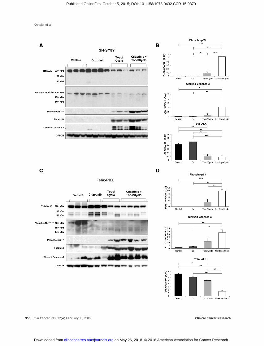
To next explore the in vivo effects of chemotherapy and crizo-tinib on p53 activation, mice bearing SH-SY5Y and Felix-PDXtumors were treated with crizotinib, topo/cyclo, and the combi-nation for 3 days. Tumors were collected 4 hours after lasttreatment, lysed and analyzed by immunoblotting. In SH-SY5Y,the standard dose of crizotinib (100mg/kg) did not alter levels ofphosphorylated-ALK (Fig. 5A), in agreement with previous resultsindicating that this dose is insufficient for abrogating ALK signal-ing (16).Unexpectedly, topo/cyclo alone reducedprotein levels ofboth total and phosphorylated ALK (Fig. 5A and B).Mirroring theeffects described in vitro, levels of total, phosphorylated p53, andcleaved caspase-3 were greater in the combination therapy com-pared with topo/cyclo and crizotinib agent alone (Fig. 5A).Quantitative analysis showed that the combination inducedsignificantly greater levels of phospho-p53 compared with allother treatments, including topo/cyclo alone (Fig. 5B, top).
Finally, in line with the SH-SY5Y results, treatment with topo/cyclo � crizotinib showed a reduction in total ALK expression inthe Felix-PDX patient derived-tumors (Fig. 5C and D). Upregula-tion of both phosphorylated p53 and caspase-3 was observed intopo/cyclo and topo/cyclo þ crizotinib-treated tumors but levelsof these two proteins were not markedly greater in the combina-tion therapy compared with topo/cyclo alone (Fig. 5C and D).
The combination of crizotinib and chemotherapy enhancedcytotoxicity in neuroblastoma cell lines carrying wild-type p53
To explore the potential role of p53 in mediating the synergybetween crizotinib and chemotherapy, we established p53
Figure 5.Combination of crizotinib with topo/cyclo induces downstream markers of cell death in vivo. Once tumors reached 200 mm3, mice bearing (A) SH-SY5Y and(C) Felix-PDX human neuroblastoma xenografts were treated for 3 consecutive days as follows: vehicle, crizotinib (Cz, 100 mg/kg, oral), topotecan (Topo,0.05mg/kg) and cyclophosphamide (Cyclo, 20mg/kg), and combination of both. Tumorswere harvested 4 hours after last treatment. Cell lysateswere analyzed byimmunoblotting with the indicated antibodies. Bands corresponding to phosphorylated-p53-ser15 (P-p53, top), cleaved caspase-3 (cc3, middle), andtotal ALK (bottom) of (B) SH-SY5Y and (D) Felix-PDX were quantified by densitometric analysis using the software ImageJ. Results are expressed asarbitrary units (A.U.) and were normalized against the loading control GAPDH. The bands from each treatment group were averaged and compared using anANOVA and a Tukey HSD test. � , P < 0.05; �� , P < 0.01; ��� , P < 0.001.
www.aacrjournals.org Clin Cancer Res; 22(4) February 15, 2016 957
Crizotinib and Chemotherapy Synergize in Neuroblastoma
on May 26, 2018. © 2016 American Association for Cancer Research. clincancerres.aacrjournals.org Downloaded from
Published OnlineFirst October 5, 2015; DOI: 10.1158/1078-0432.CCR-15-0379
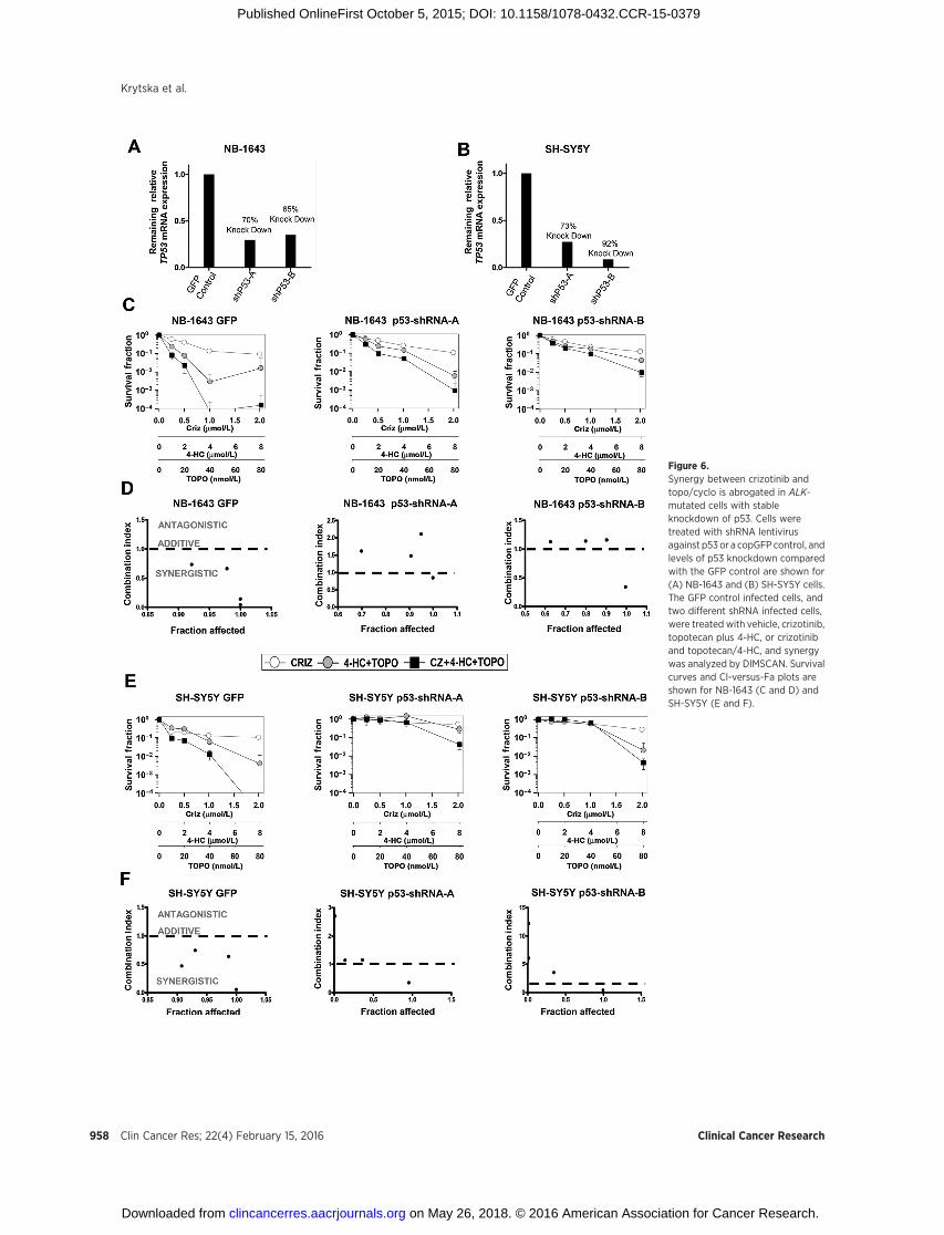
Figure 6.Synergy between crizotinib andtopo/cyclo is abrogated in ALK-mutated cells with stableknockdown of p53. Cells weretreated with shRNA lentivirusagainst p53 or a copGFP control, andlevels of p53 knockdown comparedwith the GFP control are shown for(A) NB-1643 and (B) SH-SY5Y cells.The GFP control infected cells, andtwo different shRNA infected cells,were treated with vehicle, crizotinib,topotecan plus 4-HC, or crizotiniband topotecan/4-HC, and synergywas analyzed by DIMSCAN. Survivalcurves and CI-versus-Fa plots areshown for NB-1643 (C and D) andSH-SY5Y (E and F).
Clin Cancer Res; 22(4) February 15, 2016 Clinical Cancer Research958
Krytska et al.
on May 26, 2018. © 2016 American Association for Cancer Research. clincancerres.aacrjournals.org Downloaded from
Published OnlineFirst October 5, 2015; DOI: 10.1158/1078-0432.CCR-15-0379
knockdown NB-1643 and SH-SY5Y cells using two differentshRNA vectors (referred to as shRNA "A" and "B"). Both cell linesshowed greater than 60% p53 knockdown compared with theirrespective GFP-transduced control with both vectors (Fig. 6A andB). Testing the combination of crizotinib þ topotecan/4-HC inthese lines by DIMSCAN revealed synergy in the GFP-transducedcontrol lines (Fig. 6C and E, left), consistent with the previousDIMSCAN results using the parental NB-1643 and SH-SY5Y lines(Fig. 3A). Notably, synergy was abrogated in the NB-1643 or SH-SY5Y p53 knockdown lines (Fig. 6C–F, respectively).
DiscussionNeuroblastoma remains a leading cause of childhood cancer
death despite progress made in the field of chemoradiotherapy,surgery, and immunotherapy. Optimizing treatment for thesepatients has become increasingly complex, requiring an approachthat allows us to determine when it is best to incorporate targeteddrugs and with what chemotherapy to combine targeted agents,and in which patients. Rapid advances in the development oftargeted therapies have provided the unprecedented ability totherapeutically exploit oncogenic drivers.ALK is a tractablemolec-ular target in neuroblastoma, with aberrations in this geneaccounting for over 80% of hereditary cases, and present in14%of high-risk cases (6, 29).We previously reported differentialsensitivity of themost common ALK kinase domainmutations inneuroblastoma (16, 29), suggesting that ALK inhibitors as a singleagent are likely to fail in such patients. However, there remains thepotential for ALK inhibitors to enhance traditional cytotoxicagents in tumors carrying ALKmutations that confer single-agentALK inhibitor resistance.
Here, we report that a novel therapeutic combination of theALK inhibitor, crizotinib, with conventional chemotherapeutics(topotecan plus cyclophosphamide), has marked antitumoractivity and substantially prolongs EFS in both crizotinib-sensi-tive and crizotinib-resistant models of ALK-mutant neuroblasto-ma. We also show evidence for in vitro synergy with this combi-nation in one neuroblastoma cell line harboring high-level ALKamplification. Although our data suggest that ALK-amplifiedtumors will be responsive to crizotinib combined with topotecanand cyclophosphamide, additional nonclinical studies arewarranted in models that represent this infrequent subset ofneuroblastomas with ALK aberrations. Importantly, synergywas seen with the combination in settings where crizotinib orchemotherapy alone induced only mild or no activity. Forinstance, xenografts harboring the resistant F1174L mutationshowed complete remission of tumors for an additional 24 weeksafter discontinuation of therapy. Interestingly, this sustainedantitumor activity was accompanied by striking upregulation ofthe tumor suppressor protein, p53, and its downstream effectorsp21, PUMA, and cleaved-caspase 3 in ALK aberrant and p53functional neuroblastomamodels treatedwith combination ther-apy. This observation prompted us to investigate whether activa-tion of the p53 pathway could be a determinant of the synergyobserved when combining crizotinib and genotoxic agents. Thelack of response to the combination therapy in neuroblastomamodels carrying mutated TP53 provided the initial evidencethat this synergy relies, at least in part, on a functional p53pathway. In addition, in two ALK-mutated neuroblastoma cellswith genetically ablated p53, we found an abrogation of synergyupon combination treatment compared to the respective control
lines. Taken together, we show here that activation of the p53pathway plays a role in mediating the synergy observed betweencrizotinib and chemotherapy in neuroblastoma models withactivated ALK.
Our findings are supported by recent studies suggesting acorrelation between inhibition of ALK signaling and reactivationof p53-mediated cell death. In addition, synthetic peptides cor-responding to the proapoptotic domain of ALK caused p53-mediated cytotoxicity in ALCL and neuroblastoma cells (30). Inthe same study, ALK peptides interacted with proteins that havebeen previously reported to interact with the p53 gene andprotein. Similar to our findings, p53 knockdown rescued bothALCL and neuroblastoma cells from ALK peptide-mediated celldeath. The precise mechanism of how ALK inhibition impacts thep53 pathway and promotes synergy between crizotinib andchemotherapy has not been fully elucidated. Studies are ongoingto determine whether ALK inhibitor-mediated cell death is due toa dual function of ALK in regulating not only the canonical RTKpathway but also the p53 pathway. This could inform the designof drug combinations targeting ALK and other specific p53-tar-geted therapies such as MDM2 inhibitors. We observed a markedreduction in total ALK levels in the chemotherapy and combina-tion-treated xenograft tumors of unclear significance, but wepostulate this could be secondary to decreased transcriptionupon chemotherapy treatment or a potential feedback mecha-nism between ALK and p53 in which ALK, serving as a negativeregulator of p53, is then decreased by transcriptional activityof p53.
Altogether, we demonstrate that combining crizotinib withconventional chemotherapeutic agents is effective in ALK-drivenmodels of neuroblastoma that show differential crizotinib sen-sitivity, providing the rationale for the currently accruing COGphase I trial (NCT01606878). Within the high-risk subset ofnewly diagnosed neuroblastoma patients, 14% will harbor anALK aberration and will have inferior outcome, providing aunique opportunity for clinical studies testing the integration ofcrizotinib into the backbone of contemporary chemotherapyregimens for these patients. The absence of synergy in neuroblas-toma models harboring wild-type ALK and/or loss-of-functionTP53mutations supports a responder hypothesis that ALK statusin addition to a functional p53 pathway is a determinant ofresponse to this therapeutic strategy. Systematic investigation ofcrizotinib and other ALK inhibitors with DNA-damaging chemo-therapy combinations is ongoing to rigorously evaluate theimportance of timing and sequence of these regimens in orderto best guide clinical trial design.
Disclosure of Potential Conflicts of InterestNo potential conflicts of interest were disclosed.
Authors' ContributionsConception and design: H.T. Ryles, R. Sano, C.P. Reynolds, Y.P. Moss�eDevelopment of methodology: H.T. Ryles, R. Sano, M.R. Makena, C.P. Rey-nolds, Y.P. Moss�eAcquisition of data (provided animals, acquired and managed patients,provided facilities, etc.): K. Krytska, T.D. Hansel, M.R. Makena, M.M. Song,C.P. Reynolds, Y.P. Moss�eAnalysis and interpretation of data (e.g., statistical analysis, biostatistics,computational analysis): K. Krytska, H.T. Ryles, P. Raman, N.R. Infarinato,M.R. Makena, M.M. Song, C.P. Reynolds, Y.P. Moss�eWriting, review, and/or revision of the manuscript: K. Krytska, H.T. Ryles,R. Sano, M.R. Makena, M.M. Song, C.P. Reynolds, Y.P. Moss�e
www.aacrjournals.org Clin Cancer Res; 22(4) February 15, 2016 959
Crizotinib and Chemotherapy Synergize in Neuroblastoma
on May 26, 2018. © 2016 American Association for Cancer Research. clincancerres.aacrjournals.org Downloaded from
Published OnlineFirst October 5, 2015; DOI: 10.1158/1078-0432.CCR-15-0379
Administrative, technical, or material support (i.e., reporting or organizingdata, constructing databases): K. Krytska, Y.P. Moss�eStudy supervision: K. Krytska, R. Sano, Y.P. Moss�eOther (execution of experiments): N.R. Infarinato
Grant SupportThis work was supported by NIH Grant R01CA140198 (to Y.P. Moss�e).
The costs of publication of this article were defrayed in part by thepayment of page charges. This article must therefore be hereby markedadvertisement in accordance with 18 U.S.C. Section 1734 solely to indicatethis fact.
Received February 16, 2015; revised September 21, 2015; accepted September21, 2015; published OnlineFirst October 5, 2015.
References1. Maris JM, Hogarty MD, Bagatell R, Cohn SL. Neuroblastoma. Lancet
2007;369:2106–20.2. Maris JM. Recent advances in neuroblastoma. N Engl J Med 2010;362:
2202–11.3. Chen Y, Takita J, Choi YL, Kato M, Ohira M, Sanada M, et al. Oncogenic
mutations of ALK kinase in neuroblastoma. Nature 2008;455:971–4.4. George RE, Sanda T, Hanna M, Frohling S, Luther W II, Zhang J, et al.
Activatingmutations in ALKprovide a therapeutic target in neuroblastoma.Nature 2008;455:975–8.
5. Janoueix-Lerosey I, Lequin D, Brugieres L, Ribeiro A, de Pontual L, Com-baret V, et al. Somatic and germline activating mutations of the ALK kinasereceptor in neuroblastoma. Nature 2008;455:967–70.
6. Mosse YP, Laudenslager M, Longo L, Cole KA, Wood A, Attiyeh EF, et al.Identification of ALK as a major familial neuroblastoma predispositiongene. Nature 2008;455:930–5.
7. Iwahara T, Fujimoto J, Wen D, Cupples R, Bucay N, Arakawa T, et al.Molecular characterization of ALK, a receptor tyrosine kinase expressedspecifically in the nervous system. Oncogene 1997;14:439–49.
8. Yao S, Cheng M, Zhang Q, Wasik M, Kelsh R, Winkler C. Anaplasticlymphoma kinase is required for neurogenesis in the developing centralnervous system of zebrafish. PLoS One 2013;8:e63757.
9. Chiarle R, Voena C, Ambrogio C, Piva R, Inghirami G. The anaplasticlymphoma kinase in the pathogenesis of cancer. Nat Rev Cancer 2008;8:11–23.
10. Morris SW, Kirstein MN, Valentine MB, Dittmer KG, Shapiro DN, SaltmanDL, et al. Fusion of a kinase gene, ALK, to a nucleolar protein gene, NPM, innon-Hodgkin's lymphoma. Science 1994;263:1281–4.
11. Soda M, Choi YL, Enomoto M, Takada S, Yamashita Y, Ishikawa S, et al.Identification of the transforming EML4-ALK fusion gene in non-small-celllung cancer. Nature 2007;448:561–6.
12. Mosse YP, Wood A, Maris JM. Inhibition of ALK signaling for cancertherapy. Clin Cancer Res 2009;15:5609–14.
13. Christensen JG. Proof of principle for crizotinib in anaplastic lymphomakinase-positive malignancies was achieved in ALK-positive nonclinicalmodels. Mol Cancer Ther 2011;10:2024.
14. Camidge DR, Bang YJ, Kwak EL, Iafrate AJ, Varella-Garcia M, Fox SB, et al.Activity and safety of crizotinib in patients with ALK-positive non-small-cell lung cancer: updated results from a phase 1 study. Lancet Oncol2012;13:1011–9.
15. Kwak EL, Bang YJ, Camidge DR, Shaw AT, Solomon B, Maki RG, et al.Anaplastic lymphoma kinase inhibition in non-small-cell lung cancer. NEngl J Med 2010;363:1693–703.
16. Bresler SC, Wood AC, Haglund EA, Courtright J, Belcastro LT, PlegariaJS, et al. Differential inhibitor sensitivity of anaplastic lymphoma
kinase variants found in neuroblastoma. Sci Transl Med 2011;3:108ra14.
17. Mosse YP, LimMS, Voss SD,Wilner K, Ruffner K, Laliberte J, et al. Safety andactivity of crizotinib for paediatric patients with refractory solid tumours oranaplastic large-cell lymphoma: a Children's Oncology Group phase 1consortium study. Lancet Oncol 2013;14:472–80.
18. Komuro H, Hayashi Y, Kawamura M, Hayashi K, Kaneko Y, Kamoshita S,et al.Mutations of the p53 gene are involved in Ewing's sarcomas but not inneuroblastomas. Cancer Res 1993;53:5284–8.
19. Pugh TJ, Morozova O, Attiyeh EF, Asgharzadeh S, Wei JS, Auclair D, et al.The genetic landscape of high-risk neuroblastoma. Nat Genet 2013;45:279–84.
20. Carr-Wilkinson J, O'Toole K, Wood KM, Challen CC, Baker AG, Board JR,et al. High frequency of p53/MDM2/p14ARF pathway abnormalities inrelapsed neuroblastoma. Clin Cancer Res 2010;16:1108–18.
21. Keshelava N, Zuo JJ, Chen P, Waidyaratne SN, Luna MC, Gomer CJ, et al.Loss of p53 function confers high-level multidrug resistance in neuroblas-toma cell lines. Cancer Res 2001;61:6185–93.
22. Attiyeh EF, Diskin SJ, Attiyeh MA, Mosse YP, Hou C, Jackson EM, et al.Genomic copy number determination in cancer cells from single nucle-otide polymorphism microarrays based on quantitative genotyping cor-rected for aneuploidy. Genome Res 2009;19:276–83.
23. Frgala T, Kalous O, Proffitt RT, Reynolds CP. A fluorescence microplatecytotoxicity assay with a 4-log dynamic range that identifies synergisticdrug combinations. Mol Cancer Ther 2007;6:886–97.
24. Chou TC, Talaly P. A simple generalized equation for the analysis ofmultiple inhibitions of Michaelis-Menten kinetic systems. J Biol Chem1977;252:6438–42.
25. Sano R, Hou YC, Hedvat M, Correa RG, Shu CW, Krajewska M, et al.Endoplasmic reticulum protein BI-1 regulates Ca(2)(þ)-mediated bioen-ergetics to promote autophagy. Genes Dev 2012;26:1041–54.
26. Hothorn T, Bretz F, Westfall P. Simultaneous inference in general paramet-ric models. Biom J 2008;50:346–63.
27. Pinheiro J, Bates D, DebRoy S, Sarkar D, Team RC. nlme: Linear andNonlinearMixed EffectsModels. R package version. R package version 3.1–120 ed2015.
28. Therneau T, Grambsch P.Modeling survival data: extending the coxmodel.New York, NY: Springer; 2000.
29. Bresler SC, Weiser DA, Huwe PJ, Park JH, Krytska K, Ryles H, et al. ALKmutations confer differential oncogenic activation and sensitivity to ALKinhibition therapy in neuroblastoma. Cancer Cell 2014;26:682–94.
30. Aubry A, Galiacy S, Ceccato L, Marchand C, Tricoire C, Lopez F, et al.Peptides derived from the dependence receptor ALK are proapoptotic forALK-positive tumors. Cell Death Dis 2015;6:e1736.
Clin Cancer Res; 22(4) February 15, 2016 Clinical Cancer Research960
Krytska et al.
on May 26, 2018. © 2016 American Association for Cancer Research. clincancerres.aacrjournals.org Downloaded from
Published OnlineFirst October 5, 2015; DOI: 10.1158/1078-0432.CCR-15-0379
2016;22:948-960. Published OnlineFirst October 5, 2015.Clin Cancer Res Kateryna Krytska, Hannah T. Ryles, Renata Sano, et al. NeuroblastomaCrizotinib Synergizes with Chemotherapy in Preclinical Models of
Updated version
10.1158/1078-0432.CCR-15-0379doi:
Access the most recent version of this article at:
Material
Supplementary
http://clincancerres.aacrjournals.org/content/suppl/2015/10/03/1078-0432.CCR-15-0379.DC1
Access the most recent supplemental material at:
Cited articles
http://clincancerres.aacrjournals.org/content/22/4/948.full#ref-list-1
This article cites 28 articles, 10 of which you can access for free at:
Citing articles
http://clincancerres.aacrjournals.org/content/22/4/948.full#related-urls
This article has been cited by 2 HighWire-hosted articles. Access the articles at:
E-mail alerts related to this article or journal.Sign up to receive free email-alerts
Subscriptions
Reprints and
To order reprints of this article or to subscribe to the journal, contact the AACR Publications Department at
Permissions
Rightslink site. Click on "Request Permissions" which will take you to the Copyright Clearance Center's (CCC)
.http://clincancerres.aacrjournals.org/content/22/4/948To request permission to re-use all or part of this article, use this link
on May 26, 2018. © 2016 American Association for Cancer Research. clincancerres.aacrjournals.org Downloaded from
Published OnlineFirst October 5, 2015; DOI: 10.1158/1078-0432.CCR-15-0379