ctcl: important diagnostic tools - stanford...
TRANSCRIPT

CTCL: Important Diagnostic Tools
Uma Sundram, MD, PhDAssistant ProfessorDepartments of Pathology and DermatologyStanford University

Diagnosis of CTCL
Clinical Information and Histology Immunohistochemistry
identify neoplastic lymphocyte population subtype CTCL according to WHO-EORTC criteria
T cell receptor gene rearrangement studies by polymerase chain reaction (PCR) or Southern blot hybridization identify clonal population
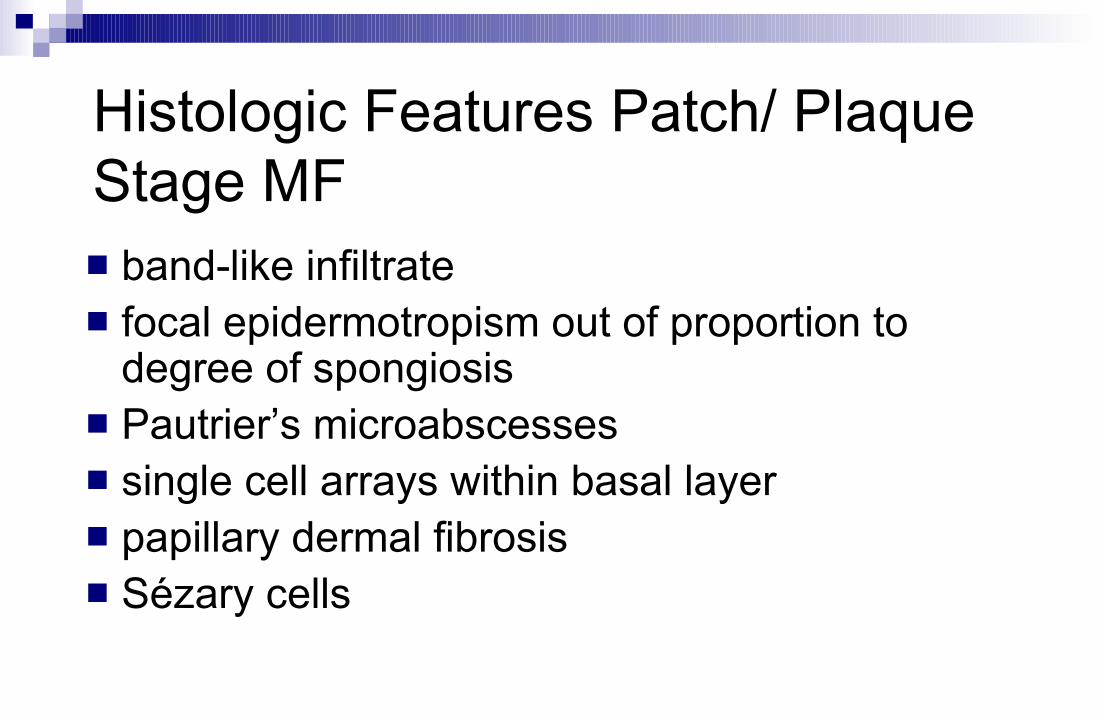
Histologic Features Patch/ Plaque Stage MF band-like infiltrate focal epidermotropism out of proportion to
degree of spongiosis Pautrier’s microabscesses single cell arrays within basal layer papillary dermal fibrosis Sézary cells

Diagnosis of Mycosis fungoides
H&E morphology and clinical correlation

DD of Bandlike Dermal Infiltrates
lymphomatoid drug eruptiontreated spongiotic dermatitisinterface dermatitislichenoid purpuraactinic reticuloidlichen striatusLS & APityriasis lichenoidestype B lymphomatoid papulosisCD8+ epidermotropic CTCL

Immunophenotyping
ImmunophenotypeCD4+, CD7 – (reactive infiltrates can show loss of
CD7)CD4/CD8 ratio (reactive infiltrates can have CD4/CD8
ratios of up to 6:1)

Immunophenotyping
Usually confirmatory in morphologically unequivocal cases Least helpful in ambiguous cases
Scant infiltrate Intraepidermal neoplastic population with variable reactive
infiltrate CD4/ CD8 ratios not well established for entities in DD
(Stanford study is unique)

Molecular Studies
Molecular studies T cell receptor clonally rearranged by PCR or Southern Blotting
in > 80% of MF Up to 30% false positive rate with PCR (GeneScan analysis) Compare the clone in multiple biopsies (sequential or
multifocal)

Diagnosis of Patch/Plaque Mycosis Fungoides
H&E is gold standard biopsies from several sites serial biopsies close clinicopathologic correlation biopsy performed after adequate time off topical therapy (1-2
weeks)

CD8+ Epidermotropic CTCL
mycosis fungoides (hypopigmented variant) Woringer Kolopp disease CD8+ epidermotropic T-cell lymphoma
(provisional entity)

CD8+ Epidermotropic CTCL
generalized patches, plaques, nodules spread to visceral organs, but not LN aggressive clinical course (32 months average
survival)
Berti et al. Am J Pathol 1999,155:483-492

CD8+ Epidermotropic CTCL
band-like infiltrate Medium or large sized cells pronounced epidermotropism in acanthotic
epidermis neoplastic cells CD8+

Phenotype
CD3+ CD8+ TIA-1+ proliferation rate >> MF (approx. 40%)

DD of Dense Dermal Lymphoid Infiltrates
CD20, CD3
CD20+, CD3-CD20-, CD3+CD20-, CD3-
Cutaneous B-cell lymphomaCTCLNK/T- cell lymphoma

DD Nodular Dermal T-cell Infiltrate tumor stage MF/transformed MF Type C lymphomatoid papulosis CD30 positive anaplastic large cell lymphoma (primary cutaneous/
secondary) CD4+ small/medium-sized T-cell lymphoma (provisional entity) Peripheral T cell lymphoma, NOS Extranodal NK/T lymphoma, nasal-type CD8+ aggressive epidermotropic lymphoma Cutaneous gamma/delta TCL (provisional entity) cutaneous lymphoid hyperplasia Leukemia If within the subcutaneous tissue only: subcutaneous panniculitis like T cell
lymphoma

Nodular Dermal T-cell infiltrate
Does the patient have a history of MF?

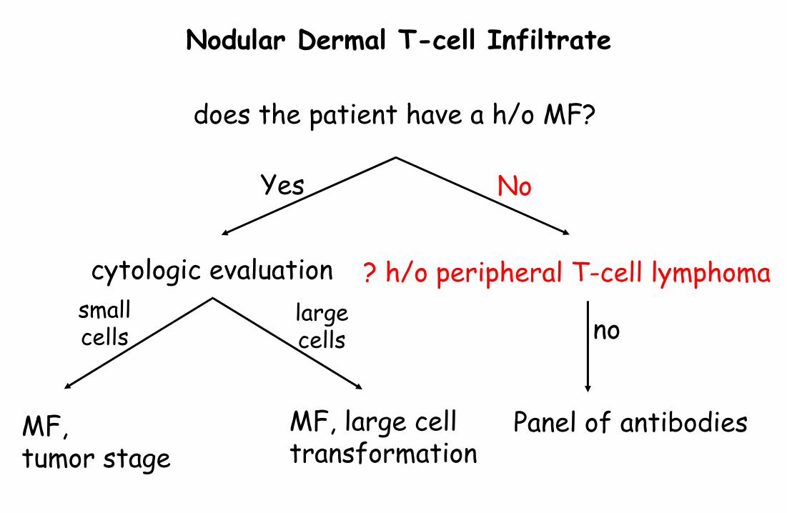
Nodular Dermal T-cell Infiltrate
does the patient have a h/o MF?
MF, tumor stage
Yes
cytologic evaluation
MF, large celltransformation
smallcells
largecells

Tumor Stage/Transformed MF
Antibodies: Ki 67 CD30
Diagnosis of Transformed MF: Infiltrate has to contain >25% large cells

Nodular Dermal T-cell Infiltrate
does the patient have a h/o MF?
MF, tumor stage
Yes No
cytologic evaluation
MF, large celltransformation
smallcells
largecells
Panel of antibodies
? h/o peripheral T-cell lymphoma
no
Nodular Dermal T-cell Infiltrate
does the patient have a h/o MF?
MF, tumor stage
Yes No
cytologic evaluation
MF, large celltransformation
smallcells
largecells
Panel of antibodies
? h/o peripheral T-cell lymphoma
no

First Line Panel of Antibodies
CD30 CD56
CD4 CD8 TIA-1 Ki-67 β-F1 (Important in
gamma/delta T cell lymphoma)

CD30+, CD56-, CD3+/-CD30+ Lymphoproliferative Disorder more than 75% of cells CD30 positive
lymphomatoid papulosis type C primary cutaneous anaplastic large cell lymphoma systemic anaplastic large cell lymphoma,
alk-negative systemic anaplastic large cell lymphoma,
alk-positive (transformed MF)

CD30-, CD56-, CD3+,CD4+/-, CD8+/-, CD4+ small/medium-sized T-cell lymphoma Peripheral T cell lymphoma, NOS CD8+ epidermotropic lymphoma cutaneous gamma/delta TCL

CD4+ Small/Medium-sized T-Cell Lymphoma
Solitary (usually) or multiple tumor nodules not preceded by patches or plaques of MF Relatively favorable clinical course 60% 5 year survival

CD4+ Small/Medium-sized T-Cell Lymphoma deep and diffuse infiltrate small-to-medium sized cells <30% large cells By definition, CD4+ CD30- CD56-

Peripheral T Cell Lymphoma, NOS
single or multiple tumor nodules not preceded by patches or plaques of MF aggressive clinical course 10-20% 5 year survival

Peripheral T Cell Lymphoma, NOS
deep and diffuse infiltrate >30% large cells CD30- CD56-

CD30-, CD56+, CD3+/-, CD4-, CD8+/-
cell membrane staining with CD56 in > 50% of cells
NK/T-cell lymphomaGamma delta T-cell lymphoma

NK Cells
10-15% of human peripheral blood lymphocytes Non-T/ non-B lineage, more closely related to T-
lineage

NK Cells
morphology: large granular lymphocyte kills target cell through cytolysis (perforin,
granzyme, TIA-1) BM derived (CD34+ stem cells)

Histology dense dermal infiltrate columns of lymphoid cells centered on skin
appendages often angiocentric/ angiodestructive fat commonly involved areas of necrosis high mitotic and apoptotic rate
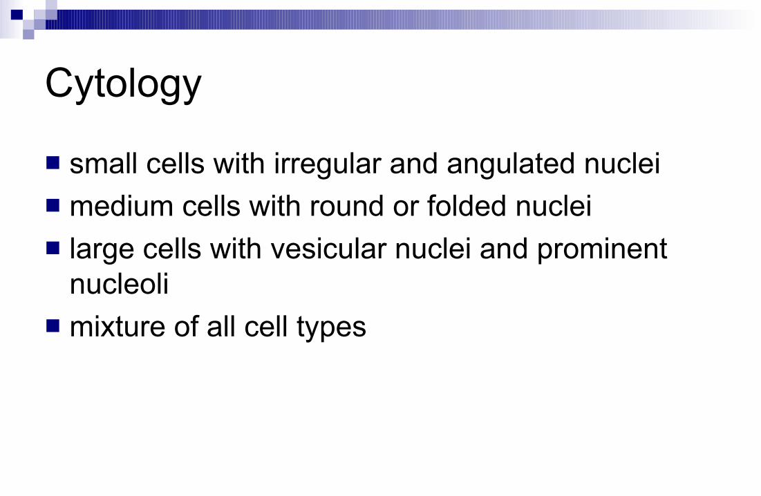
Cytology
small cells with irregular and angulated nuclei medium cells with round or folded nuclei large cells with vesicular nuclei and prominent
nucleoli mixture of all cell types

Immunophenotype
CD56 (N-CAM) + CD43 + TCRβ - CD20 - CD30-/+ CD4 -/+ CD8 -/+
TIA-1 +/- CD3 surface - CD3 (ε) cytoplasmic +/- MDR +/-

CD56 N-CAM: neural cell adhesion molecule adhesion molecule of immunoglobulin superfamily expressed on: NK cells, NK/T cells, neural and
neuroendocrine tissues tumors: NK neoplasms, neuroblastoma, small cell
carcinoma (Merkel cell carcinoma), medullary thyroid carcinoma, peripheral nerve tumors
CD3
discrepant results in frozen and paraffin embedded tissue (negative in frozen, positive in paraffin)

Ancillary Studies
clonality cannot be demonstrated with Southern blotting or PCR for TCR subset of NK/T is clonal with these methods and will also
expresss bF1 Distinction from gamma delta T cell lymphoma: these will
show a T cell clone EBV mRNA often positive (in situ hybridization) less frequently associated with EBV than nasal type
T/NK lymphomas

Summary
patch and plaque stage MF are best diagnosed with HE
immunophenotyping is essential in the diagnosis and classification of dermal nodular T-cell infiltrates
CD56 and CD 30 should be included in the routine panel for immunophenotyping

Acknowledgements
Sabine Kohler, MD Youn Kim, MD